Page 1
Copyright © 2017 The Authors; exclusive licensee Bio-protocol LLC. 1
www.bio-protocol.org/e2107 Vol 7, Iss 02, Jan 20, 2017 DOI:10.21769/BioProtoc.2107
Aggregation Prevention Assay for Chaperone Activity of Proteins Using Spectroflurometry Manish Bhuwan1, #, Madhuri Suragani2, #, §, Nasreen Z. Ehtesham3, * and Seyed E. Hasnain1, 4, 5, *
1Molecular Infection and Functional Biology Laboratory, Kusuma School of Biological Sciences, Indian
Institute of Technology, New Delhi, India; 2Department of Biochemistry, School of Life Sciences,
University of Hyderabad, Professor C.R. Rao Road, Hyderabad, India; 3Inflammation Biology and Cell
Signaling Laboratory, National Institute of Pathology, New Delhi, India ; 4Dr. Reddy’s Institute of Life
Sciences, University of Hyderabad Campus, Professor C.R. Rao Road, Hyderabad, India; 5JH Institute
of Molecular Medicine, Jamia Hamdard, Hamdard Nagar, New Delhi, India
*For correspondence: [email protected] ; [email protected] #Contributed equally to this work §Deceased
[Abstract] The ability to stabilize other proteins against thermal aggregation is one of the major
characteristics of chaperone proteins. Molecular chaperones bind to nonnative conformations of
proteins. Folding of the substrate is triggered by a dynamic association and dissociation cycles which
keep the substrate protein on track of the folding pathway (Figure 1). Usually molecular chaperones
exhibit differential affinities with different conformations of the substrate. With the exception of the sHsp
family of molecular chaperones, the shift from a high-affinity binding state to the low-affinity release state
is triggered by ATP binding and hydrolysis (Haselback and Buchner, 2015). Aggregation prevention
assay is a simple, yet definitive assay to determine the chaperone activity of heat labile proteins such
as Maltodextrin glucosidase (MalZ), Citrate Synthase (CS) and NdeI. This is based on the premise that
proteins with chaperone like activity should prevent protein substrates (MalZ, CS and NdeI) from thermal
aggregation. Here, we describe a detailed protocol for aggregation prevention assay using two different
chaperone proteins, resistin and MoxR1, identified from our lab. Resistin, a human protein (hRes) and
MoxR1 a Mycobacterium tuberculosis protein were analysed for their effect on prevention of
MalZ/Citrate Synthase (CS)/NdeI aggregation.
Page 2
Copyright © 2017 The Authors; exclusive licensee Bio-protocol LLC. 2
www.bio-protocol.org/e2107 Vol 7, Iss 02, Jan 20, 2017 DOI:10.21769/BioProtoc.2107
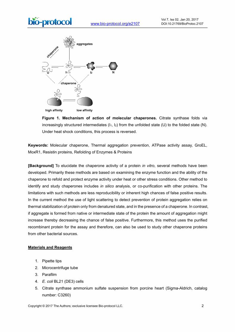
Figure 1. Mechanism of action of molecular chaperones. Citrate synthase folds via
increasingly structured intermediates (I1, I2) from the unfolded state (U) to the folded state (N).
Under heat shock conditions, this process is reversed.
Keywords: Molecular chaperone, Thermal aggregation prevention, ATPase activity assay, GroEL,
MoxR1, Resistin proteins, Refolding of Enzymes & Proteins [Background] To elucidate the chaperone activity of a protein in vitro, several methods have been
developed. Primarily these methods are based on examining the enzyme function and the ability of the
chaperone to refold and protect enzyme activity under heat or other stress conditions. Other method to
identify and study chaperones includes in silico analysis, or co-purification with other proteins. The
limitations with such methods are less reproducibility or inherent high chances of false positive results.
In the current method the use of light scattering to detect prevention of protein aggregation relies on
thermal stabilization of protein only from denatured state, and in the presence of a chaperone. In contrast,
if aggregate is formed from native or intermediate state of the protein the amount of aggregation might
increase thereby decreasing the chance of false positive. Furthermore, this method uses the purified
recombinant protein for the assay and therefore, can also be used to study other chaperone proteins
from other bacterial sources.
Materials and Reagents
1. Pipette tips
2. Microcentrifuge tube
3. Parafilm
4. E. coli BL21 (DE3) cells
5. Citrate synthase ammonium sulfate suspension from porcine heart (Sigma-Aldrich, catalog
number: C3260)
Page 3
Copyright © 2017 The Authors; exclusive licensee Bio-protocol LLC. 3
www.bio-protocol.org/e2107 Vol 7, Iss 02, Jan 20, 2017 DOI:10.21769/BioProtoc.2107
6. Ammonium sulphate
7. Tris (pH 8.0) (AMRESCO, catalog number: 0497)
8. Luria Bertani agar plates
9. Ampicillin (Sigma- Aldrich, catalog number: A9518)
10. Isopropyl β-D-1-thiogalactopyranoside, IPTG (Sigma-Aldrich, catalog number: I6758)
11. DNAse I (Sigma-Aldrich, catalog number: AMPD1)
12. Magnesium chloride hexahydrate (MgCl2·6H2O) (Sigma-Aldrich, catalog number: M2670)
13. Phenylmethylsulfonyl fluoride (PMSF) (Sigma-Aldrich, catalog number: 7626)
14. Bradford reagent (Bio-Rad Laboratories, catalog number: 5000006)
15. Bovine serum albumin (BSA) (Sigma-Aldrich, catalog number: A7906)
16. Recombinant protein MoxR1 and hRes (1 mg/ml) (Purified in the laboratory)
17. Purified recombinant protein GroEL (0.5 mg/ml) (Purified in the laboratory)
18. Lysozyme (1 mg/ml) (Sigma-Aldrich, catalog number: 4919)
19. Milli-Q water
20. NdeI (20,000 U/ml) (New England BioLab, catalog number: R0111)
21. Maltodextrin glucosidase protein (1 mg/ml) (Purified in the laboratory)
22. EDTA (Sigma-Aldrich, catalog number: E9884)
23. Sodium phosphate (dibasic) heptahydrate (Na2HPO4·7H2O) (AMRESCO, catalog number: 0348)
24. Sodium phosphate (monobasic) anhydrous (NaH2PO4) (AMRESCO, catalog number: 0571)
25. Sodium chloride (NaCl) (AMRESCO, catalog number: 0241)
26. Imidazole (AMRESCO, catalog number: 0527)
27. Glycerol (AMRESCO, catalog number: 0854)
28. TE buffer (see Recipes)
29. 20 mM sodium phosphate buffer pH 7.4 (see Recipes)
30. Binding buffer (see Recipes)
31. Washing buffer (see Recipes)
32. Elution buffer (see Recipes)
33. Dialysis buffer (see Recipes)
Equipment
1. Pipette (Gilson, model: Pipetman Neo®)
2. Centrifuge (Hermle Labor Technik, model: Z 326 K)
3. PerkinElmer spectrofluorometer (PerkinElmer, model: LS55) with controlled temperature peltier
block
4. Incubator (Eppendorf, BrunswickTM, model: 44/44R)
5. Quartz cuvette (PerkinElmer, Suprasil®, catalog number: B0631071) 6. pH meter (Thermo Fisher Scientific, Thermo Scientific, model: CyberScan Ph 510) 7. Fluorescence spectrophotometer (Agilent Technologies, model: Cary Eclipse)
Page 4
Copyright © 2017 The Authors; exclusive licensee Bio-protocol LLC. 4
www.bio-protocol.org/e2107 Vol 7, Iss 02, Jan 20, 2017 DOI:10.21769/BioProtoc.2107
Software
1. Microsoft Excel
2. Graphpad Prism 5
Procedure A. Preparation of citrate synthase and maltodextrin glucosidase
1. Commercially available citrate synthase is supplied in ammonium sulphate suspension to keep
the enzyme in an inactive state. The suspension is then pipetted into a fresh microcentrifuge
tube and centrifuge at 15,000 x g, for about 10 min at 4 °C. The enzyme CS is present in a solid
state and therefore will be collected in the pellet fraction.
2. Discard the supernatant and dissolve the pellet containing enzyme CS in sterile water at a
concentration of 1 mg/ml.
3. The CS should be further purified by size exclusion chromatography and dialysed against 50
mM Tris (pH 8.0). For refolding assays, CS stock solutions at concentration of 150 μM is
prepared in TE buffer by concentrating CS using Amicon Ultra microconcentrator with 30 kDa
cutoff before use.
4. The expression plasmids pCS19MalZ containing (His)6 tagged coding region of malz gene are
transformed into E. coli BL21 (DE3) competent cells and plating is performed on Luria Bertani
agar plates containing 100 µg/ml ampicillin for selecting pCS19MalZ.
5. A single colony from the transformed bacterial cells grown on Luria Bertani agar plate containing
100 µg/ml ampicillin is picked and grown in the Luria Bertani broth containing 100 µg/ml
ampicillin.
6. The tube is incubated for overnight at 37 °C with constant shaking at a rate of 150 rpm.
7. About 2% of the overnight culture is inoculated in fresh Luria Bertani broth containing ampicillin
100 µg/ml and then incubate it for 2 h at 37 °C with constant shaking at a rate of 150 rpm.
8. The expression of MalZ protein is then induced with 100 µM IPTG and left for 12 h.
9. The IPTG induced culture is pelleted through centrifugation at 11,510 x g for 50 min. The
supernatant is discarded, wash the cell pellet with binding buffer and resuspend the cells in
binding buffer, along with DNAse I (1 µl/ml of cell lysate), 0.5 mM MgCl2 and 1 mM PMSF (a
serine protease inhibitor).
10. The cells are disrupted through sonication on ice, and the supernatant is collected after
centrifugation. (Sonication was performed for 5 min at 1 sec on and 2 sec off cycle and at 30%
amplitude)
11. The protein is further purified by Ni-NTA affinity chromatography. The column is washed with 5
column volumes of washing buffer and the protein is eluted with 5 column volumes of elution
buffer (Goyal et al., 2014).
Page 5
Copyright © 2017 The Authors; exclusive licensee Bio-protocol LLC. 5
www.bio-protocol.org/e2107 Vol 7, Iss 02, Jan 20, 2017 DOI:10.21769/BioProtoc.2107
Note: Nickel nitrilotriacetic [Ni-NTA] chromatography is used for purification of 6x His-tagged
recombinant proteins overproduced in bacteria. The Ni-NTA agarose resins possess high affinity
and specificity for 6x His-tagged recombinant fusion proteins. The proteins bound to the resin
are eluted by competition with imidazole.
12. The concentration of the protein is determined using Bradford reagent. The standard curve is
plotted using different concentration of bovine serum albumin by determining the Optical Density
using spectrophotometer and plotting the 595 nm values (y-axis) versus their concentration in
µg/ml (x-axis).The concentration of dialyzed protein is then determined using the standard curve.
B. Thermally induced aggregation assay
1. To elucidate the chaperonic function of hRes protein on the thermal aggregation of CS,
recombinant GroEL protein is used as a positive control and lysozyme as a negative control.
Note: Chaperone to substrate ratio is critical and initially different ratios should be used to
determine the optimal ratio for the effect of chaperone activity.
2. We maintained 1:1 ratio between hRes (chaperone) vs. CS (Suragani et al., 2013). The proteins
are used at 0.15 µM concentration.
3. Similarly, to analyze the chaperone function of MoxR1 protein on thermal aggregation of MalZ,
GroEL is used as a positive control and lysozyme as a negative control. GroEL and lysozyme
are used at 2 µM concentration. In this case, we maintained the initial 12:1 ratio between MoxR1
(chaperone) and MalZ (Bhuwan et al., 2016) (see Note 1).
4. Stirred 0.75 ml quartz cuvettes are to be used in a fluorescence spectrophotometer. The stirring
is achieved by removing the cuvettes out of the spectrophotometer and sealing it by Parafilm
and mixing the solution by see-saw movement during the time interval of each data point. The
emission and excitation slits should be set to 2-5 nm.
5. Pre-equilibrate 50 mM Tris (pH 8.0) with and without chaperones at 45-47 °C for 50 min. The
chaperone dialysis buffer without chaperones is used as a control which is included in the assay
to rule out the possibility of stabilizing effects of buffer.
Note: After each sample analysis the quartz cuvettes should be thoroughly washed with a strong
jerk using MilliQ water to remove the traces of previous protein samples and any film formed on
the side of the cuvettes and after washing, the cuvette should be dried before using it at the next
step.
6. The initial light scattering signals are monitored at a constant wavelength of 320 nm for CS and
NdeI. Here, the data points should be taken at every 5 min and the kinetics are constantly
measured for 45 min at 45 °C. The increase in light scattering is observed to visualize the
thermal aggregation of CS.
7. The light scattering signal for MalZ is to be recorded at 500 nm wavelength.
Note: The range of absorbance for dynamic light scattering to detect aggregation is 300 nm to
600 nm and can also be optimized for different chaperone analysis.
8. Take the data at 5 min interval and the kinetics is monitored for 15 min at 47 °C (see Note 2).
Page 6
Copyright © 2017 The Authors; exclusive licensee Bio-protocol LLC. 6
www.bio-protocol.org/e2107 Vol 7, Iss 02, Jan 20, 2017 DOI:10.21769/BioProtoc.2107
9. The signal prior to CS or NdeI and MalZ addition is set to 0 (= no aggregation).
10. The readings are exported in a Microsoft Excel format and graph.
11. Plot the data for Optical Density on x-axis vs. Time on y-axis using Origin or Graphpad Prism 5
software to obtain a graph.
12. The resulting sigmoidal graphs will allow identifying the chaperonic function of a protein by
preventing thermal aggregation of CS or NdeI and MalZ.
Data analysis
The experimental data showing recombinant hRes and MoxR1 proteins mediated prevention of
thermal aggregation of CS or NdeI and MalZ proteins are depicted in Figures 2 and 3.
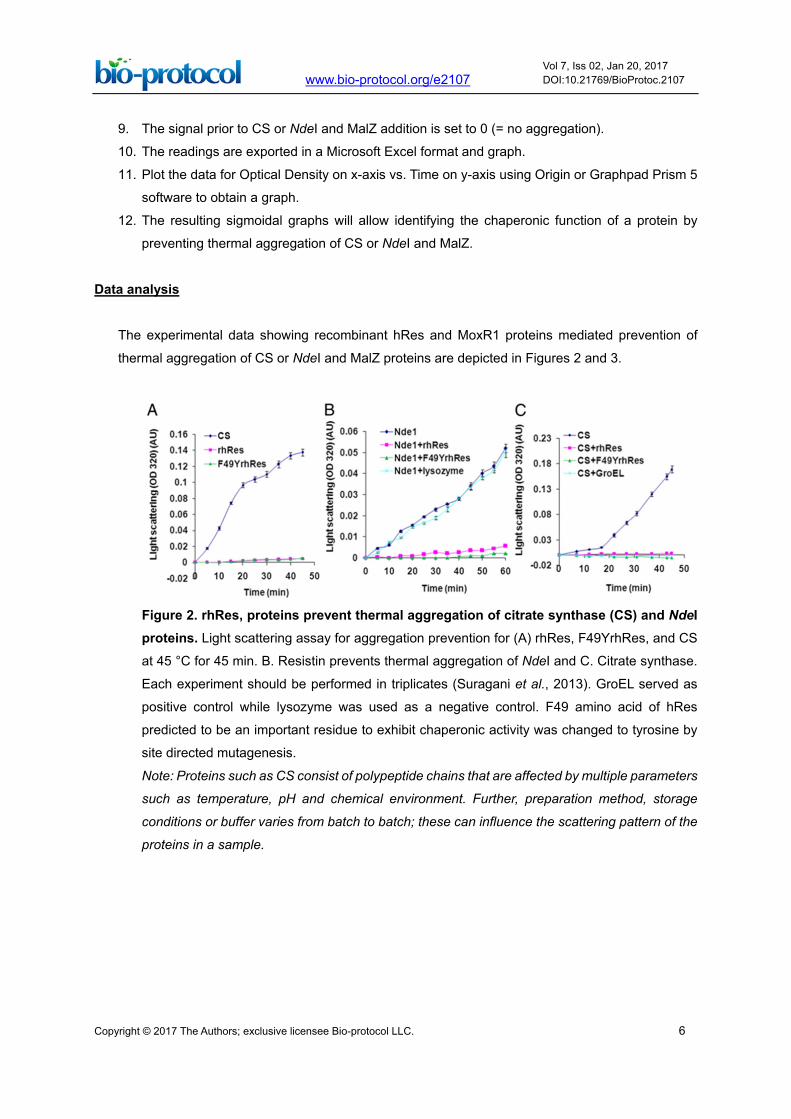
Figure 2. rhRes, proteins prevent thermal aggregation of citrate synthase (CS) and NdeI proteins. Light scattering assay for aggregation prevention for (A) rhRes, F49YrhRes, and CS
at 45 °C for 45 min. B. Resistin prevents thermal aggregation of NdeI and C. Citrate synthase.
Each experiment should be performed in triplicates (Suragani et al., 2013). GroEL served as
positive control while lysozyme was used as a negative control. F49 amino acid of hRes
predicted to be an important residue to exhibit chaperonic activity was changed to tyrosine by
site directed mutagenesis.
Note: Proteins such as CS consist of polypeptide chains that are affected by multiple parameters
such as temperature, pH and chemical environment. Further, preparation method, storage
conditions or buffer varies from batch to batch; these can influence the scattering pattern of the
proteins in a sample.
Page 7
Copyright © 2017 The Authors; exclusive licensee Bio-protocol LLC. 7
www.bio-protocol.org/e2107 Vol 7, Iss 02, Jan 20, 2017 DOI:10.21769/BioProtoc.2107
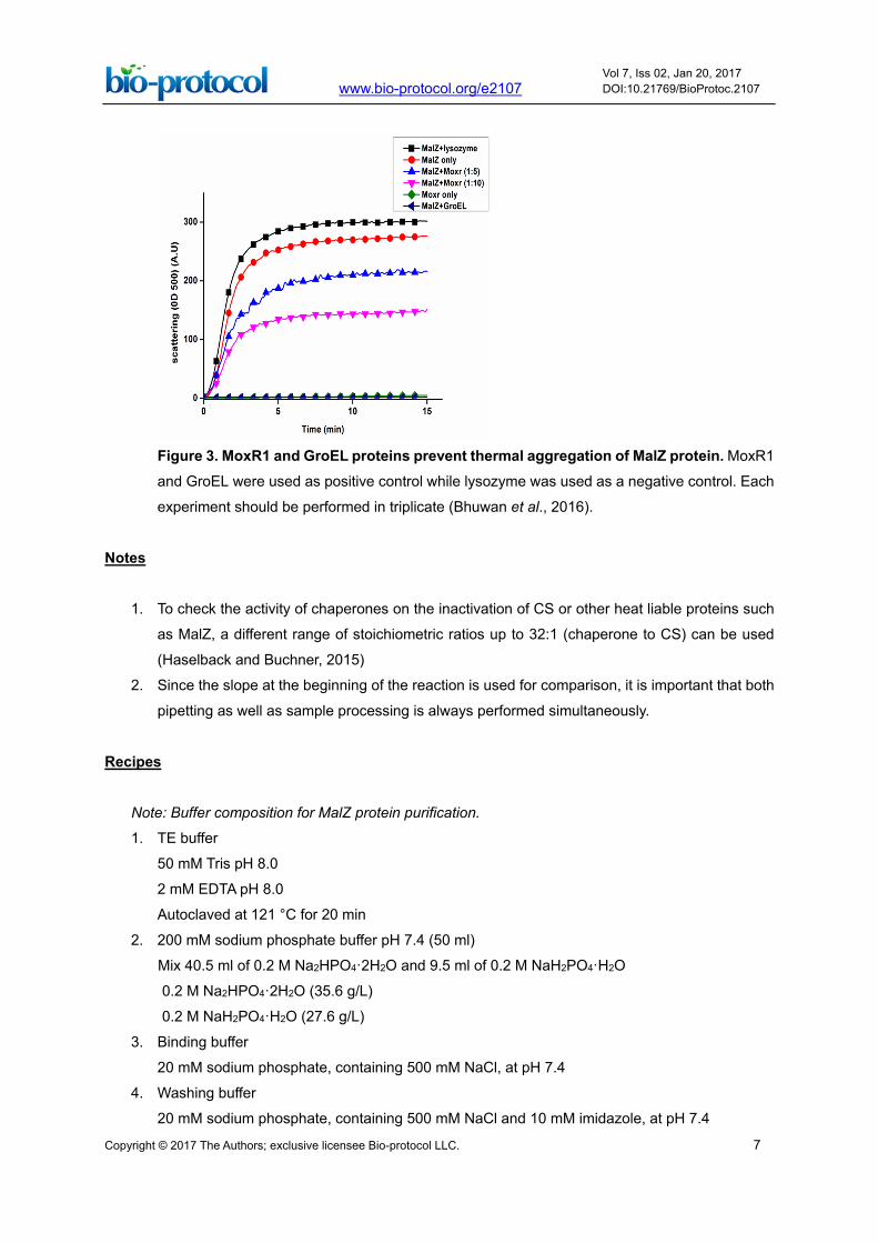
Figure 3. MoxR1 and GroEL proteins prevent thermal aggregation of MalZ protein. MoxR1
and GroEL were used as positive control while lysozyme was used as a negative control. Each
experiment should be performed in triplicate (Bhuwan et al., 2016).
Notes
1. To check the activity of chaperones on the inactivation of CS or other heat liable proteins such
as MalZ, a different range of stoichiometric ratios up to 32:1 (chaperone to CS) can be used
(Haselback and Buchner, 2015)
2. Since the slope at the beginning of the reaction is used for comparison, it is important that both
pipetting as well as sample processing is always performed simultaneously.
Recipes
Note: Buffer composition for MalZ protein purification.
1. TE buffer
50 mM Tris pH 8.0
2 mM EDTA pH 8.0
Autoclaved at 121 °C for 20 min
2. 200 mM sodium phosphate buffer pH 7.4 (50 ml)
Mix 40.5 ml of 0.2 M Na2HPO4·2H2O and 9.5 ml of 0.2 M NaH2PO4·H2O
0.2 M Na2HPO4·2H2O (35.6 g/L)
0.2 M NaH2PO4·H2O (27.6 g/L)
3. Binding buffer
20 mM sodium phosphate, containing 500 mM NaCl, at pH 7.4
4. Washing buffer
20 mM sodium phosphate, containing 500 mM NaCl and 10 mM imidazole, at pH 7.4
Page 8
Copyright © 2017 The Authors; exclusive licensee Bio-protocol LLC. 8
www.bio-protocol.org/e2107 Vol 7, Iss 02, Jan 20, 2017 DOI:10.21769/BioProtoc.2107
5. Elution buffer
20 mM sodium phosphate, containing 500 mM NaCl and 500 mM imidazole, at pH 7.4
6. Dialysis buffer
20 mM sodium phosphate, containing 500 mM NaCl and 10% glycerol, at pH 7.4
Acknowledgments
This protocol was adapted from our earlier publications (Suragani et al., 2013 and Bhuwan et al.,
2016). The work was supported by a Centre of Excellence Phase 2 Grant
(BT/R12817/COE/34/23/2015) from the Department of Biotechnology, Ministry of Science &
Technology, Government of India to S.E.H. and N.Z.E.
References 1. Bhuwan, M., Arora, N., Sharma, A., Khubaib, M., Pandey, S., Chaudhuri, T. K., Hasnain, S. E.
and Ehtesham, N. Z. (2016). Interaction of Mycobacterium tuberculosis virulence factor RipA
with chaperone MoxR1 is required for transport through the TAT secretion system. mBio 7(2):
e02259.
2. Goyal, M., Chaudhuri, T. K. and Kuwajima, K. (2014). Irreversible denaturation of maltodextrin
glucosidase studied by differential scanning calorimetry, circular dichroism, and turbidity
measurements. PLoS One 9(12): e115877.
3. Haslbeck, M. and Buchner, J. (2015). Assays to characterize molecular chaperone function in
vitro. Methods Mol Biol 1292: 39-51.
4. Suragani, M., Aadinarayana, V. D., Pinjari, A. B., Tanneeru, K., Guruprasad, L., Banerjee, S.,
Pandey, S., Chaudhuri, T. K. and Ehtesham, N. Z. (2013). Human resistin, a proinflammatory
cytokine, shows chaperone-like activity. Proc Natl Acad Sci U S A 110(51): 20467-20472.