115 Radiographically, UA often has a unilocular radiolucency surrounding the crown of an unerupted tooth, although it may also appear as a well-defined intraosseous radiolucent area 1,4 . These findings resemble cysts in the jaws and, hence, the final diagnosis is only confirmed through histopathological analysis 2,4 . Therefore, UA tends to present as a well-defined unilocular lesion; nevertheless, a multilocular appearance is also reported 4,5 . In the early phase, UA may be routinely found in panoramic radiographs 4 . Nevertheless, computed to- mography (CT) scans are highly recommended to determine details in relation to tumor extension and bone and teeth in- volvement 6 . Here, we described a case of UA not found during orth- odontic treatment planning. Although, some unilocular radio- lucent lesions in the posterior mandible areas present sign and symptoms, others are completely asymptomatic and thorough imaging examination is necessary to avoid diagnosis negli- gence. II. Case Report A 16-year-old Caucasian male patient was submitted to radiographic examination for orthodontic planning in 2004. (Fig. 1) On the panoramic radiograph, a circumscribed uni- I. Introduction In spite of its benign nature, the ameloblastoma is a slowly growing, locally invasive, and epithelial odontogenic tumor 1 . Multicystic ameloblastoma is the most common variant and 5% to 15% of all ameloblastomas are of the unicystic type 2 . This lesion is a less clinically aggressive variant of amelo- blastoma 3 . More than 90% of unicystic ameloblastoma (UA) involve the mandible, usually the posterior region; and up to 80% are associated with an unerupted mandibular third molar 2 . The mean age of UA occurrence is around 16 years, as opposed to 35 years, in the absence of an unerupted tooth without gender predilection 1,2 . CASE REPORT Isadora Luana Flores Department of Dentistry, Federal University of Juiz de Fora, Campus Governador Valadares, Rua Israel Pinheiro 2000, Bairro Universitário, Governador Valadares 35020-220, Brazil TEL: +55-33-3301-1000 FAX: +55-33-3301-1000 E-mail: [email protected]ORCID: http://orcid.org/0000-0002-6628-2122 This is an open-access article distributed under the terms of the Creative Commons Attribution Non-Commercial License (http://creativecommons.org/licenses/by-nc/4.0/), which permits unrestricted non-commercial use, distribution, and reproduction in any medium, provided the original work is properly cited. CC Aggressive unicystic ameloblastoma affecting the posterior mandible: late diagnosis during orthodontic treatment Sérgio Lúcio Pereira de Castro Lopes 1 , Isadora Luana Flores 2 , Thiago de Oliveira Gamba 3 , Rivea Ines Ferreira-Santos 4 , Mari Eli Leonelli de Moraes 1 , Aline Alvarez Cabello 1 , Paula Nascimento Moutinho 1 1 Department of Diagnosis and Surgery, São José dos Campos Dental School, São Paulo State University, São José dos Campos, 2 Department of Dentistry, Federal University of Juiz de Fora, Campus Governador Valadares, Governador Valadares, 3 Department of Oral Diagnosis, Piracicaba Dental School, University of Campinas, Piracicaba, 4 Department of Orthodontics, City University of São Paulo, Tatuapé, Brazil Abstract (J Korean Assoc Oral Maxillofac Surg 2017;43:115-119) Maxillofacial images must be examined to find pathologies not identified during clinical examination. Unicystic ameloblastoma (UA) extending to the mandibular body and ramus was neglected on initial panoramic radiographic examination. After orthodontic therapy, a huge lesion was observed clini- cally and through imaging exams. After the conservative surgery, no recurrence was observed during five years of follow-up. This case emphasized the need for careful evaluation of patient images focusing on the oral diagnosis before any dental treatment planning, including orthodontic therapy. Key words: Oral diagnosis, Dentistry, Diagnostic imaging, Ameloblastoma [paper submitted 2016. 2. 29 / revised 2016. 5. 4 / accepted 2016. 5. 18] Copyright Ⓒ 2017 The Korean Association of Oral and Maxillofacial Surgeons. All rights reserved. https://doi.org/10.5125/jkaoms.2017.43.2.115 pISSN 2234-7550 · eISSN 2234-5930
Transcript
115
Radiographically, UA often has a unilocular radiolucency
surrounding the crown of an unerupted tooth, although it may
also appear as a well-defined intraosseous radiolucent area1,4.
These findings resemble cysts in the jaws and, hence, the
final diagnosis is only confirmed through histopathological
analysis2,4. Therefore, UA tends to present as a well-defined
unilocular lesion; nevertheless, a multilocular appearance
is also reported4,5. In the early phase, UA may be routinely
found in panoramic radiographs4. Nevertheless, computed to-
mography (CT) scans are highly recommended to determine
details in relation to tumor extension and bone and teeth in-
volvement6.
Here, we described a case of UA not found during orth-
odontic treatment planning. Although, some unilocular radio-
lucent lesions in the posterior mandible areas present sign and
symptoms, others are completely asymptomatic and thorough
imaging examination is necessary to avoid diagnosis negli-
gence.
II. Case Report
A 16-year-old Caucasian male patient was submitted to
radiographic examination for orthodontic planning in 2004.
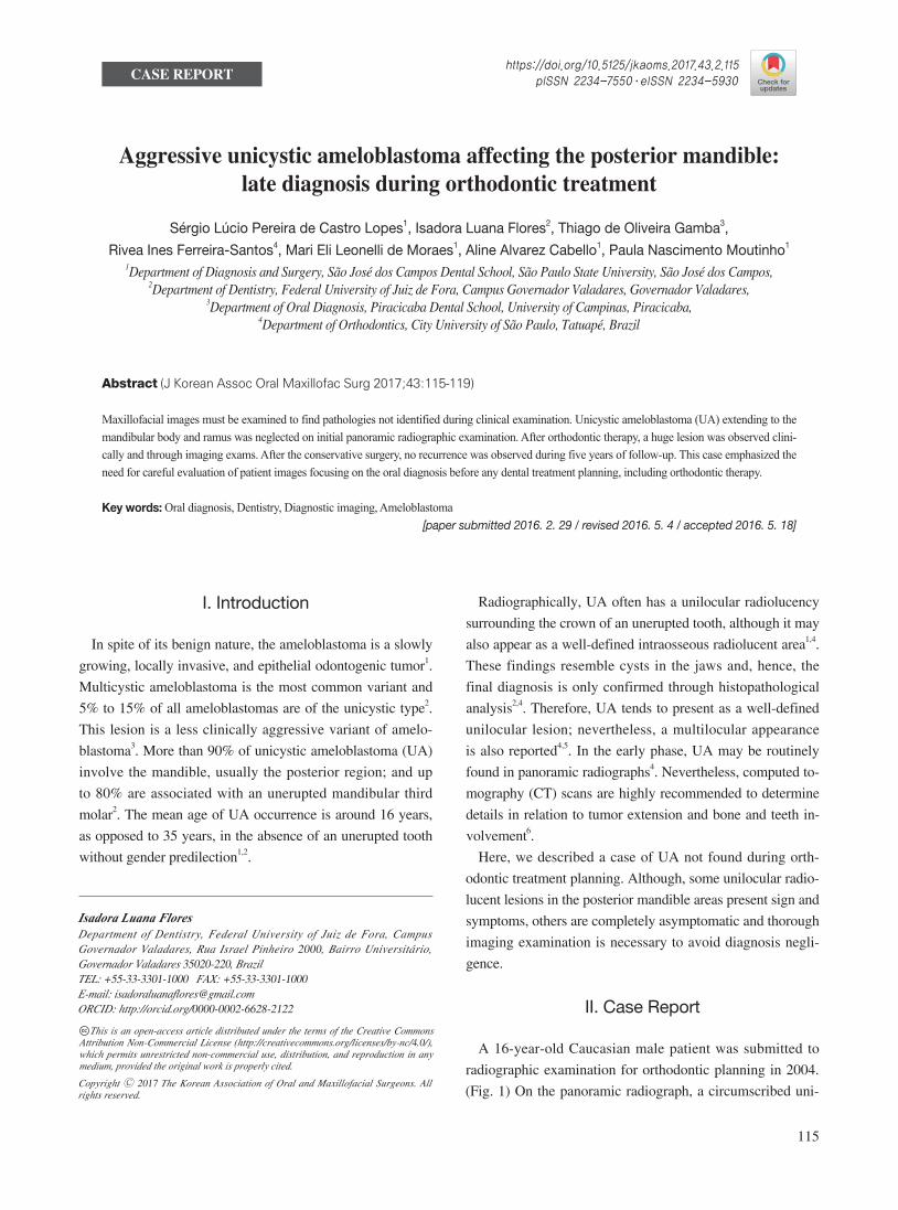
(Fig. 1) On the panoramic radiograph, a circumscribed uni-
I. Introduction
In spite of its benign nature, the ameloblastoma is a slowly
growing, locally invasive, and epithelial odontogenic tumor1.
Multicystic ameloblastoma is the most common variant and
5% to 15% of all ameloblastomas are of the unicystic type2.
This lesion is a less clinically aggressive variant of amelo-
blastoma3. More than 90% of unicystic ameloblastoma (UA)
involve the mandible, usually the posterior region; and up
to 80% are associated with an unerupted mandibular third
molar2. The mean age of UA occurrence is around 16 years,
as opposed to 35 years, in the absence of an unerupted tooth
without gender predilection1,2.
CASE REPORT
Isadora Luana FloresDepartment of Dentistry, Federal University of Juiz de Fora, Campus Governador Valadares, Rua Israel Pinheiro 2000, Bairro Universitário, Governador Valadares 35020-220, BrazilTEL: +55-33-3301-1000 FAX: +55-33-3301-1000E-mail: [email protected]: http://orcid.org/0000-0002-6628-2122
This is an open-access article distributed under the terms of the Creative Commons Attribution Non-Commercial License (http://creativecommons.org/licenses/by-nc/4.0/), which permits unrestricted non-commercial use, distribution, and reproduction in any medium, provided the original work is properly cited.
CC
Aggressive unicystic ameloblastoma affecting the posterior mandible: late diagnosis during orthodontic treatment
Sérgio Lúcio Pereira de Castro Lopes1, Isadora Luana Flores2, Thiago de Oliveira Gamba3,
Rivea Ines Ferreira-Santos4, Mari Eli Leonelli de Moraes1, Aline Alvarez Cabello1, Paula Nascimento Moutinho1
1Department of Diagnosis and Surgery, São José dos Campos Dental School, São Paulo State University, São José dos Campos, 2Department of Dentistry, Federal University of Juiz de Fora, Campus Governador Valadares, Governador Valadares,
3Department of Oral Diagnosis, Piracicaba Dental School, University of Campinas, Piracicaba, 4Department of Orthodontics, City University of São Paulo, Tatuapé, Brazil
Abstract (J Korean Assoc Oral Maxillofac Surg 2017;43:115-119)
Maxillofacial images must be examined to find pathologies not identified during clinical examination. Unicystic ameloblastoma (UA) extending to the mandibular body and ramus was neglected on initial panoramic radiographic examination. After orthodontic therapy, a huge lesion was observed clini-cally and through imaging exams. After the conservative surgery, no recurrence was observed during five years of follow-up. This case emphasized the need for careful evaluation of patient images focusing on the oral diagnosis before any dental treatment planning, including orthodontic therapy.
J Korean Assoc Oral Maxillofac Surg 2017;43:115-119
116
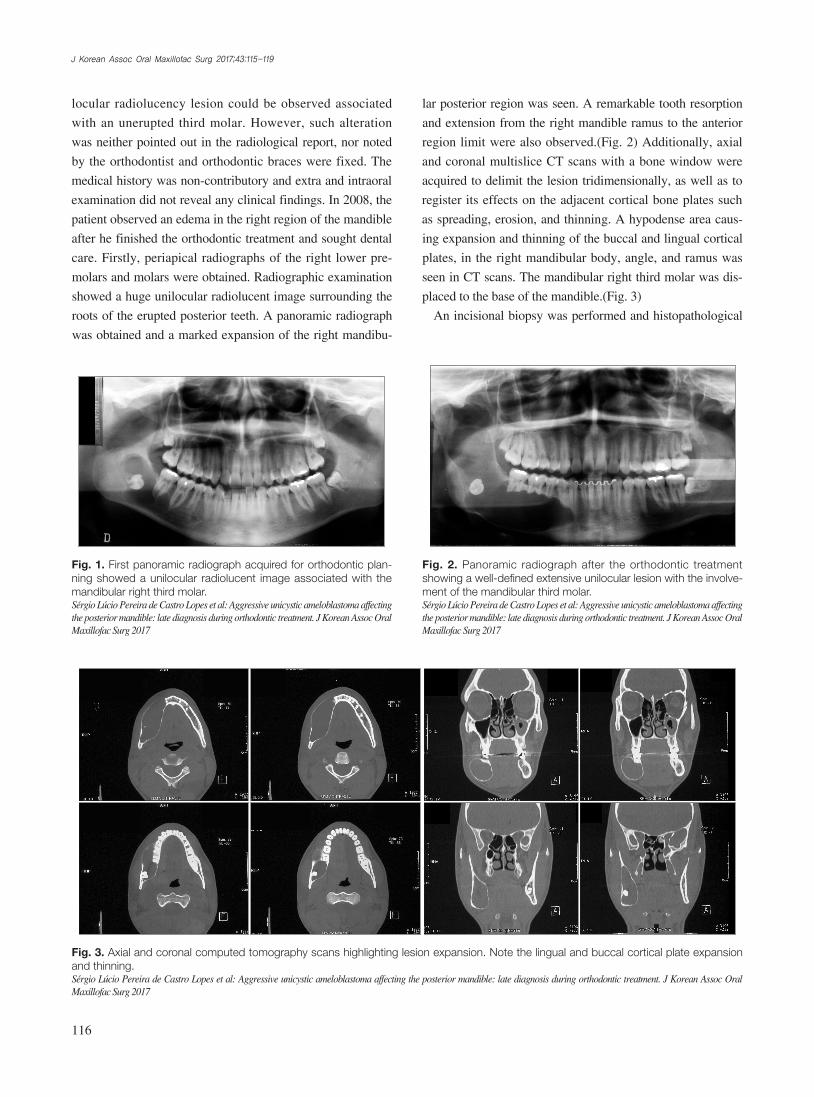
lar posterior region was seen. A remarkable tooth resorption
and extension from the right mandible ramus to the anterior
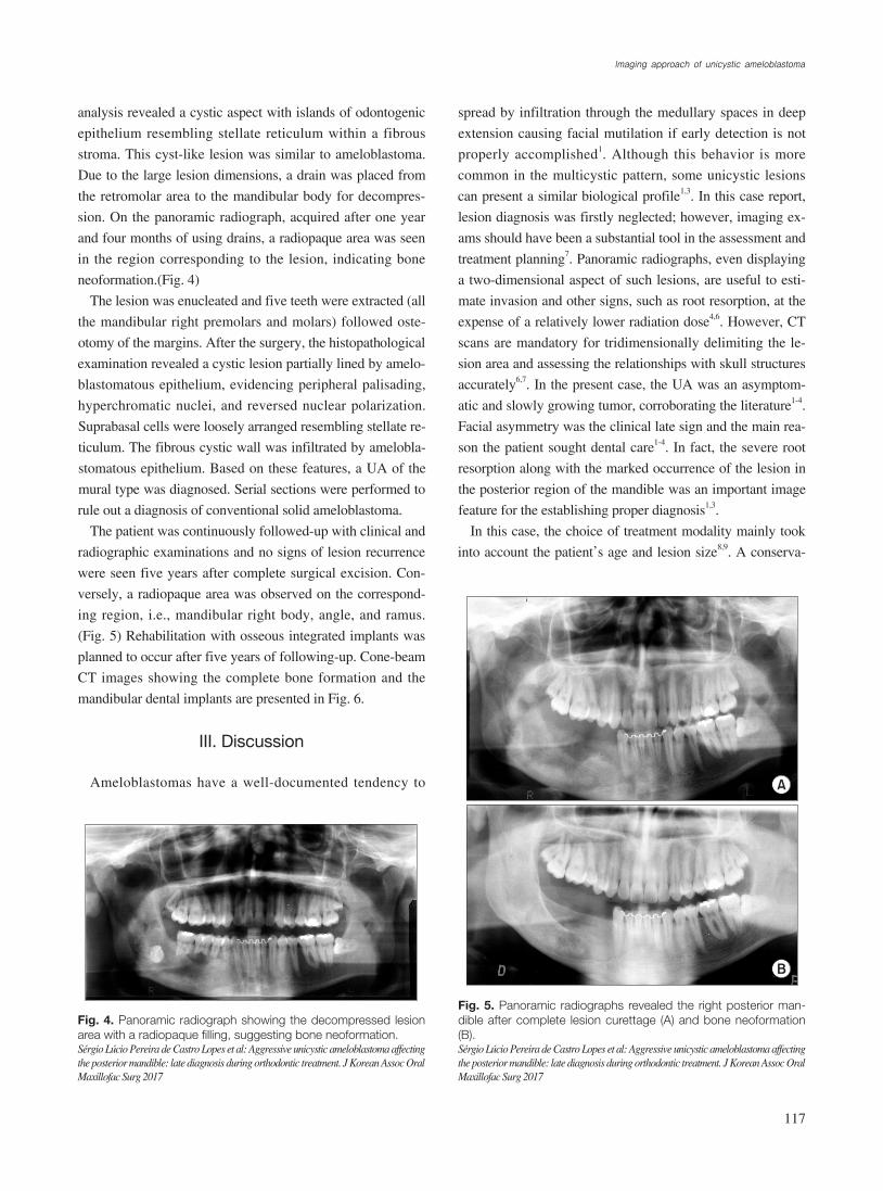
region limit were also observed.(Fig. 2) Additionally, axial
and coronal multislice CT scans with a bone window were
acquired to delimit the lesion tridimensionally, as well as to
register its effects on the adjacent cortical bone plates such
as spreading, erosion, and thinning. A hypodense area caus-
ing expansion and thinning of the buccal and lingual cortical
plates, in the right mandibular body, angle, and ramus was
seen in CT scans. The mandibular right third molar was dis-
placed to the base of the mandible.(Fig. 3)
An incisional biopsy was performed and histopathological
locular radiolucency lesion could be observed associated
with an unerupted third molar. However, such alteration
was neither pointed out in the radiological report, nor noted
by the orthodontist and orthodontic braces were fixed. The
medical history was non-contributory and extra and intraoral
examination did not reveal any clinical findings. In 2008, the
patient observed an edema in the right region of the mandible
after he finished the orthodontic treatment and sought dental
care. Firstly, periapical radiographs of the right lower pre-
molars and molars were obtained. Radiographic examination
showed a huge unilocular radiolucent image surrounding the
roots of the erupted posterior teeth. A panoramic radiograph
was obtained and a marked expansion of the right mandibu-
Fig. 3. Axial and coronal computed tomography scans highlighting lesion expansion. Note the lingual and buccal cortical plate expansion and thinning.Sérgio Lúcio Pereira de Castro Lopes et al: Aggressive unicystic ameloblastoma affecting the posterior mandible: late diagnosis during orthodontic treatment. J Korean Assoc Oral Maxillofac Surg 2017
Fig. 1. First panoramic radiograph acquired for orthodontic plan-ning showed a unilocular radiolucent image associated with the mandibular right third molar.Sérgio Lúcio Pereira de Castro Lopes et al: Aggressive unicystic ameloblastoma affecting the posterior mandible: late diagnosis during orthodontic treatment. J Korean Assoc Oral Maxillofac Surg 2017
Fig. 2. Panoramic radiograph after the orthodontic treatment showing a well-defined extensive unilocular lesion with the involve-ment of the mandibular third molar. Sérgio Lúcio Pereira de Castro Lopes et al: Aggressive unicystic ameloblastoma affecting the posterior mandible: late diagnosis during orthodontic treatment. J Korean Assoc Oral Maxillofac Surg 2017
Imaging approach of unicystic ameloblastoma
117
spread by infiltration through the medullary spaces in deep
extension causing facial mutilation if early detection is not
properly accomplished1. Although this behavior is more
common in the multicystic pattern, some unicystic lesions
can present a similar biological profile1,3. In this case report,
lesion diagnosis was firstly neglected; however, imaging ex-
ams should have been a substantial tool in the assessment and
treatment planning7. Panoramic radiographs, even displaying
a two-dimensional aspect of such lesions, are useful to esti-
mate invasion and other signs, such as root resorption, at the
expense of a relatively lower radiation dose4,6. However, CT
scans are mandatory for tridimensionally delimiting the le-
sion area and assessing the relationships with skull structures
accurately6,7. In the present case, the UA was an asymptom-
atic and slowly growing tumor, corroborating the literature1-4.
Facial asymmetry was the clinical late sign and the main rea-
son the patient sought dental care1-4. In fact, the severe root
resorption along with the marked occurrence of the lesion in
the posterior region of the mandible was an important image
feature for the establishing proper diagnosis1,3.
In this case, the choice of treatment modality mainly took
into account the patient’s age and lesion size8,9. A conserva-
analysis revealed a cystic aspect with islands of odontogenic
epithelium resembling stellate reticulum within a fibrous
stroma. This cyst-like lesion was similar to ameloblastoma.
Due to the large lesion dimensions, a drain was placed from
the retromolar area to the mandibular body for decompres-
sion. On the panoramic radiograph, acquired after one year
and four months of using drains, a radiopaque area was seen
in the region corresponding to the lesion, indicating bone
neoformation.(Fig. 4)
The lesion was enucleated and five teeth were extracted (all
the mandibular right premolars and molars) followed oste-
otomy of the margins. After the surgery, the histopathological
examination revealed a cystic lesion partially lined by amelo-
hyperchromatic nuclei, and reversed nuclear polarization.
Suprabasal cells were loosely arranged resembling stellate re-
ticulum. The fibrous cystic wall was infiltrated by amelobla-
stomatous epithelium. Based on these features, a UA of the
mural type was diagnosed. Serial sections were performed to
rule out a diagnosis of conventional solid ameloblastoma.
The patient was continuously followed-up with clinical and
radiographic examinations and no signs of lesion recurrence
were seen five years after complete surgical excision. Con-
versely, a radiopaque area was observed on the correspond-
ing region, i.e., mandibular right body, angle, and ramus.
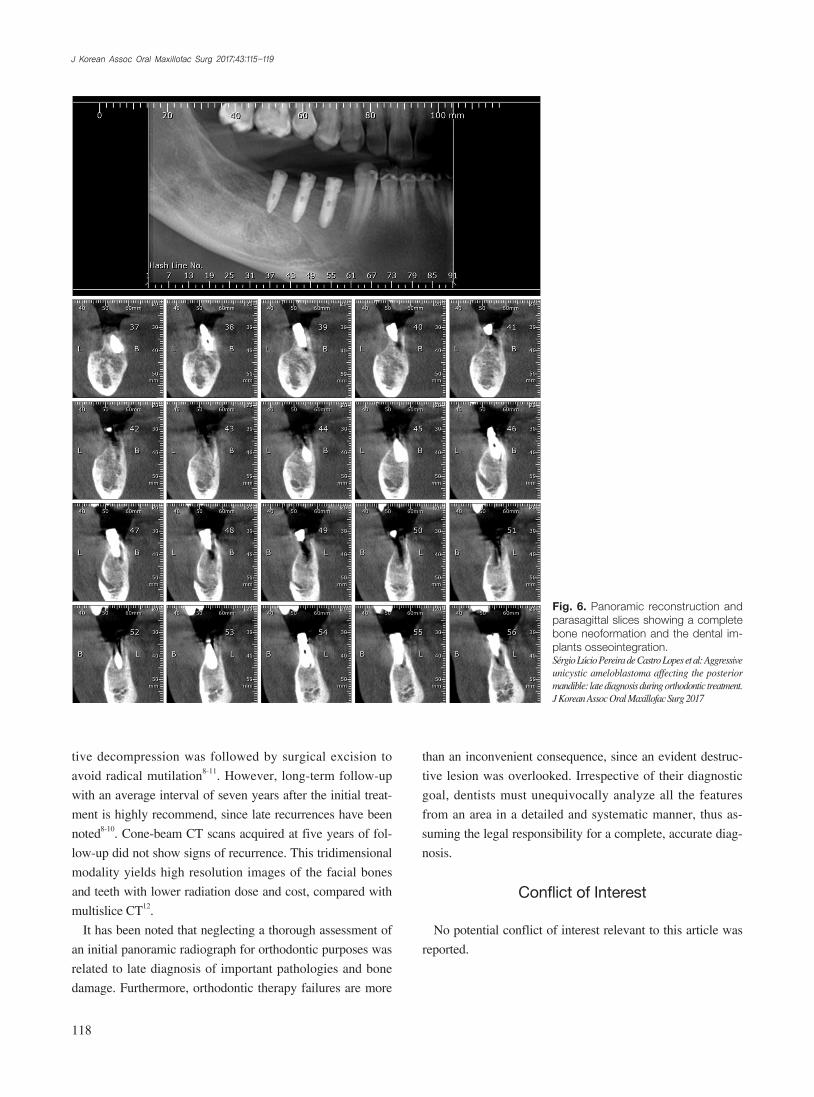
(Fig. 5) Rehabilitation with osseous integrated implants was
planned to occur after five years of following-up. Cone-beam
CT images showing the complete bone formation and the
mandibular dental implants are presented in Fig. 6.
III. Discussion
Ameloblastomas have a well-documented tendency to
Fig. 4. Panoramic radiograph showing the decompressed lesion area with a radiopaque filling, suggesting bone neoformation. Sérgio Lúcio Pereira de Castro Lopes et al: Aggressive unicystic ameloblastoma affecting the posterior mandible: late diagnosis during orthodontic treatment. J Korean Assoc Oral Maxillofac Surg 2017
A
B
Fig. 5. Panoramic radiographs revealed the right posterior man-dible after complete lesion curettage (A) and bone neoformation (B).Sérgio Lúcio Pereira de Castro Lopes et al: Aggressive unicystic ameloblastoma affecting the posterior mandible: late diagnosis during orthodontic treatment. J Korean Assoc Oral Maxillofac Surg 2017
J Korean Assoc Oral Maxillofac Surg 2017;43:115-119
118
than an inconvenient consequence, since an evident destruc-
tive lesion was overlooked. Irrespective of their diagnostic
goal, dentists must unequivocally analyze all the features
from an area in a detailed and systematic manner, thus as-
suming the legal responsibility for a complete, accurate diag-
nosis.
Conflict of Interest
No potential conflict of interest relevant to this article was
reported.
tive decompression was followed by surgical excision to
avoid radical mutilation8-11. However, long-term follow-up
with an average interval of seven years after the initial treat-
ment is highly recommend, since late recurrences have been
noted8-10. Cone-beam CT scans acquired at five years of fol-
low-up did not show signs of recurrence. This tridimensional
modality yields high resolution images of the facial bones
and teeth with lower radiation dose and cost, compared with
multislice CT12.
It has been noted that neglecting a thorough assessment of
an initial panoramic radiograph for orthodontic purposes was
related to late diagnosis of important pathologies and bone
damage. Furthermore, orthodontic therapy failures are more
Fig. 6. Panoramic reconstruction and parasagittal slices showing a complete bone neoformation and the dental im-plants osseointegration.Sérgio Lúcio Pereira de Castro Lopes et al: Aggressive unicystic ameloblastoma affecting the posterior mandible: late diagnosis during orthodontic treatment. J Korean Assoc Oral Maxillofac Surg 2017
Imaging approach of unicystic ameloblastoma
119
ORCID
Sérgio Lúcio Pereira de Castro Lopes, http://orcid.org/0000-0002-0882-5862
Isadora Luana Flores, http://orcid.org/0000-0002-6628-2122Thiago de Oliveira Gamba, http://orcid.org/0000-0002-
1. Masthan KM, Anitha N, Krupaa J, Manikkam S. Ameloblastoma. J Pharm Bioallied Sci 2015;7(Suppl 1):S167-70.
2. Barnes L, Eveson JW, Reichart P, Sidransky D. Pathology and ge-netics of head and neck tumours. 3rd ed. Lyon: IARC Press; 2005.
3. Philipsen HP, Reichart PA. Unicystic ameloblastoma. A review of
193 cases from the literature. Oral Oncol 1998;34:317-25.4. Volpi L, Ferreli F, Karligkiotis A, Pistochini A, Meloni F, Castelnu-
ovo P. Radiology quiz case 1. Diagnosis: unicystic ameloblastoma (UA). Arch Otolaryngol Head Neck Surg 2012;138:973-4.
5. Bajpai M, Agarwal D, Bhalla A, Kumar M, Garg R, Kumar M. Multilocular unicystic ameloblastoma of mandible. Case Rep Dent 2013. doi: 10.1155/2013/835892.
6. Crusoé-Rebello I, Oliveira C, Campos PS, Azevedo RA, dos San-tos JN. Assessment of computerized tomography density patterns of ameloblastomas and keratocystic odontogenic tumors. Oral Surg Oral Med Oral Pathol Oral Radiol Endod 2009;108:604-8.
7. De Melo WM, Pereira-Santos D, Sonoda CK, Pereira-Freitas SA, de Moura WL, de Paulo Cravinhos JC. Large unicystic ameloblas-toma of the mandible: management guided by biological behavior. J Craniofac Surg 2012;23:e499-502.
8. Dolanmaz D, Etoz OA, Pampu A, Kalayci A, Gunhan O. Marsupi-alization of unicystic ameloblastoma: a conservative approach for aggressive odontogenic tumors. Indian J Dent Res 2011;22:709-12.
9. de Paulo LF, Oliveira MT, Rodrigues ÁR, Zanetta-Barbosa D. Treatment of an extensive unicystic ameloblastoma in a 7-year-old child: the best approach? Br J Oral Maxillofac Surg 2015;53:292-4.
10. Ramesh RS, Manjunath S, Ustad TH, Pais S, Shivakumar K. Uni-cystic ameloblastoma of the mandible--an unusual case report and review of literature. Head Neck Oncol 2010;2:1.
11. Park HS, Song IS, Seo BM, Lee JH, Kim MJ. The effectiveness of decompression for patients with dentigerous cysts, keratocystic odontogenic tumors, and unicystic ameloblastoma. J Korean Assoc Oral Maxillofac Surg 2014;40:260-5.
12. Luo J, You M, Wen C, Xu L, Zheng G. Cone-beam CT features of ameloblastomas. Hua Xi Kou Qiang Yi Xue Za Zhi 2014;32:373-7.