ASSOCIATION OF PLURIPOTENCY GENE PROMOTER METHYLATION WITH THE CHROMOSOMAL STATUS OF PRODUCTS OF CONCEPTION by AGNIESZKA LONCZAK Thesis submitted to the Graduate School-Newark Rutgers, The State University of New Jersey in partial fulfillment of the requirements for the degree of Master of Science Graduate Program in Biology written under the direction of Nathan R. Treff, Ph.D. and Nan Gao, Ph.D. and approved by _______________________________ _______________________________ _______________________________ Newark, New Jersey January, 2013
Transcript
ASSOCIATION OF PLURIPOTENCY GENE PROMOTER METHYLATION WITH
THE CHROMOSOMAL STATUS OF PRODUCTS OF CONCEPTION
by
AGNIESZKA LONCZAK
Thesis submitted to the
Graduate School-Newark
Rutgers, The State University of New Jersey
in partial fulfillment of the requirements
for the degree of
Master of Science
Graduate Program in Biology
written under the direction of
Nathan R. Treff, Ph.D. and Nan Gao, Ph.D.
and approved by
_______________________________
_______________________________
_______________________________
Newark, New Jersey
January, 2013
ii
ABSTRACT OF MASTER THESIS
ASSOCIATION OF PLURIPOTENCY GENE PROMOTER METHYLATION WITH
THE CHROMOSOMAL STATUS OF PRODUCTS OF CONCEPTION
By AGNIESZKA LONCZAK
Master Thesis Director:
Nathan R. Treff, Ph.D.
Infertility affects one in six couples and often necessitates the use of assisted reproductive
technology (ART). While ART is the most effective treatment, efficiency remains poor
with less than 13% of transferred in vitro fertilization (IVF) derived embryos resulting in
a live birth according to the Center for Disease Control. This has led to routine use of
multiple embryo transfer to increase pregnancy rates. However, as a result of multiple
embryo transfer, a significant proportion of IVF pregnancies involve multiples. Indeed,
multiple gestation is the most common complication associated with ART and is now the
primary focus of research and development in reproductive medicine. The ability to
identify the embryo with true reproductive potential could overcome the need for
multiple embryo transfer in order to achieve reasonable pregnancy rates from IVF.
Differentiation and establishment of the trophectoderm lineage during preimplantation
embryo development represents a potential target to identify new biomarkers of
reproductive potential. Several gene promoters have already been shown to be
iii
differentially methylated in pluripotent versus differentiated cells. These promoters
include: NANOG, PTPN6, RAB25, LYST, GBP3, MGMT, Oct4 and Elf5. The extent of
methylation of these promoters was characterized after the development of a
methodology for methylation sensitive restriction enzyme digestion followed by
quantitative real-time PCR. Chromosomal aneuploidy is a well characterized marker of
reproductive potential. The level of differentiation inferred from methylation status of
these promoters was used to evaluate whether aneuploid and euploid conceptions possess
unique levels of differentiation. Results indicate that GBP3 promoter methylation is
significantly different in aneuploid relative to euploid conceptions supporting the concept
that chromosomally normal embryos may differentiate more successfully than
chromosomally abnormal embryos.
iv
Acknowledgements
I would like to gratefully acknowledge the help, support, guidance and supervision of
Dr. Nathan Treff during this work. I am very grateful to him and his team: especially Xin
Tao, Dr. Lesley Northrop, Annie Fedick, Margaret Lebiedzinski, Oksana Bendarsky,
Jessyca Campos, Leslie Duffy and Anna Czyrsznic for encouragement, guidance,
technical assistance and moral support.
I would also like to thank Dr. Jennifer Moore from Rutgers University for letting me
obtain induced pluripotent cells which was a critical factor in this work, and Dr. Manuel
Krispin from Zymo Research Corporation and his team for technical assistance and
discussion and for providing me with the reagents imperative for my work.
I would like to take this opportunity to thank Dr. Gao and members of his group, in
whose lab I worked for a very short duration, for his encouragement and helpful advice
during my work there.
Finally, I am forever indebted to my family, especially my husband Krzysztof, for their
understanding, endless patience and encouragement when it was most required.
v
Table of Contents
Abstract ............................................................................................................................... ii
Acknowledgments.............................................................................................................. iv
List of Tables ..................................................................................................................... vi
List of Illustrations ............................................................................................................ vii
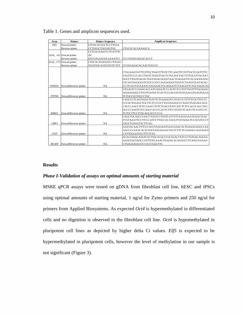
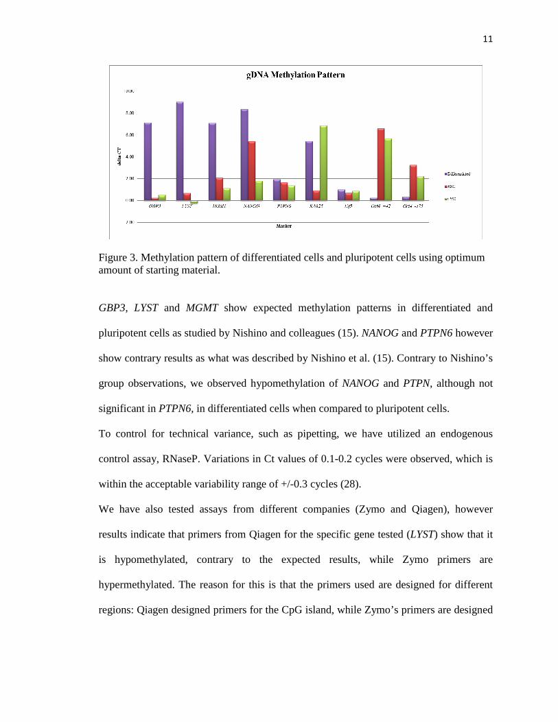
Phase I-Validation of assays on optimal amounts of starting material
MSRE qPCR assays were tested on gDNA from fibroblast cell line, hESC and iPSCs
using optimal amounts of starting material, 1 ng/ul for Zymo primers and 250 ng/ul for
primers from Applied Biosystems. As expected Oct4 is hypermethylated in differentiated
cells and no digestion is observed in the fibroblast cell line. Oct4 is hypomethylated in
pluripotent cell lines as depicted by higher delta Ct values. Elf5 is expected to be
hypermethylated in pluripotent cells, however the level of methylation in our sample is
not significant (Figure 3).
11
Figure 3. Methylation pattern of differentiated cells and pluripotent cells using optimum amount of starting material.
GBP3, LYST and MGMT show expected methylation patterns in differentiated and
pluripotent cells as studied by Nishino and colleagues (15). NANOG and PTPN6 however
show contrary results as what was described by Nishino et al. (15). Contrary to Nishino’s
group observations, we observed hypomethylation of NANOG and PTPN, although not
significant in PTPN6, in differentiated cells when compared to pluripotent cells.
To control for technical variance, such as pipetting, we have utilized an endogenous
control assay, RNaseP. Variations in Ct values of 0.1-0.2 cycles were observed, which is
within the acceptable variability range of +/-0.3 cycles (28).
We have also tested assays from different companies (Zymo and Qiagen), however
results indicate that primers from Qiagen for the specific gene tested (LYST) show that it
is hypomethylated, contrary to the expected results, while Zymo primers are
hypermethylated. The reason for this is that the primers used are designed for different
regions: Qiagen designed primers for the CpG island, while Zymo’s primers are designed
12
for CpG sites in the promoter. Therefore, Zymo and Qiagen’s primers study different
sites of the markers. Zymo's site is at 29514651-2914650 while Qiagen's is at
234096210-234097220 for LYST.
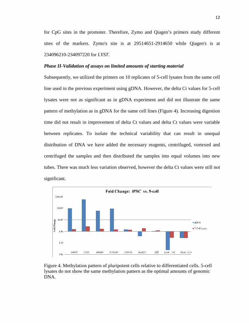
Phase II-Validation of assays on limited amounts of starting material
Subsequently, we utilized the primers on 10 replicates of 5-cell lysates from the same cell
line used in the previous experiment using gDNA. However, the delta Ct values for 5-cell
lysates were not as significant as in gDNA experiment and did not illustrate the same
pattern of methylation as in gDNA for the same cell lines (Figure 4). Increasing digestion
time did not result in improvement of delta Ct values and delta Ct values were variable
between replicates. To isolate the technical variability that can result in unequal
distribution of DNA we have added the necessary reagents, centrifuged, vortexed and
centrifuged the samples and then distributed the samples into equal volumes into new
tubes. There was much less variation observed, however the delta Ct values were still not
significant.
Figure 4. Methylation pattern of pluripotent cells relative to differentiated cells. 5-cell lysates do not show the same methylation pattern as the optimal amounts of genomic DNA.
13
Efficiency of digestion was controlled for by adding an internal positive control to each
sample. Low delta Ct values in the positive control indicate inefficient digestion on the
5-cell lysates (Table 2).
Table 2. Delta Ct values of samples with an internal positive control.
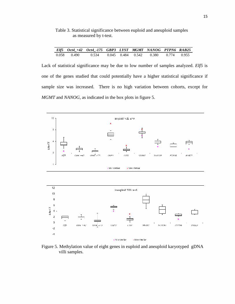
Lack of statistical significance may be due to low number of samples analyzed. Elf5 is
one of the genes studied that could potentially have a higher statistical significance if
sample size was increased. There is no high variation between cohorts, except for
MGMT and NANOG, as indicated in the box plots in figure 5.
Figure 5. Methylation value of eight genes in euploid and aneuploid karyotyped gDNA villi samples.
16
GBP3, LYST, MGMT and Elf5 gene promoters are expected to be hypermethylated in
pluripotenet cells and hypomethylated in differentiated cells. Gene promoters of
NANOG, PTPN6, RAB25 and Oct4 are expected to be hypomethylated in pluripotent
cells. Elf5 was reported to be more strongly expressed in the first trimester and was
down-regulated towards term (20). Consistent with this observation, our results indicate
hypomethylation in villi samples from first trimester in both cohorts (Figure 6). As
expected Oct4_-175 shows hypermethylation in villi samples (Figure 6). LYST, however,
although insignificant, unexpectedly shows a tendency toward hypermethylation, in the
samples analyzed (Figure 6).
17
0
1
2
3
4
20 30 40
delt
a C
T
Gestation Age (Days)
Elf5
0
1
2
3
20 30 40
delt
a C
T
Gestation Age (Days)
Oct4_+42
-1
0
1
2
3
4
5
20 30 40
delt
a C
T
Gestation Age (Days)
Oct4_-175
0
2
4
6
8
10
20 30 40
delt
a C
T
Gestation Age (Days)
GBP3
0
1
2
3
4
20 30 40
delt
a C
T
Gestation Age (Days)
LYST
02468
101214
20 30 40
delt
a C
T
Gestation Age (Days)
MGMT
0
1
2
3
4
5
6
7
20 30 40
delt
a C
T
Gestation Age (Days)
NANOG
0
1
2
3
4
20 30 40
delt
a C
T
Gestation Age (Days)
PTPN6
0
1
2
3
4
5
6
20 30 40
delt
a C
T
Gestation Age (Days)
RAB25
Figure 6. Relationship of euploid (blue diamond) or aneuploid (red circles) villi samples
to gestational age measured in days. Euploid samples show a statistically differential
methylation status within the GBP3 promoter compared to aneuploid samples.
*P < 0.05
18
LYST is hypomethylated in differentiated cells when compared to iPSCs (15).Our results
indicate hypomethylation of LYST in fibroblasts which represent differentiated samples
(Figure 3). However, in villi samples LYST shows a tendency toward hypermethylation
(Figure 6). LYST is a lysosomal trafficking regulator gene and was implicated to function
as an adaptor protein that affects proteins involved in intracellular membrane fusion
reactions (29).
Of the nine gene promoters studied, GBP3 shows differential methylation status between
euploid and aneuploid villi samples. As expected GBP3 gene promoter is hypomethylated
in differentiated cells. Results indicate hypomethylation in euploid samples versus
aneuploid samples.
Discussion
In this study, we have begun to develop a method to screen preimplantation embryos for
their reproductive potential using several candidate gene promoter methylation markers
of pluripotency. We characterized the methylation patterns in products of conception
using a set of gene promoter methylation sites. We found that euploid samples have a
statistically different methylation status within the GBP3 promoter compared to
aneuploid villi samples. The guanylate binding proteins (GBPs) belong to a family of
cytokine-induced GTPases and are located on chromosome 1 (30). Moon et al. (2011)
identified GBP3 as one of the genes that is upregulated in early stages of induced
abortion in mice (31). Methylation regulates gene expression and can be used as a
molecular marker for such. Our results indicate hypomethylation of the GBP3 gene
promoter in the euploid compared to aneuploid products of conception. In addition,
19
aneuploid samples in our study aborted at a later time point than euploid samples and
hypothetically they should have been more differentiated. GBP3 is a marker of
differentiation and should therefore be less methylated in tissue that is more
differentiated. Our outcomes indicate that aneuploid tissue possesses significantly more
methylation of the GBP3 gene promoter indicating that euploid tissue is better
differentiated. It is therefore possible to utilize GBP3 as a diagnosis marker of
differentiation level in embryos. However, we have only studied GBP3 in tissues from
spontaneous abortions making further experimentation necessary to determine the
applicability to preimplantation embryo reproductive potential.
Indeed, one of the original aims of this study was to characterize the methylation status of
products of conception on small amounts of starting material (i.e. trophectoderm biopsy)
from DNA left over from clinical genetic testing. We have noted that very low amounts
of DNA (30pg) do not behave in the expected manner as optimal amounts of DNA do.
This observation could be due to non-optimal ratio of enzyme to DNA concentration. The
observed outcome could also be due to the fact that cell line samples were at different
passage numbers (P25 and P32) when tested at optimal versus limited amounts of starting
material . It has been acknowledged previously that passage number does play a role in
the methylation status, where the number of differentially methylated regions
dramatically decreased from an early passage (P10 to P20) to late passage (P30 to P40).
(15). We have identified that using available excess DNA from leftover samples was not
possible and conclude that this is likely due to the composition of the lysis
reagents/protocol used rather than a limitation of the small amounts of starting material.
Unfortunately, modifying the lysis protocol was not feasible within the present study as it
20
will require considerable amounts of testing and validation, and acquisition of new
research materials. Alternatively, we utilized samples with large amounts of starting
material that have been prepared in a way that enzyme digestion is not inhibited. This
allowed us to study the methylation status in products of conception to identify possible
associations between the methylation status and the chromosomal normalcy status of
post-implantation samples.
Future studies will aim to characterize the methylation status of discarded embryos,
rather than from excess DNA from embryos used in clinical practice. This will provide an
opportunity to investigate alternative methods of lysis and represents an area of active
investigation in collaboration with Zymo Research Inc. Another area of investigation
stemming from the present study involves characterizing the genome wide methylation
status of the human preimplantation embryo. This project, unlike the present study, will
not be limited to evaluating a subset of targeted gene promoters (i.e. pluripotency gene
promoters). In addition, it may allow for the identification of differential methylation in
gene promoters which may have otherwise not been hypothesized to be of importance to
differentiation or reproductive potential in the human embryo.
21
References
1. Marikawa Y, Alarcon VB (2009) Establishement of trophectoderm and inner cell mass lineages in the mouse embryo. Mol Reprod Dev 76: 1019-1032.
2. Cross JC, Werb Z, Fisher SJ (1994) Implantation and the placenta: key pieces of the developmental puzzle. Science 266: 1508-1518.
3. Dean W, Lucifero D, Santos F (2005) DNA methylation in mammalian development and disease. Birth Defects Res 75: 98-111.
4. Antequera F (2003) Structure, function and evolution of CpG island promoters. Cell Mol Life Sci 60: 1647-1658.
5. Schultz MR (2002) The molecular foundations of the maternal to zygotic transition in the preimplantation embryo. Hum Reprod Update 8: 323-331.
6. Reik W, Dean W, Walter J (2001) Epigenetic reprogramming in mammalian development. Science. 293: 1089-1093.
7. Smith ZD, Chan MM, Mikkelsen TS, Gu H, Gnirke H, et al. (2012) A unique regulatory phase of DNA methylation in the early mammalian embryo. Nature 484:339-344.
8. Latham KE, Schultz RM (2003) Preimplantation embryo development. Fauser, B. C. J M., et al. eds. Reproductive Medicine: molecular, cellular, and genetic fundamentals. New York: The Parthenon Publishing Group, 421-438.
9. Sudheer S, Adjaye J (2007) Functional genomics of human preimplantation development. Briefings in Functional Genomics and Proteomics 6: 120-132.
10. Aplin JD (1991) Implantation, trophoblast differentiation and haemochorial placentation: mechanistic evidence in vivo and in vitro. J Cell Science 99: 681-692.
11. Fleming TP, Sheth B, Fesenko I (2001) Cell adhesion in the preimplantation mammalian embryo and its role in trophectoderm differentiation and blastocyst morphogenesis. Front Biosci 6: 1000-1007.
12. Malassine A, Frendo JL, Evain-Brion D (2010) Trisomy 21-affected placenta highlight prerequisite factors for human trophoblast fusion and differentiation. Int J Dev Biol 54: 475-482.
22
13. Roberts L, Sebire NJ, Fowler D, Nicolaides KH (2000) Histomorphological features of chorionic villi at 10-14 weeks of gestation in trisomic and chromosomally normal pregnancies. Placenta 21: 678-683.
14. Minguillon C, Eiben B, Bahr-Porsch S, Vogel M, Hansmann I (1989) The predictive value of chorionic villus histology for identifying chromosomally normal and abnormal spontaneous abortions. Hum Genetics 82: 373-376.
15. Nishino K, Toyoda M, Yamazaki-Inoue M, Fukawatase Y, Chikazawa E, et al. (2011) DNA methylation dynamicsin human induced pluripotent stem cells over time. PLoS Genetics 7: 1-14.
16. Kaneko T, Saito Y, Kotani T, Okazawa H, Iwamura H, et al. (2012) Dendritic cell-specific ablation of the protein tyrosine phosphatase Shp1 promotes Th1 cell differentiation and induces autoimmunity. J Immun 188: 5397-5407.
17. Wang H, Dey SK (2006) Roadmap to embryo implantation: clues from mouse models. Nature Reviews 7: 185-199.
18. Takeda J, Seino S, Bell GI (1992) Human Oct3 gene family: cDNA sequences, alternative splicing, gene organization, chromosomal location, and expression at low levels in adult tissues. Nucleic Acids Res 20: 4613-4620.
19. Pan G, Thomson JA (2007) NANOG and transcriptional networks in embryonic stem cell pluripotency. Cell Research 17: 42-49.
20. Hemberger M, Udayashankar, Tesar P, Moore H, Burton GJ (2010) Elf5-enforced transcriptional networks define an epigenetically regulated trophoblast stem cell compartment in the human placenta. Hum Mol Genetics 19: 2456-2467.
21. Calvanese V, Horrillo A, Hmadcha A, Suazer-Alvarez B, Fernandez AF, et al. (2008) Cancer genes hypermethylated in human embryonic stem cells. Proc Natl Acad Sci USA 3: e3294.
22. Papageorgiou EA, Fiegler H, Rakyan V, Beck S, Hulten M, et al. (2009) Sites of differential DNA methylation between placenta and peripheral blood molecular markers for noninvasive prenatal diagnosis of aneuploidies. Am J Pathol 174: 1609-1618.
24. Land JA, Evers JLH (2003) Risks and complications in assisted reproduction techniques: report of an ESHRE consensus meeting. Hum Reprod 18:455-457.
23
25. Cui X, Li H, Goradia TM, Lange K, Kazazian HH Jr., et al. (1989) Single-sperm typing: Determination of genetic distance between the G
�� –globin and parathyroid hormone loci by using the polymerase chain reaction and allele-specific oligomers. Proc Natl Acad Sci U S A 86: 9389-9393. .
26. Frebeerg CT, Dahl JA, Timoskainen S, Collas, P (2007) Epigenetic reprogramming of Oct4 and NANOG regulatory regios by embryonal carcinoma cell extract. Mol Biol Cell 18:1543-1553.
27. Melnikov AA, Gartenhaus RB, Levenson AS, Motchoulskaia AS, Levenson VV (2005) MSRE-PCR for analysis of gene specific DNA methylation. Nucleic Acids Res 33: e93.
28. Oakes CO, La Salle S, Trasler JM, Robaire B, (2009) Restriction digestion and real-time PCR (qAMP). Tost J ed. DNA methylation: methods and protocols 507: 271-280.
29. Tchernev TT, Mansfield TA, Giot L, Kumar M, Nandabalan K, et al. (2002) The Chediak-Higashi protein interacts with the SNARE complex and signal transduction proteins. Mol Med 8: 56-64.
30. Vestal DJ, Jeyaratnam JA, (2011) The Guanylate-binding proteins: Emerging insights into the biochemical properties and functions of this family of large interferon-induced guanosine triphosphtase. J Intereferon Cytokine Res. 31: 89-97.
31. Moon JM, Lee SE, Min YI, Jung C, Ahn KY et al. (2011) Gene expression profiling of mouse aborted uterus induced by lipopolysaccharide. Anat Cell Biol 44: 98-105.
24
DATE: 09/20/12
CURRICULUM VITAE
NAME: AGNIESZKA LONCZAK
Date of Birth October 17, 1982
Place of Birth Poland
ADDRESS: 321 South Street
Morristown, NJ 07960 201-321-5176
UNDERGRADUATE EDUCATION: Queens College-City University of New York Flushing, NY Bachelor of Arts/ Biology 2000-2005 GRADUATE CERTIFICATE: Montclair State University Montclair, NJ Graduate Certificate/Molecular Biology 2006-2007
PRINICPAL OCCUPATIONS: Maternal Fetal Medicine Associates, LLC. 2006-2006 Medical Biller Client Associated Businesses, Inc. 2006-2007 Clinical Data Associate Reproductive Medicine Associates of New Jersey, LLC. 2007-present Research Assistant
PUBLICATIONS:
Treff, N.R., Tao, X., Su, J., Lonczak, A., Northrop, L.E., Ruiz, A.A., Scott, R.T. Tracking embryo implantation using cell-free fetal DNA enriched from maternal circulation at 9 weeks gestation. Molecular Human Reproduction 2011; 17(7): 434-8. Patounakis, G., Treff, N.R., Tao, X., Lonczak, A., Scott, R.T., and Frattarelli, J.L. The p53 codon 72 single nucleotide polymorphism lacks a significant effect on implantation rate in fresh in vitro fertilization cycles: an analysis of 1,056 patients. Fertil Steril 2009; 92:1290-6.