Prediction of Enophthalmos by Computer-Based Volume Measurement of Orbital Fractures in a Korean Population. Ahn HB, et. al. Ophthalmic Plastic and Reconstructive Surgery. 2008 ; 24 : 36–9. Presenter: Che-Hao Chuang Professor: Dr. Yen-Ting Chen Date: 2009/12/08. Outline. Introduction - PowerPoint PPT Presentation
1 Prediction of Enophthalmos by Computer-Based Volume Measurement of Orbital Fractures in a Korean Population Ahn HB, et. al. Ophthalmic Plastic and Reconstructive Surgery. 2008 ; 24 : 36–9. Presenter: Che-Hao Chuang Professor: Dr. Yen-Ting Chen Date: 2009/12/08
Transcript
1
Prediction of Enophthalmos by Computer-Based Volume Measurement of Orbital
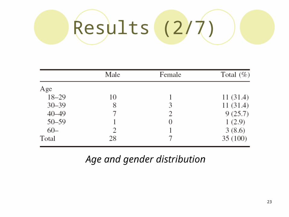
The patient's gender classification Men: 28 of 35 patientsWomen: 7 of 35 patients
The patient‘s age classificationMean age: 37.06 yearsLess than 50 years: 31 of 35 patients
23
Results (2/7)
Age and gender distribution
24
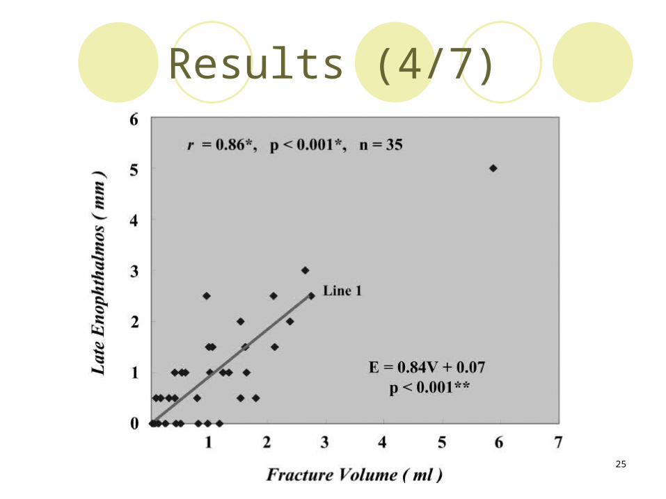
Results (3/7)
Fracture volume and enophthalmos
0 - 1.0 (ml) 54.3%
25
Results (4/7)
26
Results (5/7)
Mathematical formula of linear regression analysis:
E = enophthalmos (mm)
V = fractured site volume (ml)
SEE = standard error of estimate
It was used to calculate the expected degree of late enophthalmos.
27
Results (6/7)
Volume of the orbital fractureLess than 1 ml 0.91 mm2 ml 1.74 mm
Depth of enophthalmos2 mm 2.30 ml0.84 mm for every 1.0 ml increase in volume
28
Results (7/7)
Amount of late enophthalmos predicted from the fracture volume
Enophthalmos of 2 mm or more was predicted with an orbital fracture volume of 2.30 ml.
29
Discussions
30
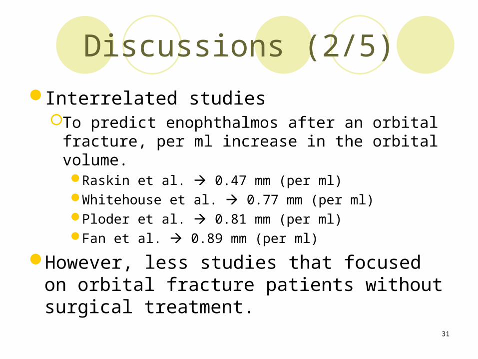
Discussions (1/5)
Why use CT?Many studies have mentioned
E.X. Gilbard et al.
Best means of observing the bone structure and soft tissues of the orbit.
It’s great help in predicting the prognosis of a patient with a fracture in the orbital floor.
31
Discussions (2/5)
Interrelated studiesTo predict enophthalmos after an orbital fracture,
per ml increase in the orbital volume.Raskin et al. 0.47 mm (per ml)Whitehouse et al. 0.77 mm (per ml)Ploder et al. 0.81 mm (per ml)Fan et al. 0.89 mm (per ml)
However, less studies that focused on orbital fracture patients without surgical treatment.
32
Discussions (3/5)
Attentive itemCompared with the orbital volume of the normal eye
A tilted head positioning during CT scanning
Normally, the volume between the right and left orbit may differ by approximately 7% to 8%.
33
Discussions (4/5)
In the present studyMeasuring the volume of the orbital fracture
site.Source: the consecutive coronal CTRapidia software
2D 3DArea and volume
34
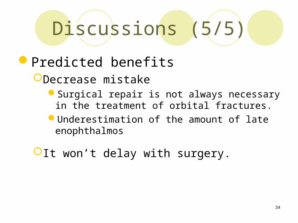
Discussions (5/5)
Predicted benefitsDecrease mistake
Surgical repair is not always necessary in the treatment of orbital fractures.
Underestimation of the amount of late enophthalmos
It won’t delay with surgery.
35
Conclusions
36
Conclusions (1/1)
A significant correlationThe fracture site volume and the degree of late
enophthalmos.
Predicting overall enophthalmos and provide useful information to surgeons.
37
Future works
38
Future works (1/1)
Proof of orbital symmetryFind out the relevant parametersIncrease the number of sampleStatistical verification
39
Reference
40
Reference (1/1)
Ahn HB, et. al. Prediction of Enophthalmos by Computer-Based Volume Measurement of Orbital Fractures in a Korean Population. Ophthalmic Plastic and Reconstructive Surgery. 2008 ; 24 : 36–9.