AJCC Staging Moments AJCC TNM Staging 7th Edition Melanoma Case #3 Contributors: Jeffrey E. Gershenwald, MD University of Texas MD Anderson Cancer Center, Houston, Texas Daniel G. Coit, MD Memorial Sloan-Kettering Cancer Center, New York, New York Charles M. Balch, MD Johns Hopkins Medical Center, Baltimore, Maryland
Transcript
AJCC Staging Moments
AJCC TNM Staging 7th Edition
Melanoma Case #3
Contributors: Jeffrey E. Gershenwald, MD University of Texas MD Anderson Cancer Center, Houston, Texas Daniel G. Coit, MD Memorial Sloan-Kettering Cancer Center, New York, New York Charles M. Balch, MD Johns Hopkins Medical Center, Baltimore, Maryland David R. Byrd, MD University of Washington Medical Center, Seattle, Washington
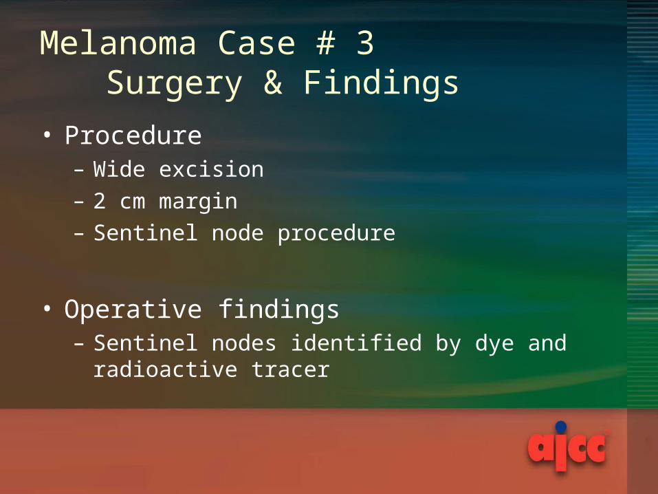
Melanoma Case # 3Presentation of New Case
• Newly diagnosed melanoma patient
• Presentation at Cancer Conference for treatment recommendations and clinical staging
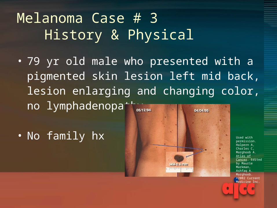
Melanoma Case # 3History & Physical
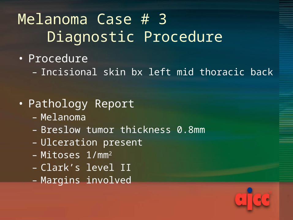
• 79 yr old male who presented with a pigmented skin lesion left mid back, lesion enlarging and changing color, no lymphadenopathy
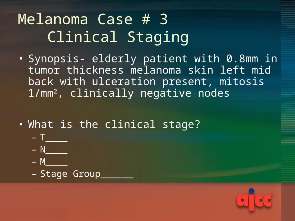
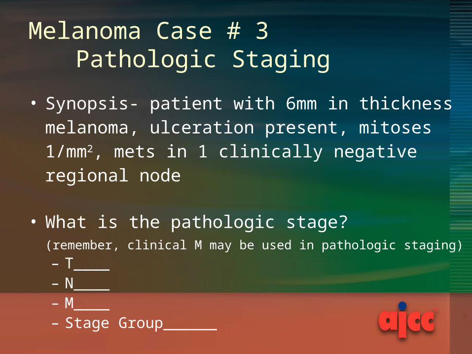
• Synopsis- elderly patient with 0.8mm in tumor thickness melanoma skin left mid back with ulceration present, mitosis 1/mm2, clinically negative nodes
• What is the clinical stage?– T____– N____– M____– Stage Group______
• Based on pathologic stage, there is more information to estimate prognosis and adjuvant treatment is selected
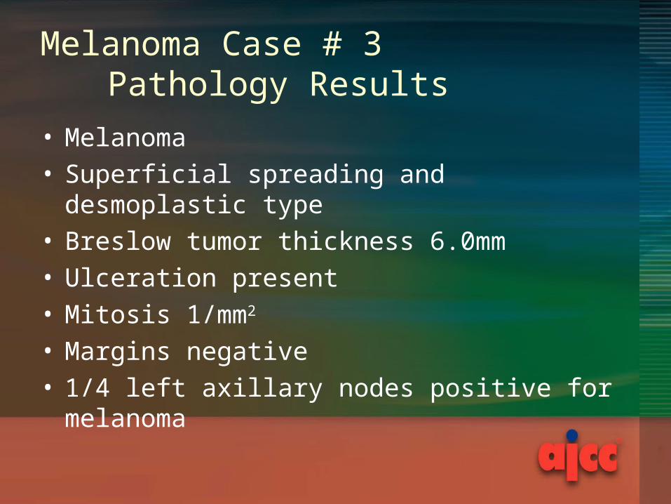
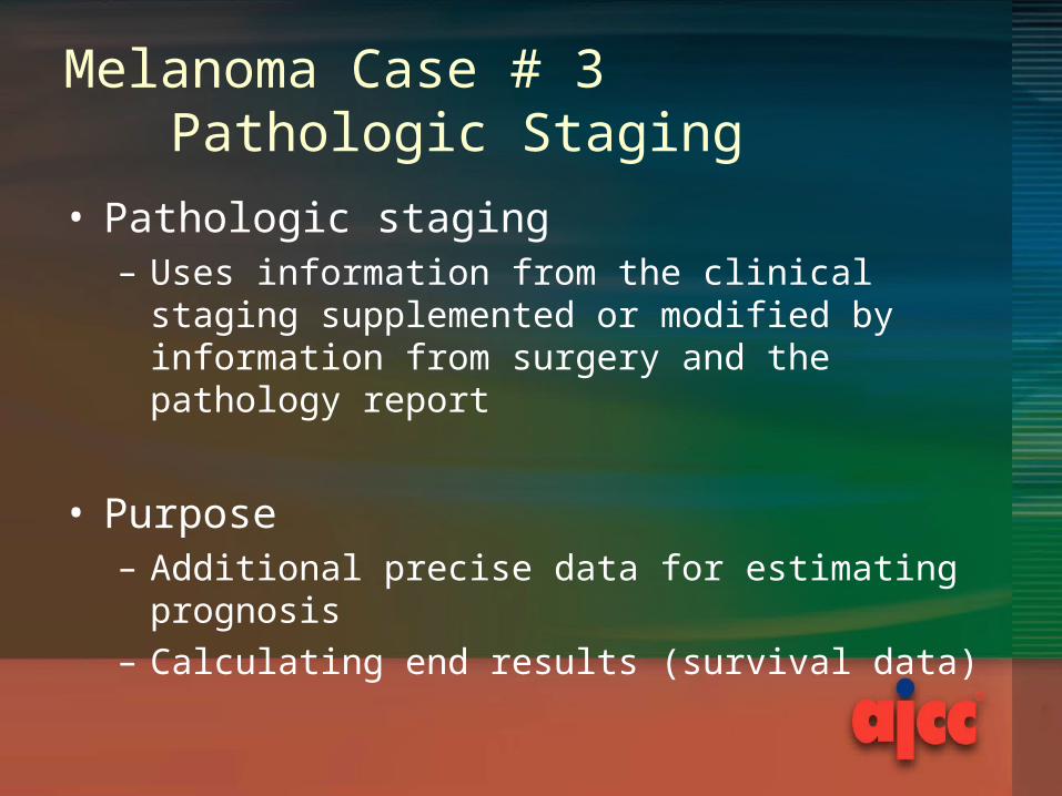
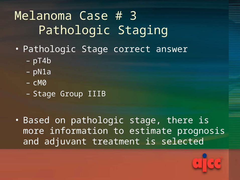
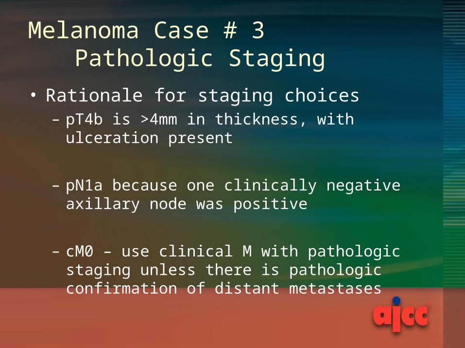
Melanoma Case # 3Pathologic Staging
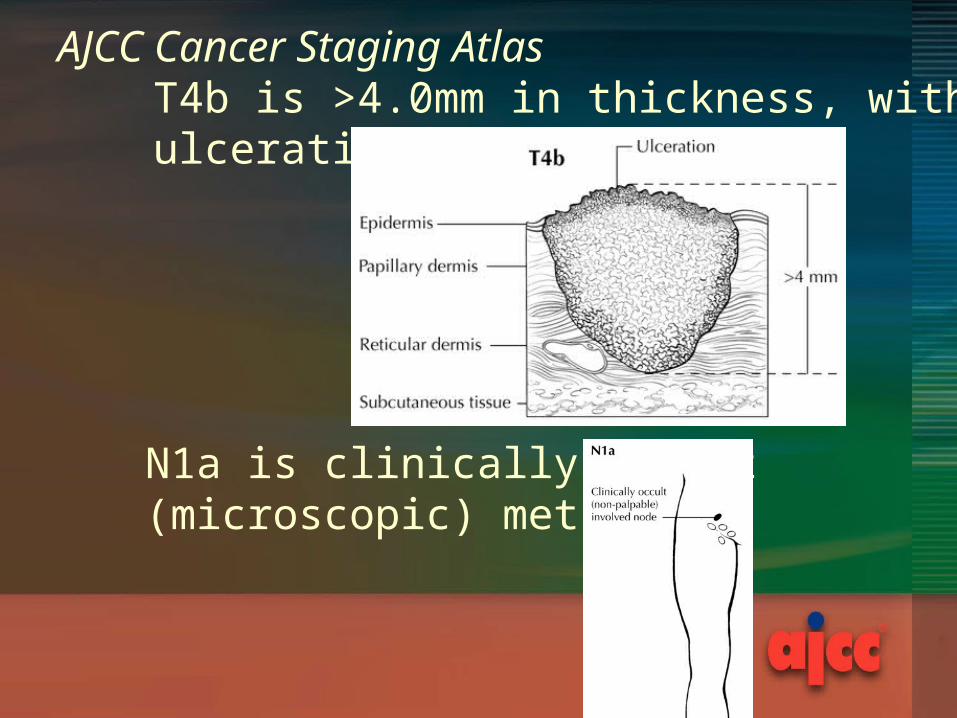
• Rationale for staging choices– pT4b is >4mm in thickness, with ulceration present
– pN1a because one clinically negative axillary node was positive
– cM0 – use clinical M with pathologic staging unless there is pathologic confirmation of distant metastases
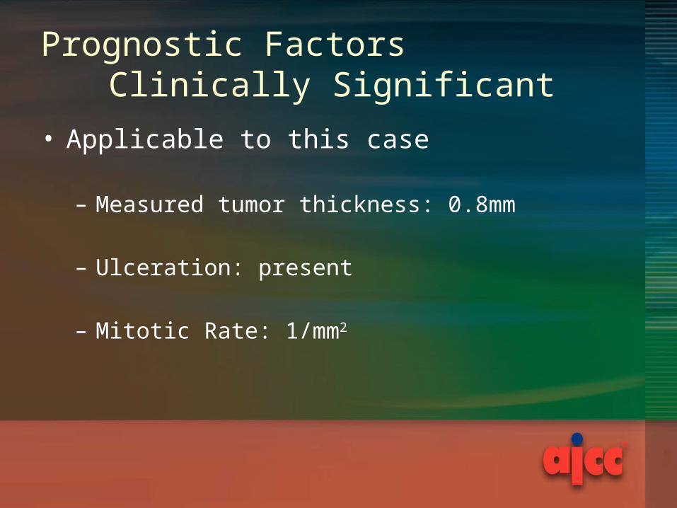
Prognostic FactorsClinically Significant
• Applicable to this case
– Measured thickness: 6.0mm
– Ulceration: present
– Mitotic Rate: 1/mm2
AJCC Cancer Staging AtlasT4b is >4.0mm in thickness, with
ulceration
N1a is clinically occult(microscopic) mets
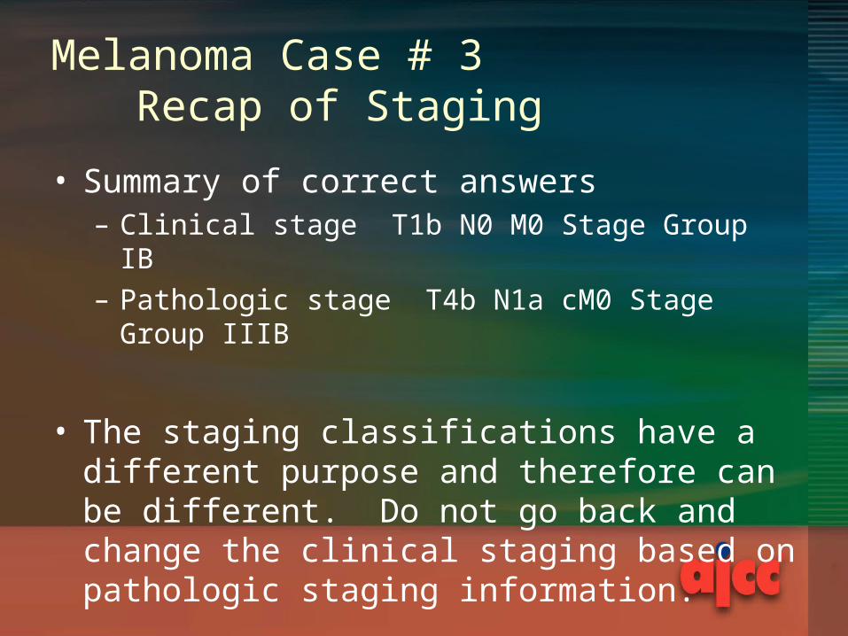
Melanoma Case # 3Recap of Staging
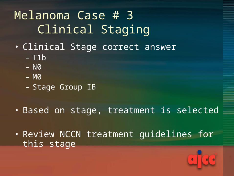
• Summary of correct answers– Clinical stage T1b N0 M0 Stage Group IB– Pathologic stage T4b N1a cM0 Stage Group IIIB
• The staging classifications have a different purpose and therefore can be different. Do not go back and change the clinical staging based on pathologic staging information.
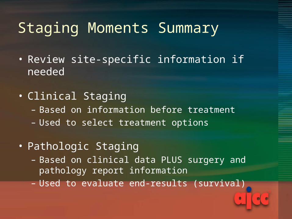
Staging Moments Summary
• Review site-specific information if needed
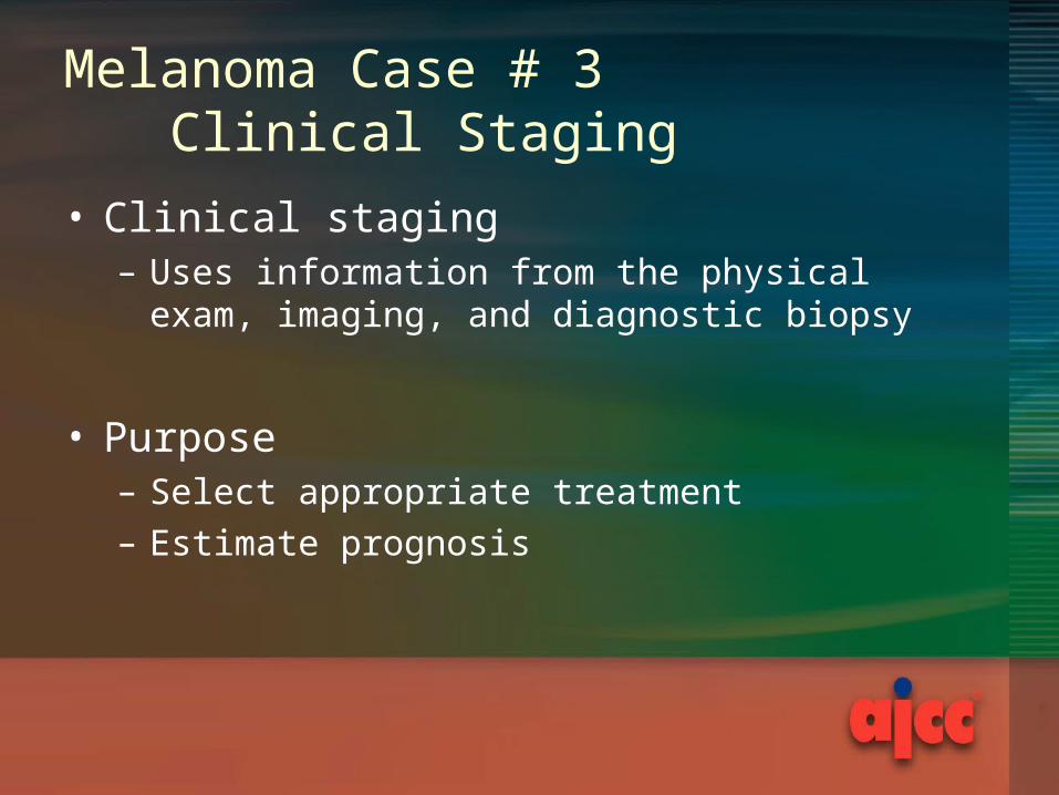
• Clinical Staging– Based on information before treatment– Used to select treatment options
• Pathologic Staging– Based on clinical data PLUS surgery and pathology
report information– Used to evaluate end-results (survival)


![[PPT]What is t,n,m staging and summary staging? Staging for... · Web viewWhat are we discussing? What is AJCC Staging Purpose of staging General rules for clinical and pathological](https://static.documents.pub/doc/80x56/5b1cc7cc7f8b9a8c5a8ba42e/pptwhat-is-tnm-staging-and-summary-staging-staging-for-web-viewwhat.jpg)