People in the Kashmir, like rest of the world are living longer with each passing decade and remain healthier and more active as they age. The prevalence of musculoskeletal problems in the population is increasing with longevity and may continue to increase over time. An increased morbidity by virtue of musculoskeletal disorders may compromise their quality of life and impose burden on health economics. The cost of treatment and inability to remain employed full time will have a significant financial impact on the economy of a resource constrained developing country. It warrants that trainee physicians should recognize and appreciate fully the importance of common musculoskeletal conditions. “…although the diseases that kill attract much of the public’s attention, musculoskeletal or rheumatic diseases are the major cause of morbidity throughout the world, having a substantial influence on health and quality of life, and inflicting an enormous burden of cost on health systems …” (World Health Organization 2003) Undergraduate Medical Education Programs are primarily responsible in Pakistan for preparing medical graduates/doctors to care for common clinical disorders prevalent in the country. Therefore, it is mandatory for medical schools to provide learning experiences that allow students to gain an appreciation of the importance of these conditions and the challenges inherent in caring for those patients. Traditional Medical Schools may not be accomplishing this educational goal since the attention paid to the conditions in the usual medical school curriculum is not commensurate with the prevalence of these conditions. We have developed LMR Module, which would provide appropriate learning experiences necessary for effective training of future physicians’ knowledge, skills, and attitudes relevant to musculoskeletal conditions that all medical students should acquire prior to graduation. This module has been designed to unfold the structural organization, functions, congenital anomalies and some of the disorders of the limbs and back. It explains the mechanism of neuromuscular transmission, its biochemical basis and the importance of Ca++ in the body along with neurotransmitters/drugs acting at this level. It also highlights the main components of primary survey in a trauma patient along with identification of common fractures of long bones on radiographs and examination of musculoskeletal system along with joint examination. Teaching methodology includes lectures, PBLs, SGD and demonstrations on models and dissection of the limbs along with teaching in Histology labs to enable the students to recognize different types of muscle and bone tissues under microscope.
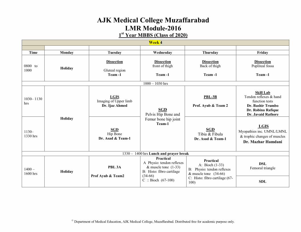
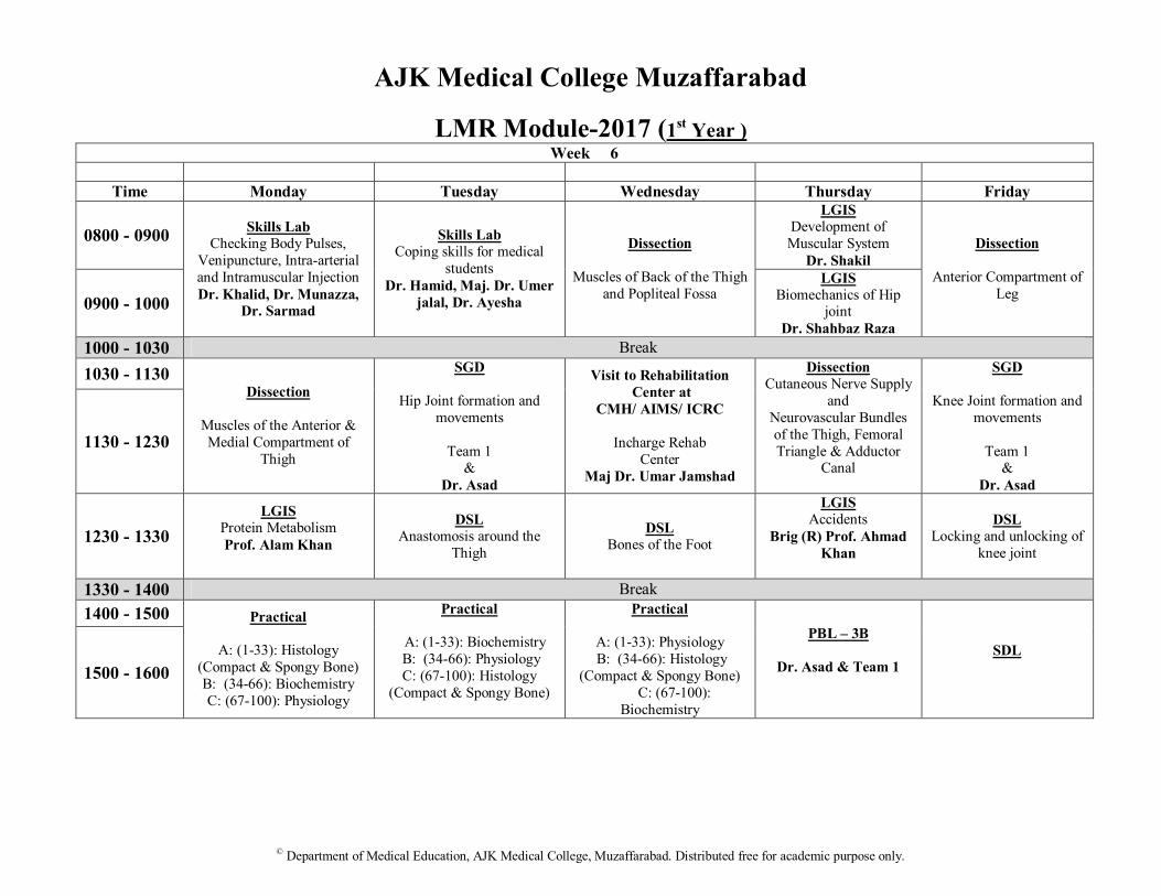
Teaching Strategy The content of this module will be delivered by a combination of different teaching strategies. These include small group discussions (SGD), large group interactive sessions (LGIS), demonstrations in dissection hall, lab practical, general club and clinical skill sessions at skill lab. Group projects will be assessed at the end of the block. Organization of Module The module consists of seven themes, and 8 PBLs each based on a real life situation. Each theme has its explicate LOs. The module will employ different modes of instruction, briefly described below. Major emphasis will be on discussion, analysis and deductions; all by the students and guided by the faculty. Content Delivery Entire curriculum will be delivered by clinical case scenarios each covering a theme. Read the cases and the objectives of the theme which you are supposed to encounter next day, understand and explain the case to yourself and read the relevant information. Following learning/teaching strategies will be employed to discuss the cases: Small Group Discussion Main bulk of the course content will be delivered in small group sessions. Each theme has an associated case. The case will be the centre around which learning will take place. Depending on the case you might be required to deduce objectives and learning issues or only learning issues. Every group will have a facilitator assigned to it. The facilitator will be there to keep you on track, giving you maximum liberty to discuss and achieve the objectives as a group. Small groups will be followed by a wrap up session to standardize learning. Rest of the information will be in the schedule/ time table. Large group Large group instruction will be employed at times sparingly. Attend large group sessions with the following focus a. Identify important points b. Ask questions on concepts not well understood in the text books c. Measure your learning comprehension Videos Video demonstrations on dissection history taking and clinical examination, will be shown to give you an idea into the disease process, clinical testing and practical aspect of communication with the patients. Hands-on Activities/ Practical Practical activities, linked with the case, will take place. Lab: Attend your scheduled lab and take advantage of free time for study .Use your labs to correlate text structures to actual specimens in lab practice.
Self Directed Learning (SDL) / Directed Self Learning (DSL) A few SDLs and DSLs have been added in between to create an environment for you to search literature as well as to deduce and synthesize information from different sources to meet the learning objectives. It will also help in breaking the monotonous / strenuous schedule and make you life- long learner. Assessment In this 6-weeks duration module, you will have formative surprise quizzes/intermittent short tests. A full-fledged summative assessment will be conducted at the end of module. This will give you an idea about the format of the examination to be held at the end of the year. This will be followed by feedback on your performance in the exam. Marks obtained in the module examination will contribute 30% (internal assessment) towards end of year Professional University Examination. There is no re-sit exam for module written assessment and block IPE. If you miss them, your internal assessment will be recorded as zero. No excuse of any kind is permissible for absence in module or IPE assessment.
Table of Specifications
Sr.# Theme % Weightage
1 Skeletal supports & Body movements
30
2 Short Limbs in Locomotive apparatus & consequent dysfunctions 10
By the end of session, the students must be able to: 1. Classify muscles of upper and lower limbs according to their functions; and give origin,
insertion and nerve supply of each group. 2. Describe Histological structure of bone and cartilage 3. Identify the type of bone on given slides. 4. Interpret/illustrate histological features of Cartilage and Bone. 5. Describe biochemical composition of bone; Bone matrix, minerals, mucopolysaccharide
and proteoglycans. 6. Illustrate microscopic features of bone and cartilage. 7. Correlate structural organization of human body with locomotion. 8. Identify each of the bones of upper and lower limbs and point out their important features
with demonstration on bones. 9. Correlate important fractures of limb bones to their clinical manifestations and
radiological findings. 10. Demonstrate range of movement of upper and lower limb joints and correlate with
muscles responsible for the said movements 11. Differentiate various fractures of upper and lower limb bones and relate with clinical
manifestations. 12. Describe the fasciae in the upper and lower limbs (including retinaculae). 13. Describe the facial spaces of upper and lower limbs. 14. Identify dislocations of limb joints and correlate with their clinical manifestations on
patients/SP/video/X-ray 15. Describe the blood and nerve supply of shoulder, elbow, hip and knee and ankle joints;
and the mechanics of all large joints in the limbs with emphasis on the special features that help to maintain their stability and movements.
16. Demonstrate movements of the hand and foot joints (with movements of fingers and toes) in relation to the insertions and actions of the muscles and tendons which affect them.
17. Describe/demonstrate the arches of the foot and their shock absorption role in walking, jumping & running.
18. Identify components/parts of clinically important bones of limbs on imaging modalities. 19. Outline plan for taking history and performing examination of Musculoskeletal system on
SP/peer. 20. Demonstrate cultural sensitivity while taking history and performing examination of the
Musculoskeletal system on SP/peer of opposite gender.
2. Short Limbs & consequent dysfunctions 1. Describe general and special connective tissues, giving examples of each. 2. Interpret different steps in synthesis of collagen and its role in health and disease. 3. Draw and label Connective & Fibrous Tissues, Cartilage and Bone as on Light
Microscope. 4. Compare and contrast different types of collagen fibers and their location in different
body tissues. 5. Describe Embryonic and fetal development of limbs along with their congenital
anomalies. 6. Describe the development of muscles. Correlate the process of osteogenesis and
myogenesis to locomotor apparatus. 7. Describe Embryonic and fetal development of bones. 8. Compare and contrast metaphysis, epiphysis and diaphysis. 9. Describe postnatal growth stages of bone. 10. Explain the biochemical factors affecting bone formation and growth. 11. Describe the role of vitamin D, Parathormone and calcitonin in regulating the calcium
metabolism. 12. Interpret role of calcium and phosphorus in bone mineral homeostatsis. 13. Correlate biochemical basis of Rickets and osteomalacia on SP/video/lab
investigations/Xray. 14. Identify osteogenic imperfecta and developmental dysplasia of hip (DDH) on
radiographs/pictures/videos. 15. Identify and interpret congenital anomalies of the limbs and correlate with their
embryological basis. 16. Describe Radiological findings in osteomyelitis, & osteomalacia.
Theme 3: Painful Joints
1. Describe special features of main/major joints of upper and lower limbs. 2. Identify/illustrate components of clinically important joints on imaging modalities. 3. Illustrate and differentiate immunological manifestations of Rheumatic and rheumatoid
arthritis in limb joints. 4. Interpret biochemical basis of immunological and degenerative arthritis and correlate
with their clinical presentation. 5. Compare and contrast gouty arthritis from traumatic and rheumatoid arthritis. 6. Compare morphological and histological findings of osteoarthritis, Rheumatic,
Rheumatoid, metabolic, infectious and neoplastic arthritis. 7. Outline plan for taking history of joint disorder and the important components of their
examination. 8. Council the patient suffering from immunological and/or degenerative arthritis for
4. Myopathy 1. Correlate the histological features to the molecular basis of skeletal muscle contraction. 2. Describe different phases of action potential in skeletal and smooth muscle cells. 3. Illustrate and interpret the various components of the neuromuscular junction in skeletal
and smooth muscles. 4. Interpret the role of neurotransmitters in transmission of nerve impulse at the
neuromuscular junction. 5. Illustrate the muscle spindle and Golgi tendon organs and correlate with their role in
tendon reflexes and muscle tone. 6. Enumerate the various components of the skeletal muscle fibers. Interpret the role of
calcium in the walk along theory of muscle contraction 7. Enlist the steps in excitation contraction coupling. 8. Demonstrate isotonic and isometric contraction on yourself/SP/video. 9. Identify the histological features of skeletal muscles on light microscope. 10. Differentiate between the fast glycolytic and slow oxidative fiber types in skeletal
muscles. Correlate their uses in sprinters and marathon runners. 11. Interpret/Correlate hypertrophy and atrophy of muscle with its use. 12. Discuss the Biochemical and Physiological basis of different Myopathies (myasthenia
gravis, polio, Guillain-Barre syndrome, upper and lower motor neuron lesions) and correlate with their clinical presentation.
13. Differentiate between UMNL and LMNL. 14. Illustrate/interpret the various steps of protein metabolism [Anabolism/Catabolism] in
health and disease.
5. Painful Swollen limb
1. Correlate the venous and lymphatic drainage of upper and lower limb to their clinical significance.
2. Explain the organization of arterial system supplying the limbs and vertebral column. 3. Identify major veins of upper and lower limb on cadaver/peers/ simulated patients. 4. Mark the surface anatomy of the major arteries of upper and lower limb on
SP/cadaver/angiogram. 5. Mark the surface anatomy of the major veins of upper and lower limb on
SP/cadaver/venogram. 6. Demonstrate intra-arterial, intra-venous injection on mannequin. 7. Demonstrate venepuncture on mannequin.
6. Numb limb
1. Describe the origin, formation and distribution of the brachial plexus highlighting their important relations and neurological deficits.
2. Describe the origin, course, clinically important relationships and distribution of the axillary, radial, median and ulnar nerves in the upper limb.
3. Describe the origin and important relationships of lumbo- sacral plexuses. 4. Describe the origin, course, relationships and distribution of the sciatic, femoral, common
peroneal, deep and superficial peroneal, and sural nerves in the lower limb. 5. Indicate the nerve supply of the muscles of the upper and lower limbs and the effect of
their denervation on movement and action across joints. 6. Describe the origin, course, branches and relationships of the sciatic,Tibial, Peroneal,
obturator and femoral nerves; and the distribution of their important branches in the leg and foot.
7. Identify Anatomical land marks and mark surface Anatomy of major nerves in the upper and lower limbs.
8. Correlate the common injuries of brachial & lumbosacral plexus to their clinical presentation.
9. Demonstrate the integrity/examination of clinically important upper and lower limb nerves.
10. Demonstrate/illustrate dermatomes of UL & LL on manikin/ SP/cadaver.
7. Hunched Back 1. Describe the development of vertebral column and axial musculature, giving their
common anomalies. 2. Differentiate various muscles responsible for movements of back. 3. Differentiate Vertebrae in different segments of vertebral column. 4. Identify specifically the atlas and the axis and the characteristic features which
distinguish cervical, thoracic and lumbar vertebrae on anatomical specimen. 5. Describe the structure and function of intervertebral discs and vertebral ligaments. 6. Using diagrams or dissected specimens, identify the muscles which support and act upon
the vertebral column. 7. Identify on the bones, the features typical to all vertebrae and those unique to each type
of vertebra (including sacrum and coccyx) 8. Identify primary and secondary curves of spine. 9. Differentiate abnormal curvatures of spine and correlate with their
Clinical Case history 35 years old lady presented to orthopaedic outpatient clinic of AIMS with the complaints of pain in right shoulder with decreased movements on this joint. She also gave history of house decoration and dusting in weekend. No history of trauma or any major illness in the past. The patient denies any significant drug or environmental allergies. There is no history of neck pain, numbness in the right upper limb and restriction of neck movements Examination: GPE: PULSE 76/min BP 120/85 Temp 98F Respiratory rate : normal Pallor –ve Cyanosis-ve Clubbing –ve Local examination: Tenderness in sub-acromial region is positive. There is full range of movement at the right shoulder joint, but there is pain during abduction from 60o to 120o. Decreased ROM (active) in right shoulder joint especially abduction and forward flexion. Systemic review was unremarkable.
Scenario-2
A 64-year-old, right-handed, retired woman presents with intermittent numbness, tingling, and burning pain in the three radial digits of both hands. She has had these symptoms for three months, which awaken her several times each night. She has no atrophy of the thenar muscles. Sensation to light touch is intact. How should she be evaluated and treated? Her symptoms were exaggerated with repetitive activities of the hand and wrist, particularly with a combination of forceful and repetitive activities. Nerve conduction studies worsened over time. Critical Questions:
1. Why we need skeletal supports and joints in human body? 2. What are components of axial and appendicular skeleton in human body? 3. What constitutes the rotator cuff and how it facilitates ROM in shoulder joint? 4. What is a bursa? Why it gets inflamed? 5. What factors/structures provide stability to shoulder joint? 6. What is the relationship between stability and range of movement (ROM) at any synovial
joint? 7. What is a frozen shoulder? 8. Why shoulder joint injury is more common in comparison to that of hip joint? 9. How would you interview, investigate and manage such cases in your real life practice? 10. What is the prevalence of such disorders in our local population?
11. Demonstrate range of movement of shoulder joint and correlate with muscles responsible for the said movements.
12. Describe the blood and nerve supply of shoulder joint with emphasis on the special features that help to maintain the stability and movements.
13. Which imaging modalities are frequently used for Shoulder joint investigations? 14. What is arthroscopy? Does it have any role in traumatic shoulder arthritis? 15. What kind of implants is used to treat the diseased bones and joints in human being? 16. What are the consequences of immobilization of fractured bones and/or joints?
Common Injuries for the Major Joints During medical school, students should be taught to understand and assess some of the more common injuries for the major joints. • Neck: mechanical/myofascial neck pain, cervical radiculopathy, (cervical myelopathy) • Shoulder: rotator cuff tendinitis/opathy, AC joint problems (arthritis, separation), anterior dislocation, biceps tendinitis/rupture • Elbow: lateral and medial epicondylitis, radial head fracture, olecranon bursitis • Wrist: DeQuervain's tenosynovitis, carpal tunnel syndrome, scaphoid fracture • Hip: trochanteric bursitis, osteoarthritis • Lumbar spine: mechanical low back pain, lumbar radiculopathy, lumbar spinal stenosis, lumbar spondylolysis, and listhesis • Knee: ligament sprains, including anterior cruciate ligament, meniscal tears, patellofemoral pain • Ankle/foot: inversion sprains, Achilles tendinitis, plantar fasciitis
The “Anatomy” of a 3-minute Clerk’s Musculoskeletal Examination in the Routine Screening Physical Examination
The musculoskeletal exam can be divided into three broad sections: visual inspection, palpation, and the evaluation of joint motion (LOOK, FEEL and MOVE.) First, the examiner evaluates the patient visually for signs of deformity, swelling, scars, inflammation or muscle atrophy. Second, surface anatomy landmarks are used to evaluate for localization of points of tenderness or fluid collection. Third, involved joints are moved actively by the patient, then passively by the examiner. If indicated, stress maneuvers are performed on joints in order to evaluate stability. By means of an orderly physical assessment of the axial and the appendicular skeleton, an anatomically detailed picture of musculoskeletal injury or illness is discerned. The musculoskeletal physical examination is performed following elicitation of a chief complaint and a history of the presenting condition, along with a review of systems, past medical and surgical history, review of medications and drug allergies, and a social history.
Similar Symptoms, Different Diagnosis Along the Age Spectrum Age is an important variable to consider when diagnosing a musculoskeletal condition. What more commonly manifests in the first two decades of life will not necessarily be the same problem that is common for adults. For example: When a patient presents with hip pain, common underlying conditions include, for an • 80 year-old, Fractured hip • 50 year-old, Osteoarthritis or disc disease • 25 year-old, Bursitis • 7–11 year-old, Osteonecrosis or slipped capital-femoral epiphysis
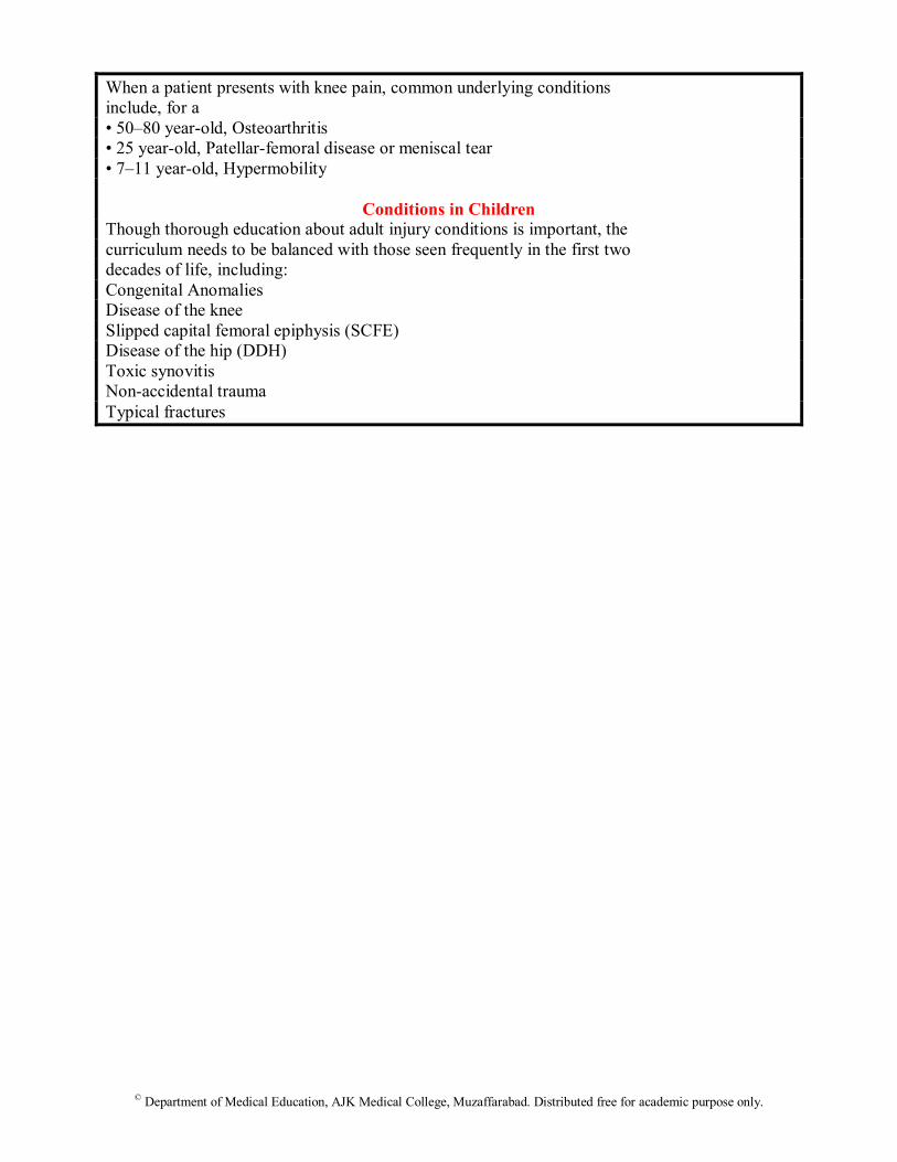
When a patient presents with knee pain, common underlying conditions include, for a • 50–80 year-old, Osteoarthritis • 25 year-old, Patellar-femoral disease or meniscal tear • 7–11 year-old, Hypermobility
Conditions in Children
Though thorough education about adult injury conditions is important, the curriculum needs to be balanced with those seen frequently in the first two decades of life, including: Congenital Anomalies Disease of the knee Slipped capital femoral epiphysis (SCFE) Disease of the hip (DDH) Toxic synovitis Non-accidental trauma Typical fractures
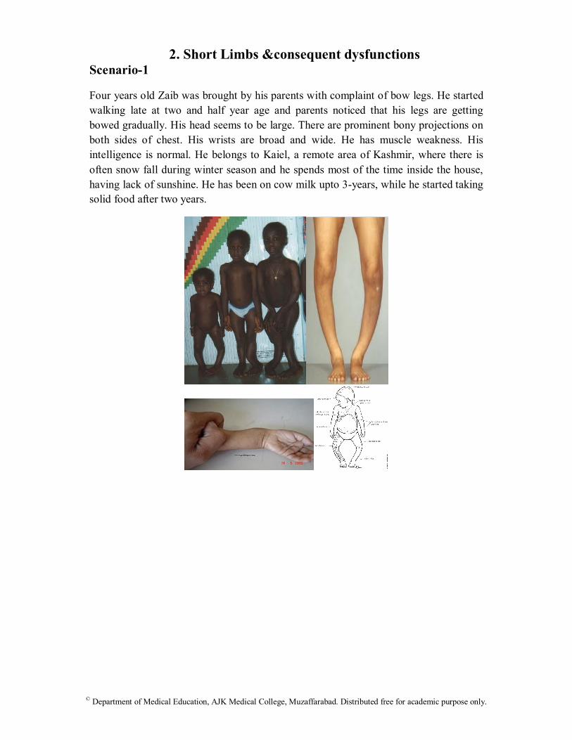
2. Short Limbs &consequent dysfunctions Scenario-1
Four years old Zaib was brought by his parents with complaint of bow legs. He started walking late at two and half year age and parents noticed that his legs are getting bowed gradually. His head seems to be large. There are prominent bony projections on both sides of chest. His wrists are broad and wide. He has muscle weakness. His intelligence is normal. He belongs to Kaiel, a remote area of Kashmir, where there is often snow fall during winter season and he spends most of the time inside the house, having lack of sunshine. He has been on cow milk upto 3-years, while he started taking solid food after two years.
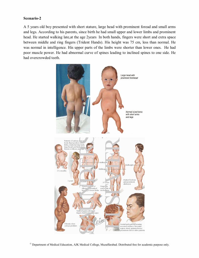
A 5 years old boy presented with short stature, large head with prominent foread and small arms and legs. According to his parents, since birth he had small upper and lower limbs and prominent head. He started walking late,at the age 2years In both hands, fingers were short and extra space between middle and ring fingers (Trident Hands). His height was 75 cm, less than normal. He was normal in intelligence. His upper parts of the limbs were shorter than lower ones. He had poor muscle power. He had abnormal curve of spines leading to inclined spines to one side. He had overcrowded teeth.
A 38-year-old woman with a five-year history of rheumatoid arthritis had complete remission after 17 months of weekly monotherapy with 4 mg of methotrexate. Radiographs of both hands and feet were taken to assess the effectiveness of the treatment. In contrast to the radiograph that had been taken one year before the initiation of treatment, healing of the erosions at the metatarsal head and at the base of the proximal phalanx in the fifth metatarsophalangeal joint of the right foot were observed. The patient’s symptoms in her hands, wrists, and feet (including joint pain, tenderness, swelling, and redness) had begun to improve during the first six months after the initiation of therapy. Her only remaining symptom was a limitation in the range of motion of both wrists. Typically, the goal of treatment of rheumatoid arthritis is a reduction in the pace of the destructive process. Rarely, as was noted in this case, healing of arthritic changes in bone may occur, especially in patients treated with methotrexate or, recently, with an anti–tumor necrosis factor antibody.
When Rheumatoid Arthritis Entered My Life: A Personal Narrative
It has been 12 years since being diagnosed with rheumatoid arthritis. I was 29, with three young children ages 9, 12, and 14, working full time as a dietician and going to nursing school. The swelling in my hands wouldn’t go away. I lost weight, my body was always sore, and I was always tired. I was in denial and didn’t understand what was happening to me. I felt depressed, but knew I had to keep going every time I looked at my kids. I was exhausted but I kept a smile on. My kids were preteens. It was impossible for me even to take them to the mall because I was too weak and tired. I called family or neighbors and asked them to drive my car so my kids could go to activities. I kept secrets. I withdrew from nursing school and changed my phone number. I didn’t want my friends to see my condition. I didn’t date. I thought, “Who would possibly want to date me?” I was ashamed of how I looked— my hands, and scars from surgeries. I wore long-sleeved shirts so the scar from my elbow replacement wouldn’t show. I was in financial turmoil because I had to stop work. And I couldn’t afford my medicines. I was told to bring in receipts so that I could apply for spin down. I just couldn’t—my body was always sore and I was always tired. Eventually I entered some drug studies, received medications through the studies, and improved. I had always loved to work out but I gave up going to the gym. Even now I miss running and speed walking with friends. As my daughters matured, they took control of the house, the shopping, and the banking. My older daughter who volunteered at a local children’s hospital saw a child with rheumatoid arthritis. She was scared. Was it hereditary and would she get it? I really needed a job. I tried to find one I could handle but found myself turning down offers saying, “I’m sorry. I really don’t think I can do that job.” Or being turned down because I was overqualified. Two years after my diagnosis and after reconstructive surgery on my left foot, I returned to work—this time in airport security. My feet hurt— the only comfortable shoes were clogs. The job required black boots or shoes. I explained about my rheumatoid arthritis and was transferred to an office position. My friends found out about my rheumatoid arthritis. They piled in and out of the house and refused to stay away. “You need us,” they told me. My kids are interested in health-related fields. I‘m sure the life we have led has been a factor. My older daughter is a paramedic. My middle daughter is in college completing pre-med requirements. Eventually I returned to nursing school. I knew it was something I really wanted to do. I did finish my program and received my R.N. degree. I just wanted to have that degree—to have it on my wall and to know that I accomplished something. It was in my soul. —Anonymous (Abstract derived from AAMC Publications for teaching purpose only)
Scenario-1 39 years old lady presented with intermittent muscle weakness in her face for the past three months. She complains that swallowing has become difficult. She also noted double vision (diplopia) which becomes worse at night and improves during the day. On physical examination, she has notable ptosis (“drooping”) of both eyelids after repeated blinking exercises. Electromyographic (EMG) testing suggested progressive weakness of distal arm muscles. Both her symptoms and electromyographic findings were reversed within 40 seconds of intravenous administration of acetylcholinesterase inhibitor. Blood testing revealed high levels of an anti-acetylcholine receptor antibody in her plasma, and a diagnosis of myasthenia gravis was made.
THEME 5: Painful Swollen limb
Humaira Kiyani, a 35-year old City School teacher ,presented in OPD at AIMS Hospital with H/O dull ache in both lower limbs for the last four months. There is no H/O any medical disorder. Examination: Pulse 80/min Bp 110/70 mm of hg She is obese. There are multiple dilated tortuous veins in both lower limbs. The skin on shins is hyperpigmented. Upon pressing the Sephno-femoral junction, there is no refilling (Tourniquet test). Peripheral pulsations are well felt, hand held Doppler revealed good Doppler signals. A venous Doppler was advised by Consultant Surgeon, and the radiologist reported that the Sephano-femoral junction on both side is incompetent. There are few perforators too below the knee joint. Venogram confirmed incompetent perforators. After blood investigations, surgery was planned.
Scenario-2 Maria Jan, a 48-year old NBP Executive, was admitted in Sheikh Khalifa Bin Zaiyed Hospital. Her Hystrectomy was done last week. Now she is complaining of painful swelling of the left leg. Examination: Pulse: 95/min BP 120/80 mm of hg Temp:37.5 Abdominal wound is clean. There is diffuse swelling of left leg. The skin is shining. Peripheral pulsations are well felt. Calf tenderness is positive. Upon forcible dorsiflextion of foot (Homen’s sign), the pain gets aggravated (though this test is not encouraged now a days).Venous Doppler suggests a big thrombus in the femoral vein.
Theme 6: Numb limb
Scenario-1 A young man while driving a car without seat belt had a head on collosion with another car sustained whiplash injury to his neck resulting in weakness of all four limbs, pain neck and retention of urine. On examination he is conscious with shallow rapid abdominal breathing, anxious, cold clammy skin with blood pressure 80/60, pulse is 60 per min. He is paralyzed in all his four limbs with power G 0/5. He has loss of pin prick sensation from C6 and below. Scenario-2 A young soldier involved in bomb blast received splinter injury to right side of his neck. He developed numbness over the lateral side of upper half of the upper arm and he is unable to abduct and externally rotate his right shoulder.
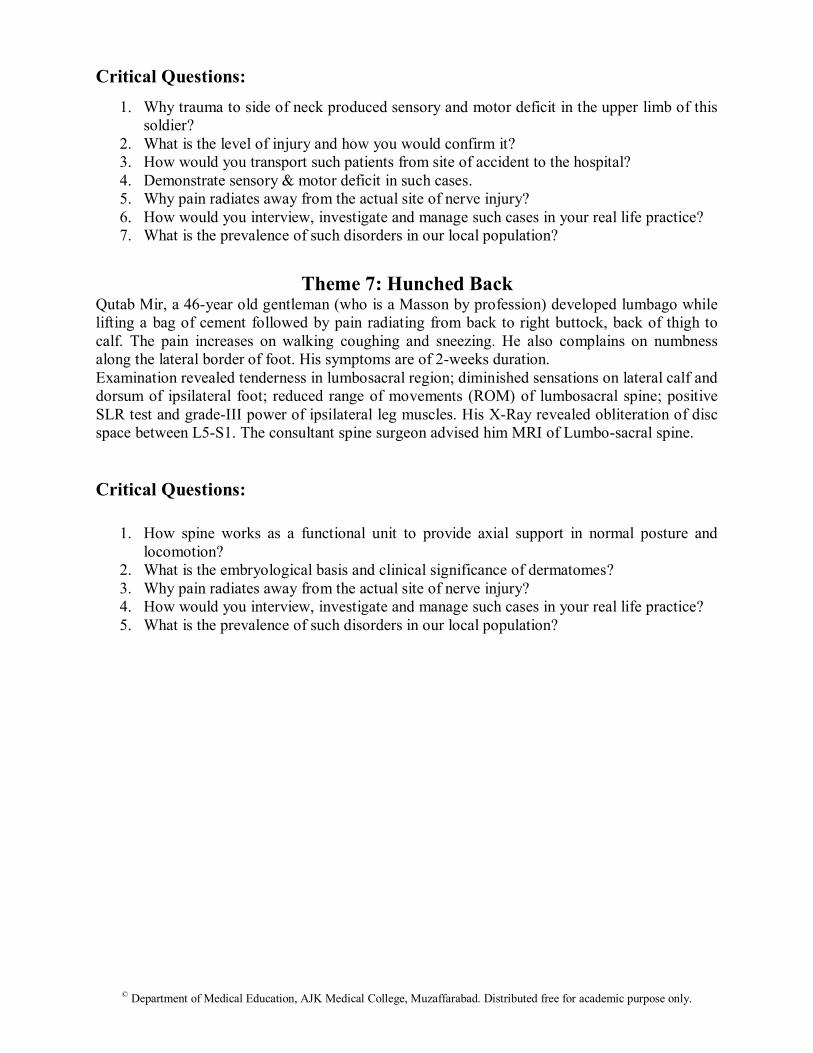
Critical Questions: 1. Why trauma to side of neck produced sensory and motor deficit in the upper limb of this
soldier? 2. What is the level of injury and how you would confirm it? 3. How would you transport such patients from site of accident to the hospital? 4. Demonstrate sensory & motor deficit in such cases. 5. Why pain radiates away from the actual site of nerve injury? 6. How would you interview, investigate and manage such cases in your real life practice? 7. What is the prevalence of such disorders in our local population?
Theme 7: Hunched Back Qutab Mir, a 46-year old gentleman (who is a Masson by profession) developed lumbago while lifting a bag of cement followed by pain radiating from back to right buttock, back of thigh to calf. The pain increases on walking coughing and sneezing. He also complains on numbness along the lateral border of foot. His symptoms are of 2-weeks duration. Examination revealed tenderness in lumbosacral region; diminished sensations on lateral calf and dorsum of ipsilateral foot; reduced range of movements (ROM) of lumbosacral spine; positive SLR test and grade-III power of ipsilateral leg muscles. His X-Ray revealed obliteration of disc space between L5-S1. The consultant spine surgeon advised him MRI of Lumbo-sacral spine.
Critical Questions:
1. How spine works as a functional unit to provide axial support in normal posture and locomotion?
2. What is the embryological basis and clinical significance of dermatomes? 3. Why pain radiates away from the actual site of nerve injury? 4. How would you interview, investigate and manage such cases in your real life practice? 5. What is the prevalence of such disorders in our local population?
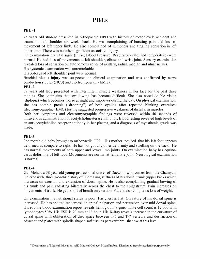
25 years old student presented in orthopaedic OPD with history of motor cycle accident and trauma to left shoulder six weeks back. He was complaining of bursting pain and loss of movement of left upper limb. He also complained of numbness and tingling sensation in left upper limb. There was no other significant associated injury. On examination his vital signs (Pulse, Blood Pressure, Respiratory rate, and temperature) were normal. He had loss of movements at left shoulder, elbow and wrist joint. Sensory examination revealed loss of sensation on autonomous zones of axillary, radial, median and ulnar nerves. His systemic examination was unremarkable. His X-Rays of left shoulder joint were normal. Brachial plexus injury was suspected on clinical examination and was confirmed by nerve conduction studies (NCS) and electromyogram (EMG). PBL-2 39 years old lady presented with intermittent muscle weakness in her face for the past three months. She complains that swallowing has become difficult. She also noted double vision (diplopia) which becomes worse at night and improves during the day. On physical examination, she has notable ptosis (“drooping”) of both eyelids after repeated blinking exercises. Electromyographic (EMG) testing suggested progressive weakness of distal arm muscles. Both her symptoms and electromyographic findings were reversed within 40 seconds of intravenous administration of acetylcholinesterase inhibitor. Blood testing revealed high levels of an anti-acetylcholine receptor antibody in her plasma, and a diagnosis of myasthenia gravis was made. PBL-3 0ne month old baby brought to orthopaedic OPD. His mother noticed that his left foot appears deformed as compare to right. He has not got any other deformity and swelling on the back. He has normal movements of both upper and lower limb joints. On examination baby has equino-varus deformity of left foot. Movements are normal at left ankle joint. Neurological examination is normal. PBL-4 Gul Mehar, a 38-year old young professional driver of Daewoo, who comes from the Chamyati, Dhirkot with three months history of increasing stiffness of his dorsal trunk (upper back) which increases on exertion and extension of dorsal spine. He is also complaining gradual bowing of his trunk and pain radiating bilaterally across the chest to the epigastrium. Pain increases on movements of trunk. He gets short of breath on exertion. Patient also complains loss of weight.
On examination his nutritional status is poor. His chest is flat. Curvature of his dorsal spine is increased. He has spotted tenderness on spinal palpation and percussion over mid dorsal spine. His routine blood examination report reveals hemoglobin 8-gms, white cell count is 12,000 with lymphocytes 50%. His ESR is 70 mm at 1st hour. His X-Ray reveals increase in the curvature of dorsal spine with obliteration of disc space between T-6 and T-7 vertebra and destruction of adjacent end plates with spindle shaped soft tissues paravertebral shadow at this level.
RECOMMENDED BOOKS Clinical Anatomy by regions (Richard S. Snell) Gray’s Anatomy for students Last’s Anatomy KLM Text book of Clinically Oriented Anatomy Textbook of Human Physiology by Guyton and Hall Biochemistry – Lippincott’s Illustrated Reviews (4th/5th edition) (Champe, Harvey and
Ferrier) Marks’ Essentials of Medical Biochemistry – A Clinical Approach (Lieberman, Marks
and Smith) Harper’s Illustrated Biochemistry (Murray, Bender, Botham, Kennelly, Rodwell and Weil) Medical Histology by Laiq Hussain Siddiqui DiFore atlas of histology. The Developing Human: Clinically Oriented Embryology (by Moore and Persaud) Langman’s Medical Embryology (by T.W Sadler)
Learning Resources 1. McInnes IB, Schett G. The Pathogenesis of Rheumatoid Arthritis. NEJM 2011;
365:2205-19. 2. Gelfand EW. Intravenous Immune Globulin in Autoimmune and Inflammatory Diseases.
NEJM 2012; 367:2015-25. 3. McCool FD, Tzelepis GE. Dysfunction of the Diaphragm. NEJM 2012; 366:932-942. 4. Bernstein J (ed). Musculoskeletal Medicine. Rosemont, IL, American Academy of
Orthopaedic Surgeons; 2003. 5. Boyer M. Objectives of undergraduate medical education in musculoskeletal surgery and
medicine. J Bone Joint Surg. 2005; 87(3): 684-5. 6. Dicaprio MR, Covey A, Bernstein J. Curricular requirements for musculoskeletal
medicine. American medical schools. J Bone Joint Surg. 2003; 85-A(3): 565-567. 7. Freedman KB, Bernstein J. The adequacy of medical school education in musculoskeletal
medicine. J Bone Joint Surg.1998; 80-A(10): 1421-1427. 8. Laskowski ER, Moutvic M, Smith J, Newcomer-Aney K, Showalter CJ. Integration of
physical medicine and rehabilitation into a medical school curriculum: musculoskeletal evaluation and rehabilitation. Am J Phys Med Rehab. 2000; 79(6): 551-7.
9. Lidgren, L. The Bone and Joint Decade 2000–2010. Bull World Health Organ. 2003; 81(9): 629-629.
10. Saleh K,Messner R, Axtell S, Harris I, Mahowald M. Development and evaluation of an integrated musculoskeletal disease course for medical students. J Bone Joint Surg. 2004: 86-A(8): 1653-1658.
11. World Health Organization. The Burden of Musculoskeletal Conditions at the Start of the New Millennium: report of a WHO scientific group. 2003: Geneva,Switzerland.