13
AKABANE STANDARD OPERATING PROCEDURES: 1. OVERVIEW OF ETIOLOGY AND ECOLOGY DRAFT JANUARY 2015
AKABANE STANDARD OPERATING PROCEDURES:1. OVERVIEW OF ETIOLOGY AND ECOLOGY
DRAFT JANUARY 2015
SOP Manual ii AKAV Etiology and Ecology
The Foreign Animal Disease Preparedness and Response Plan (FAD PReP) Standard Operating
Procedures (SOPs) provide operational guidance for responding to an animal health emergency
in the United States.
These draft SOPs are under ongoing review. This document was last updated in January 2015.
Please send questions or comments to:
National Preparedness and Incident Coordination
Veterinary Services
Animal and Plant Health Inspection Service
U.S. Department of Agriculture
4700 River Road, Unit 41
Riverdale, Maryland 20737
Telephone: (301) 851-3595 Fax: (301) 734-7817
E-mail: [email protected]
While best efforts have been used in developing and preparing the FAD PReP SOPs, the U.S.
Government, U.S. Department of Agriculture (USDA), and the Animal and Plant Health
Inspection Service and other parties, such as employees and contractors contributing to this
document, neither warrant nor assume any legal liability or responsibility for the accuracy,
completeness, or usefulness of any information or procedure disclosed. The primary purpose of
these FAD PReP SOPs is to provide operational guidance to those government officials
responding to a foreign animal disease outbreak. It is only posted for public access as a reference.
The FAD PReP SOPs may refer to links to various other Federal and State agencies and private
organizations. These links are maintained solely for the user's information and convenience. If
you link to such site, please be aware that you are then subject to the policies of that site. In
addition, please note that USDA does not control and cannot guarantee the relevance, timeliness,
or accuracy of these outside materials. Further, the inclusion of links or pointers to particular
items in hypertext is not intended to reflect their importance, nor is it intended to constitute
approval or endorsement of any views expressed, or products or services offered, on these
outside websites, or the organizations sponsoring the websites.
Trade names are used solely for the purpose of providing specific information. Mention of a
trade name does not constitute a guarantee or warranty of the product by USDA or an
endorsement over other products not mentioned.
USDA prohibits discrimination in all its programs and activities on the basis of race, color,
national origin, sex, religion, age, disability, political beliefs, sexual orientation, or marital or
family status. (Not all prohibited bases apply to all programs.) Persons with disabilities who
require alternative means for communication of program information (Braille, large print,
audiotape, etc.) should contact USDA’s TARGET Center at (202) 720-2600 (voice and
telecommunications device for the deaf [TDD]).
To file a complaint of discrimination, write USDA, Director, Office of Civil Rights, Room
326-W, Whitten Building, 1400 Independence Avenue SW, Washington, DC 20250-9410 or call
(202) 720-5964 (voice and TDD). USDA is an equal opportunity provider and employer.
SOP Manual iii AKAV Etiology and Ecology
Contents
1.1 Introduction ...................................................................................................................... 1-2
1.1.1 Goals…….. ............................................................................................................. 1-2
1.1.2 Further Information ................................................................................................. 1-2
1.2 Purpose ............................................................................................................................. 1-2
1.3 Etiology ............................................................................................................................ 1-3
1.3.1 Name…….. ............................................................................................................. 1-3
1.3.2 Virus Characteristics ............................................................................................... 1-3
1.3.3 Morphology............................................................................................................. 1-3
1.3.4 AKAV Strains and Isolates ..................................................................................... 1-3
1.4 Ecology ............................................................................................................................ 1-4
1.4.1 General Overview ................................................................................................... 1-4
1.4.2 Susceptible Species ................................................................................................. 1-4
1.4.3 Introduction and Transmission of AKAV............................................................... 1-4
1.4.3.1 Vertical Transmission .................................................................................. 1-4
1.4.3.2 Vector Transmission .................................................................................... 1-5
1.4.3.3 Wildlife ........................................................................................................ 1-5
1.4.4 Incubation Period .................................................................................................... 1-5
1.4.5 Morbidity and Mortality ......................................................................................... 1-5
1.4.5.1 Clinical Signs ............................................................................................... 1-5
1.5 Environmental Persistence ............................................................................................... 1-7
1.6 Risk of Introduction into the United States ...................................................................... 1-7
Attachment A. References and Selected Resources .................................................................... 1-8
Attachment B. Abbreviations..................................................................................................... 1-10

SOP Manual 1-1 AKAV Etiology and Ecology
Akabane
Etiology & Ecology Quick Summary
Disease
Akabane disease, congenital arthrogryposis-hydranencephaly syndrome, congenital bovine epizootic A-H syndrome, acorn calves, curly lamb disease, curly calf disease.
Mortality and Morbidity
Morbidity ranges from 5 to 80 percent in cattle and 15 to 80 percent in sheep; mortality is very high in newborns.
Susceptible Species
Cattle, sheep, and goats experience clinical disease; wild ruminants also are infected, but the effects on their offspring are unknown.
Zoonotic Potential
Not a threat to public health.
Transmission
The virus is both vectorborne, by Culicoides biting midges and mosquitoes, and tranmitted vertically across the placenta where it infects the fetus.
Persistence in the Environment
Akabane virus can be destroyed by common disinfectants, including bleach, detergents, chlorhexidine, alcohol, and phenols, and heat above 50°C; it does not persist when exposed in the environment.
Animal Products and By-Products
Akabane virus is not maintained in animal tissues.
SOP Manual 1-2 AKAV Etiology and Ecology
1.1 Introduction
Akabane disease, caused by an arbovirus, is mainly characterized by congenital defects in
ruminants. First isolated in 1959 from mosquitoes in Japan, it is named for the village where it
was discovered. Akabane disease has also been detected in Australia, Israel, and Korea, and
reported throughout much of the African continent1 as well as in Israel and Turkey.
2,3
Akabane virus (AKAV), which causes Akabane disease, is spread by biting midges and/or
mosquitoes. It can also be transmitted vertically from mother to fetus. Adult ruminants typically
experience inapparent infections, though certain strains cause encephalomyelitis in cattle.4 The
disease is considered endemic over two ranges: one extending from Japan to Australia and the
other from the Middle East to South Africa.5
1.1.1 Goals
As a preparedness goal, the Animal and Plant Health Inspection Service (APHIS) will provide
etiology and ecology summaries for Akabane disease and update these summaries at regular
intervals.
As a response goal, the Unified Command and stakeholders will have a common set of etiology
and ecology definitions and descriptions, to ensure proper understanding of AKAV when
establishing or revising goals, objectives, strategies, and procedures.
1.1.2 Further Information
This document is intended to be an overview, focusing on Akabane disease in domestic
ruminants. Resources are listed in Attachment A.
Publicly available documents are available at http://www.aphis.usda.gov/fadprep or on the
APHIS Intranet at http://inside.aphis.usda.gov/vs/em/fadprep.shtml for APHIS employees.
1.2 Purpose
This document provides responders and stakeholders with a common understanding of the
disease agent.
1 Al-Busaidy S, Hamblin C, Taylor WP. 1987. “Neutralising antibodies to Akabane virus in free-living wild animals
in Africa”. Tropical Animal Health and Production. 19(4): 197–202. 2 Merck Veterinary Manual. 2012. “Akabane Virus Infection.” Accessed from
http://www.merckmanuals.com/vet/generalized_conditions/congenital_and_inherited_anomalies/akabane_virus_infe
ction.html (January 21, 2015). 3 Geoghegan JL, Walker PJ, Duchemin JB, Jeanne I, Holmes EC. 2014. “Season Drivers of the Epidemiology of
Arthropod-Borne Viruses in Australia.” PLoS Neglected Tropical Diseases. Online before print. 4 Oem JK, Kim YH, Kim SH, Lee MH, Lee KK. 2014. “Serological characteristics of affected cattle during an
outbreak of bovine enzootic encephalomyelitis caused by Akabane virus. Trop Anim Health Prod. 46: 261–263. 5 Center for Food Security and Public Health, Iowa State University (CFSPH). 2009. “Akabane Disease”. Technical
Factsheet. www.cfsph.iastate.edu.
SOP Manual 1-3 AKAV Etiology and Ecology
1.3 Etiology
1.3.1 Name
This disease is most commonly known as Akabane disease, but it may also be referred to as
congenital arthrogryposis-hydraencephaly syndrome, congenital bovine epizootic A-H
syndrome, acorn calves, curly lamb disease, and curly calf disease. 6
1.3.2 Virus Characteristics
According to the International Committee on Taxonomy of Viruses, this disease has the
following characteristics:7
Family: Bunyaviridae
Genera: Orthobunyavirus
Baltimore Classification: Group V (-) ssRNA.
1.3.3 Morphology
AKAV is enveloped with a genome made up of three distinct single strands of RNA. These are
designated the large (L), medium (M), and small (S) RNA, and code for the large virion protein,
two envelope glycoproteins, and a nucleoprotein and nonstructural protein, respectively. 8,9
1.3.4 AKAV Strains and Isolates
AKAV belongs to the Simbu serogroup of the Bunyaviridae family. There are many different
strains and isolates of AKAV10
, including Tinaroo virus, Sabo virus, Yaba-7 virus, and the Iriki
strain; groupings of AKAV are geographically distinct and highly variable in their levels of
virulence.11
The Aino virus, a member of the same serogroup as AKAV, has a nearly identical etiology and
ecology, and observations of infection with both viruses have been made in cattle.12
Aino virus
appears to occur more rarely.13
Though sometimes referred to interchangeably, AKAV and Aino
viruses can be differentiated by real-time reverse-transcriptase polymerase chain reaction.14
In
6 CFSPH, 2009. 7 International Committee on Taxonomy of Viruses. 2012. ICTV Taxonomy History for Akabane virus.
http://ictvonline.org. 8 Akashi H, Inaba Y. 1997. “Antigenic diversity of Akabane virus detected by monoclonal antibodies”. Virus
Research. 47: 187–196. 9 Stram Y, Kuznetzova L, Guini M, Rogel A, Meirom R, Chai D, Yadin H, Brenner J. 2004. “Detection and
quantitation of Akabane and Aino viruses by multiplex real-time reverse transcriptase PCR”. Journal of Virological
Methods. 116(2): 147–154. 10 Oem JK, Lee KH, Kim HR, Bae YC, Chung JY et al. 2012. “Bovine Epizootic Encephalomyelitis caused by
Akabane Virus infection in Korea.” Journal of Comparative Pathology. 147(2-3):101–105. 11 Kono R, Hirata M, Kaji M, Goto Y, Ikeda S et al. 2008. “Bovine epizootic encephalomyelitis caused by Akabane
virus in southern Japan.” BMC Veterinary Research. 4:20. 12 Stram et al., 2004. 13 Akashi H, Onuma S, Nagano H, Ohta M, Fukutomi T. 1999. “Detection and differentiation of Aino and Akabane
Simbu serogroup bunyaviruses by nested polymerase chain reaction”. Archives of Virology. 144: 2101–2109. 14 Stram et al., 2004.
SOP Manual 1-4 AKAV Etiology and Ecology
addition to AKAV and Aino virus, the Schmallenberg virus is also a member of the Simbu
serogroup.
1.4 Ecology
1.4.1 General Overview
AKAV is endemic over two distinct geographic ranges. One stretches over east and southeast
Asia south to Australia, affecting Australia as well as Japan, Korea, and Taiwan. In the other
range, AKAV is endemic from the Middle East to South Africa, where reporting countries also
include Turkey, Cyprus, and Israel. Outbreaks of clinical disease tend to occur at the edges of the
virus’ endemic ranges, often where climatic variability stretches the area where conditions for
the disease are favorable. Epizootics tend to occur seasonally.15
1.4.2 Susceptible Species
Clinical signs have been reported exclusively in cattle, sheep, and goats. Akabane disease
primarily presents a danger to ruminant neonates. It is possible that wild ruminants may be
affected by congenital defects associated with AKAV, but this has not been confirmed. Viral
antibodies have also been isolated from horses, donkeys, buffalo, deer, and camels.16,17
1.4.3 Introduction and Transmission of AKAV
AKAV is transmitted by Culicoides biting midges and mosquitoes, and vertically across the
placenta during pregnancy. In areas where the disease is endemic, ruminants typically are
immune by the time they reach sexual maturity, so congenital defects are not observed.
However, when environmental conditions are favorable, the vector and virus can spread outside
of its normal range and affect ruminants with no immunity.18
AKAV is not known to be spread
through contact with infected animals or their tissues, sera, or excretions; accordingly, fomites
and mechanical vectors are not considered important in transmission.
1.4.3.1 Vertical Transmission
Akabane is most damaging as a disease of fetal ruminants; transmission across the placenta is an
important route of infection. Vertical transmission of the virus occurs when a pregnant animal is
bitten by an infected insect and develops a viremia; the virus is transmitted across the placenta to
the fetus. Past studies indicate that bulls infected with AKAV were not likely to pass the virus to
their offspring.19
15 Kono R. et al. 2008. 16 Cybinski DH, St George TD, Paull NI. 1978. “Antibodies to Akabane virus in Australia.” Aust Vet J. 54(1): 1–3. 17 Yang DK, Kim BH, Kweon CH, Nah JJ, Kim HJ et al. 2008. “Serosurveillance for Japanese encephalitis,
Akabane, and Aino viruses for Thoroughbread horses in Korea.” J Vet Sci. 9(4):381–385. 18 Merck Veterinary Manual. 2012. 19 Parsonson IM, Della-Porta AJ, Snowdon WA, O’Halloran ML. 1981. Experimental infection of bulls with
Akabane virus”. Research in Veterinary Science. 31(2): 157–160.
SOP Manual 1-5 AKAV Etiology and Ecology
1.4.3.2 Vector Transmission
Multiple species within the Culicoides genus of midges are known vectors, and several mosquito
species have been identified to carry AKAV. In Japan, horizontal transmission is mainly by
Culicoides oxystoma, but the mosquitoes Aedes vexans and Culex tritaeniorhyncus have been
implicated as well. In Australia, the vector of note is Culicoides brevitarsis, but C. wadei, C.
fulvus, and C. actoni may also contribute.20
AKAV is reported to be transmitted more efficiently
in Australia than bluetongue viruses and can be spread by relatively small vector populations.21
African AKAV is transmitted by the midges C. milnei and C. imicola as well as the mosquito
Anopheles funestus. Culicoides nubeculosus and C. variipennis have also been found competent
to transmit AKAV in experimental settings.22
1.4.3.3 Wildlife
Wild species of ruminants can become infected with AKAV. No fetal defects in wildlife have
been observed, but the occurrence remains possible due to the difficulty of witnessing wild
births. Antibodies to the virus have been recovered from buffalo, deer, and various African
wildlife species.23
1.4.4 Incubation Period
Infection is typically inapparent in adult animals, but viremia, which can lead to vertical
transmission, occurs 1 to 6 days after infection.24
1.4.5 Morbidity and Mortality
Where AKAV is endemic, susceptible animals attain immunity by the time they reach sexual
maturity. The morbidity and mortality rates differ by species, viral strain, and the timing of an
infection with AKAV. A morbidity rate of 5 to 50 percent can be observed in cattle, whereas in
sheep the rate is between 15 and 80 percent. Infected newborns experience very high mortality,
as almost none are viable and either die soon after birth or must be euthanized.
1.4.5.1 Clinical Signs
AKAV is most commonly associated with fetal and postnatal complications, which are
frequently severe enough to cause abortions, stillbirths, premature births, and congenital defects.
Cases involving affected fetuses typically involve malformations of the joints, muscles, and
brain, and pregnant animals do not show signs of illness. Infected fetuses are only known at birth
or in the event of disease-induced abortion.
The effects of AKAV infection of a fetus depend on when during gestation the infection occurs;
this is more evident in cattle, due to their longer gestation periods:
20 Geoghegan JL et al. 2014. 21 Kirkland PD. 2012. “Akabane Virus– Epidemiology, Pathogenesis and Impact”. Scientific Seminar on
Management of Schmallenberg Virus, Brussels, Belgium.
http://ec.europa.eu/food/animal/diseases/schmallenberg_virus/docs/akabane_other_sbv_en.pdf. 22 CFSPH, 2009. 23 Al-Busaidy S, et al. 1987. 24 CFSPH, 2009.
SOP Manual 1-6 AKAV Etiology and Ecology
Congenital lesions in the brain may be present in calves born to cows infected early in
pregnancy. These calves exhibit behavioral abnormalities even though their motor
faculties seem normal. Neurological signs may occur, and calves may be blind, deaf,
depressed, or unaware of surroundings; they may have difficulty suckling and generally
die soon after birth or must be euthanized.
Infection in the second trimester of pregnancy is likely to result in a calf born with
arthrogryposis, or rigid joints with atrophied muscles.
Fetuses infected at a later stage tend to be aborted, stillborn, or premature; often signs of
disease are not noticeable until examination of the cranial cavity reveals
hydranencephaly.
Newborn calves may be alert but typically cannot stand, and they are likely to be
uncoordinated, partially or fully paralyzed, have bulging or tearing eyes, and make
unusual vocalizations.25
The Iriki strain, in particular, is known to cause intense nervous
signs in calves.26
In cases where AKAV infection causes clinical disease in adult or postnatal ruminants,
encephalomyelitis in cattle may result in tremors, ataxia, lameness, paralysis, involuntary eye
movement, muscle spasms, and hypersensitivity. However, it is common for adult cattle to be
infected without demonstrating clinical signs.27
In sheep and goats, similar signs are seen in fetuses and newborn animals, but they often occur
together. Lambs and kids are particularly likely to experience defects and contortions of the neck
and spine as well as having incompletely developed lungs.28
Lambs and kids are typically
stillborn or die soon after birth.29
Other diseases presenting with similar signs to Akabane include:
Schmallenberg virus;
disease caused by Aino or Cache Valley (Chuzan) viruses;
bovine viral diarrhea virus;
border disease;
Wesselsbron disease;
bluetongue (in sheep); and
other nutritional, genetic, and toxic diseases.
25 CFSPH, 2009. 26 Miyazato S, Miura Y, Hase M, Kubo M, Goto Y, Kono Y. 1989. “Encephalitis of cattle cause by Iriki isolate, a
new strain belonging to Akabane virus”. Japanese Journal of Veterinary Science.51(1): 128–136. 27 Oem JK, Lee KH, et al. 2012. 28 CFSPH, 2009. 29 Merck Veterinary Manual. 2012.
SOP Manual 1-7 AKAV Etiology and Ecology
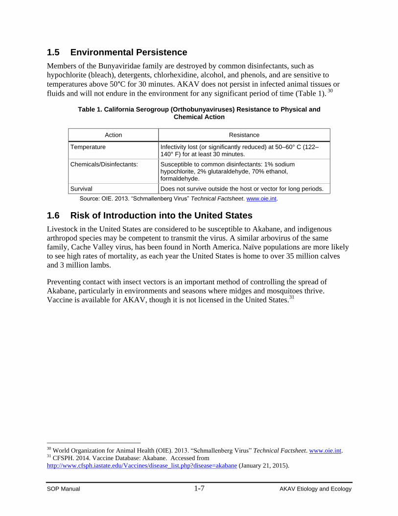
1.5 Environmental Persistence
Members of the Bunyaviridae family are destroyed by common disinfectants, such as
hypochlorite (bleach), detergents, chlorhexidine, alcohol, and phenols, and are sensitive to
temperatures above 50°C for 30 minutes. AKAV does not persist in infected animal tissues or
fluids and will not endure in the environment for any significant period of time (Table 1). 30
Table 1. California Serogroup (Orthobunyaviruses) Resistance to Physical and Chemical Action
Action Resistance
Temperature Infectivity lost (or significantly reduced) at 50–60° C (122–140° F) for at least 30 minutes.
Chemicals/Disinfectants: Susceptible to common disinfectants: 1% sodium hypochlorite, 2% glutaraldehyde, 70% ethanol, formaldehyde.
Survival Does not survive outside the host or vector for long periods.
Source: OIE. 2013. “Schmallenberg Virus” Technical Factsheet. www.oie.int.
1.6 Risk of Introduction into the United States
Livestock in the United States are considered to be susceptible to Akabane, and indigenous
arthropod species may be competent to transmit the virus. A similar arbovirus of the same
family, Cache Valley virus, has been found in North America. Naïve populations are more likely
to see high rates of mortality, as each year the United States is home to over 35 million calves
and 3 million lambs.
Preventing contact with insect vectors is an important method of controlling the spread of
Akabane, particularly in environments and seasons where midges and mosquitoes thrive.
Vaccine is available for AKAV, though it is not licensed in the United States.31
30 World Organization for Animal Health (OIE). 2013. “Schmallenberg Virus” Technical Factsheet. www.oie.int. 31 CFSPH. 2014. Vaccine Database: Akabane. Accessed from
http://www.cfsph.iastate.edu/Vaccines/disease_list.php?disease=akabane (January 21, 2015).
SOP Manual 1-8 AKAV Etiology and Ecology
Attachment A. References and Selected Resources
Akashi H, Inaba Y. 1997. “Antigenic diversity of Akabane virus detected by monoclonal
antibodies”. Virus Research. 47: 187–196.
Akashi H, Onuma S, Nagano H, Ohta M, Fukutomi T. 1999. “Detection and differentiation of
Aino and Akabane Simbu serogroup bunyaviruses by nested polymerase chain reaction”.
Archives of Virology. 144: 2101–2109.
Al-Busaidy S, Hamblin C, Taylor WP. 1987. “Neutralising antibodies to Akabane virus in free-
living wild animals in Africa”. Tropical Animal Health and Production. 19(4): 197–202.
Center for Food Security and Public Health, Iowa State University (CFSPH). 2009. “Akabane
Disease”. Technical Factsheet. www.cfsph.iastate.edu.
Center for Food Security and Public Health, Iowa State University (CFSPH). 2014. “Vaccine
Database: Akabane.”
http://www.cfsph.iastate.edu/Vaccines/disease_list.php?disease=akabane.
Cybinski DH, St George TD, Paull NI. 1978. “Antibodies to Akabane virus in Australia.” Aust
Vet J. 54(1): 1–3.
Geoghegan JL, Walker PJ, Duchemin JB, Jeanne I, Holmes EC. 2014. “Season Drivers of the
Epidemiology of Arthropod-Borne Viruses in Australia.” PLoS Neglected Tropical
Diseases. Online before print. doi: 10.1371/journal.pntd.0003325.
International Committee on Taxonomy of Viruses. 2012. ICTV Taxonomy History for Akabane
virus. http://ictvonline.org.
Kirkland PD. 2012. “Akabane Virus– Epidemiology, Pathogenesis and Impact”. Scientific
Seminar on Management of Schmallenberg Virus, Brussels, Belgium.
http://ec.europa.eu/food/animal/diseases/schmallenberg_virus/docs/akabane_other_sbv_en.p
df.
Kono R, Hirata M, Kaji M, Goto Y, Ikeda S et al. 2008. “Bovine epizootic encephalomyelitis
caused by Akabane virus in southern Japan.” BMC Veterinary Research. 4:20.
Merck Veterinary Manual. 2012. “Akabane Virus Infection.”
http://www.merckmanuals.com/vet/generalized_conditions/congenital_and_inherited_anom
alies/akabane_virus_infection.html.
Miyazato S, Miura Y, Hase M, Kubo M, Goto Y, Kono Y. 1989. “Encephalitis of cattle cause by
Iriki isolate, a new strain belonging to Akabane virus”. Japanese Journal of Veterinary
Science.51(1): 128–136.
SOP Manual 1-9 AKAV Etiology and Ecology
Oem JK, Kim YH, Kim SH, Lee MH, Lee KK. 2014. “Serological characteristics of affected
cattle during an outbreak of bovine enzootic encephalomyelitis caused by Akabane virus.
Trop Anim Health Prod. 46: 261–263.
Oem JK, Lee KH, Kim HR, Bae YC, Chung JY et al. 2012. “Bovine Epizootic
Encephalomyelitis caused by Akabane Virus infection in Korea.” Journal of Comparative
Pathology. 147(2-3):101–105.
Parsonson IM, Della-Porta AJ, Snowdon WA, O’Halloran ML. 1981. Experimental infection of
bulls with Akabane virus”. Research in Veterinary Science. 31(2): 157–160.
Stram Y, Kuznetzova L, Guini M, Rogel A, Meirom R, Chai D, Yadin H, Brenner J. 2004.
“Detection and quantitation of Akabane and Aino viruses by multiplex real-time reverse
transcriptase PCR”. Journal of Virological Methods. 116(2): 147–154.
World Organization for Animal Health (OIE). 2013. “Schmallenberg Virus” Technical
Factsheet. www.oie.int.
Yang DK, Kim BH, Kweon CH, Nah JJ, Kim HJ et al. 2008. “Serosurveillance for Japanese
encephalitis, Akabane, and Aino viruses for Thoroughbread horses in Korea.” J Vet Sci.
9(4):381–385.
SOP Manual 1-10 AKAV Etiology and Ecology
Attachment B. Abbreviations
APHIS Animal and Plant Health Inspection Service
FAD PReP Foreign Animal Disease Preparedness and Response Plan
AKAV Akabane virus
RNA ribonucleic acid
SOP standard operating procedure
TDD telecommunications device for the deaf
USDA United States Department of Agriculture


