68

Contents
Introduction 3
Overview of submission rates, animal demographics and weather 4
Submission rates to the AFBI and DAFF veterinary laboratories in 2010 4
Irish animal demographics 5
Weather 6
Diseases of cattle 7
Neonatal calves (birth to one-month-old) 7
Calves 9
Weanlings 10
Adult cattle 11
Clostridial diseases in cattle 13
Fatal poisonings in cattle 15
Introduction 15
Lead 15
Ragwort 16
Copper 17
Bovine neonatal pancytopaenia – an update 17
Bovine neonatal enteritis 18
Zinc sulphate turbidity test results 20
Bovine abortion 20
Bovine mastitis 23
Bovine respiratory disease 25
Johne’s disease 28
Biosecurity 30
Diseases of sheep 31
Parasitic disease in sheep 32
Clostridial disease in sheep 32
Other findings of interest in sheep 33
Ovine abortion 35
Diseases of pigs 36
Diseases of poultry 38
Backyard poultry 39
Wildlife surveillance 40
Corkscrew injuries in harbour seals 40
Suspected cases of wildlife poisoning in Ireland 41
Trichinella surveillance in wildlife (foxes) 41
Bovine tuberculosis (bTB) surveillance in badgers 42
Parasitic diseases 42
Liver and rumen fluke infections 42
Gastro-intestinal parasitic infections 43
Lungworm infections 43
Coccidiosis 44
Other parasitic diseases 44
All-island Animal Disease Surveillance Report 2010
Contents1
Report 2010:Layout 1 07/09/2011 13:29 Page 2
All-island Animal Disease Surveillance Report 2010
Contents2
Contents
Psoroptic mange in a beef herd 44
Antimicrobial susceptibility profiles 45
Staphylococcus aureus 46
Streptococcus spp. 47
Escherichia coli 48
Clinical chemistry 49
Copper analyses 49
Mineral deficiency-related neonatal mortality in a suckler herd 49
Selenium analyses 50
Iodine analyses 50
Haematology testing in the veterinary laboratories 51
Proficiency testing in AFBI and DAFF veterinary laboratories 52
Procedures for the submission of samples for laboratory investigation 52
Surveillance for Office International des Epizooties (OIE) listed disease 53
Foot-and-Mouth disease 53
Bluetongue 54
Avian influenza 54
Porcine influenza 54
Newcastle disease 54
Classical swine fever 55
Bovine Spongiform Encephalopathy (BSE) 55
Scrapie 55
A selection of farm investigations 56
Dwarfism outbreaks in calves 56
Mortality after acute disease in lambs 57
An investigation of milk drop in a dairy herd. 57
An investigation of recurring milk drop in a dairy herd 58
Neonatal calf diarrhoea and forelimb paresis 59
Pathological fractures in calves 60
Copper deficiency leading to scour and stunting in dairy calves 60
Increased cell count and increased clinical mastitis incidence 61
Periparturient neonatal mortality in a dairy herd 61
An outbreak of ill-thrift in a dairy herd 62
A selection of abstracts from published scientific papers 62
Control of caseous lymphadenitis in six sheep flocks using clinical examination 62
and regular ELISA testing
Fasciola hepatica: Histological changes in the reproductive structures of triclabendazole 63
(TCBZ)-sensitive and TCBZ-resistant flukes after treatment in vivo with TCBZ and the related
benzimidazole derivative, Compound Alpha
Detection and quantification of Toxoplasma gondii in ovine maternal and foetal tissues 63
from experimentally infected pregnant ewes using real-time PCR.
Identification of immunologically relevant proteins of Chlamydophila abortus using sera 64
from experimentally infected pregnant ewes.
Report 2010:Layout 1 07/09/2011 13:29 Page 3

Introduction
The need for a high level animal health and welfare status
throughout the island led the North South Ministerial Council
(NSMC) to commission in late 2001 a programme of work to
develop closer co-operation and joint strategies for the
improvement of animal health on both sides of the border.
This led to the development of the All-Island Animal Health and
Welfare Strategy, which was agreed by NSMC Ministers in
March 2010. The ultimate objective of the strategy is the
development of policies which facilitate the free movement of
animals on the island.
This is the first All-island Animal Disease Surveillance Report,
prepared by the veterinary diagnostic laboratories operated by
the Agri-Food and Biosciences Institute (AFBI) in Northern
Ireland, and by the Department of Agriculture, Fisheries and
Food (DAFF) in Ireland and is part of the actions agreed by the
Department of Agriculture and Rural Development and DAFF
to help deliver the All-Island Animal Health and Welfare
Strategy.
The island of Ireland trades in livestock produce on the basis of
environmentally sustainable grass-based production systems.
Livestock health is a difficult concept to encapsulate and
quantify over a sizeable animal population in diverse
management systems. In this report we seek to do this by
quantifying these diseases and monitoring the trends of their
occurrence in Irish livestock.
Food security is now a pressing global concern and the
increasing requirement for animal protein affords the agri-food
industry on this island with an opportunity to expand
production and trade in livestock produce. This opportunity is
encapsulated in DAFF’s “Food Harvest 2020” report which
envisages sectoral expansion, especially in dairying as quota
restrictions are relaxed post-2013. Likewise, work is ongoing in
Northern Ireland to develop the current Focus on Food Strategy
into a longer term strategic vision for the sector. Sustainable
expansion of the livestock sector and export trade will require
greater attention to the provision of credible data on the
frequency and patterns of disease in farmed animals, as
achieved by surveillance.
If a farmer anywhere on the island has unexplained illness or
deaths in farmed stock, their veterinary practitioner can refer
them to avail of the services of the state supported veterinary
diagnostic laboratories of AFBI or DAFF. These centres accept
carcases of dead animals and samples from live animals, and
offer a wide range of diagnostic test methods. The results of
the tests and post-mortem examinations performed at these
regional centres are reported to the farmer through his/her
veterinary practitioner, who is in a position to complement these
findings with appropriate advice. Laboratory-based veterinary
staff may also undertake follow-up field investigations in certain
cases as outlined in page 56 of this report. Both agencies enter
all of the results from these investigations into their respective
computerised databases, from which data can be extracted to
provide detail on various aspects of food animal morbidity and
mortality.
Jointly reporting the data generated by these activities in both
jurisdictions during 2010 is a natural progression in the
development of the island-wide animal health strategy. It is an
essential step in signalling the commitment of parent
Government Departments, DAFF and DARD (NI) to reducing
restrictions on trade and movement of livestock across the
border and ensuring that this is reflected in the revision of EU
legislation on animal health currently underway in Europe which
in turn may help deliver the aims envisaged in the All-Island
Animal Health and Welfare Strategy.
The sea that surrounds the island of Ireland provides a natural
defence to the introduction of many diseases of livestock.
However as both jurisdictions operate open trading economies
within the EU-wide single market; we need to remain vigilant,
to the threats of the introduction of novel or exotic diseases.
Our ability to detect and identify such threats depends on a
steady throughput of diagnostic material ensuring both services
have a well-trained and practiced staff of veterinary
diagnosticians, laboratory scientists and support staff. Closer
alignment of surveillance activities and collaboration between
the staff in both agencies has the potential to increase the
efficiency and credibility with which we substantiate freedom
from specific diseases on this island. It also allows us to identify
knowledge deficits that might be addressed through
collaborative research initiatives.
This report assembled by frontline staff of both agencies, is the
first of what is hoped will be a series of Annual All-island Animal
Disease Surveillance Reports into the future, and it is intended
that the scope and reach of these reports will expand as this
initiative develops.
All-island Animal Disease Surveillance Report 2010
Introduction3
Report 2010:Layout 1 07/09/2011 13:29 Page 4

All-island Animal Disease Surveillance Report 2010
Overview of submissions rates, demographics and weather4
Submission rates to the AFBI and DAFFveterinary laboratories in 2010
In a continuation of the trends identified in recent years, the
number of carcases submitted to the DAFF Regional Veterinary
Laboratories (RVLs) and to the AFBI Veterinary Laboratories has
continued to increase annually. While well chosen clinical
pathology samples from animals early in the clinical course of a
disease can help to reach a diagnosis, if an animal dies, then
the submission of entire carcases offers the best opportunity for
the achievement of a conclusive diagnosis in the investigation
of a fatal disease outbreak on farm.
Figure 1: Trends in the submission of carcases for post-mortemexamination to the DAFF Regional Veterinary Laboratories over thefour years 2007 to 2010.
In 2010 DAFF RVLs processed a total of 9,396 carcases (Figure
1) which represented an increase of 39.7 per cent in submission
numbers since 2007 while the AFBI veterinary laboratories
processed a further 5,937 carcases in 2010 (Figure 2) reflecting
an increase of 32.5 per cent in submission numbers during the
same period.
Figure 2: Trends in the submission of carcases for post-mortemexamination to the AFBI Veterinary Laboratories over the four years2007 to 2010.
The increase in the submission of clinical diagnostic samples
over the last four years in both the AFBI and DAFF veterinary
laboratories has been remarkable. In 2010 AFBI veterinary
laboratories processed a total of 103,811 clinical diagnostic
samples, an increase of 77.6 per cent on the submission
numbers in 2007 (Figure 3).
Figure 3: Trends in the submission of clinical pathology samples tothe AFBI Veterinary Laboratories over the four years 2007 to 20101.
Since 2007, the DAFF regional veterinary laboratories (RVLs)
have recorded a two and a half fold increase in clinical
diagnostic samples submitted with an increase of 48.3 per cent
(to 143,947 samples) in the year 2010 alone (Figure 4). This was
a staggering increase in the demand for laboratory analyses and
led to a reassessment by DAFF RVLs of our sampling and testing
protocols in early 2011. The changes which were made at this
time, of which veterinary practitioners were advised, sought to
find the most appropriate way for DAFF RVLs to deliver quality
animal disease surveillance, and to provide quality diagnostic
services to Irish livestock farmers through their veterinary
practitioners in the face of an inordinate increase in demand
coupled with finite resources.
The significant increase in the demand for the diagnostic
services of both AFBI and DAFF laboratories reflects an
increasing awareness on the part of veterinary practitioners and
their farming clients of the invaluable role which laboratory
examination can play in the diagnosis, prevention and
treatment of animal disease.
10000
9000
8000
7000
6000
5000
4000
3000
2000
1000
0
2007
Athlone Cork Dublin Kilkenny Limerick Sligo
Stormont Omagh
2008 2009 2010
7000
6000
5000
4000
3000
2000
1000
0
2007 2008 2009 2010
120,000
100,000
80,000
60,000
40,000
20,000
0
2008 2009 2010
1 Figures presented are the combined totals for AFBI Stormont and AFBI Omagh laboratories2 Dublin RVL numbers exclude parasitological and clinical chemistry submissions.
Overview of submission rates, animal demographics and weather
Report 2010:Layout 1 07/09/2011 13:29 Page 5

Figure 4: Trends in the submission of clinical pathology samples tothe DAFF Regional Veterinary Laboratories over the four years 2007to 2010 2.
Irish animal demographics
The national populations of cattle and sheep for both Northern
Ireland and Ireland are shown in Figure 5. While the cattle
population has shown little change in both jurisdictions (1.6
million cattle in Northern Ireland and 6.6 million cattle in
Ireland) during the last four years, the sheep population has
suffered a decline, particularly so south of the border. In 2010
the DAFF sheep census recorded 4.6 million sheep in Ireland
which represents a decline of 15.9 per cent since 2007. This
was mainly due to reductions in ewes aged two years and over.
Figure 5: The national cattle and sheep populations of NorthernIreland and Ireland as measured in June each year for the years 2007to 2010 (Source: NI data from The Agricultural Census in NorthernIreland - Results for June 2010; IRL data from the Central StatisticsOffice http://www.cso.ie)
Figure 6: The national herds of both jurisdictions have shown littlechange in size during the last four years.
In Northern Ireland the decline during the same period was less
marked (8.7 per cent) with a population of 1.8 million sheep
recorded in 2010. While this decline reflected reduced numbers
of ewes (now at their lowest level since 1984) and lambs in the
Northern Ireland flock, the numbers of fattening sheep aged
over one year of age increased by 50.8 per cent during the
same period.
Figure 7: Lamb prices remained buoyant in both Northern Ireland andIreland in 2010 (Photo: Declan Murray).
This shrinkage in the national breeding flocks of both
jurisdictions was a significant contributor to improved farm gate
prices in 2010. The overall average price paid by processors for
lambs in Ireland in 2010 was 460 cents per kilogram, an
increase of 17 per cent on 2009 and the second highest
average price paid for the last fifteen years. In Northern Ireland
the average price paid by processors was 375 pence per
kilogram which was the highest average price paid over the last
fifteen years. A reversal in the reduction of sheep numbers on
the island of Ireland is expected in 2011.
All-island Animal Disease Surveillance Report 2010
Overview of submissions rates, demographics and weather5
8
7
6
5
4
3
2
1
0
2007
Cattle NI Sheep NI
Cattle IRL Sheep IRL
2008 2009 2010
Nationa
lpop
ulationin
millions
160000
140000
120000
100000
80000
60000
40000
20000
0
2007
Athlone Cork Dublin Kilkenny Limerick Sligo
2008 2009 2010
Report 2010:Layout 1 07/09/2011 13:29 Page 6
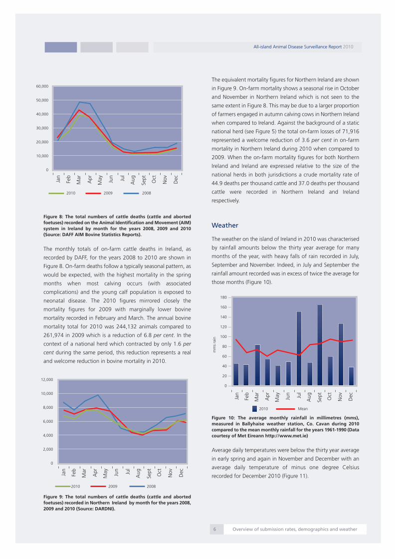
Figure 8: The total numbers of cattle deaths (cattle and abortedfoetuses) recorded on the Animal Identification and Movement (AIM)system in Ireland by month for the years 2008, 2009 and 2010(Source: DAFF AIM Bovine Statistics Reports).
The monthly totals of on-farm cattle deaths in Ireland, as
recorded by DAFF, for the years 2008 to 2010 are shown in
Figure 8. On-farm deaths follow a typically seasonal pattern, as
would be expected, with the highest mortality in the spring
months when most calving occurs (with associated
complications) and the young calf population is exposed to
neonatal disease. The 2010 figures mirrored closely the
mortality figures for 2009 with marginally lower bovine
mortality recorded in February and March. The annual bovine
mortality total for 2010 was 244,132 animals compared to
261,974 in 2009 which is a reduction of 6.8 per cent. In the
context of a national herd which contracted by only 1.6 per
cent during the same period, this reduction represents a real
and welcome reduction in bovine mortality in 2010.
Figure 9: The total numbers of cattle deaths (cattle and abortedfoetuses) recorded in Northern Ireland by month for the years 2008,2009 and 2010 (Source: DARDNI).
The equivalent mortality figures for Northern Ireland are shown
in Figure 9. On-farm mortality shows a seasonal rise in October
and November in Northern Ireland which is not seen to the
same extent in Figure 8. This may be due to a larger proportion
of farmers engaged in autumn calving cows in Northern Ireland
when compared to Ireland. Against the background of a static
national herd (see Figure 5) the total on-farm losses of 71,916
represented a welcome reduction of 3.6 per cent in on-farm
mortality in Northern Ireland during 2010 when compared to
2009. When the on-farm mortality figures for both Northern
Ireland and Ireland are expressed relative to the size of the
national herds in both jurisdictions a crude mortality rate of
44.9 deaths per thousand cattle and 37.0 deaths per thousand
cattle were recorded in Northern Ireland and Ireland
respectively.
Weather
The weather on the island of Ireland in 2010 was characterised
by rainfall amounts below the thirty year average for many
months of the year, with heavy falls of rain recorded in July,
September and November. Indeed, in July and September the
rainfall amount recorded was in excess of twice the average for
those months (Figure 10).
Figure 10: The average monthly rainfall in millimetres (mms),measured in Ballyhaise weather station, Co. Cavan during 2010compared to the mean monthly rainfall for the years 1961-1990 (Datacourtesy of Met Eireann http://www.met.ie)
Average daily temperatures were below the thirty year average
in early spring and again in November and December with an
average daily temperature of minus one degree Celsius
recorded for December 2010 (Figure 11).
All-island Animal Disease Surveillance Report 2010
Overview of submission rates, demographics and weather6
60,000
50,000
40,000
30,000
20,000
10,000
0
180
160
140
120
100
80
60
40
20
0
Jan
Feb
Mar
Apr
May Jun Jul
Aug
Sept
Oct
Nov
Dec
2010 2009 2008
12,000
10,000
8,000
6,000
4,000
2,000
0
Jan
Feb
Mar
Apr
May Jun Jul
Aug
Sept
Oct
Nov
Dec
2010 2009 2008
mmsrain
2010 Mean
Jan
Feb
Mar
Apr
May Jun Jul
Aug
Sept
Oct
Nov
Dec
Report 2010:Layout 1 07/09/2011 13:29 Page 7

Figure 11: The average monthly temperature in degrees Celsius (C)measured in Ballyhaise weather station, Co. Cavan during 2010compared to the mean monthly temperature for the years 1961-1990(Data courtesy of Met Eireann http://www.met.ie)
Wet summers provide the optimal conditions for the survival of
the mud snail, the intermediate host for liver fluke (Fasciola
hepatica) (Figure 12) and the aquatic snail that is the
intermediate host for rumen fluke (Paramphistomum cervi). This
may explain the rise in rumen fluke detection in the third
quarter of the year in both cattle and sheep (see Figure 91 and
Figure 93).
Figure 12: A liver fluke (Fasciola hepatica) (Photo: Cosme Sánchez-Miguel).
In spite of the low temperatures experienced in the winter of
2009/10, the reduction in liver fluke cercariae on pasture was
not as significant as might have been expected, and liver fluke
caused significant losses again in 2010. This finding seems to
contradict previously held beliefs that very low temperatures in
winter reduce liver fluke levels in the spring and may warrant
further investigation. Certainly, the number of liver fluke deaths
was reduced in 2010 when compared to 2009 (see Figure 24),
but much of this reduction could be attributed to increased
awareness on the part of herdowners and their consequent
adherence to preventative measures.
Diseases of cattle
In spite of the wide spectrum of possible causes of mortality in
cattle the most common causes of death remain remarkably
consistent from year to year and from each jurisdiction. The
causes of mortality in cattle diagnosed in both the AFBI and
DAFF veterinary laboratory post-mortem rooms are presented in
this section. Owing to minor differences in the age categories
used for classifying calves and weanlings in the AFBI and DAFF
data management systems, the data for each jurisdiction is
displayed in separate graphs. In interpreting the diagnoses
presented it is important to acknowledge that many of those
diagnoses listed represent a number of similar or related
entities. For ease of presentation, many similar conditions are
subsumed into a more general categorisation. More specific
data on many of these conditions can be obtained in the other
sections of this report.
Neonatal calves (birth to one-month-old)
Figure 13: The conditions most frequently diagnosed on post-mortemexaminations of neonatal (birth to one-month-old) calves in NorthernIreland in 2010 (n= 478).
Enteritis continues to be the most commonly identified cause of
mortality in neonatal calves on the island of Ireland (Figure 13
and Figure 14). In Northern Ireland, among carcases in which
enteritis was diagnosed as the cause of death, Cryptosporidium
parvum was the enteric pathogen identified with greatest
frequency (18.4 per cent) while rotavirus was identified in 17.1
per cent of cases.
All-island Animal Disease Surveillance Report 2010
Disease of cattle7
Jan
Feb
Mar
Apr
May Jun Jul
Aug
Sept
Oct
Nov
Dec
18
16
14
12
10
8
6
4
2
0
-2
2010 Mean
Tempe
rature
inde
greesC
Enteric infections
Navel ill/Joint ill
Respiratory infections
Septicaemia
Circulatory non-infectious
Nutritional/Metabolic
Hereditary & developmental
GIT torsion/obstruction
Various other diagnoses
Diagnosis not reached
5% 10% 15% 20% 25%
Report 2010:Layout 1 07/09/2011 13:29 Page 8

In Ireland the situation was remarkably similar with rotavirus
identified in 18.6 per cent of neonatal carcases and
Cryptosporidium parvum from a further 17.5 per cent of
carcases. Further analysis of findings among clinical cases of
enteritis is presented in the Neonatal enteritis section on page
18.
Hypogammaglobulinaemia (low blood immunoglobulins) was
recorded as being the predisposing cause of death in twenty
nine neonatal calves in Northern Ireland and in thirty four calves
in Ireland. This occurs when calves fail to receive adequate
amounts of protective antibodies from their mothers in
colostrum. These results underline the role played by
appropriate colostrum management in the prevention of
neonatal disease.
Figure 14: The conditions most frequently diagnosed on post-mortemexaminations of neonatal (birth to one-month-old) calves in Irelandin 2010 (n= 1214).
The majority of the circulatory non-infectious conditions in
neonatal calves were due to bovine neonatal pancytopaenia
(BNP) (thirty six in Northern Ireland and sixteen in Ireland). An
update on BNP in calves is presented on page 17 of this report.
Bacteraemia and septicaemia were relatively common findings
among neonatal calves in 2010. Bacteraemia and septicaemia
often represent the end-stage of a disease process when
pathogenic organisms and their toxins enter the bloodstream,
ultimately leading to shock. In many cases, owing to
unsuccessful antimicrobial treatment before death, routine
culture of the organ tissues may fail to isolate the causal
pathogen.
Figure 15: Atresia of a section of jejunum (arrow) in a newborn calf(Photo: Ger Murray).
In Ireland there were seventy one cases of hereditary and
developmental abnormalities recorded, representing 5.8 per
cent of neonatal mortality in 2010. Intestinal atresia (Figure 15)
was the most frequently recorded (twenty six carcases)
congenital abnormality while there were also twenty three cases
of disproportionate dwarfism. Skeletal deformities were
recorded in five carcases - three affecting the hind limbs and
one each affecting the vertebral column and the mandible.
There were also five cases of cardiac defects recorded on post-
mortem examination – two carcases with an atrial septal defect,
an interventricular septal defect in two animals and patent
ductus arteriosus in one animal. In addition there were two
cases of hydrocephalus (Figure 16) and two of renal deformities.
Arthrogryposis and palatoschisis (SAP) of Charolais cattle were
diagnosed in two calves from the same herd. Hereditary
abnormalities accounted for 2.5 per cent of mortality (twelve
cases) in Northern Ireland in this age category.
Figure 16: A case of hydranencephaly in a neonatal calf. The cranialcavity is filled with fluid and very little recognisable brain tissue isevident (arrow) (Photo: Mícheál Casey).
All-island Animal Disease Surveillance Report 2010
Disease of cattle8
Enteric infections
Septicaemia/Bacteraemia
Respiratory infections
Hereditary & developmental abnormalities
Navel ill/ Joint ill
Nutritional/Metabolic conditions
GIT torsion/obstruction
Dystocia/Anoxia
Various other diagnoses
Diagnosis not reached
0% 10% 20% 30%
Report 2010:Layout 1 07/09/2011 13:29 Page 9

Navel ill and joint ill continue to be a significant cause of death
in this age group. In Northern Ireland it was the second most
frequent diagnosis (14.6 per cent) in neonatal calves in 2010,
while in Ireland it accounted for 3.5 per cent of deaths. The
agents isolated on bacteriological culture from such cases vary
widely but Arcanobacterium pyogenes is common.
Figure 17 : Terminal dry gangrene in the hind limbs of a 6-week-oldcalf following Salmonella Dublin infection (Photo: Dónal Toolan).
Salmonella Dublin is commonly associated with enteric
infections and septicaemia in calves (Figure 17), but it was also
occasionally associated with gross lesions of nephritis,
pericarditis and peritonitis. In Ireland, Salmonella Dublin was
isolated from 5.9 per cent of neonatal carcases in 2010 and
from 5.0 per cent of neonatal carcases in Northern Ireland.
Calves
The age categorisation of calves on the data management
systems of AFBI and DAFF differ somewhat, with AFBI recording
diagnoses for calves in the one to five-month-old bracket while
DAFF records diagnoses for calves in the one to three-month-
old bracket. The data from the respective age groups therefore
are not exactly comparable. Nevertheless, allowing for this
difference in how ages are categorised there is considerable
similarity in the most frequently diagnosed causes of mortality
in calves in Northern Ireland (Figure 18) and Ireland (Figure 20).
Figure 18: The conditions most frequently diagnosed on post-mortemexaminations in juvenile (one to five-months-old) calves in NorthernIreland in 2010 (n= 336).
As with all age categories of cattle other than neonatal animals,
respiratory disease was the most frequently diagnosed cause of
mortality in calves in Northern Ireland (34.2 per cent) and
Ireland (29.6 per cent) in 2010. Further details of respiratory
diagnoses in cattle are available on page 25 of this report.
Figure 19: Fluorescence of the cerebrum under the Woods lamp (UV)in a three-month-old calf diagnosed with cerebrocortical necrosis(CCN) (Photo: Jim O' Donovan).
The category ‘nutritional/metabolic conditions’ in calves includes
diseases such as cerebrocortical necrosis (Figure 19), bloat,
ruminal acidosis and mineral deficiencies. In 2010 this grouping
of diagnoses accounted for 5.7 per cent of diagnoses among one
to five-months-old calves in Northern Ireland and 2.4 per cent of
one to three-month-old calves in Ireland. This category of
diagnoses was relatively more common in Ireland among
neonatal calves (3.5 per cent).
All-island Animal Disease Surveillance Report 2010
Disease of cattle9
Respiratory infections
Enteric infections
GIT torsion/obstruction
Nutritional/Metabolic conditions
Clostridial disease
Abomasal ulcer/Perf/Peritonitis
Various other diagnoses
Diagnosis not reached
0% 10% 20% 30% 40%
Report 2010:Layout 1 07/09/2011 13:29 Page 10

Figure 20: The conditions most frequently diagnosed on post-mortemexaminations in calves (one to three-months-old) in Ireland in 2010(n=510).
Among calves in Northern Ireland, enteric infections accounted
for fifty six (16.7 per cent) deaths in this age category with
coccidiosis accounting for twenty one (37.5 per cent) of these
diagnoses. Salmonella Dublin was isolated from fifteen of these
fifty six (26.8 per cent) cases. Pathological signs of septicaemia
were also described in some of these carcases in addition to
enteric infections. In Ireland, enteric infections were diagnosed in
seventy (13.7 per cent) calves in the one to three-months-old
category. Salmonella Dublin was isolated from thirty of these calf
carcases; however septicaemia, rather than enteritis, was the
cause of death in some of these cases.
Figure 21: Abomasal ulceration and perforation in a young calf(Photo: Colm Ó Muireagáin).
Abomasal Ulcer/Perforation/Peritonitis’ is a category which
includes abomasal ulcers, some of which may have perforated
through the abomasal wall causing peritonitis (Figure 21),
accounting for approximately 3 to 4 per cent of mortality in young
calves. The causes of these abomasal ulcers are often not
immediately apparent in young calves but occasionally, their
occurrence may be associated with BVD virus infection, dietary
mismanagement or, in some cases, due to animals being dosed
with acidic solutions (such as copper sulphate or cobalt sulphate).
Peritonitis, due to causes other than abomasal perforation, was
diagnosed in a further sixteen calves (3.1 per cent) in Ireland.
Figure 22: The characteristic finding of diffuse congestion of theintestines associated with mesenteric torsion in a three-month-old calf(Photo: Colm Ó Muireagáin).
The disease category “GIT torsion/obstruction” includes intestinal
torsion, mesenteric torsion (Figure 22), and intestinal obstruction.
Intestinal torsion is a relatively common diagnosis in calves
accounting for 7.7 per cent and 6.1 per cent of deaths in calves
in Northern Ireland and Ireland respectively in 2010.
Weanlings
In spite of the differences in the age categorisation used for
weanlings in both jurisdictions (six to twelve-months-old in
Northern Ireland and three to twelve-months-old in Ireland), the
relative frequency of the most common diagnoses are quite
similar.
Respiratory infections continue to be the most significant cause of
death among weanlings on the island of Ireland, accounting for
31.0 per cent of deaths in this age category in Northern Ireland
(Figure 23) and 31.3 per cent in Ireland (Figure 24).
All-island Animal Disease Surveillance Report 2010
Disease of cattle10
Respiratory infections
Enteric infections
Septicaemia/Bacteraemia
GIT torsion/obstruction
Abomasal ulcer/Perf/Peritonitis
Clostridial disease
Peritonitis
Various other diagnoses
Diagnosis not reached
0% 10% 20% 30% 40%
Report 2010:Layout 1 07/09/2011 13:29 Page 11

Figure 23: The most frequently diagnosed conditions following post-mortem examinations of weanlings (six- to twelve-months-old) inNorthern Ireland in 2010 (n= 174).
Clostridial disease (14.3 per cent) was the second most common
cause of death recorded in this age category in Northern Ireland
while in Ireland it accounted for 6.3 per cent of deaths. Losses
due to diseases caused by clostridial infections continue to occur
despite being preventable by the use of a multivalent clostridial
vaccine in the herd. This is an area where much improvement in
survival rates of cattle should be achievable.
Figure 24: The most frequently diagnosed conditions following post-mortem examinations of weanlings (three- to twelve-months-old) inIreland in 2010 (n= 568).
Figure 25: Ulceration of the oesophagus associated with BVD virusinfection in a ten-month-old heifer (Photo: Ger Murray).
BVD virus was detected in thirty one weanling carcases in Ireland
by PCR methodology in 2010. This was the age category in which
it was most frequently detected on post-mortem examination. It
was also detected in twenty three adults. In Northern Ireland, it
was detected most frequently in adult carcases (fourteen cases),
and considerably less frequently in weanling carcases (4 cases). In
addition to the classical lesions of mucosal disease (Figure 25) it
was also associated with lesions of pneumonia in a number of
these carcases, suggesting its probable role as a risk factor in the
development of respiratory disease.
Adult cattle
Figure 26: The most frequently diagnosed conditions following post-mortem examinations of adult (greater than twelve-months-old)cattle in Northern Ireland in 2010 (n= 610).
All-island Animal Disease Surveillance Report 2010
Disease of cattle11
Respiratory infections
Clostridial disease
Enteric infections
Nutritional/Metabolic conditions
GIT torsion/obstruction
Circulatory infections
Various other diagnoses
Diagnosis not reached
0% 10% 20% 30% 40%
Respiratory infections
Enteric infections
Fasciolosis
Clostridial disease
BVD
Septicaemia/Bacteraemia
GIT torsion/obstruction
Poisoning
Various other diagnoses
Diagnosis not reached
0% 10% 20% 30% 40%
Respiratory infections
Clostridial disease
Circulatory infections
Abscessation
Nutritional/Metabolic conditions
Enteric infections
Abomasal ulcer/Perf/Peritonitis
Urinary tract infections
Various other diagnoses
Diagnosis not reached
0% 10% 20% 30% 40%
Report 2010:Layout 1 07/09/2011 13:29 Page 12

In both jurisdictions, bovine animals over twelve months of age
are classified as adults. In this age group, the variety of
diagnosed causes of death tends to be greater and while
respiratory infections are still the most common cause of
mortality, the relative frequency of this diagnosis is less than in
other age categories (Figure 26 and Figure 28). Further analysis
of respiratory infections in cattle is presented on page 25.
Again, as in weanlings, clostridial disease featured prominently
as a cause of death in adult cattle. Northern Ireland recorded
clostridial involvement in 10.0 per cent of adult deaths while
5.7 per cent of deaths in Ireland among adults were due to
these pathogens. Further analysis of clostridial infections in
cattle on the island of Ireland is presented on page 13.
Figure 27: ‘Bread and butter’ pericarditis caused by the puncturingof the pericardial sac by a piece of metal wire (arrow) which wasfound in situ (Photo: Jim O Donovan).
The category ‘Circulatory Infections’ includes conditions such
as vegetative endocarditis, pericarditis (Figure 27), vena cava
thrombosis, babesiosis, and myocarditis. This group of broadly
similar infections was much more common in adult cattle than
in the other age categories and accounted for 6.1 per cent and
3.3 per cent of adult bovine mortality in Northern Ireland and
Ireland respectively in 2010.
Figure 28: The most frequently diagnosed conditions following post-mortem examinations of adult (greater than twelve-months-old)cattle in Ireland in 2010 (n= 564).
Each year there are a significant number of carcases in which
abscessation is considered a very significant finding. The category
of ‘abscessation’ included all abscesses recorded from multiple
sites on post-mortem examination where the finding was
considered to be the cause of death. Among adult cattle, there
were twenty eight cases of abscessation recorded in Northern
Ireland in 2010 and thirteen cases in Ireland.
Figure 29: Abscessation of the interventricular septum of a two-year-old heifer (Photo: Ger Murray).
Arcanobacterium pyogenes was the pathogen most commonly
isolated from these abscesses on bacteriological culture. Cardiac
abscesses (Figure 29) were the most common in Ireland and
accounted for five of the thirteen cases in adult bovines while
brain abscessation (nine cases) and cardiac abscessation (eight
cases) were most frequently recorded in Northern Ireland.
All-island Animal Disease Surveillance Report 2010
Disease of cattle12
Respiratory infections
Septicaemia/Bacteraemia
Fasciolosis
Enteric infections
Nutritional/Metabolic conditions
Clostridial disease
Poisoning
BVD/Mucosal disease
Various other diagnoses
Diagnosis not reached
0% 10% 20% 30% 40%10%
Report 2010:Layout 1 07/09/2011 13:29 Page 13
Hepatic abscessation (five cases and three cases in Northern
Ireland and Ireland respectively) is commonly associated with
ruminal acidosis. It is important that good hygienic practice is
adhered to when injecting cattle, as the use of non-sterile
needles may be a significant contributing factor to the
development of all types of abscessation, in particular intra-
muscular abscessation.
The category ‘nutritional/metabolic conditions’ accounted for
approximately 5 per cent of diagnoses among adult cattle in
both Northern Ireland and Ireland. This category includes a
number of conditions, of which the most frequently diagnosed
were ruminal/metabolic acidosis, bloat, fatty liver and
hypocalcaemia.
Fasciolosis was diagnosed in thirty seven adult cattle in Ireland,
representing 6.5 per cent of adult mortality in 2010. In Northern
Ireland, fasciolosis was recorded as the cause of death of
eighteen adults (3.0 per cent), although it was recorded as a
secondary finding in a further seventeen carcases. These results
probably reflect the high rainfall recorded in some months in
2010 (see Figure 10) and also suggest that some liver fluke
control regimens that were employed were insufficient to
adequately control the parasite. Further details on the frequency
of detection of fasciolosis are available on page 42 of this
report.
Clostridial diseases in cattle
Figure 30: A classical blackleg lesion of haemorrhagic myositis in themusculature from the hind leg of a bovine animal (Photo: CosmeSánchez-Miguel).
Blackleg (Figure 30) was the most frequently diagnosed
clostridial disease in cattle in 2010 - accounting for fifty two
deaths in cattle in Northern Ireland (almost half of them in
weanlings) and twenty five deaths in Ireland, seven of which
were in calves aged one to three-months-old (Figure 32 and
Figure 33).
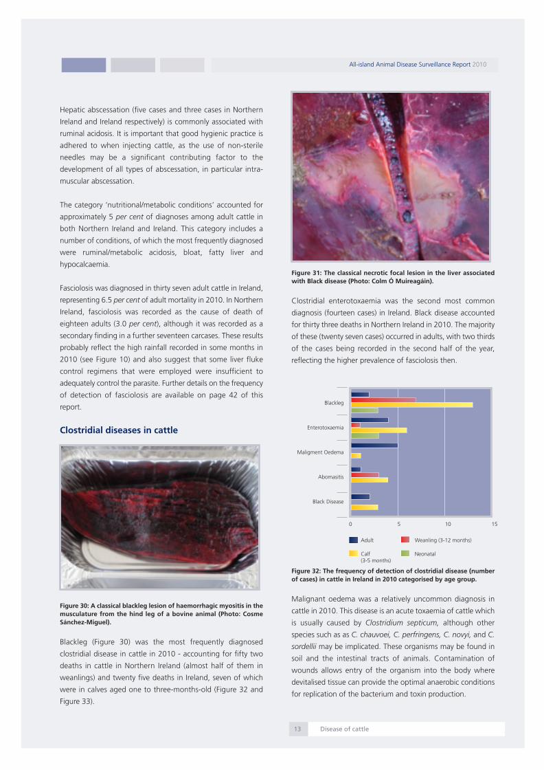
Figure 31: The classical necrotic focal lesion in the liver associatedwith Black disease (Photo: Colm Ó Muireagáin).
Clostridial enterotoxaemia was the second most common
diagnosis (fourteen cases) in Ireland. Black disease accounted
for thirty three deaths in Northern Ireland in 2010. The majority
of these (twenty seven cases) occurred in adults, with two thirds
of the cases being recorded in the second half of the year,
reflecting the higher prevalence of fasciolosis then.
Figure 32: The frequency of detection of clostridial disease (numberof cases) in cattle in Ireland in 2010 categorised by age group.
Malignant oedema was a relatively uncommon diagnosis in
cattle in 2010. This disease is an acute toxaemia of cattle which
is usually caused by Clostridium septicum, although other
species such as as C. chauvoei, C. perfringens, C. novyi, and C.
sordellii may be implicated. These organisms may be found in
soil and the intestinal tracts of animals. Contamination of
wounds allows entry of the organism into the body where
devitalised tissue can provide the optimal anaerobic conditions
for replication of the bacterium and toxin production.
All-island Animal Disease Surveillance Report 2010
Disease of cattle13
Blackleg
Enterotoxaemia
Maligment Oedema
Abomasitis
Black Disease
0 5 10 15
Adult
Calf(3-5 months)
Neonatal
Weanling (3-12 months)
Report 2010:Layout 1 07/09/2011 13:29 Page 14

All-island Animal Disease Surveillance Report 2010
Disease of cattle14
Figure 33: The frequency of detection of clostridial disease (numberof cases) in cattle in Northern Ireland in 2010 categorised by agegroup.
Botulism in cattle
Botulism is caused by Clostridium botulinum, a bacterium that
produces toxins under certain environmental conditions.
Botulism remains an occasionally diagnosed disease on the
island of Ireland and is normally associated with areas where
spreading of broiler litter on farm land occurs. Testing of all
samples on the island is carried out by the AFBI Veterinary
Sciences Division (VSD) in conjunction with the DAFF
laboratories. The 'Type D' toxin of C. botulinum is the most
commonly identified botulinum toxin in cattle. In 2010 the toxin
was not identified in any samples submitted via the DAFF
laboratories and was identified in fourteen suspect botulism
cases by AFBI, almost entirely in adult cattle.
C. botulinum bacteria are commonly found in the environment
and will grow rapidly in decaying organic matter, including
animal and bird carcases. The spreading on pasture of broiler
litter which has been contaminated with the carcases of
chickens that have died from various causes during production
is a common risk factor in Irish botulism outbreaks in cattle.
Transfer of even small fragments of carcases onto adjoining or
nearby pasture by scavenger animals, such as foxes, dogs or
crows can potentially lead to exposure of cattle to the toxin.
It is important to note that the spreading of poultry litter
sourced from egg laying hen units is not usually associated with
outbreaks of botulism in cattle. A possible reason for this is that
husbandry arrangements for layers reduce the likelihood of
contamination of litter with carcases, except on rare occasions.
A joint investigation by veterinarians from DAFF and AFBI into
a botulism outbreak in a poultry flock in 2010 was the first
reported case of botulism in laying hens in the British Isles.
(Sharpe et al.,2011)
Cattle and sheep of all ages are susceptible to botulism, which
is characterised by a progressive muscle weakness (paralysis).
Cattle characteristically display flaccid paralysis, and become
recumbent. Occasionally flaccid protrusion of the tongue
caused by a reduction of muscle tone (Figure 34) is seen, and
while this is almost pathognomonic of the disease, it is only
seen in approximately five per cent of cases clinically examined.
Signs in sheep and goats are similar to those seen in cattle, but
protrusion of the tongue, if it occurs, may not be as obvious. In
most cases the disease is fatal, although some animals may
recover. In many cases of botulism, euthanasia is necessary on
welfare grounds. Cattle are extremely sensitive to the effects of
the toxin, such that ingestion of very small amounts can result
in clinical disease. The progression and severity of the disease
depend on the amount of toxin ingested – the ingestion of a
large quantity may lead to apparent sudden death.
Figure 34: Tongue protrusion and recumbency in a cow with botulism(Photo: AFBI).
The diagnosis of botulism is based primarily on the clinical signs
and a history of known exposure to contaminated broiler litter
or carcase material. Laboratory confirmation is frequently
difficult and relies on detection of the toxin in samples
harvested from suspect cases, and the elimination of other
possible causes of disease. Practitioners and herdowners are
urged to submit the carcase for post-mortem examination so
that other differential diagnoses can be ruled out. In suspected
botulism cases where the submission of a carcase is not
possible, samples of rectal, small intestinal, and abomasal
contents should be submitted, together with a comprehensive
history of the clinical signs and any other relevant information.
Adult
Calf(1-5 months)
Neonatal
Weanling (6-12 months)
0 10 20 30
Blackleg
Enterotoxaemia
MalignantOedema
Black Disease
Botulism
Report 2010:Layout 1 07/09/2011 13:29 Page 15

All-island Animal Disease Surveillance Report 2010
Disease of cattle15
Careful disposal of all animal or bird carcases and poultry litter
is essential to minimise the risk of botulism to livestock. Poultry
carcases should be promptly removed and disposed of by
incineration, or by rendering as required by EU Regulations No.
1069/2009 and 142/2011. At no time should broiler litter be
accessible to dogs, foxes, crows or other scavengers that may
carry carcases onto adjacent pasture or into livestock housing.
Washings from poultry houses and yards should be collected in
tanks rather than be allowed to flow onto adjacent land.
C. botulinum toxin may persist on pasture for a considerable
time if there is ongoing production of new toxin within the
anaerobic environment of a contaminated carcase. Poultry litter
should not be spread on agricultural land that is to be grazed,
or from which silage or hay is to be harvested, in the same year.
If litter must be spread, it should be deep-ploughed into arable
ground. Spreading litter on a windy day may also pose a risk of
contaminating adjacent fields.
No vaccine is available under general licence for the protection
of cattle against botulism. However, veterinary surgeons in
Northern Ireland may apply to the Veterinary Medicines
Directorate (VMD) to obtain and use vaccines under “special
treatment certification”, to protect animals at risk of botulism.
In Ireland, botulism vaccine is not available for cattle.
Vaccination should not be used as a substitute for the hygiene
and biosecurity measures described above.
The UK Food Standards Agency’s Advisory Committee on the
Microbiological Safety of Food has concluded that the risks
posed to the human food chain by outbreaks of botulism in
cattle, sheep or goats, associated with broiler litter, are very low
as the toxin types involved in such outbreaks have only rarely
been associated with human disease.
Further information and advice may be obtained from
http://www.afbini.gov.uk or
http://www.agriculture.gov.ie/animalhealthwelfare/diseasecon
trol/botulism/
Reference:
A. E. Sharpe, E. J. Sharpe, E. D. Ryan, H. J. Clarke and S. A.
McGettrick (2011) An outbreak of type C botulism in laying
hens. Veterinary Record 168, 669
Fatal poisonings in cattle
Introduction
The common toxicities in cattle carcases identified on post-
mortem examination by the AFBI and DAFF laboratories in 2010
are presented in Table 1.
Table 1: The frequency of detection of various toxic agents in bovinecarcases where poisoning was diagnosed in 2010.
Lead
Lead poisoning is the most common cause of fatal poisoning of
cattle submitted for post-mortem examination in farm animals
and was identified in a combined total of forty six animals in
2010. Lead poisoning occurs most commonly during the spring
and early summer when cattle are turned out to pasture (Figure
35).
Figure 35: Combined data from AFBI and DAFF showing theseasonality of lead poisoning in cattle in 2010 (n=46).
Owing to their curious nature and tendency to nuzzle and lick
at what they find on grazing pasture, cattle often ingest lead
from old batteries, sump oil, flaking paint, paint cans and
rubbish fire ash that contains lead. It is highly advisable that
herdowners walk their fields before turnout of animals to
identify if any items have been dumped on their land that could
potentially poison their animals. Feed can also be contaminated
although this is a less frequent finding. Approximately fifty
deaths occurred on one farm in Ireland in 2008 when animals
were exposed to silage that was contaminated when a car
battery was inadvertently chopped by a feed mixer.
Poisonous Agent Ireland Northern
Ireland
Yew 0 2
Oak/acorn 1 0
Ragwort 8 1
Lead 31 15
Copper 3 2
Cobalt 1 0
Other plants 0 1
Totals 44 21
14
12
10
8
6
4
2
0
Jan
Feb
Mar
Apr
May Jun Jul
Aug
Sept
Oct
Nov
Dec
Num
berof
cases
Report 2010:Layout 1 07/09/2011 13:29 Page 16

To prevent lead poisoning in livestock
• Dispose of used car batteries and motor oil through official
local authority collection/recycling facilities
• Keep rubbish out of pastures and areas used by animals
• Prevent access to refuse, landfill sites (even if disused), old
machinery, vehicles
• Service farm machinery away from animals
• Remove all lead paint and treat all painted objects, especially
pallets, as potential sources of lead.
• Check carefully before introducing animals to pasture, yards
or housing
• Do not overgraze areas that have potentially high soil lead
Ragwort
Ragwort (Senecio spp.) poisoning was diagnosed as the cause
of death in one animal in Northern Ireland and eight animals in
Ireland in 2010. Ragwort is a highly poisonous plant and cattle
in particular are highly sensitive to its active compounds,
pyrrolizidine alkaloids. These are cumulative toxins, which
damage the liver leading to a variety of clinical signs which may
include jaundice, diarrhoea, generalised oedema or
photosensitisation. On post mortem examination the liver tissue
may be hard due to fibrosis and, histologically, the characteristic
changes of bile duct proliferation, portal fibrosis and
megalocytosis (Figure 36) may be seen. In animals which display
clinical signs the disease is almost always fatal. Sheep tend to be
more tolerant but losses will still occur. Ragwort is a biennial
plant in which the typical yellow flowers occur during the
second year of growth. The plant is poisonous through both
years of growth. While the growing plant tends to be
unpalatable to cattle, they will ingest the plant when pastures
are bare. Dead or dying ragwort is considerably more palatable
to cattle and poses a greater risk. If the plant is conserved as hay
or ensiled the risk of poisoning remains. In the case of silage
the whole silage pit may be contaminated. Control requires the
pulling rather than cutting of plants or alternatively spraying
with herbicide.
The risk that ragwort poisoning poses to livestock was
highlighted by a field investigation conducted by Athlone RVL
presented below.
Figure 36: A photomicrograph showing the histological findings ofmegalocytosis, portal fibrosis and bile duct proliferation which areconsistent with a diagnosis of ragwort poisoning (Photo: CosmeSánchez-Miguel).
Athlone RVL investigated a number of cattle deaths on a
holding in the summer of 2010. Deaths, predominantly among
yearlings, were first recorded in June, and by mid-August,
twelve animals had died. The clinical signs recorded in these
animals included fluid green diarrhoea, trembling of the limbs,
stiffness, stupor, photosensitisation on the muzzle (Figure 37)
and rectal prolapse in some animals due to tenesmus (intense
and repetitive straining to pass faeces, often seen in ragwort
poisoning). Three carcases were examined at Athlone RVL and
a diagnosis of ragwort toxicity was made following gross and
histopathological examinations. On post-mortem examination
the gross lesions noted were diffuse enteritis, the liver tissue
consistency was firm, and oedema of the abomasal folds,
mesentery and intestinal wall. There was also spongy change
(status spongiosus) of the brain of one of the animals. Spongy
change occurs when the liver function is reduced to the extent
that toxins are not filtered from the blood stream resulting in
damage to brain tissue.
Figure 37: Erythema of the muzzle caused by photosensitisation in ayearling with ragwort poisoning (Photo: John Fagan).
All-island Animal Disease Surveillance Report 2010
Disease of cattle16
Report 2010:Layout 1 07/09/2011 13:29 Page 17

Other apparently normal cattle in the herd were blood sampled
and showed hypoalbuminaemia which was a consistent finding
(arising from liver malfunction) and raised liver enzymes
(indicating hepatocyte damage) in some samples. The Woolfe
test (Iodine flocculation test for liver function) was used in this
case and proved useful as a predictor of liver damage among
clinically normal animals many of which subsequently
developed clinical signs of ragwort toxicity. On further
investigation, it was discovered that there had been ragwort
ensiled in the silage eaten during the previous winter/spring.
The slow but progressive nature of the disease was
demonstrated by the fact that the first clinical signs didn’t
develop until June, four weeks after the animals were let out
onto grass. Failure to control ragwort on land can have serious
economic consequences for a farmer.
Copper
Figure 38: Blue- green staining material on the abomasal mucosa ofa calf in which copper sulphate poisoning was diagnosed (Photo: GerMurray).
Copper toxicity was responsible for five deaths in cattle in 2010.
While sheep are particularly prone to copper poisoning, it is a
relatively uncommon finding in cattle and is normally associated
with over-zealous supplementation of animals which are assumed
to be deficient. Copper sulphate is sometimes administered by
herdowners to animals with diarrhoea, as a traditional remedy,
occasionally with fatal consequences (Figure 38).
Figure 39: Diffuse cortical haemorrhages in the kidney of a calfdiagnosed with copper sulphate poisoning (Photo: Ger Murray).
With cases of acute copper sulphate intoxication there can be
irritation of the lining of the abomasum and duodenum due to
the corrosive nature of the copper sulphate solution, while the
subsequent loss of fluid into the intestinal lumen can lead to
hypovolaemic shock. Animals which survive this initial phase
may progress to develop haemolysis, haemoglobinuria and
diffuse haemorrhages (Figure 39).
Haphazard supplementation of animals with minerals should
be avoided and supplementation should be undertaken only
where a deficiency has been definitively identified.
Bovine neonatal pancytopaenia – an update
Bovine neonatal pancytopaenia (BNP) is a disease of calves
characterised by bleeding in young calves following minor
injuries, causing haemorrhages in the body or from the skin.
The condition is associated with severe bone marrow damage,
which is believed to be caused by antibodies the calf receives
from the colostrum consumed during the first twenty four
hours of life. Normally the antibodies absorbed from colostrum
by the calf provide it with protection from disease; however in
a few rare diseases antibodies can attach to cells and tissues in
the body, leading to cell damage.
It should be noted that BNP is a rare condition. The feeding of
colostrum to a newborn calf is a vital step in the prevention of
neonatal disease and the survival of calves and should not,
under any circumstances, be discontinued in a healthy herd.
Herdowners with confirmed cases of BNP in their herd should
seek veterinary advice on colostrum management.
The first cases of BNP on the island of Ireland were diagnosed
in October 2009 in Northern Ireland, while the first three cases
of BNP in Ireland were diagnosed in May 2010. Since the initial
cases, the condition has been diagnosed in sixty two calves from
forty nine farms in Northern Ireland (thirty six calves in 2010),
and in sixteen calves from fourteen farms in Ireland. These
calves, of both sexes and various breed types, were apparently
normal at birth. Typically calves became ill at around sixteen
days of age with recognisable signs of BNP: fever, anaemia,
bleeding and shock. In some cases the calves were found dead.
Agreeing a case definition for a novel disease can be difficult.
For BNP the case definition which has been used to identify
suspect cases is as follows:
- calves with widespread haemorrhages
- less than one month of age
- thrombocytopaenia (platelet count less than 100X109)
- leukopaenia (white blood cell count less than 3 X 10⁹)
- BVD virus negative
- calf is not septicaemic
All-island Animal Disease Surveillance Report 2010
Disease of cattle17
Report 2010:Layout 1 07/09/2011 13:29 Page 18

As the disease progresses, reduction in the white cell counts
can leave a calf susceptible to septicaemia such that fulfilling
the final criterion of the case definition can be difficult.
Figure 40: An intestinal blood cast in the lumen of the jejunum of acalf diagnosed with bovine neonatal pancytopaenia (BNP) (Photo:Pauline Baird).
At post-mortem examination, there were numerous
haemorrhages observed on the surface of various organs, in
muscles, or in the lumen of the intestines, where a firm cast
may form (Figure 40). The bone marrow was grossly pale in
many cases and histologically the (normally highly cellular)
marrow of a young calf had severely reduced numbers of cells
or was replaced by fatty tissue. Bacteriology, virology including
culture, immunofluorescence, PCR and serology failed to
identify an alternative infectious cause in these animals,
although secondary bacterial infections were detected.
In some countries it has been reported in occasional cases that
cows have had more than one affected calf but not in
consecutive years (i.e. affected calves were born to the cow in
year one and year three with a ‘normal’ calf being born to the
cow in year two). In view of this finding, farms with confirmed
cases of BNP should not use the colostrum from cows which
have had previous calves with the condition.
Farms with BNP cases are advised to store colostrum from
healthy cows without a history of having affected calves to
supply the newborn calves of affected dams. It is good practice
to record the identity of the donating cow against the calf
receiving the colostrum in case the calf unexpectedly develops
signs of BNP. All farms should avoid the use of pooled colostrum
due to the risk of spreading infectious diseases such as Johne's
disease.
As a precaution farms with affected calves should not supply
colostrum or blood for commercial use such as for the
production of ‘artificial colostrum’, but there is no evidence to
suggest that milk or meat from the affected cows or recovered
calves is unsafe for human consumption.
Bovine neonatal enteritis
Neonatal enteritis is responsible for a high proportion of
mortalities in calves less than one month of age throughout the
island of Ireland (see Figure 13 and Figure 14). In order to
identify the enteric pathogens involved in cases of neonatal calf
diarrhoea a series of tests are performed on faecal samples from
these calves.
To aid the achievement of a diagnosis, faecal samples from
neonatal calves with enteric infections should be taken prior to
the administration of treatment.
Figure 41: The relative frequency of calf faecal pathogens detected infaecal samples from calves less than one-month-old in Ireland (DAFF:n=3157) and Northern Ireland (AFBI: n=1863) in 2010.
The relative frequency of identification of enteric pathogens in
calf faecal samples in 2010 is shown in Figure 41.
Cryptosporidium parvum and rotavirus were the most common
enteropathogens identified. Cryptosporidium parvum (40.5 per
cent) was the most frequently identified pathogen in Northern
Ireland in this age group and the diagnostic frequency was
similar to recent years. Rotavirus (36.2 per cent) was the most
frequently identified pathogen in Ireland, a consistent finding in
recent years, with the frequency of identification ranging from
26 – 36 per cent between 2005 and 2010. Results between
laboratories may not be directly comparable due to differences
in test selection protocols and methodologies.
Calves are most susceptible to rotavirus enteritis between one
and three-weeks-old. Adult animals are the primary source of
rotavirus infection for neonatal calves. Rotavirus targets the
upper small intestine causing shortening and fusion of the
intestinal villi causing malabsorption and leading to diarrhoea.
Death may ensue due to acidosis, dehydration (Figure 42) and
starvation.
All-island Animal Disease Surveillance Report 2010
Disease of cattle18
DAFF
AFBI
Rotavirus
Cryptosporidium parvum
Coronavirus
Salmonella spp.
E. coli K99
0% 10% 20% 30% 40% 50%
Report 2010:Layout 1 07/09/2011 13:29 Page 19

Figure 42: Enopthalmus (sunken eye) and anaemia in a calf.Enopthalmus is a typical clinical and/or post-mortem finding indehydrated calves (Photo: Dónal Toolan).
Cryptosporidiosis is a common cause of enteritis in calves
between one and three-weeks-old. Affected calves excrete large
numbers of oocysts that are resistant to many disinfectants.
Control of the parasite is best achieved by strict maintenance of
good calf housing hygiene practices and avoidance of mixing
animals of different ages. The prophylactic use of drugs such
as halofuginone lactate may also be useful where a disease risk
has been identified. In addition to causing disease in animals,
Cryptosporidium parvum has the potential to cause zoonotic
disease especially in immunocompromised humans; therefore
farm workers should take appropriate hygiene precautions
when handling calves.
As with disease associated with rotavirus and Cryptosporidium
parvum, calves are most susceptible to coronavirus enteritis
between one and three-weeks-old. Coronavirus preferentially
infects enterocytes in the lower small intestine and colon
typically resulting in blunting and fusion of villi and mild colitis.
E. coli K99 is an enterotoxigenic E. coli (ETEC) and is an
important cause of neonatal enteritis in young calves, typically
less than three-days-old. These strains of E. coli preferentially
colonise the lower small intestine and produce toxins that cause
hypersecretion of water and electrolytes from the intestinal
mucosa, resulting in rapid dehydration. The percentage
prevalence of E. coli K99 would likely be higher if testing for
this enteric pathogen was restricted to animals less than one-
week-old but as the data presented includes calves up to one-
month-old the proportion of calves from which E. coli K99 is
identified is somewhat diluted.
Salmonella Dublin accounted for 2.6 per cent and 2.9 per cent
of enteritis cases in neonatal calves in Northern Ireland and
Ireland respectively in 2010. As well as enteritis (Figure 43),
Salmonella Dublin is invasive and can cause a number of other
conditions in young calves such as septicaemia and pneumonia.
Figure 43: Fibrino-necrotising enteritis associated with SalmonellaDublin infection in a two-month-old calf (Photo: Dónal Toolan).
Campylobacter jejuni is an important bacterial enteric pathogen
in humans although it is not generally pathogenic in cattle. It
was identified in 10.4 per cent of faecal samples from calves
less than one-month-of-age in Ireland in 2010, an increase from
7.9 per cent in 2009. This highlights again the importance of
adherence to good hygiene practices by calf handlers.
The risk of neonatal enteritis in housed calves increases as the
calving season progresses primarily due to inadequate calf
house hygiene procedures leading to the build up of infectious
agents in the calves' environment.
The basic principles for the prevention and control of neonatal
enteritis include:
• Feeding an adequate quantity and quality of colostrum at, or
very soon after, birth (3 litres within 2 hours of birth).
• Grouping calves according to their age and avoiding high
stocking densities.
• Provision of dry, clean bedding for calves.
• Good hygienic practices including appropriate disinfection
of housing between batches of calves.
• Rapid isolation and treatment of sick calves.
• Appropriate nutrition of young calves including diarrhoeic
calves.
• Vaccination of dams may also play a role in the control of
some enteric pathogens.
The age of the calf must be included on all laboratory
submission forms accompanying faecal samples for neonatal
enteritis testing, to allow meaningful interpretation of the
laboratory findings. The significance of the results of faecal
analyses is dependent on the age of the calf.
All-island Animal Disease Surveillance Report 2010
Disease of cattle19
Report 2010:Layout 1 07/09/2011 13:29 Page 20

Zinc sulphate turbidity test results
The zinc sulphate turbidity (ZST) test can be performed on calf
serum to give an indirect measure of immunoglobulin
concentrations which are essential to prevent establishment of
infectious disease in calves during early life. In calves less than
two-weeks-old this concentration can be used to evaluate the
adequacy of the passive transfer of maternal immunity to the
calf via the colostrum. The ZST test is reported in units of
turbidity with a result of twenty units or greater considered
indicative of the adequate transfer of immunity.
In 2010, a combined total of 1,746 ZST tests was performed by
the DAFF and AFBI laboratories, on blood samples submitted
by veterinary practitioners, as well as on samples taken from
carcases examined post mortem. Of these, 1,040 samples
(almost 60 per cent) recorded results of less than twenty units
(Figure 44).
Figure 44: The results of ZST tests performed in 2010, presented asreflecting adequate (≥20 units) or inadequate (<20 units) colostrumconsumption (n=1746).
When the results of samples taken post mortem alone in the
DAFF Regional Veterinary Laboratories were examined, the
proportion of inadequate results increased to 71 per cent
(Figure 45). This underlines the link between the inadequate
transfer of maternal immunity to the neonatal calf through
colostrum consumption and neonatal mortality.
Farms should have measures in place to ensure that all calves
receive adequate colostrum early enough for absorption to take
place (i.e. ideally in the first six to twelve hours). Inadequate
amounts of colostrum ingested, poor quality colostrum and
delayed colostrum feeding can all lead to failure of passive
transfer.
Figure 45: The results of ZST tests performed on samples taken fromneonatal calves at post-mortem examination in DAFF RVLs in 2010,presented as reflecting adequate (≥twenty units) or inadequate (<twenty units) colostrum consumption (n=642).
Failure of passive transfer of immunity, via colostrum, increases
the risk to calves from diseases, particularly enteritis and
septicaemia. The high incidence of failure of passive transfer
reflected by the results of samples tested by the laboratory
services suggests that inadequate colostrum is a common factor
in the disease processes which bring these animals to veterinary
attention. Many of these disease processes could be readily
prevented by adherence to good colostrum management.
Bovine abortion
Bovine abortion is a significant cause of loss of productivity and
profitability on farms. The cost of a single bovine abortion can
be difficult to quantify but in a dairy herd it is estimated to cost
approximately £630 (€700) (Cabell, 2007). Occasional abortion
is a normal occurrence in any herd; however when the abortion
rate exceeds 3 per cent or a number of abortions occur over a
short period of time, they should be a cause for concern. All
bovine abortions should be notified to the veterinary services
and aborted foetuses and placentas may be submitted to the
veterinary laboratory for a diagnostic workup; where available,
maternal serology may also be informative.
Figure 46: A mummified foetus and autolysed placenta (Photo: DónalToolan)
All-island Animal Disease Surveillance Report 2010
Disease of cattle20
40%
60%
Adequate
Inadequate
71%
29%
Inadequate
Adequate
Report 2010:Layout 1 07/09/2011 13:29 Page 21

The diagnostic rate achieved for abortions in cattle can vary
depending on the preservation of the carcase. Often in cattle
there may be a delay between foetal death and expulsion,
resulting in advanced autolysis (Figure 46), with a significant
deleterious effect on the sensitivity of the diagnostic tests
employed.
Abortions in cattle may result from a broad range of causes –
both infectious and non-infectious. Non-infectious causes
include trauma, nutritional deficiency and genetic defects while
infectious causes include bacterial, viral, fungal and parasitic
agents. Among bovine abortions in which an aetiological
diagnosis is achieved, bacterial agents represent the most
frequently identified group. Many of the bacterial abortions are
sporadic in nature and are caused by organisms that are
ubiquitous in the environment of the cow. These agents may
gain access to the bloodstream and consequently the placenta
of the cow eventually reaching the immature foetus which may
lack the immunological capability to eliminate them.
Figure 47: A second trimester foetus submitted for post-mortemexamination, from which Salmonella Dublin was isolated. (Photo:Dónal Toolan).
Figure 48 Salmonella Dublin abortions as a percentage of all bovinefoetal submissions during 2010 in Northern Ireland (AFBI: n=571) andIreland (DAFF: n=2608).
Contagious bacterial abortion agents, such as Brucella abortus
and Salmonella Dublin (Figure 47), have the potential to cause
abortion storms. While brucellosis-free status has been achieved
in Ireland, Brucella abortus was identified in one foetus in
Northern Ireland in 2010. Brucellosis poses a serious zoonotic
risk to animal handlers and may be transmitted by contact with
foetal tissues. Salmonella spp. are also zoonotic although
Salmonella Dublin is rarely so. Cows suffering Salmonella spp.
induced abortion may also show signs of enteritis and
septicaemia, although the abortion is often the only clinical sign
observed. The monthly distribution of Salmonella Dublin
associated abortion, for both AFBI and DAFF laboratories,
follows the characteristic seasonal distribution, increasing
steadily in frequency towards October and November (Figure
48).
Figure 49: A bovine placenta showing cotyledonary necrosis (arrow)with extension to the intercotyledonary areas, caused by Aspergillusspp. (Photo: Dónal Toolan).
Mycotic (fungal) abortion is relatively uncommon and is a result
of fungal invasion of the placenta and foetus. The characteristic
gross lesions on the placenta or foetus are generally evident
(Figure 49) although on occasion live infected calves are born.
As the infection is of haematogenous origin, it typically infects
placentomes (cotyledonary areas) initially and then proceeds to
the intercotyledonary areas. Fungal abortion tends to be more
prevalent in the winter months when animals are housed and
exposed to preserved fodder – particularly mouldy hay or silage.
The fungi most frequently isolated are Aspergillus spp. In 2010
Aspergillus spp. were isolated by AFBI and DAFF laboratories in
1.4 per cent and 1.0 per cent of cases respectively.
Arcanobacterium pyogenes is a common cause of sporadic
abortion in cattle. A. pyogenes is a ubiquitous organism and
normally reaches the placenta following bacteraemia in the
cow, resulting in placentitis and subsequent abortion. Bacillus
licheniformis similarly is a cause of sporadic abortion in herds.
Spoiled forage and feed often acts as a vehicle for the
introduction of this organism to the herd.
All-island Animal Disease Surveillance Report 2010
Disease of cattle21
Jan
Feb
Mar
Apr
May Jun Jul
Aug
Sept
Oct
Nov
Dec
40%
35%
30%
25%
20%
15%
10%
5%
0%
DAFF AFBI
Report 2010:Layout 1 07/09/2011 13:29 Page 22

Figure 50: A comparison of selected bovine foetal culture results inthe AFBI (n=571) and DAFF (n=2608) laboratories in 2010.
Infections with bovine abortion agents during early pregnancy
may result in early embryonic death and return to service
without other visible signs. Infections at a later stage may lead
to abortion, stillbirths or the birth of live but weak calves. In
both jurisdictions a combined total of three thousand one
hundred and seventy nine (two thousand six hundred and eight
by DAFF and five hundred and seventy one by AFBI) foetal
cultures was undertaken in 2010. The results of these
bacteriological cultures are shown in Figure 50. The prevalence
of Salmonella Dublin in Ireland (14.0 per cent) was significantly
higher than in Northern Ireland (7.4 per cent) and doubled in
2010 when compared to 2009 (6.0 per cent).
Other microorganisms isolated from foetal cultures conducted
by both DAFF and AFBI were Escherichia coli and other
coliforms (three hundred and thirteen isolates), Streptococcus
spp. (sixty two), Bacillus spp. (nineteen), Staphylococcus spp.
(twelve), Pasteurella spp. (eleven), Fungal species (nine),
Campylobacter spp. (seven), S. Typhimurium (two). Yersinia
pseudotuberculosis (one) and Pseudomonas aeruginosa (one).
Neospora caninum is a protozoan parasite first identified in
1989. Dogs are the definitive host and excrete oocysts in their
faeces, normally only for a short period of time. When ingested
by the intermediate hosts, such as cattle, tachyzoites may
develop in the placenta while tissue cysts (containing
bradyzoites) may develop within the central nervous system of
the calf. Infected foetuses may be aborted (typically at six to
seven months of gestation), may be born with neurological
deficits or may be clinically normal at birth. While dogs may be
responsible for the introduction of the parasite into cattle herds,
the vertical route of transmission appears to be the most
significant mode of transmission in cattle. Cows which abort
may do so again and the retention of the female offspring of
infected cows for replacement purposes is not recommended.
The finding of Neospora caninum antibodies in a blood sample
from a cow which has aborted or in the foetal pleural fluid is
not sufficient to confirm Neospora-induced abortion.
Confirmation requires the supporting evidence of protozoal
encephalitis or myocarditis on histopathology of foetal tissues.
Table 2: The frequency of detection of Leptospira Hardjo antibodies,Neospora caninum antibodies and BVD virus in foetal carcases in AFBIand DAFF veterinary laboratories in 2010 (*Comb titre of >1/100; **placenta samples; *** MAT titre of >1/30)
Serological results in addition to the results of specific tissue
analyses from foetal carcases for Leptospira Hardjo, Neospora
caninum and BVD virus are shown in Table 2. Foetal serological
and maternal serological results need to be interpreted with
caution in the investigation of bovine abortion. Rising maternal
titres to BVD virus or BHV-1 may be difficult to demonstrate due
to the time delay between infection and abortion in some cases
and maternal titres may remain high for an extended period
after the initial infection. Maternal vaccination and its timing
can also complicate the interpretation of results. Leptospira spp.
titres can be equally difficult to interpret for these reasons.
Serology therefore should be used as supporting evidence in
the investigation of bovine abortion but the submission of a
foetal carcase (preferably accompanied by the foetal
membranes) to the veterinary laboratory is central to identifying
the causal agent and is the submission of choice.
Comparison of the frequency of detection of Leptospira Hardjo,
Neospora caninum and BVD virus in AFBI and DAFF laboratories
(Table 2) shows that the incidence of detection of these
pathogens in foetal carcases is broadly similar in both Northern
Ireland and Ireland. The 2010 figures for Ireland show a
marginal increase in the detection of all three pathogens relative
to 2009 and a return to levels of detection witnessed in 2008.
Differences in detection frequency between Northern Ireland
and Ireland may be due to differences in the sample sizes tested
in each jurisdiction or possibly due to differences in the tests
employed.
All-island Animal Disease Surveillance Report 2010
Disease of cattle22
DAFF
AFBI
0% 5% 10% 15%
Agent Test Positive Total %
Tested Positive
DAFF BVD Ag PCR + ELISA 81 1241 6.5%
Neospora Ab Comb+ELISA 119 1751 6.8%
Leptospira Ab Comb 60* 1157 5.2%
AFBI BVD Ag ELISA + 31 637 4.9%
Immunofluorescence
Neospora Ab ELISA 14 439 3.2%
Leptospira Ag FAT** 8 161 5.0%
Leptospira Ab MAT 4*** 441 0.9%
Salmonella Dublin
Arcanobacterium pyogenes
Bacillus licheniformis
Listeria moncytogenes
Aspergillus spp.
Report 2010:Layout 1 07/09/2011 13:29 Page 23

Where the submission of a foetus carcase to the laboratory is
not possible, the veterinary practitioner should submit as many
of the following tissues as possible:
• Stomach fluid collected and submitted in a sterile manner
for culture
• Pleural fluid (5mls where possible) for serology
• Brain – a section fixed in 10 per cent formalin
• Placenta – a section including a cotyledon both fresh and
fixed
• Thyroid gland - a section fixed in 10 per cent formalin
• A fresh section of thymus or spleen – for BVD virus PCR
• A maternal blood sample, from the dam of the aborted
foetus, as well as from any other cows that aborted
or have proven to be non-pregnant.
Details of maternal vaccination and the timing of vaccination
should also be provided.
See Packaging Requirements for the submission of samples
outlined on page 52.
References:
Cabell, E (2007) In Practice 29, 455-463
Bovine mastitis
The submission of milk samples for bacteriological culture is an
important step in the diagnosis, treatment and control of both
clinical and subclinical bovine mastitis. Mastitis is the second
most common cause, after infertility, of culling of cows from
dairy herds and leads to both direct costs (discarded milk,
veterinary treatment, processor penalties etc.) and indirect costs
(increased mortality and extended calving intervals) which have
been estimated at approximately €125 to €400 per case
depending on the severity (Esslemont and Kossaibati, 2002).
The control of mastitis in cows is based on six fundamental
principles:
1) Hygienic teat management (and housing management)
2) Prompt treatment of mastitis cases
3) Appropriate dry cow therapy
4) Culling of chronic cases
5) Proper milking machine maintenance
6) Proper record keeping
Figure 51: The frequency of submission of milk samples by month toAFBI and DAFF veterinary laboratories in 2010.
The submission of milk samples to the veterinary laboratories
follows a predictably seasonal pattern from year to year in
Ireland (DAFF) with an increase in sample submissions in the
autumn months when most cows are ‘dried off’. The seasonal
distribution of milk sample submission to the AFBI laboratories
in Northern Ireland differs with the autumn rise in sample
submission not being observed, and the overall distribution
shows a more consistent pattern of submission throughout the
year (Figure 51). Examination of the pattern of milk sample
submissions to both AFBI and DAFF RVLs in 2010 shows a rise
in the numbers submitted in July. In 2009, a marked increase in
sample submissions to the Regional Veterinary Laboratories was
also noted in July of that year which was attributed to the heavy
rainfall experienced. The heavy rainfall experienced in July 2010
(see Figure 10) which was more than double the July average
probably resulted in the re-housing of stock in some parts of
the island, creating the necessary conditions in which mastitis
pathogens can flourish.
Figure 52: A case of `summer mastitis' in a cow (Photo: Brian Flynn)
All-island Animal Disease Surveillance Report 2010
Disease of cattle23
Jan
Feb
Mar
Apr
May Jun Jul
Aug
Sept
Oct
Nov
Dec
1000
900
800
700
600
500
400
300
200
100
0
DAFF AFBI
Report 2010:Layout 1 07/09/2011 13:29 Page 24
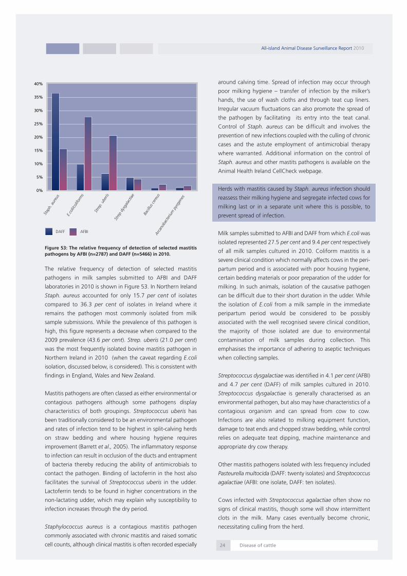
Figure 53: The relative frequency of detection of selected mastitispathogens by AFBI (n=2787) and DAFF (n=5466) in 2010.
The relative frequency of detection of selected mastitis
pathogens in milk samples submitted to AFBI and DAFF
laboratories in 2010 is shown in Figure 53. In Northern Ireland
Staph. aureus accounted for only 15.7 per cent of isolates
compared to 36.3 per cent of isolates in Ireland where it
remains the pathogen most commonly isolated from milk
sample submissions. While the prevalence of this pathogen is
high, this figure represents a decrease when compared to the
2009 prevalence (43.6 per cent). Strep. uberis (21.0 per cent)
was the most frequently isolated bovine mastitis pathogen in
Northern Ireland in 2010 (when the caveat regarding E.coli
isolation, discussed below, is considered). This is consistent with
findings in England, Wales and New Zealand.
Mastitis pathogens are often classed as either environmental or
contagious pathogens although some pathogens display
characteristics of both groupings. Streptococcus uberis has
been traditionally considered to be an environmental pathogen
and rates of infection tend to be highest in split-calving herds
on straw bedding and where housing hygiene requires
improvement (Barrett et al., 2005). The inflammatory response
to infection can result in occlusion of the ducts and entrapment
of bacteria thereby reducing the ability of antimicrobials to
contact the pathogen. Binding of lactoferrin in the host also
facilitates the survival of Streptococcus uberis in the udder.
Lactoferrin tends to be found in higher concentrations in the
non-lactating udder, which may explain why susceptibility to
infection increases through the dry period.
Staphylococcus aureus is a contagious mastitis pathogen
commonly associated with chronic mastitis and raised somatic
cell counts, although clinical mastitis is often recorded especially
around calving time. Spread of infection may occur through
poor milking hygiene – transfer of infection by the milker’s
hands, the use of wash cloths and through teat cup liners.
Irregular vacuum fluctuations can also promote the spread of
the pathogen by facilitating its entry into the teat canal.
Control of Staph. aureus can be difficult and involves the
prevention of new infections coupled with the culling of chronic
cases and the astute employment of antimicrobial therapy
where warranted. Additional information on the control of
Staph. aureus and other mastits pathogens is available on the
Animal Health Ireland CellCheck webpage.
Herds with mastitis caused by Staph. aureus infection should
reassess their milking hygiene and segregate infected cows for
milking last or in a separate unit where this is possible, to
prevent spread of infection.
Milk samples submitted to AFBI and DAFF from which E.coli was
isolated represented 27.5 per cent and 9.4 per cent respectively
of all milk samples cultured in 2010. Coliform mastitis is a
severe clinical condition which normally affects cows in the peri-
partum period and is associated with poor housing hygiene,
certain bedding materials or poor preparation of the udder for
milking. In such animals, isolation of the causative pathogen
can be difficult due to their short duration in the udder. While
the isolation of E.coli from a milk sample in the immediate
peripartum period would be considered to be possibly
associated with the well recognised severe clinical condition,
the majority of those isolated are due to environmental
contamination of milk samples during collection. This
emphasises the importance of adhering to aseptic techniques
when collecting samples.
Streptococcus dysgalactiae was identified in 4.1 per cent (AFBI)
and 4.7 per cent (DAFF) of milk samples cultured in 2010.
Streptococcus dysgalactiae is generally characterised as an
environmental pathogen, but also may have characteristics of a
contagious organism and can spread from cow to cow.
Infections are also related to milking equipment function,
damage to teat ends and chopped straw bedding, while control
relies on adequate teat dipping, machine maintenance and
appropriate dry cow therapy.
Other mastitis pathogens isolated with less frequency included
Pasteurella multocida (DAFF: twenty isolates) and Streptococcus
agalactiae (AFBI: one isolate, DAFF: ten isolates).
Cows infected with Streptococcus agalactiae often show no
signs of clinical mastitis, though some will show intermittent
clots in the milk. Many cases eventually become chronic,
necessitating culling from the herd.
All-island Animal Disease Surveillance Report 2010
Disease of cattle24
Stap
h.au
reus
E.co
li/co
lifor
ms
Stre
p.ub
eris
Stre
pdy
sgal
actia
e
Bacil
lus ce
reus
Arcan
obac
teriu
mpy
ogen
es
40%
35%
30%
25%
20%
15%
10%
5%
0%
40%
35%
30%
25%
20%
15%
10%
5%
0%
DAFF AFBI
Report 2010:Layout 1 07/09/2011 13:29 Page 25

Aseptic technique for milk sample collection
1. Take the samples before milking.
2. Soak a number of cotton wool balls in alcohol.
3. Using a hand or paper towel, brush any loose dirt, straw or
hair from the underside of the udder and teats. Washing
should be avoided if possible, but if teats are very dirty they
should be washed and carefully dried with paper towels.
4. Dip all four teats with teat dip and leave for at least one
minute.
5. Wear gloves if available. If not, then wash and dry the hands
thoroughly and use some of the cotton wool balls to wipe
them with alcohol.
6. Beginning with teats on the far side of the udder, scrub the
ends thoroughly with the cotton wool and alcohol until the
teats are very clean. Spend at least ten seconds on each teat.
Do not use the same cottonwool ball on more than one teat.
7. Begin sampling with the teats on the near side of the udder.
Remove the cap of the sampling tube and keep the top face
down in the palm. Hold the open tube (in the same hand as
the top) at an angle of forty five degrees (holding it straight
up will allow dust etc. to fall inside). Using the free hand,
discard a few streams of milk on to the ground before
collecting three or four streams in the tube. Do not allow
the teat ends to make contact with the tube. Move quickly
onto the next teat. When the four teats have been sampled
close the tube.
8. Put the tubes in a fridge and cool to 4°C. This is very
important.
9. The samples should be taken to the laboratory as soon as
possible.
Further Reading:
Animal Health Ireland CellCheck webpage at:
http://www.animalhealthireland.ie/scc.php
Barrett, D.J., Healy, A.M., Leonard, F.C. Doherty, M.L. (2005)
Prevalence of pathogens causing subclinical mastitis in 15 dairy
herds in the Republic of Ireland Irish Veterinary Journal, 58:333-
337
Esslemont R.J. and Kossaibati M.A. (2002). Mastitis: How do
we get out of the dark ages? The Veterinary Journal 164, 85-
86
Bovine respiratory disease
Respiratory disease is a major problem in all bovine husbandry
systems, is associated with production losses and mortalities
and can have a long-lasting impact on growth in young stock
In this section, two main types of data are presented:
- Post-mortem examinations: agents that were identified as
the principal pathogen isolated from bovine lungs at post-
mortem examination, where the diagnosis was respiratory
disease.
- Clinical pathology: viral agents demonstrated in samples
submitted for PCR examination to the DAFF Veterinary
Laboratory Service.
Figure 54: Fibrinous (yellow deposit) pleuropneumonia in a yearlingwith Mannheimia haemolytica infection (Photo: Colm Ó Muireagáin).
Pasteurella multocida and Mannheimia haemolytica (Figure 54)
continue to be the major pathogens associated with fatal
respiratory disease in Irish cattle (Figure 55). Although both of
these agents are capable of precipitating fatal disease,
parainfluenza 3 (PI3) virus is believed to play an important role
in facilitating the rapid invasion of the lungs by these agents.
The very low number of fatal respiratory disease cases in 2010
where PI3 virus was the only pathogen identified (1.8 per cent)
supports the view that it is a minor pathogen in its own right on
Irish farms.
Arcanobacterium pyogenes is not considered a primary
pathogen in bovine pneumonia, but is a common secondary
invader, when tissues have already been damaged by an acute
episode caused by any one of a range of infectious agents. It is
particularly common in cases of chronic suppurative
pneumonia, often a chronic sequel to incompletely resolved
pneumonia in young calves. It was isolated from 14.0 per cent
of carcases with a diagnosis of respiratory disease in 2010.
All-island Animal Disease Surveillance Report 2010
Disease of cattle25
Report 2010:Layout 1 07/09/2011 13:29 Page 26

Figure 55: The relative frequency of post-mortem detection ofprimary respiratory pathogens associated with fatal respiratorydisease in AFBI and DAFF veterinary laboratories in 2010 (n=602).
Mycoplasma bovis can play a primary role in the development
of respiratory disease and was identified in 9.0 per cent of
necropsy cases of respiratory disease in 2010. It can also be
implicated in mastitis, meningitis, otitis and arthritis in cattle.
Mycoplasma bovis can be introduced into a herd by subclinical
carriers which may shed the organism through nasal discharges.
The impairment of the mucocillary clearance in the airways or
the immune defence can lead to the introduction of the
organism into the lower respiratory tract of an animal. At
necropsy, the cranioventral areas of lungs are red-blue in colour,
consolidated and occasionally exhibit the characteristic ‘rice
grain’ multifocal abscessation. The treatment of Mycoplasma
bovis is difficult as Mycoplasma spp. lack a cell wall, and are
thus resistant to some commonly used antibiotics.
Histophilus somni is a commensal of the mucous membranes of
cattle which is occasionally associated with pneumonia,
particularly in feedlot cattle. It is also associated with other
conditions of cattle such as reproductive disorders, neurological
signs and septicaemia. It is a primary respiratory pathogen
although co-infection by opportunist pathogens such as
Pasteurella multocida and Mannheimia haemolytica is often
recorded. Occasionally rapid death of the animal can occur
before clinical signs have been detected.
Parasitic pneumonia – hoose - caused by Dictyocaulus viviparus
has a highly seasonal pattern and is also much more common
in young stock. However the frequency and severity of disease
varies from year to year, often occurring as explosive outbreaks
associated with extended periods of moist mild weather.
Figure 56: Severe tracheitis associated with Bovine herpesvirus – 1infection in a two-year-old bull (Photo: John Fagan).
Deaths due to bovine herpesvirus 1 (BHV-1, causing infectious
bovine rhinotracheitis - IBR) (Figure 56) and respiratory syncytial
virus (RSV) (Figure 57) are more usually seen in younger stock,
with RSV usually being more important than IBR in the youngest
groups of calves. Many RSV-related deaths in calves occur
before they are let out to grass. Post-mortem findings typically
include diffuse pneumonia, consolidation, oedema and
emphysema, the severity of which will vary depending on the
clinical course of the disease. BHV-1 and RSV were identified in
6.0 per cent and 5.0 per cent of cases of respiratory disease
diagnosed on post-mortem examination in 2010.
Figure 57: A photomicrograph of a bovine lung showing syncytialcells (arrow) which are a characteristic finding in RSV pneumonia(Photo: John Fagan).
All-island Animal Disease Surveillance Report 2010
Disease of cattle26
Pasteurellamultocida
Mannheimiahaemolytica
Arcanobacteriumpyogenes
Dictyocaulusviviparus
Mycoplasmabovis
Bovineherpesvirus 1(BHV 1/IBR)
Respiratorysyncytial virus (RSV)
Pasteurella spp.
Histophilus somni
Miscellaneousaetiologies
0% 5% 10% 15% 20% 25%
Report 2010:Layout 1 07/09/2011 13:29 Page 27

The category ‘miscellaneous aetiologies’ encompasses a range
of minor infectious agents associated with respiratory disease
including Pseudomonas spp., Staphylococcus spp.,
Streptococcus spp. and Salmonella Dublin.
An analysis of the relative frequency of the infectious agents
associated with fatal respiratory disease in any particular year
tells only part of a complex story. All infectious diseases occur
as a result of the complex interplay of the animal, the agent
and the environment, and few conditions illustrate the interplay
of these factors more than respiratory disease. Stress, transport,
stocking densities, and air quality are among the main
environmental factors, while animal age, nutrition and immune
status also determine the incidence and final outcome of
respiratory disease in any management group when any
particular infectious agent enters the herd.
Table 3: The relative frequency of detection of primary respiratoryvirus pathogens on PCR tests in DAFF laboratories in necropsy casesand clinical cases of respiratory disease in 2010.
Table 3 shows the relative frequency of the detection of primary
respiratory virus pathogens in all samples tested from cases of
respiratory disease in 2010. This includes samples from post-
mortem examinations in the Regional Veterinary Laboratories
in Ireland, as well as clinical samples (swabs) submitted by
private veterinary practitioners from clinical cases of respiratory
disease. The results are from animals spanning a wide range of
age groups. These figures provide a snapshot of the range of
bovine respiratory viruses confirmed in affected cattle on farms
in Ireland in 2010. The relatively high detection rate for PI3
virus and bovine coronavirus, when compared to the other viral
pathogens, should be considered against the comparatively
small sample size tested for these two agents.
Table 4: The relative frequency of detection of primary respiratoryvirus pathogens on fluorescent antibody tests (FAT) among necropsycases in AFBI laboratories in 2010.
Table 4 outlines the results of fluorescent antibody tests (FAT) for
respiratory pathogens conducted on tissue from necropsy cases
by AFBI in 2010. The differences in the relative frequency of
detection of the various pathogens when compared to Table 3
is possibly due to differences in methodologies but is more likely
to be accounted for by the fact that these tests were performed
entirely on necropsy cases, which often represent the end-stage
of respiratory disease when the availability of virus for detection
can be considerably less than in acute clinical cases.
Figure 58: The relative frequency of detection of viruses implicated inbovine respiratory disease by month during 2010 as a percentage ofall nasal swabs tested by polymerase chain reaction (PCR) at theCVRL, Backweston.
Figure 58 shows the seasonal patterns observed in the detection
of these agents. As well as the expected winter peak associated
with housing, it is clear that BHV1 (IBR) is detected with
increasing frequency from late summer onwards. Presented
below is a farm investigation into an outbreak of IBR conducted
by Dublin RVL in December 2010.
An investigation into an infectious bovinerhinotracheitis (IBR) outbreak in a beef unit
Dublin RVL diagnosed IBR as the cause of pneumonia in a large
feedlot in December 2010. At the time of the outbreak there
were nine hundred and fifty animals in the herd. Fourteen
animals had died, but over eight hundred animals had shown
signs of respiratory disease (84 per cent morbidity).
Cattle were bought in September and October at about
fourteen months of age, and fattened over the following one
hundred and fifty days or so. Animals were housed on arrival
and vaccinated with an IBR intra-nasal vaccine within twenty
four hours. The farmer made an effort not to mix groups, but
given that they were bought at marts they would have been
mixing with cattle from a diverse range of sources anyway.
There were five separate cattle sheds in the feedlot and animals
were not mixed between these sheds. Cattle were fed on
maize silage.
All-island Animal Disease Surveillance Report 2010
Disease of cattle27
Number Number Percentage
positive tested positive
Bovine 55 291 18.9
coronavirus
PI3 89 360 10.1
BHV1 176 2017 8.7
BVD 82 1192 6.9
BRSV 92 1551 5.9
Number Number Percentage
positive tested positive
PI3 7 352 2.0%
BHV1 21 404 5.2%
BVD 9 444 2.0%
BRSV 7 352 2.0%
Jan
Feb
Mar
Apr
May Jun Jul
Aug
Sept
Oct
Nov
Dec
BHV1 BRSV BVDV PI3V
45%
40%
35%
30%
25%
20%
15%
10%
0%
Report 2010:Layout 1 07/09/2011 13:29 Page 28

Following the very cold weather in early December, cattle in two
of the five sheds showed clinical signs of IBR. This was
subsequently confirmed by the detection of bovine herpes virus
(BHV-1) by PCR on nasal swabs.
All affected cattle in both sheds were administered antimicrobial
therapy, with more severely affected animals receiving non-
steroidal anti-inflammatory drugs. Animals in the remaining three
sheds were given a second dose of IBR intranasal vaccine. Animals
in these three sheds developed clinical signs of IBR, but not as
severely as the animals in the first two sheds. During the course
of the outbreak average feed intake fell by over 80 per cent.
The investigation concluded that extreme weather at the start of
December caused physiological stress to some of the stock. A
proportion of these was BHV-1-infected, and started re-shedding
virus due to this stress. It is more likely that virus was re-activated
in several animals in each shed than the alternative theory that
one shedder caused the outbreak which then spread from shed
to shed. The fact that animals in sheds at opposite ends of the
feedlot got sick first corroborates this theory. Extremely cold air
can create a “ceiling” in very still conditions (a phenomenon
known as inversion), trapping warmer air underneath it. While
this normally does not occur in sheds, it may have occurred in the
open-faced straw-bedded bull sheds, thus creating a lack of
ventilation as the humid hot air breathed by the bulls was trapped
under a colder layer. This would have exacerbated the situation
further.
Johne’s disease
Johne’s disease is a chronic disease of cattle caused by infection
with Mycobacterium avium subspecies paratuberculosis (MAP)
and is an OIE-listed disease. The clinical signs can vary but
chronic diarrhoea and weight loss are consistently recorded.
Most cattle acquire infection early in life, through ingestion of
colostrum or milk containing MAP, or by exposure to feed,
water or environments contaminated by MAP. It is also possible
for older animals exposed to a large number of MAP organisms
to become infected. In-utero infection of the foetus can also
occur and has been typically recorded in herds where the
prevalence exceeds 5 per cent. Most commonly, however, the
disease is introduced to a MAP free herd through the purchase
of an infected animal.
It is possible for infected animals to be negative on both
serology and faecal culture, but as the disease progresses MAP
will be shed in the faeces and subsequently seroconversion will
usually occur.
Owing to the nature of MAP infection and the blood testing
kits presently available, establishing the exact correlation
between MAP serological results and infection can be difficult.
The correlation between serological results and faecal culture
tends to be highest in animals showing clinical signs. Samples
tested in the MAP ELISA generate an S/P (sample divided by the
positive control) value. The higher the ELISA S/P value is, the more
likely the animal is to be infected. MAP serology is useful therefore
when employed as an annual screening test on adult animals in
herds which have a MAP control programme in place. As animals
progress to being high shedders, they are more likely to be
seropositive in the ELISA test. DAFF operates a policy of
recommending to veterinary practitioners that all seropositive
animals are subsequently sampled for confirmatory MAP faecal
culture tests as animals may display false-positive serology results.
It is important to remember that animals can shed MAP for long
periods prior to seroconverting.
Clinical signs are most frequently observed when the animal is
between two and six-years-old and relate to the development of a
granulomatous enteritis with the consequent development of
protein losing enteropathy and malabsorption. At this stage in the
disease, clinically affected animals will shed very large numbers of
MAP in their diarrhoeic faeces. Approximately two thirds of MAP
culture positive animals identified by DAFF were high shedders.
Table 5: Mycobacterium avium subsp. paratuberculosis (MAP) cultureresults from cattle for the years 2008 to 2010
In 2010, of four hundred and ten bovine samples submitted for
culture to DAFF in Ireland, MAP was isolated from eighty two
(20 per cent) animals, representing fifty five herds (Table 5). A
proportion of these samples was submitted in response to
positive serological results on the MAP ELISA, which may
account for the relatively high rate of detection of MAP on
culture which is not a reflection of the prevalence in the national
herd. Good et al. (2009) estimated the true prevalence in Ireland
of herds infected and shedding Mycobacterium avium
subspecies paratuberculosis to be 9.5 per cent for all herd types.
Around 85 per cent of infected cattle in Ireland were female
and 46 per cent of infected animals had been bought in. The
majority (>92 per cent) of infected animals were born in Ireland.
In Northern Ireland, AFBI cultured one hundred and ninety
samples using the Treks automated liquid culture system; 6.3
per cent of samples tested were positive.
All-island Animal Disease Surveillance Report 2010
Disease of cattle28
Ireland Northern Ireland
Year No. Percentage No. Percentage
cultured positive cultured positive
2008 416 22.1% 64 6.3%
2009 376 27.4% 50 4.0%
2010 410 20.0% 190 6.3%
Report 2010:Layout 1 07/09/2011 13:29 Page 29

Culture is used as a confirmatory step as part of the AFBI Cattle
Health Scheme and is typically used where animals show
unexpected seropositivity (e.g. in herds with no history of
Johne’s disease) or in animals with seropositivity values near the
test cut-off value.
Figure 59: The age profile of MAP culture positive animals identifiedby DAFF in Ireland in 2010 (n=410).
The age profile of cattle in Ireland in which DAFF cultured MAP
from their faeces is shown in Figure 59. Forty eight per cent
were aged less than four-years-old.
A survival analysis of culture-positive animals indicated that 56
per cent were dead within three months and 75 per cent were
dead within six months of the date the sample was received at
the Regional Veterinary Laboratory.
Table 6: The percentage of sera which tested positive in theMycobacterium avium subsp. paratuberculosis ELISA in Ireland3 andNorthern Ireland4 for the years 2008 to 2010.
Figure 60 A breakdown of MAP ELISA positive results identified byDAFF in Ireland in 2010 (n=302)
A total of 5,062 sera was tested by DAFF using a MAP ELISA in
2010; six per cent of these were positive (Table 6). The analysis
of the positive results is shown in Figure 60. Animals displaying
strong reactivity in the ELISA are probably infected with MAP,
but culture should be performed for confirmation if the disease
has not previously been diagnosed on the farm. Survival analysis
shows that ninety per cent of strongly seropositive animals in
Ireland were dead within one year of sample submission to a
DAFF laboratory. It is very unsatisfactory that some strongly
seropositive animals were sold into other herds in Ireland,
subsequent to their serologic status being established. Such
irresponsible activities will present a serious challenge to the
implementation of any effective or credible control programme
for the prevention of between-herd transmission of the disease.
Figure 61: Positive Ziehl-Nielson (ZN) staining (red) of themacrophages in the lamina propria in a MAP-positive animal (Photo:Cosme Sánchez-Miguel).
AFBI tested a total of twelve thousand two hundred and twenty
nine samples using a MAP ELISA in 2010 and 8.0 per cent of
these were positive. A proportion of these samples were tested
as part of the cattle health scheme operated by AFBI in
Northern Ireland. This scheme allows herdowners to
demonstrate freedom of their herds from four of the most
economically important infectious disease agents present in
herds in Ireland, or to monitor and eradicate these infections as
appropriate.
False-positive serological reactions can arise due to infection
with non-pathogenic environmental mycobacteria. Equally,
intradermal administration of tuberculin can cause certain
animals to display false-positive reactions in the MAP ELISA for
up to ninety days after exposure. It is recommended that faecal
culture is used to establish the true status of a seropositive
animal. The Central Veterinary Research Laboratory in
Backweston employs a liquid culture system where samples are
incubated for forty two days and this has led to the availability
of results in a shorter time frame than occurred previously.
All-island Animal Disease Surveillance Report 2010
Disease of cattle293 Data is from clinical submissions only. Herd health screening is also performed whichis not included in these figures.4 Data includes clinical submissions and commercial health screening samples
Ireland Northern Ireland
Year No. Percentage No. Percentage
tested positive tested positive
2008 3372 6.8% 6834 10.8%
2009 3981 6.3% 7749 8.9%
2010 5062 6.0% 12229 8.0%
Weak positive
Medium positive
Strong positive
Very strongpositive30\%
27%
24%19%
Under 4 yrs 4-6 yrs 6-8 yrs >8 years
48%
23%
24%
5%
Report 2010:Layout 1 07/09/2011 13:29 Page 30

Because the disease can spread silently and diagnosis can be
difficult, herd owners and their private veterinary practitioners
should develop a plan to prevent MAP introduction and spread
on the farm. Barrett et al.,(2011), identified importation of
animals, pooling of colostrum and the lack of individual calving
pens as risk factors for the spread of disease. Other key points
include maintaining a closed herd, feeding calves milk replacer
or pasteurised milk and keeping younger stock away from adult
faeces both at housing and at pasture. Further details on
control are available on the DAFF website at:
http://www.agriculture.gov.ie/media/migration/animalhealthwel
fare/diseasecontrols/johnes3.pdf and the AFBI website at:
http://www.afbini.gov.uk/index/services/services-diagnostic-
and-analytical/cattlehealthscheme/animal-cattle-health-
diseases/animal-cattle-health-johnes-disease.htm
References:Barrett, D., Mee, J., Mullowney, P., Good, M., McGrath, G.,
Clegg, T., More, S. (2011). Risk factors associated with Johne's
disease test status in dairy herds in Ireland. Veterinary Record
168:410
Good, M., Clegg T., Sheridan H., Yearsely D., O'Brien T., Egan
J. and Mullowney P. (2009). Prevalence and distribution of
paratuberculosis (Johne's disease) in cattle herds in Ireland. Irish
Veterinary Journal Vol. 62 No. 9: 597-606.
Biosecurity
Biosecurity is defined as the prevention of disease causing
agents entering or leaving any place where they can pose a risk
to farm animals, other animals, humans, or the safety and
quality of a food product. In any discussion of disease
prevention, farm biosecurity plays a vital role. As the single most
effective way of spreading animal disease is the movement of
infected livestock on to or off a farm, biosecurity involves more
than cleansing and disinfecting; it includes, for example the
prudent sourcing of stock, on-farm quarantine, and testing for
specific diseases/agents. Biosecurity should be part of the
general farm management to limit incursion of infectious
disease or the spread of endemic disease on the farm.
Figure 62: The use of (a) appropriate signage and (b) footbaths atthe entry points to farms are simple and effective biosecuritymeasures to prevent the transmission of disease (Photos: MichaelGormley).
The herd veterinarian has a critical role in advising on
biosecurity. Specifically, such advice refers to controlling the
movement of people (Figure 62(a)) and animals onto and within
the farm, grouping of animals according to their age, and
adherence to effective vaccination protocols.
Protection of the herd or flock begins by preventing the
introduction of infectious disease. This can best be achieved by
operating a closed herd, i.e. by not buying-in animals and
preventing contact between animals in the herd and animals
kept elsewhere. The importance of appropriate disinfection of
protective footwear at the entry point to the farm (Figure 62
(b)), as well as the disinfection of transport vehicles should not
be overlooked.
Where animals must be purchased, the provision of isolation
facilities (for quarantine and testing before introduction to the
herd) plays an important part in controlling the entry of, and
spread of disease on the farm, and is part of the process in
planning to avoid disease.
The control of endemic disease on farm requires measures such
as batch rearing animals with appropriate disinfection between
batches, isolation and prompt treatment of sick animals, and
the separation of age-groups while housed.
Additional biosecurity measures which should be adhered to
are listed below:
� Maintain clean feed and water troughs
� Try to purchase cattle from herds with a known herd status
and test purchased animals for BVD, IBR, Johne’s disease
and L. Hardjo before mixing with the herd.
� Avoid purchasing cull cows.
� Do not share bulls between herds
� Quarantine any animals returned unsold from the mart for
three weeks before reintroducing them to the herd.
� Do not use calving pens for sick animals. Disinfect
equipment (e.g. cattle tongs) before use - this is especially
important with stomach tubes. Never use a stomach tube
for colostrum feeding where it has been used on sick calves.
� Adopt a routine of wearing clean protective clothing and
footwear for use solely on your premises. Wash and
disinfect regularly.
� Discourage vermin by disposing of waste feed, and
operating vermin control; prevent dog or cat access to feed
stores.
All-island Animal Disease Surveillance Report 2010
Biosecurity30
(a) (b)
Report 2010:Layout 1 07/09/2011 13:29 Page 31

The implementation of effective biosecurity measures promotes
animal health and welfare and consequentially farm
productivity.
Additional useful information can be accessed at either of the
following websites:
http://www.agriculture.gov.ie/animalhealthwelfar
e/diseasecontrol/ or
http://www.dardni.gov.uk/biosecurity_code_book
let_for_northern_ireland_farms.pdf.
Diseases of sheep
Figure 63 shows the diagnostic analysis for the most frequent
causes of sheep mortality in Northern Ireland and Ireland during
2010. The data are presented on a disease category basis and
as a percentage of the total submissions in each catchment
area, excluding abortions.
Figure 63: The relative frequency of the causes of mortality of sheepof all ages submitted for post-mortem examination in NorthernIreland (AFBI: n=849) and in Ireland (DAFF: n=737) in 2010.
Parasitic disease, respiratory and enteric diseases were the most
commonly diagnosed causes of death in sheep of all ages in
Ireland. Mannheimia haemolytica was the most common cause
of bacterial pneumonia. Jaagsiekte, also known as ovine
pulmonary adenocarcinoma (Figure 64) was much more
commonly diagnosed in Northern Ireland (sixteen cases) than
in Ireland (one case). Enteric diseases included abomasitis and
intestinal torsion in growing lambs and enteric colibacillosis and
ovine neonatal enterotoxaemia (‘watery mouth’) in young
lambs.
Figure 64: Lesions of Jaagsiekte (OPA) in the lungs of an adult ewe(Photo: Bob Hanna).
Septicaemia / toxaemia was more commonly diagnosed in
Ireland than in Northern Ireland while the prevalence of
clostridial disease was similar in both jurisdictions. Clostridium
perfringens Type D (pulpy kidney disease in lambs,
enterotoxaemia in adult sheep) and Clostridium novyi Type B
infection (Black disease) were the most common clostridial
diseases diagnosed. Black disease is very often associated with
fasciolosis.
Figure 65: Perivascular cuffing and microabscess formation (arrow)in the midbrain of a ewe with listerial encephalitis (Photo: BobHanna).
Central nervous system (CNS) disease and poisoning were more
commonly diagnosed in Northern Ireland in 2010. Listerial
encephalitis (Figure 65), usually associated with the feeding of
silage to pregnant ewes, was the most common CNS disease
recorded. Copper and Pieris spp (Forest Flame) were the most
commonly diagnosed causes of poisoning.
All-island Animal Disease Surveillance Report 2010
Diseases of sheep31
Parasitic disease
Pneumonia/Pleurisy
Enteritis (non-parasitic)
CNS disease
Septicaemia/Toxaemia
Poisoning
Clostridial disease
Metabolic disease
Intestinal obstruction/torsion
Trauma
Other diagnoses
Diagnosis not reached
5% 10% 15% 20% 25%0%
DAFF
AFBI
Report 2010:Layout 1 07/09/2011 13:29 Page 32
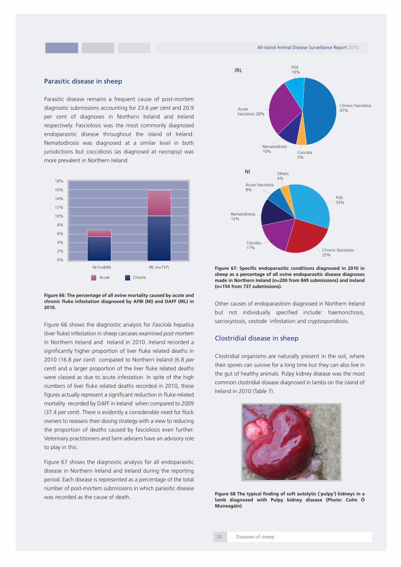
Parasitic disease in sheep
Parasitic disease remains a frequent cause of post-mortem
diagnostic submissions accounting for 23.6 per cent and 20.9
per cent of diagnoses in Northern Ireland and Ireland
respectively. Fasciolosis was the most commonly diagnosed
endoparasitic disease throughout the island of Ireland.
Nematodirosis was diagnosed at a similar level in both
jurisdictions but coccidiosis (as diagnosed at necropsy) was
more prevalent in Northern Ireland.
Figure 66: The percentage of all ovine mortality caused by acute andchronic fluke infestation diagnosed by AFBI (NI) and DAFF (IRL) in2010.
Figure 66 shows the diagnostic analysis for Fasciola hepatica
(liver fluke) infestation in sheep carcases examined post mortem
in Northern Ireland and Ireland in 2010. Ireland recorded a
significantly higher proportion of liver fluke related deaths in
2010 (16.8 per cent) compared to Northern Ireland (6.8 per
cent) and a larger proportion of the liver fluke related deaths
were classed as due to acute infestation. In spite of the high
numbers of liver fluke related deaths recorded in 2010, these
figures actually represent a significant reduction in fluke-related
mortality recorded by DAFF in Ireland when compared to 2009
(37.4 per cent). There is evidently a considerable need for flock
owners to reassess their dosing strategy with a view to reducing
the proportion of deaths caused by fasciolosis even further.
Veterinary practitioners and farm advisers have an advisory role
to play in this.
Figure 67 shows the diagnostic analysis for all endoparasitic
disease in Northern Ireland and Ireland during the reporting
period. Each disease is represented as a percentage of the total
number of post-mortem submissions in which parasitic disease
was recorded as the cause of death.
Figure 67: Specific endoparasitic conditions diagnosed in 2010 insheep as a percentage of all ovine endoparasitic disease diagnosesmade in Northern Ireland (n=200 from 849 submissions) and Ireland(n=154 from 737 submissions).
Other causes of endoparasitism diagnosed in Northern Ireland
but not individually specified include: haemonchosis,
sarcocystosis, cestode infestation and cryptosporidiosis.
Clostridial disease in sheep
Clostridial organisms are naturally present in the soil, where
their spores can survive for a long time but they can also live in
the gut of healthy animals. Pulpy kidney disease was the most
common clostridial disease diagnosed in lambs on the island of
Ireland in 2010 (Table 7).
Figure 68 The typical finding of soft autolytic (`pulpy') kidneys in alamb diagnosed with Pulpy kidney disease (Photo: Colm ÓMuireagáin)
All-island Animal Disease Surveillance Report 2010
Diseases of sheep32
18%
16%
14%
12%
10%
8%
6%
4%
2%
0%
NI (n=849)
ChronicAcute
IRL (n=737)
Chronic fasciolosis47%Acute
fasciolosis 28%
Nematodirosis10% Coccidia
5%
PGE33%
Chronic fasciolosis25%
Others4%
Acute fasciolosis8%
Nematodirosis13%
Coccidia17%
PGE10%IRL
NI
Report 2010:Layout 1 07/09/2011 13:29 Page 33
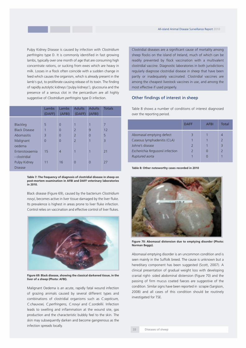
Pulpy Kidney Disease is caused by infection with Clostridium
perfringins type D. It is commonly identified in fast growing
lambs, typically over one month of age that are consuming high
concentrate rations, or sucking from ewes which are heavy in
milk. Losses in a flock often coincide with a sudden change in
feed which causes the organism, which is already present in the
lamb's gut, to proliferate causing release of its toxin. The finding
of rapidly autolytic kidneys (‘pulpy kidneys’), glucosuria and the
presence of a serous clot in the pericardium are all highly
suggestive of Clostridium perfringins type D infection.
Table 7: The frequency of diagnosis of clostridial disease in sheep onpost-mortem examination in AFBI and DAFF veterinary laboratoriesin 2010.
Black disease (Figure 69), caused by the bacterium Clostridium
novyi, becomes active in liver tissue damaged by the liver fluke.
Its prevalence is highest in areas prone to liver fluke infection.
Control relies on vaccination and effective control of liver flukes.
Figure 69: Black disease, showing the classical darkened tissue, in theliver of a sheep (Photo: AFBI).
Malignant Oedema is an acute, rapidly fatal wound infection
of grazing animals caused by several different types and
combinations of clostridial organisms such as C.septicum,
C.chauvoei, C.perfringens, C.novyi and C.sordellii. Infection
leads to swelling and inflammation at the wound site, gas
production and the characteristic bubbly feel to the skin. The
skin may subsequently darken and become gangrenous as the
infection spreads locally.
Clostridial diseases are a significant cause of mortality among
sheep flocks on the island of Ireland, much of which can be
readily prevented by flock vaccination with a multivalent
clostridial vaccine. Diagnostic laboratories in both jurisdictions
regularly diagnose clostridial disease in sheep that have been
partly or inadequately vaccinated. Clostridial vaccines are
among the cheapest livestock vaccines in use, and among the
most effective if used properly.
Other findings of interest in sheep
Table 8 shows a number of conditions of interest diagnosed
over the reporting period.
Table 8: Other noteworthy cases recorded in 2010
Figure 70: Abomasal distension due to emptying disorder (Photo:Norman Beggs).
Abomasal emptying disorder is an uncommon condition and is
seen mainly in the Suffolk breed. The cause is unknown but a
hereditary component has been suggested (Scott, 2007). A
clinical presentation of gradual weight loss with developing
cranial right- sided abdominal distension (Figure 70) and the
passing of firm mucus coated faeces are suggestive of the
condition. Similar signs have been reported in scrapie (Sargison,
2008) and all cases of this condition should be routinely
investigated for TSE.
All-island Animal Disease Surveillance Report 2010
Diseases of sheep33
Lambs Lambs Adults Adults Totals
(DAFF) (AFBI) (DAFF) (AFBI)
Blackleg 5 0 1 1 7
Black Disease 1 0 2 9 12
Abomasitis 3 0 2 0 5
Malignant 0 0 2 1 3
oedema
Enterotoxaemia 15 4 1 1 21
- clostridial
Pulpy Kidney 11 16 0 0 27
Disease
DAFF AFBI Total
Abomasal emptying defect 3 1 4
Caseous lymphadenitis (CLA) 1 1 2
Johne’s disease 2 1 3
Escherichia fergusonii infection 2 0 2
Ruptured aorta 1 0 1
Report 2010:Layout 1 07/09/2011 13:29 Page 34

Figure 71: (a) A lymph node granuloma in a case of CLA; (b) A cultureof Corynebacterium pseudotuberculosis on blood agar from the samelesion(Photos: Frank Malone).
Caseous lymphadenitis (CLA) is due to infection with
Corynebacterium pseudotuberculosis and presents as an
enlargement of the superficial lymph nodes (Figure 71) with
internal abscessation being recorded in some cases. The disease
has become important in the pedigree sector but has little
impact in commercial flocks in the Northern hemisphere.
Serological tests exist but are unreliable in individual animals
while their use for control of the disease on a group or flock
basis is somewhat more promising. Diagnosis continues to rely
primarily on clinical examination, culture of suspect lesions and
necropsy. Affected animals should be culled and control can be
achieved by:
1. Adherence to strict biosecurity measures
2. Scrupulous hygiene in routine operations which
involve injections or may cause wounds (like shearing,
tagging)
3. Possibly the use of vaccines under Special Treatment
Authorisation from the Regulatory Authorities
Figure 72: Acid fast MAP organisms within macrophages in thelamina propria of the terminal ileum, in a case of Johne’s disease insheep (Photo: Frank Malone).
Johne’s disease due to Mycobacterium avium subspecies
paratuberculosis (MAP) is an important cause of slow
progressive weight loss in adult sheep. The condition should be
considered in any cases of weight loss in adequately fed adult
sheep which are not affected by poor dentition, chronic
parasitism or lameness (Scott, 2007). Full necropsy,
histopathology (Figure 72) or culture of the organism is required
for diagnosis.
Escherichia fergusonii has been isolated from sheep with clinical
signs including abortion, diarrhoea, septicaemia and sudden
death (Voigt et al, 2009). In a number of cases where this
organism has been isolated, the significance of the isolate is not
clear. A primary or secondary pathogenic role is likely especially
in association with chronic parasitic disease. Healthy carrier
sheep have also been shown to exist (Voigt et al, 2009)
The cause of aortic rupture in sheep is unknown and the
condition is not reported in the literature.
References:
Sargison, N., 2008: Sheep Flock Health Blackwell Publishing,
Oxford, UK
Scott, P. R., 2007: Sheep Medicine, Manson Publishing, London,
UK
Voigt, K., Evans, J., MacArthur, I., Foster, G., 2009: Escherichia
fergusonii in the Scottish Highlands: Prevalence and Potential
Pathogenicity factors: Proceedings of the Sheep Veterinary
Society, 33 p 81 – 82.
All-island Animal Disease Surveillance Report 2010
Diseases of sheep34
(a)
(b)
Report 2010:Layout 1 07/09/2011 13:29 Page 35

Ovine abortion
Sporadic abortions around the beginning of the lambing period
are expected by most shepherds and their incidence can
increase with flock size. Some of these abortions can readily
be attributed to external events such as rough handling or
fighting between ewes. However, a few isolated abortions can
sometimes precipitate quickly into an outbreak or ‘abortion
storm’. Where the incidence rapidly surpasses 5 per cent of the
ewe flock or where there are a number of abortions in a short
time period, then a laboratory determination of the cause
should be attempted.
There was an increase in the number of laboratory submissions
to the DAFF laboratories, in 2010 compared to 2009, with a 33
per cent increase in the numbers of individual foetuses
examined. Attributing a reason for this significant increase is
difficult but it is likely that the high average lamb price in 2010
(as discussed on page 5) possibly played a role. Submissions for
ovine abortion frequently include multiple lamb carcases and
placental material when available.
Veterinary surgeons and shepherds are strongly urged to submit
diagnostic material from flocks experiencing abortion. Not least
because the two most common causes of abortion (enzootic
abortion and toxoplasmosis), if diagnosed, are largely
preventable by vaccination. Outlined below are the guidelines
for sampling during an abortion investigation.
Guidelines for sampling for an abortion investigation are
as follows:
1) The foetus and placenta should be submitted when
both are available
2) More than one submission is normally required to
investigate an outbreak of abortion.
3) Maternal blood samples may be useful (EAE,
Toxoplasmosis, Border Disease, Leptospira Hardjo),
to demonstrate recent acute infection. Ideally a
minimum of at least 10 per cent of the ewe flock or
10 ewes (whichever is the greater) should be blood
sampled
4) If a full carcase submission is not possible the samples
of choice are placenta (fixed and fresh), foetal
stomach contents (collected in an aseptic manner),
liver and foetal pleural fluid.
Figure 73: The relative frequency of detection of the agents whichcause ovine abortions in foetal carcase submissions in NorthernIreland (n=317) and Ireland (n=402) in 2010.
Figure 73 shows the relative frequency of detection of ovine
infectious abortifacients in foetal carcases on post-mortem
examination in Northern Ireland and Ireland in 2010. Enzootic
abortion of ewes (EAE) was the most frequently diagnosed
cause of ovine abortion in Northern Ireland, accounting for 17
per cent of diagnoses while Toxoplasmosis was the most
frequently diagnosed cause of ovine abortion in Ireland (21.4
per cent). Toxoplasmosis was a relatively frequent diagnosis in
Northern Ireland too accounting for a further 16 per cent of
diagnoses while leptospirosis (9 per cent) was also a relatively
common diagnosis in aborted ewes.
Salmonella spp. were isolated relatively frequently from ovine
foetal carcases (7.5 per cent), in the DAFF laboratories, with
Salmonella Dublin being identified in twenty nine foetal
carcases while Salmonella Typhimurium was identified in one
carcase. In Northern Ireland, the Salmonella spp. identified
included Salmonella Dublin (three cases), Salmonella
Typhimurium (one case) and Salmonella Arizonae (one case).
The category ‘other diseases’ include sporadic bacterial abortion
due to Arcanobacterium pyogenes, Bacillus licheniformis,
Mannheimia haemolytica, Staph. aureus, Streptococcal spp.
and Yersinia pseudotuberculosis.
Chlamydophila abortus infection is transmitted to uninfected
female sheep by contact with contaminated foetal membranes,
foetal fluids, discharges or bedding material. Infection in these
animals usually becomes latent but may reactivate during the
subsequent pregnancy.
All-island Animal Disease Surveillance Report 2010
Diseases of sheep35
EAE
Toxoplasma gondii
Leptospirosis
E coli
Campylobacter spp.
Listeria spp.
Salmonella spp.
Other diagnoses
Diagnosis not reached
DAFF
AFBI
5% 10% 15% 20% 25%0%
Report 2010:Layout 1 07/09/2011 13:29 Page 36

Figure 74: (a) Foetus and placenta from a case of Toxoplasmaabortion showing pinpoint necrotic foci on the placental cotyledons(Photo: AFBI). (b) A photomicrograph showing a Toxoplasma gondiiorganism (arrow) in the placental cotyledon (Photo: Cosme Sánchez-Miguel).
Both Toxoplasma gondii and Chlamydophila abortus are
zoonotic pathogens which can pose a risk to the unborn child.
Pregnant women should avoid all contact with sheep, especially
at lambing time.
Toxoplasmosis, caused by infection with Toxoplasma gondii is a
commonly diagnosed cause of abortion especially in ewe lambs
and hoggets/shearlings (Figure 74). Sheep are infected by
ingestion of oocysts present in cat faeces, usually contaminating
concentrate feed. Infection outside pregnancy usually causes
no more than a subclinical febrile condition which often escapes
detection. The consequence of infection during pregnancy
depends on the stage of gestation at which exposure occurs.
The likely consequences of T. gondii infection in sheep
Non pregnant: Acute, subclinical febrile condition
Early-mid pregnancy (<110 days): Foetal death and abortion.
Late pregnancy: Birth of a live but weak lamb.
A solid, probably life-long, immunity is established after natural
infection; hence the higher level of disease in young breeding
sheep, as older sheep tend to become exposed and resistant.
Good control can be achieved by the use of a live tachyzoite
vaccine.
Diseases of pigs
The pig sector is an important part of Irish agriculture, and the
laboratory services throughout the island provide diagnostic
support to pig producers to assist disease control, fulfil export
requirements, and improve productivity. The DAFF laboratories
are currently undertaking a programme, with expert assistance
from AFBI colleagues, of increasing and developing our existing
capacity to provide diagnostic support to pig practitioners in
Ireland. In 2010 AFBI received four hundred and twenty nine
pig carcases for examination, while the DAFF received ninety,
totalling five hundred and nineteen carcases for the island.
The most common diagnoses were pneumonia (19.7 per cent)
and gastrointestinal disease (9.2 per cent). Figure 75 and Figure
76 show the aetiological agents implicated for each,
respectively.
Figure 75: Causes of pneumonia in pigs diagnosed in 2010 (n=102).
As detailed in Figure 75 and in line with current worldwide
trends the most common respiratory pathogen isolated from
pigs was Pasteurella multocida. P. multocida is considered an
opportunistic respiratory pathogen in pigs, as pathological
infection has only been reproduced experimentally using very
high inoculations. It is hypothesised that clinical pneumonic
pasteurellosis arises where pre-existing lung lesions are
colonised by P. multocida. This secondary infection increases the
severity of pneumonia and worsens the clinical signs. This is
particularly common in cases of Mycoplasma hyopneumoniae
infection. Other primary respiratory pathogens of pigs that can
be complicated by secondary P. multocida infection include;
Swine influenza virus, porcine reproductive and respiratory
syndrome (PRRS) virus and Bordetella bronchiseptica. Therefore,
a diagnosis of P. multocida pneumonia should be considered as
an indicator of the presence of other respiratory pathogens
within the herd.
All-island Animal Disease Surveillance Report 2010
Diseases of pigs36
Pasteurellamultocida48%
Streptococcusspp4%
Mycoplasmaspp1%
Streptococcussuis13%
Mannheimia haemolytica1%
Actinobacilluspleuropneumoniae10%
H1N18%
Bordetellabronchiseptica6%
Other8%
Mycoplasmahyopneumoniae1%
(a)
(b)
Report 2010:Layout 1 07/09/2011 13:29 Page 37

Actinobacillus pleuropneumoniae was the third most common
respiratory pathogen isolated from porcine lungs and is a
primary respiratory pathogen of major importance in porcine
pneumonia worldwide. It most commonly affects growing pigs
up to six-months-old and is spread by direct contact between
infected pigs. Although the pathognomic lesions of
pleuropneumonia allow rapid diagnosis, even with treatment
A. pleuropneumoniae infection within a herd will adversely
affect growth rates as there is often incomplete resolution of
the associated pathology. In addition, treated and recovered
animals can maintain subclinical infection in the tonsils for up
to six months, acting as a continuing source of infection within
the herd. A diagnosis of A. pleuropneumoniae disease should
be complemented by serotyping of the agent, as immunity to
one serotype is only partially protective against other serotypes.
This is particularly relevant in vaccinated herds as vaccines are
only protective against homologous serotypes.
Figure 76: Causes of enteric disease in pigs diagnosed in 2010 (n=48).
Figure 77: An ulcer (arrow) in the stomach of a 22-month-old pig(Photo: Colm Ó Muireagáin).
Porcine circovirus 2 (PCV2), the aetiologic agent of postweaning
multisystemic wasting syndrome (PMWS), was detected in thirty
two cases by AFBI, while one PMWS case was diagnosed at
post-mortem examination by DAFF. PCV2 infection alone is not
confirmatory of PMWS; clinical and histopathological findings
are also required to confirm a diagnosis.
Figure 78: Severe congestion of the intestines in a pig due tointestinal torsion (Photo: AFBI)
Diagnosis DAFF AFBI Total
Endocarditis 1 1 2
Meningitis 4 13 17
Nephritis 1 2 3
Pericarditis 1 0 1
Polyserositis 2 8 10
Septicaemia 9 4 13
Exudative dermatitis 1 0 1
PDNS 1 2 3
Streptococcus suis 6 21 27
Mulberry heart disease 0 3 3
Liver Lobe Torsion 0 2 2
Table 9: Other pig diagnoses made in 2010.
Streptococcus suis is a common but serious infection of pigs;
serotypes two and nine are particularly severe and can be
zoonotic. Twenty seven cases of Streptococcus suis infection
(5.2 per cent of cases) were detected in 2010 (Table 9).
Twenty cases of abortion were submitted to AFBI; two were
caused by parvovirus, two by Leptospira, while no significant
pathogen was identified in the remainder. The DAFF
laboratories identified Leptospira canicola as the cause of
abortion in one case.
All-island Animal Disease Surveillance Report 2010
Diseases of pigs37
Other4%
Oedema disease9%
E coli (0148)20%
Coliforms 7%
Rotavirus5%
Cryptosporidiumparvum7%
Gastric ulcer13%
Intestinal torsion22%
Coccidiosis 2%
Escherichia coli11%
Report 2010:Layout 1 07/09/2011 13:29 Page 38

Diseases of poultry
The majority of poultry submissions to the laboratories come
from commercial poultry enterprises and usually consist of
multiple carcases. For this reason the diagnoses and numbers
given here relate to the numbers of submissions with a
particular diagnosis and not to individual carcase diagnoses.
In 2010 there were one thousand and eighty five chicken
submissions to the AFBI laboratories; five hundred and three
were examined for intestinal parasites, three hundred and
seventeen underwent statutory Salmonella spp. sampling and
two hundred and sixty five had post-mortem examinations
performed. There were one hundred and sixty two submissions
representing two hundred and ninety chicken carcases to the
DAFF laboratories in 2010.
Figure 79: The most frequently diagnosed conditions on post-mortemexamination in poultry in AFBI laboratories in 2010 (n=265).
Figure 79 presents the most common post-mortem diagnoses
in poultry submissions from the AFBI laboratories in 2010.
Bacterial hepatitis was the most frequently diagnosed condition
(11.3 per cent) while colisepticaemia was diagnosed in a further
8.3 per cent of carcases examined. Respiratory disease (7.5 per
cent) encompasses a number of conditions which included
infectious laryngotracheitis (Gallid Herpesvirus 1) which was
identified in five submissions. Various bursal lesions were also
detected, which included submissions confirmed with infectious
bursal disease (Gumboro disease, Avian birnavirus).
Figure 80: The most frequently diagnosed conditions on post-mortemexamination in poultry in DAFF laboratories in 2010 (n=162).
The most common diagnoses in DAFF poultry submissions are
presented in Figure 80. Respiratory disease (14.2 per cent) and
peritonitis (11.7 per cent) were the most common findings. One
respiratory submission was confirmed as infectious
laryngotracheitis and two submissions had infectious bronchitis
virus confirmed. Infectious bronchitis is contracted by the
inhalation or conjunctival route and is highly contagious. Typically
the disease is accompanied by mild respiratory signs such as
sneezing and a watery discharge from the eyes and nostrils. Co-
infection with Mycoplasma gallisepticum or Mycoplasma synoviae
can cause more severe clinical signs.
Less frequent diagnoses were coccidiosis (2.5 per cent) and broiler
ascites syndrome (2.5 per cent). Broiler ascites syndrome is a
condition commonly referred to as ‘waterbelly’. There are many
possible causes but fibrosis of the liver, heart failure, sodium
toxicity or respiratory disease may all contribute to the occurrence
of the condition. Broilers can die suddenly, often without the
ascites being clinically apparent. On post-mortem examination
the heart may be enlarged and the right ventricle may be
thickened. Control requires examination of underlying disease or
stress factors in the birds’ environment.
AFBI offers screening for parasites in commercial poultry. A total
of five hundred and three submissions was examined in 2010 and
clinically significant levels of parasites were detected in just fifty
submissions; thirty one had Heterakis spp., twelve had Ascarid
spp. and seven had Capillaria spp. infections. These results reflect
the high levels of parasite control in commercial poultry.
The frequency of some of the other common poultry diagnoses
made by AFBI and DAFF laboratories in 2010 are presented in
Table 10.
All-island Animal Disease Surveillance Report 2010
Diseases of poultry38
Bacterial hepatitis
Colisepticaemia
Respiratory disease
Bursal lesions
Peritonitis
Rickets
Ulcerative ventriculitis
Marek's disease
Encephalomalacia
No lesions
Other diagnoses
Respiratory disease
Peritonitis
Hepatitis
Septicaemia/colisepticaemia
Enteritis
Heart Failure
Coccidiosis
Yolk Sac Infection
Broiler Ascites Syndrome
No Lesions
Other diagnoses
0% 5% 10% 15% 20% 25% 30%
0% 20% 30% 40%10%
Report 2010:Layout 1 07/09/2011 13:29 Page 39

Diagnosis AFBI DAFF
submissions submissions
Ascites 2 4
Coccidiosis 6 4
Hepatic lipidosis 5 0
Tendon rupture 4 0
Inclusion body hepatitis 4 0
Tenosynovitis 3 0
Gout 3 1
Haemorrhage 1 3
Pericarditis 0 3
Gastrointestinal impaction 0 3
Table 10: The frequency of some of the other common poultrydiagnoses to AFBI and DAFF laboratories.
Enterococcus encephalomalacia caused by Enterococcus hirae
was diagnosed in five submissions. Enterococcus spp. are
present worldwide and ubiquitous in poultry environments.
Typically encephalomalacia is seen in very young chicks
presenting with neurological signs. There are no gross lesions.
Histologically there is bilateral symmetrical encephalomalacia of
the cerebrum and pons with microthrombi within the blood
vessels. Culture of Enterococcus hirae from the brain aids
diagnosis.
Figure 81: Pathognomic intranuclear inclusion bodies in the gizzardepithelium (arrow) of a hen with adenoviral ventricular ulceration(Photo: Margaret Wilson).
Adenoviral ventricular ulceration was first described in
commercial poultry in the early part of this century by Japanese
researchers. AFBI have seen a low but increasing number of
submissions for the past five years. There were ten submissions
to AFBI in 2010 in which this condition was suspected and three
of these had confirmed adenoviral aetiology. Typically, the
disease presents as reduced growth rates with gizzard erosions
and ulcerations in young broilers. Histologically there is erosion
and ulceration of the koilin layer with transmural inflammation.
Definitive diagnosis requires recognition of pathognomic
intranuclear inclusion bodies in the gizzard epithelium (Figure
81).
Backyard poultry
The husbandry of small, usually free range backyard, poultry
flocks is less intensive and fundamentally different from
commercial poultry production. In general, backyard poultry live
and lay eggs for longer; they often have access to wild birds
and are rarely vaccinated. The types of diseases and their
implications for flock health in backyard poultry are quite
different to those of commercial poultry. An outline of some
common conditions in backyard flocks follows.
Respiratory disease can present as nasal discharge, sneezing,
listlessness and reduced egg production. There are many causes
of respiratory disease in poultry. Infectious Laryngotracheitis
(Gallid Herpesvirus 1) causes acute disease with secondary
bacterial complication. Pasteurellosis (Pasteurella multocida) is
usually subacute and can be accompanied by swollen sinuses.
Gapes , where a nematode worm (Syngamus trachea) resides in
the trachea is associated with open mouth breathing, while
Aspergillus spp. pneumonia and airsacculitis tend to have a
chronic course and are associated with dusty environments.
Figure 82: (a) The classical clinical sign of unilateral leg paralysis in ahen with Marek’s disease. (b) An enlarged sciatic nerve (top) and anormal sciatic nerve (bottom) from a hen with Marek’s Disease(Photo: Jim O Donovan).
Marek’s disease is one of the common avian viral diseases seen
in backyard flocks. Typically it affects younger birds and presents
as paralysis (Figure 82) with perineuritis or as lymphoid tumours
in organs such as liver or kidney.
All-island Animal Disease Surveillance Report 2010
Diseases of poultry39
(a)
(b)
Report 2010:Layout 1 07/09/2011 13:29 Page 40
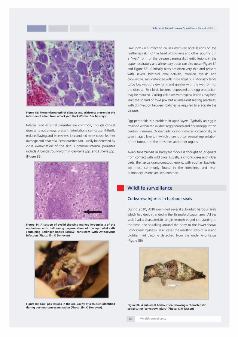
Figure 83: Photomicrograph of Eimeria spp. schizonts present in theintestine of a hen from a backyard flock (Photo: Ger Murray).
Internal and external parasites are common, though clinical
disease is not always present. Infestations can cause ill-thrift,
reduced laying and listlessness. Lice and red mites cause feather
damage and anaemia. Ectoparasites can usually be detected by
close examination of the skin. Common internal parasites
include Ascarids (roundworms), Capillaria spp. and Eimeria spp.
(Figure 83).
Figure 84: A section of eyelid showing marked hyperplasia of theepithelium with ballooning degeneration of the epithelial cellscontaining Bollinger bodies (arrow) consistent with Avipoxvirusinfection (Photo: Jim O Donovan).
Figure 85: Fowl pox lesions in the oral cavity of a chicken identifiedduring post-mortem examination (Photo: Jim O Donovan).
Fowl pox virus infection causes wart-like pock lesions on the
featherless skin of the head of chickens and other poultry, but
a "wet" form of the disease causing diptheritic lesions in the
upper respiratory and alimentary tracts can also occur (Figure 84
and Figure 85). Clinically birds are often very thin and present
with severe bilateral conjunctivitis, swollen eyelids and
conjunctival sacs distended with inspissated pus. Mortality tends
to be low with the dry form and greater with the wet form of
the disease. Sick birds become depressed and egg production
may be reduced. Culling sick birds with typical lesions may help
limit the spread of fowl pox but all-in/all-out rearing practices,
with disinfection between batches, is required to eradicate the
disease.
Egg peritonitis is a problem in aged layers. Typically an egg is
retained within the oviduct (egg bound) and fibrinosuppurative
peritonitis ensues. Oviduct adenocarcinoma can occasionally be
seen in aged layers, in which there is often serosal implantation
of the tumour on the intestines and other organs.
Avian tuberculosis in backyard flocks is thought to originate
from contact with wild birds. Usually, a chronic disease of older
birds, the typical granulomatous lesions, with acid fast bacteria,
are most commonly found in the intestines and liver;
pulmonary lesions are less common.
Wildlife surveillance
Corkscrew injuries in harbour seals
During 2010, AFBI examined several sub-adult harbour seals
which had dead stranded in the Strangford Lough area. All the
seals had a characteristic single smooth edged cut starting at
the head and spiralling around the body to the lower thorax
(‘corkscrew injuries’). In all cases the resulting strip of skin and
blubber had become detached from the underlying tissue
(Figure 86).
Figure 86: A sub adult harbour seal showing a characteristicspiral cut or ‘corkscrew injury’ (Photo: Cliff Mason)
All-island Animal Disease Surveillance Report 2010
Wildlife surveillance40
Report 2010:Layout 1 07/09/2011 13:30 Page 41

These injuries were considered to be similar to those described
in seals found on the beaches of eastern Scotland and along
the Norfolk coast in England. The injuries are considered
consistent with seals being drawn through a ducted propeller
such as a Kort nozzle or some types of Azimuth thrusters, which
are common means of propulsion and manoeuvre in a variety
of coastal and sea-going vessels.
Suspected cases of wildlife poisoning in Ireland
Figure 87: Various baits used in the poisoning of wildlife which wererecovered in the course of AFBI investigations of suspected wildlifepoisoning. The black substance was analysed and found to containcarbofuran (Photo: AFBI).
Suspected cases of wildlife poisoning in Northern Ireland are
processed and analysed by AFBI as part of the Wildlife Incident
Investigation Scheme (WIIS). The scheme is overseen and run
by the Chemical Regulations Directorate (CRD) of the United
Kingdom Health and Safety Executive. The purpose of this
scheme is to investigate deaths of wildlife and occasionally pets
where there is evidence that pesticide poisoning may have
occurred. AFBI regularly performs post-mortem examinations
on wildlife where poisoning is suspected and samples taken
from carcases are subjected to a range of toxicological analyses
(Figure 87). The results of these investigations are used to
monitor pesticide use and to enforce legislation on the
protection of humans, animals, food and the environment from
pesticides.
Figure 88: A buzzard found poisoned and submitted to Sligo RVL.Subsequent toxicological examination of tissue samples confirmeddeath was due to carbofuran poisoning (Photo: Mícheál Casey).
DAFF RVLs have provided a post-mortem diagnostic service on
wild birds of prey (both native and re-introduced) on a case-by-
case basis and hope to agree a protocol with the State
Laboratory and the National Parks and Wildlife Service in 2011
which will put this work on a more formal footing. Three state-
supported reintroduction projects have been managed by The
Golden Eagle Trust, a private charity (Red Kites in Wicklow and
Dublin, Golden Eagles in Donegal, and White-tailed Eagles in
Co. Kerry). All three projects have experienced poisoning
incidents, and DAFF RVLs have assisted by performing post-
mortem examinations and the submission of samples for
toxicology in 2010 (Figure 88). This work has highlighted the
range of poisons being used in Ireland. In all cases it was
believed that the raptors were accidentally poisoned by people
targeting other species (mainly foxes, crows), and the Golden
Eagle Trust has undertaken an education programme, including
a leaflet on the safe and legal control of foxes and crows at
lambing time. The work of the DAFF RVLs has informed this
educational work, as well as some recent changes in the
legislation to prevent the use of some of the poisons detected.
Trichinella surveillance in wildlife (foxes)
A Trichinella risk-based wildlife monitoring programme is in
place in both Northern Ireland and Ireland, both being classed
as jurisdictions in which the risk of Trichinella infestation in
domestic pigs is officially recognised as negligible.
Figure 89: A map of the island of Ireland showing the locations wherefoxes were collected for the Trichinella surveys in 2010 (n=653).Larger dots represent locations where more than one fox wascollected. The sites where the positive foxes were located are markedin red (n=4) (Map: Guy McGrath, CVERA and UCD)
All-island Animal Disease Surveillance Report 2010
Wildlife surveillance41
Report 2010:Layout 1 07/09/2011 13:30 Page 42

In 2010, six hundred and fifty three foxes were collected from
multiple locations across the island of Ireland and submitted to
contributing laboratories (one hundred and sixty seven by two
AFBI laboratories and four hundred and eighty six by six DAFF
laboratories).
Figure 89 shows the origin of the foxes collected. Muscle
samples from specific locations in the body (cheek, forelimb,
tongue and diaphragm) were collected from each fox and these
were analysed microscopically for the presence of Trichinella
spiralis larvae following a pepsin digestion procedure. Four foxes
(one each from counties Kildare, Cork, Limerick and Wicklow)
were found to be positive for Trichinella spiralis larvae.
Bovine tuberculosis (bTB) surveillance in badgers
In Ireland, the regional veterinary laboratories participate in the
screening of badgers for bovine TB. This includes the
examination of badgers found on the road (i.e. `road-kill') as
well as a proportion of those snared under licence and
submitted by district veterinary offices. In 2010 a total of two
hundred and twenty six badgers was examined post mortem
by the regional veterinary laboratories and bovine TB was
diagnosed in twenty two of these (9.7 per cent). It should be
noted that this represents only a small proportion of those
tested under the ERAD scheme.
Parasitic diseases
The number of faecal samples submitted to DAFF veterinary
laboratories for parasitological examinations increased
dramatically in 2010. As an example of this, Figure 90 shows
the rise in bovine faecal sample numbers submitted for liver
fluke egg examination in the years 2006 to 2010.
Figure 90: The number of faecal samples submitted for liver fluke eggdetection to DAFF Regional Veterinary Laboratories in the years 2006to 2010.
Liver and rumen fluke infections
In 2010 a combined total of fifteen thousand five hundred and
forty eight bovine faecal samples was analysed by AFBI and
DAFF laboratories for the presence of liver fluke eggs. Of these,
two thousand seven hundred and eleven (17.4 per cent) were
positive. A further fifteen thousand one hundred and ninety five
samples were examined for rumen fluke eggs and five thousand
six hundred and seventy nine (37.4 per cent) were positive. The
frequency of detection was remarkably similar for both liver
fluke and rumen fluke eggs in both the DAFF and AFBI
laboratories. Figure 91 illustrates the change in the proportion
of positive results by quarter during 2010.
Figure 91: The percentage of bovine faecal samples positive for flukeeggs by quarter during 2010 (AFBI: n=3,494; DAFF: n = 12,054).
The highest incidence of rumen fluke detection coincided with
the periods of highest rainfall (see Figure 10). Liver fluke
detection in cattle decreased progressively from the first to the
fourth quarter. This trend differs considerably from that
witnessed in sheep (Figure 93) and may suggest an increasing
awareness on the part of cattle herdowners of the importance
of efficient liver fluke management.
Figure 92: A rumen fluke (Paramphistomum spp.) (Photo: CosmeSánchez-Miguel)
All-island Animal Disease Surveillance Report 2010
42
18000
16000
14000
12000
10000
8000
6000
4000
2000
0
2006
Bovine Ovine
2007 2008 2009 2010
Liver Fluke Rumen Fluke
45%
40%
35%
30%
25%
20%
15%
10%
5%
0%
Q1 Q2 Q3 Q4
Parasitic diseases
Report 2010:Layout 1 07/09/2011 13:30 Page 43

It should be stressed that the finding (or failure to find) fluke
eggs in a faecal sample needs careful examination. Both liver
fluke and rumen fluke (Figure 92) are capable of causing
significant clinical disease before either parasite is mature
enough to lay eggs (pre-patent disease). On the other hand, a
large proportion of those animals with positive rumen fluke
results show few if any clinical signs of disease.
In terms of mortality, liver fluke infestation is a much more
serious threat to livestock (see Diseases of cattle on page7) than
rumen fluke infestation. The flukicides used for the treatment
of paramphistomes are not effective against immature Fasciola
hepatica. If a herd or flock owner focuses on paramphistome
control to the neglect of Fasciola hepatica control serious losses
may occur.
The clinical significance of paramphistomosis (rumen fluke
infestation) has only become apparent in recent years and
accurate data on its true prevalence and economic impact is
lacking. Heavy infestation can lead to illthrift but mortality
attributable to paramphistomosis is uncommon - in 2010 in
Northern Ireland one bovine post-mortem examination
recorded paramphistomosis as the cause of death while in
Ireland it was not recorded as the cause of death in any bovine
post-mortem examination performed. Collaborative studies
may be undertaken by AFBI and DAFF aimed at a better
understanding of the prevalence, risk factors for, and economic
impact of rumen fluke infestation in Ireland.
Figure 93: The percentage of ovine faecal samples positive for flukeeggs by quarter in 2010 (AFBI: n=1,715; DAFF: n= 965)
A combined total of two thousand six hundred and eighty ovine
faecal samples were analysed for the presence of liver fluke
eggs in 2010. Fluke eggs were detected in three hundred and
three (15.4 per cent - 14.1 per cent in AFBI labs and 23.7 per
cent in DAFF labs). A further one thousand seven hundred and
seven samples (1,450 by AFBI and 257 by DAFF) were examined
for rumen fluke eggs and four hundred and nine (24.0 per cent)
were positive (25.9 per cent in AFBI labs and 12.8 per cent in
DAFF labs). The differences in detection frequency of liver fluke
and rumen fluke eggs between the laboratories may be due, in
part, to the fact that the number of samples analysed in the
DAFF laboratories was low in comparison to the AFBI
laboratories. Figure 93 illustrates the change in the proportion
of positive results by quarter.
Gastro-intestinal parasitic infections
In 2010, a combined total of fourteen thousand nine hundred
and fifteen bovine and three thousand one hundred and twenty
four ovine faecal samples were examined by AFBI and DAFF
laboratories for strongyle eggs. A strongyle burden of greater
than five hundred eggs per gram of faeces is considered clinically
significant. Based on this, 5.1 per cent of bovine (8.2 per cent in
AFBI and 4.1 per cent in DAFF laboratories) and 20.8 per cent of
ovine samples (20.4 per cent in AFBI labs and 21.6 per cent in
DAFF labs) were recorded as having a significant strongyle worm
burden. Figure 94 illustrates the combined quarterly results of
both DAFF and AFBI for both bovine and ovine species.
Figure 94: The percentage of bovine (AFBI: n=3,593; DAFF: n=11,322)and ovine (AFBI: n=2,003; DAFF: n=1,121) faecal samples which werepositive (>=500 eggs per gram) for strongyle eggs in 2010.
The detection of strongyle eggs is consistently higher in sheep
when compared to cattle. There may be a number of reasons
for this, such as inherent resistance, age profile of the animals
sampled, type of pasture grazed etc. but it may also point
towards a greater focus on parasite control in cattle herds and
suggests that this is an area which requires further attention
among sheep producers.
Lungworm infections
The Baermann technique was used to examine a combined
total of three thousand and ninety nine bovine faecal samples
(408 by AFBI and 2,683 by DAFF) for the presence of lungworm
larvae (primarily Dictyocaulus viviparus) (Figure 95). Eighty seven
(2.8 per cent) were positive (13.7 per cent in AFBI and 1.4 per
cent in DAFF laboratories). The difference in detection rates,
while large, is most probably due to the more stringent selection
of animals to be tested by practitioners in Northern Ireland.
Often requests for this test come from practitioners treating
animals displaying clinical signs of respiratory distress such that
the proportion of positive results may be distorted due to the
type of animal sampled. Figure 96 illustrates the trend by
quarter.
All-island Animal Disease Surveillance Report 2010
Parasitic diseases43
Q1 Q2 Q3 Q4
Bovine Ovine
Q1 Q2 Q3 Q4
Liver Fluke Rumen Fluke
45%
40%
35%
30%
25%
20%
15%
10%
5%
0%
30%
25%
20%
15%
10%
5%
0%
Report 2010:Layout 1 07/09/2011 13:30 Page 44

Figure 95: A photomicrograph of a bovine bronchus showing across-section of a lungworm larva (arrow) (Photo: CosmeSánchez Miguel).
Figure 96: The percentage of bovine faecal samples positive forlungworm larvae by quarter in 2010.
Coccidiosis
Eimeria spp. are protozoan parasites that can cause significant
disease (coccidiosis) in young animals. While disease in older
bovine animals is relatively uncommon, diarrhoea caused by
Eimeria spp. is common in calves between one and six months
of age that are exposed to large numbers of oocysts. This may
occur due to poor hygiene or management practices such as
the failure to provide clean dry bedding, failure to disinfect
between batches or the mixing of different age groups during
housing.
Figure 97: The percentage of bovine faecal samples positive forcoccidial oocysts (n=12,750).
In 2010, twelve thousand seven hundred and fifty faecal
samples were tested for the presence of coccidia (three
thousand three hundred and thirteen by AFBI and nine
thousand four hundred and thirty seven by DAFF). Of these,
one thousand six hundred and seventy six samples (13.1 per
cent) were positive (20.9 per cent in AFBI and 10.4 per cent in
DAFF laboratories). However the majority of these positive
samples (66 per cent) contained very small numbers of coccidial
oocysts and are unlikely to have been solely responsible for
significant disease. Figure 97 illustrates the results.
The results of faecal coccidial oocyst counts in cattle and sheep
should be interpreted with caution. Only two (Eimeria bovis and
Eimeria zuernii) out of twelve species of bovine coocidia and
three (Eimeria crandallis, Eimeria ovinoidalis and Eimeria ahsata)
out of twelve ovine coccidia species are pathogenic. Some of
the non-pathogenic or weakly pathogenic species (e.g. E ovina)
are capable of producing massive numbers of oocysts. These
species can also produce dense focal lesions containing large
numbers of oocysts in the small intestine which are visible on
post-mortem examination. Large numbers of oocysts can be
produced daily from these focal lesions without any clinical
effect on the host. As oocysts are prevalent in faeces of sheep
of all ages, coccidiosis cannot be diagnosed based solely on the
finding of oocysts and the clinical presentation and history
should also be considered.
Other parasitic diseases
Only two outbreaks of sheep scab (psoroptic mange in sheep,
caused by Psoroptes ovis) were confirmed by laboratory testing
in 2010, both of these by DAFF laboratories in Ireland. Four
outbreaks of psoroptic mange in cattle were detected (one in
Northern Ireland and three in Ireland). Presented below is an
investigation conducted by Limerick Regional Veterinary
Laboratory (RVL) into one of these outbreaks.
Psoroptic mange in a beef herd
Limerick RVL visited a beef unit in January 2010 to investigate
a problem of severe pruritis in a group of housed bullocks. The
farmer normally purchased weanlings and store cattle in the
spring each year, grazed them and then finished the cattle over
the winter period.
In the spring of 2009 he purchased a group of cattle from a
mart. One of the animals (animal A) began to lose its hair over
the following summer and the autumn period and was housed
with other cattle in a slatted unit in early winter. Some weeks
later the owner noticed that a number of the housed cattle
were licking and scratching excessively. An ivermectin pour-on
product was administered to the group but did not appear to
remedy the situation.
All-island Animal Disease Surveillance Report 2010
Parasitic diseases44
Q1 Q2 Q3 Q4
4%
4%
3%
3%
2%
2%
1%
1%
0
Light 9.0%
Moderate 2.3%
Heavy 0.9%
Severe 0.9%
Not Detected86.8%
Positive13.1%
Report 2010:Layout 1 07/09/2011 13:30 Page 45

Animal A was clinically depressed and drooling from the mouth
on the day of the farm investigation (Figure 98).
Figure 98: Extensive hair loss in a bullock (Animal A) with psoropticmange (Photo: Alan Johnson).
Other animals affected had localised lesions of moist dermatitis
and hair loss (Figure 99), mostly on the dorsal parts of the trunk.
Figure 99: Moist dermatitis and hair loss in a bullock with psoropticmange (Photo: Alan Johnson).
Skin scrapings were taken from animal A and a small number
of the other affected cattle. A diagnosis of psoroptic mange
was made following microscopic examination of the scrapings
and identification of Psoroptes ovis mites (Figure100).
Figure 100: Psoroptes ovis mite seen in a skin scraping taken fromAnimal A (Photo: Brendan Crowe).
Treatment with two injections of 1 per cent w/v doramectin,
with a fourteen-day interval between injections, was
recommended. Animal A did not recover however, and died one
week after the farm visit. The other animals did respond well
and the scratching and licking reduced dramatically over the
following weeks. The source of the outbreak was not
determined. However an assumption was made that animal A
was the original source of the infestation. This animal had been
purchased at the mart from a farmer who was involved in the
business of purchasing, storing and selling sheep wool each
year. One theory considered during this investigation was that
the animal had come into contact with some stored wool from
a sheep infected with sheep scab. However the P. ovis mite
associated with bovine psoroptic mange is a different (cattle-
adapted) strain to that which causes sheep scab, so it was
considered unlikely that cross-infection occurred.
Anthelmintic resistance in Ireland
Anthelmintic resistance in Ireland is a growing problem. Limited
evidence derived from an AFBI questionnaire circulated in 2005,
and a field survey on eight farms conducted in 2006 suggested
that significant levels of resistance to the benzimidazoles,
levamisole and macrocyclic lactones were already established
at that time. Currently the returns from a new and extensive
Northern Ireland wide questionnaire are being analysed, and
studies on the efficacy of anthelmintic regimes used by farmers
to control gastrointestinal nematode infestations and liver fluke
on over one hundred sheep and cattle farms throughout the
province are being carried out. A detailed comparative field
study on anthelmintic efficacy against nematodes and fluke is
also in progress on a limited number of selected sheep farms
covering most of the important production systems and areas
in Northern Ireland. Our present aim is to establish a data base
of anthelmintic resistance, to develop effective protocols for
rapid diagnosis in nematode and fluke infestations and to
provide timely and up-to-date advice on effective control
strategies on a farm-to-farm basis.
Antimicrobial susceptibility profiles
Resistance to antimicrobials is a serious concern in human and
veterinary medicine. Increasing resistance within microbial
populations to commonly used antibiotics is a global problem.
Some resistant bacteria have zoonotic potential, and furthermore,
as similar bacteria are found in humans and animals, resistance
mechanisms developed by bacterial populations in animals may
be transmitted to their counterparts in humans.
The ability of micro-organisms to adapt to their environment,
together with the widespread usage of antibiotics has meant that
some bacteria have developed resistance to certain commonly
used antimicrobial drugs thereby limiting their efficacy.
All-island Animal Disease Surveillance Report 2010
Antimicrobial susceptibility profiles45
Report 2010:Layout 1 07/09/2011 13:30 Page 46
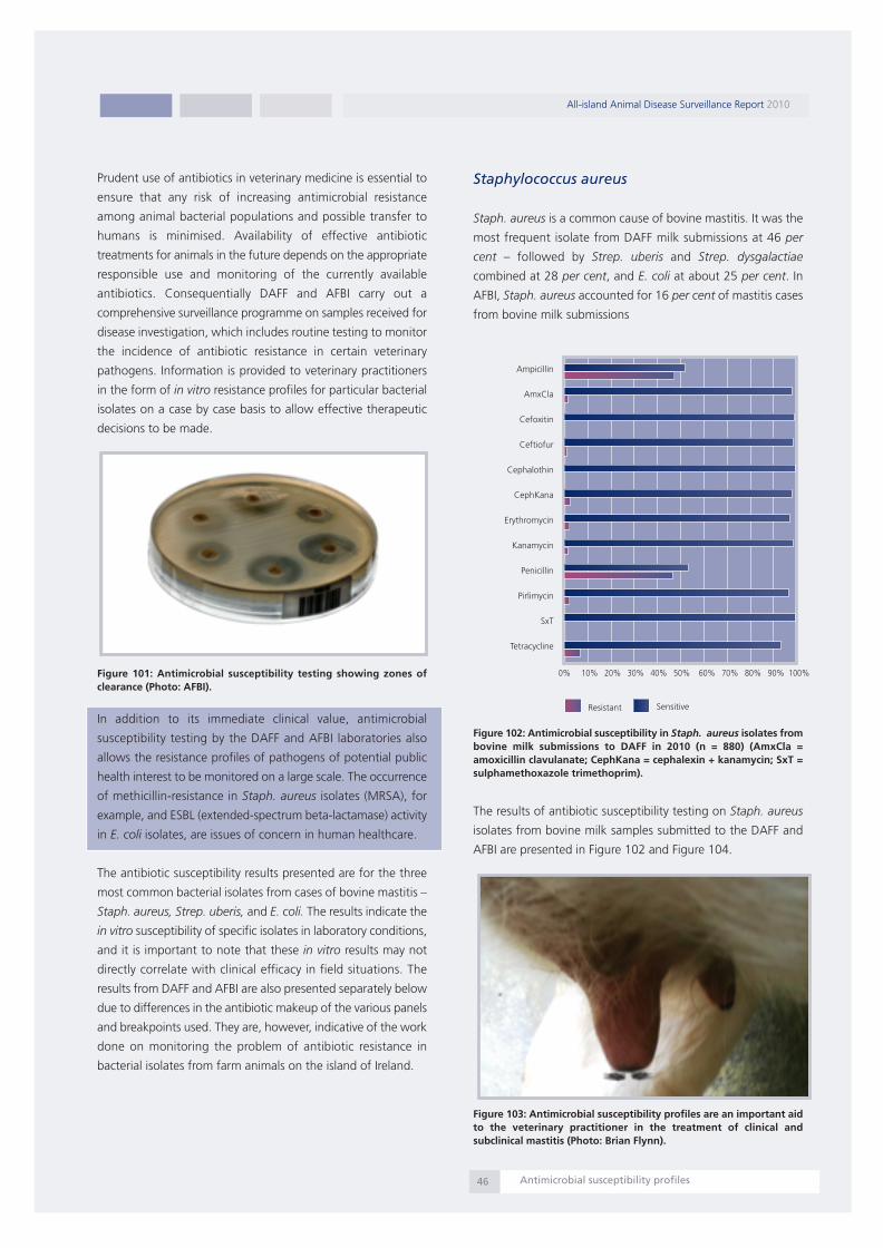
Prudent use of antibiotics in veterinary medicine is essential to
ensure that any risk of increasing antimicrobial resistance
among animal bacterial populations and possible transfer to
humans is minimised. Availability of effective antibiotic
treatments for animals in the future depends on the appropriate
responsible use and monitoring of the currently available
antibiotics. Consequentially DAFF and AFBI carry out a
comprehensive surveillance programme on samples received for
disease investigation, which includes routine testing to monitor
the incidence of antibiotic resistance in certain veterinary
pathogens. Information is provided to veterinary practitioners
in the form of in vitro resistance profiles for particular bacterial
isolates on a case by case basis to allow effective therapeutic
decisions to be made.
Figure 101: Antimicrobial susceptibility testing showing zones ofclearance (Photo: AFBI).
In addition to its immediate clinical value, antimicrobial
susceptibility testing by the DAFF and AFBI laboratories also
allows the resistance profiles of pathogens of potential public
health interest to be monitored on a large scale. The occurrence
of methicillin-resistance in Staph. aureus isolates (MRSA), for
example, and ESBL (extended-spectrum beta-lactamase) activity
in E. coli isolates, are issues of concern in human healthcare.
The antibiotic susceptibility results presented are for the three
most common bacterial isolates from cases of bovine mastitis –
Staph. aureus, Strep. uberis, and E. coli. The results indicate the
in vitro susceptibility of specific isolates in laboratory conditions,
and it is important to note that these in vitro results may not
directly correlate with clinical efficacy in field situations. The
results from DAFF and AFBI are also presented separately below
due to differences in the antibiotic makeup of the various panels
and breakpoints used. They are, however, indicative of the work
done on monitoring the problem of antibiotic resistance in
bacterial isolates from farm animals on the island of Ireland.
Staphylococcus aureus
Staph. aureus is a common cause of bovine mastitis. It was the
most frequent isolate from DAFF milk submissions at 46 per
cent – followed by Strep. uberis and Strep. dysgalactiae
combined at 28 per cent, and E. coli at about 25 per cent. In
AFBI, Staph. aureus accounted for 16 per cent of mastitis cases
from bovine milk submissions
Figure 102: Antimicrobial susceptibility in Staph. aureus isolates frombovine milk submissions to DAFF in 2010 (n = 880) (AmxCla =amoxicillin clavulanate; CephKana = cephalexin + kanamycin; SxT =sulphamethoxazole trimethoprim).
The results of antibiotic susceptibility testing on Staph. aureus
isolates from bovine milk samples submitted to the DAFF and
AFBI are presented in Figure 102 and Figure 104.
Figure 103: Antimicrobial susceptibility profiles are an important aidto the veterinary practitioner in the treatment of clinical andsubclinical mastitis (Photo: Brian Flynn).
All-island Animal Disease Surveillance Report 2010
Antimicrobial susceptibility profiles46
Ampicillin
AmxCla
Cefoxitin
Ceftiofur
Cephalothin
CephKana
Erythromycin
Kanamycin
Penicillin
Pirlimycin
SxT
Tetracycline
SensitiveResistant
0% 10% 20% 30% 40% 50% 60% 70% 80% 90% 100%
Report 2010:Layout 1 07/09/2011 13:30 Page 47
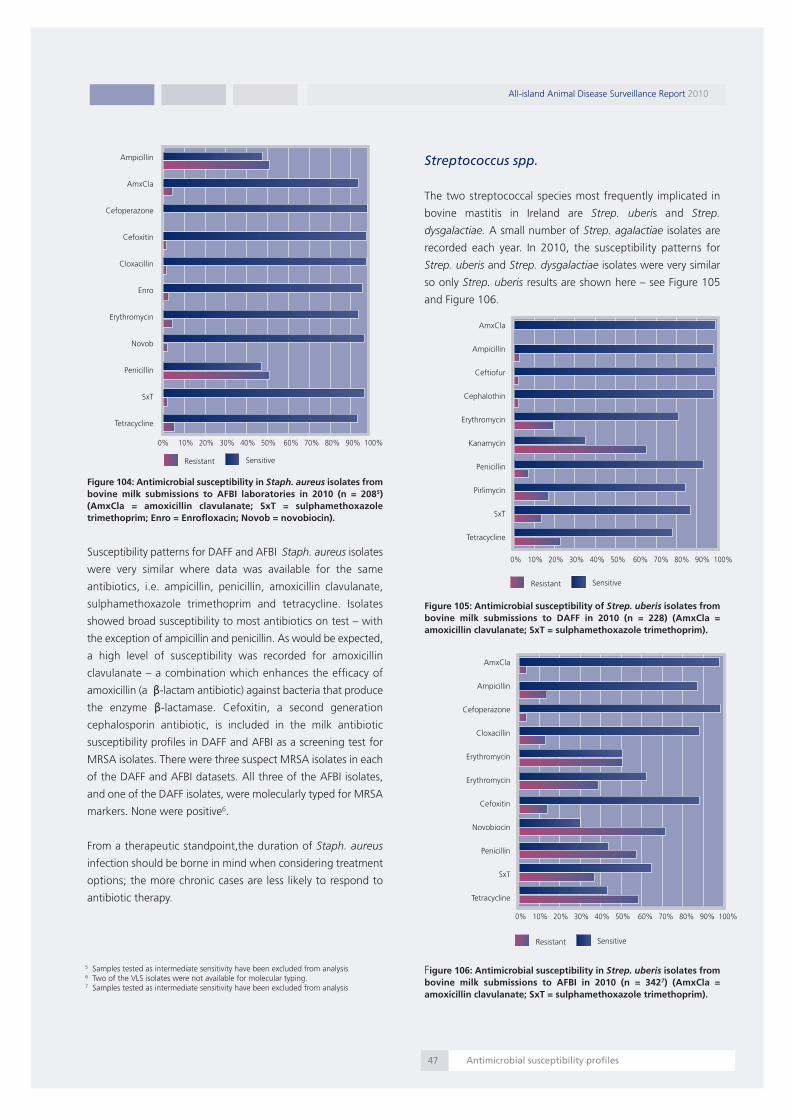
Figure 104: Antimicrobial susceptibility in Staph. aureus isolates frombovine milk submissions to AFBI laboratories in 2010 (n = 2085)(AmxCla = amoxicillin clavulanate; SxT = sulphamethoxazoletrimethoprim; Enro = Enrofloxacin; Novob = novobiocin).
Susceptibility patterns for DAFF and AFBI Staph. aureus isolates
were very similar where data was available for the same
antibiotics, i.e. ampicillin, penicillin, amoxicillin clavulanate,
sulphamethoxazole trimethoprim and tetracycline. Isolates
showed broad susceptibility to most antibiotics on test – with
the exception of ampicillin and penicillin. As would be expected,
a high level of susceptibility was recorded for amoxicillin
clavulanate – a combination which enhances the efficacy of
amoxicillin (a β-lactam antibiotic) against bacteria that produce
the enzyme β-lactamase. Cefoxitin, a second generation
cephalosporin antibiotic, is included in the milk antibiotic
susceptibility profiles in DAFF and AFBI as a screening test for
MRSA isolates. There were three suspect MRSA isolates in each
of the DAFF and AFBI datasets. All three of the AFBI isolates,
and one of the DAFF isolates, were molecularly typed for MRSA
markers. None were positive6.
From a therapeutic standpoint,the duration of Staph. aureus
infection should be borne in mind when considering treatment
options; the more chronic cases are less likely to respond to
antibiotic therapy.
Streptococcus spp.
The two streptococcal species most frequently implicated in
bovine mastitis in Ireland are Strep. uberis and Strep.
dysgalactiae. A small number of Strep. agalactiae isolates are
recorded each year. In 2010, the susceptibility patterns for
Strep. uberis and Strep. dysgalactiae isolates were very similar
so only Strep. uberis results are shown here – see Figure 105
and Figure 106.
Figure 105: Antimicrobial susceptibility of Strep. uberis isolates frombovine milk submissions to DAFF in 2010 (n = 228) (AmxCla =amoxicillin clavulanate; SxT = sulphamethoxazole trimethoprim).
Figure 106: Antimicrobial susceptibility in Strep. uberis isolates frombovine milk submissions to AFBI in 2010 (n = 3427) (AmxCla =amoxicillin clavulanate; SxT = sulphamethoxazole trimethoprim).
All-island Animal Disease Surveillance Report 2010
Antimicrobial susceptibility profiles47
5 Samples tested as intermediate sensitivity have been excluded from analysis6 Two of the VLS isolates were not available for molecular typing.7 Samples tested as intermediate sensitivity have been excluded from analysis
Ampicillin
AmxCla
Cefoperazone
Cefoxitin
Cloxacillin
Enro
Erythromycin
Novob
Penicillin
SxT
Tetracycline
AmxCla
Ampicillin
Ceftiofur
Cephalothin
Erythromycin
Kanamycin
Penicillin
Pirlimycin
SxT
Tetracycline
SensitiveResistant
0% 10% 20% 30% 40% 50% 60% 70% 80% 90% 100%
SensitiveResistant
0% 10% 20% 30% 40% 50% 60% 70% 80% 90% 100%
AmxCla
Ampicillin
Cefoperazone
Cloxacillin
Erythromycin
Erythromycin
Cefoxitin
Novobiocin
Penicillin
SxT
Tetracycline
SensitiveResistant
0% 10% 20% 30% 40% 50% 60% 70% 80% 90% 100%
Report 2010:Layout 1 07/09/2011 13:30 Page 48

Most Streptococcus uberis isolates (DAFF and AFBI) were
sensitive to the penicillin and cephalosporin classes of
antimicrobials but there was some resistance to macrolide,
aminoglycoside and tetracycline compounds.
Escherichia coli
E. coli is an environmental bacterium and an opportunistic
pathogen of the mammary gland. Laboratory antimicrobial
susceptibility testing results are only of value in cases where it
is the sole pathogen isolated. In mixed cultures and after the
first weeks of lactation, it may only be a contaminant hence the
importance of ensuring hygienic sample collection procedures
on farm. The results of in vitro antimicrobial susceptibility testing
for E. coli isolates from bovine milk samples submitted to the
DAFF and AFBI laboratories are shown in Figure 107 and Figure
108.
Figure 107: Antimicrobial susceptibility in E. coli isolates from bovinemilk submissions to DAFF in 2010 (n = 370) (AmxCla = amoxicillinclavulanate; CephKana = cephalexin + kanamycin; SxT =sulphamethoxazole trimethoprim).
Figure 108: Antimicrobial susceptibility in E. coli isolates from bovinemilk submissions to the AFBI laboratories in 2010 (n = 3558 ) (AmxCla= amoxicillin clavulanate; SxT = sulphamethoxazole trimethoprim).
DAFF isolates showed significant resistance to extended
spectrum beta-lactam (ampicillin) and first generation
cephalosporin (Cephalothin) drugs. A similar pattern of
resistance was observed in AFBI to ampicillin and other
antibiotics common to both panels. Cefpodoxime resistance
was higher in AFBI E.coli mastitis isolates. Cefpodoxime is used
as a marker antibiotic for detecting extended spectrum beta
lactamase (ESBL) resistance in E.coli (Figure 109). However
further screening and molecular based tests are required on
individual samples to confirm ESBL status. In vitro susceptibility
results for kanamycin and the combination cephalexin plus
kanamycin in the DAFF laboratories were similar at about 85
per cent.
Figure 109: ESBL E. coli isolate without a zone of clearance around acefpodoxine (cpd10) impregnated disk (Photo: Jim O' Donovan).
All-island Animal Disease Surveillance Report 2010
Antimicrobial susceptibility profiles48
8 Samples tested as intermediate sensitivity have been excluded from analysis
Ampicillin
AmxCla
Cefpodoxime
Ceftiofur
CephKana
Cephalothin
Enrofloxacin
kanamycin
Neomycin
SxT
Tetracycline
SensitiveResistant
0% 10% 20% 30% 40% 50% 60% 70% 80% 90% 100%
Ampicillin
AmxCla
Cefoperazone
Cefpodoxime
Enrofloxacin
Marbofloxacin
Spectinomycin
SxT
Tetracycline
SensitiveResistant
0% 10% 20% 30% 40% 50% 60% 70% 80% 90% 100%
Report 2010:Layout 1 07/09/2011 13:30 Page 49

Clinical chemistry
Copper analyses
Copper (Cu) is an essential trace element and a cofactor of
several enzymes and proteins. Typical clinical signs of copper
deficiency include growth retardation, increased susceptibility
to infections (due to a depressed humoral and cell mediated
immune response), anaemia, abnormal bone growth,
diarrhoea, reproductive failure, vascular abnormalities, and at
extremely low concentrations, recumbency and death may
occur. Growing cattle are particularly susceptible to copper
deficiency but older ruminants may also be affected. Secondary
deficiency may occur in the face of high molybdenum and/or
iron intakes. While the concentration of copper in liver tissue is
the best marker of the copper status of the animal, the
determination of copper in plasma or serum is a useful practical
approximation. A bovine serum copper level of nine micromoles
per litre or less is suggestive of copper deficiency.
When assessing the copper status of a herd it is important to
take samples from a number of animals in each age group.
Individual healthy rapidly growing animals may occasionally
have low blood copper levels in spite of sufficient copper
intakes. This can occur as the liberation of copper stores from
the liver may not be sufficient to maintain blood levels within
the normal range but yet is unlikely to cause disease.
Figure 110: The number of bovine samples analysed fordetermination of copper status and the numbers of those samplesidentified as deficient in AFBI (n=4,441) and VLS (n=12,912)laboratories in 2010.
The results displayed in Figure 110 show that copper deficiency
remains a problem throughout the island of Ireland. There is
some variation in results for copper analysis among laboratories
which is likely to be explained by regional differences in soil
composition and animal management practices. It is possible
that the results may indicate an increased awareness among
veterinarians and farmers of the importance of testing for
copper levels in areas where it is perceived as an issue affecting
animal performance.
Presented below is an investigation where copper deficiency
proved central to the problems experienced.
Mineral deficiency-related neonatal mortalityin a suckler herd
Athlone RVL investigated high levels of mortality in a thirty five
cow spring calving suckler herd. The problem was confined to
the two to ten-day-old age group, and was associated with
neonatal scour. Previous laboratory submissions identified
Cryptosporidium parvum, rotavirus and variable ZST results. Low
cobalt, marginal copper and low ZST were found in a three-day-
old calf which died of septicaemia.
The farmer fed minerals for a few weeks before calving, and all
cows received copper and selenium boluses. Cows were
vaccinated against leptospirosis, BVD and neonatal enteritis. All
calves normally ingest colostrum themselves, but colostrum is
administered if they do not.
Visually the stock appeared to be in good condition, apart from
calves which had been sick earlier in the spring. Those calves
failed to thrive and had dry coats. Land was considered ‘heavy’
and prone to flooding. Much of the land has been reseeded over
the last four to five years. Housing has been from early September
over the previous winters owing to the adverse weather.
Clinical pathology revealed that serum samples were low in
magnesium and copper (serum copper mean 6.0 µmols/litre,
range 1.9-13.3 µmols/litre). Previous submissions had raised
concerns in relation to poor transfer of colostral immunity.
However some of the previous necropsy submissions had been
diagnosed with septicaemic conditions despite adequate ZST
results which suggested poor colostral immunity was unlikely to
be a factor. The low serum copper concentrations encountered in
spite of supplementation were considered highly significant.
Molybedenum and sulphur were suspected as significant
antagonists to copper uptake, based on the heavy nature of the
soil. Reduced immune function is a well-recognised impact of
copper deficiency. Considering the history and poor response to
various treatments, the extremely low copper values recorded
were considered as having a major contribution to the problems
encountered. Copper oxide needles were recommended on the
basis of their relatively high bioavailability.
The low magnesium levels were considered significant and the
farmer was advised to supplement magnesium urgently. A
further concern was that concurrent copper deficiency and
hypomagnaesemia could lead to grass tetany in calves, as well
as grass tetany in cows. This case clearly shows that copper
can lead to impaired immunity. Until such impaired immunity
is addressed health problems will continue. Symptomatic
treatment will struggle to control such problems.
All-island Animal Disease Surveillance Report 2010
Clinical chemistry49
5000
4500
4000
3500
3000
2500
2000
1500
1000
500
0
AFBI
Athlon
eCo
rkDu
blin
Kilken
ny
Limerick
Sligo
Total Tested No. Deficient
Report 2010:Layout 1 07/09/2011 13:30 Page 50

Selenium analyses
Selenium (Se) is an essential trace element for mammals and
plays an important role in the cellular antioxidant defence
mechanism through selenium-dependent enzyme systems and
selenium-binding proteins. Selenium is an integral part of the
structure of the enzyme glutathione peroxidase (GSH-Px) which
plays a major role in inhibiting peroxidative cellular damage
from free radicals and maintaining the integrity of cellular
membranes. GSH-Px activity is under nutritional control through
the intake of selenium and is used as an indication of selenium
status.
Selenium deficiency in cattle can result in nutritional
myodegeneration (white muscle disease); retained placenta,
infertility, and can indirectly interfere with the normal
functioning of the immune system. Determination of blood
selenium concentrations allows identification of potential
selenium deficiencies and evaluates the adequacy of
supplementation protocols. Nutritional selenium status in cattle
can be determined by either measuring selenium concentration
in whole blood or plasma or GSH-Px activity in whole blood.
Plasma selenium indicates current dietary intake and is likely to
rise quickly following supplementation. GSH-Px activity reflects
incorporation of selenium into erythrocytes during
erythropoiesis and therefore is an indicator of selenium status
sometime earlier than that at the time of sampling.
Figure 111: The proportion of bovine blood samples recorded asselenium deficient in AFBI (n=4165) and DAFF (n=7190) laboratoriesin 2010.
Approximately 98 per cent of the GSH-Px activity in peripheral
blood is associated with the erythrocytes, and approximately 73
per cent of the blood selenium is contained in the cellular
component of blood. GSH-Px activity in red blood cells is wholly
selenium dependent and is therefore a good estimation of
selenium status. However, since the GSH-Px level takes weeks
to rise or fall following a dietary change, current dietary
deficiencies or excesses may be missed.
Selenium levels in Irish soils can be quite variable depending on
the geographic area area, resulting in animals eating grass
based diets being susceptible to selenium deficiency in some
areas. The proportion of blood samples recorded as being
indicative of deficient selenium status is displayed for each
laboratory in Figure 111. The comparison of the results of
selenium analyses needs to be interpreted with caution due to
differences in the methodologies and tests employed in each
of the laboratories; however the comparison here is of the
proportion of samples deemed deficient in each catchment area
irrespective of the methodology used (e.g. GSH-Px or whole
blood selenium analyses) and is therefore comparable on that
basis. The laboratories in Northern Ireland (AFBI) and Sligo RVL
show the highest proportion of selenium deficient samples
tested. It is probable that the marked variation in pastures, soils,
supplementation, management factors, different production
operations, etc may account for the variability in the results
obtained in different parts of the island of Ireland.
Iodine analyses
The DAFF Regional Veterinary laboratories in Ireland have
discontinued iodine analysis. Plasma inorganic Iodine is used to
assess iodine status in AFBI. It reflects current iodine intake.
Figure 112: (a) Goitre in a newborn calf (b) the enlarged thyroid glandfollowing dissection from the same calf (Photos: Colm Ó Muireagáin).
All-island Animal Disease Surveillance Report 2010
Clinical chemistry50
25%
20%
15%
10%
5%
0%
AFBI
Athlon
eCo
rk
Dublin
/CVRL
Kilken
ny
Limerick
Sligo
(a)
(b)
Report 2010:Layout 1 07/09/2011 13:30 Page 51

Deficiency of iodine in livestock leads to decreased production
of the hormone thyroxine which plays a central role in
metabolism. Iodine deficiency in Irish cattle herds is associated
primarily with reproductive disorders and impaired viability in
young calves (Figure 112). Deficient cows suffer decreased
fertility, uterine infections, embryonic loss, stillbirths and an
increased incidence of retained placenta. Calves born to
deficient cows suffer an increased incidence of neonatal disease
due to neonatal weakness, inability to suck, and decreased
immunity which often leads to a higher prevalence of other
infectious diseases such as diarrhoea or pneumonia.
It is important that animals chosen within a herd for sampling
are currently receiving a diet which is representative of the diet
of the herd. In selecting animals for sampling, animals which
have a low dry matter intake or which have received recent
supplementation should be avoided. Results should be
interpreted on a herd basis as an individual low result does not
necessarily mean an animal is deficient and has not enough
stored iodine to satisfy the needs of short term thyroxine
production. A low result does indicate however that intake on
the day of sampling was low and prolonged intakes of this level
are likely to result in deficiency, if they have not already done so.
Figure 113: Iodine status of bovine samples tested in 2010. Sampleswith values below 60 g/l are deemed to be deficient (n=3148).
Figure 113 demonstrates that plasma inorganic iodine levels
were below the reference range in 28 per cent of samples
tested in 2010. While these results do not prove that iodine
deficiency was contributing to disease in these animals, they
indicate that the diets of a high proportion of animals tested
did not contain sufficient iodine to meet their requirements on
a long term basis and would put them at higher risk of
developing signs of deficiency in the future. Supplementation in
these cases should be considered.
Haematology testing in the veterinarylaboratories
Full haematological examination of blood samples from farm
animal species is provided by all the laboratories of AFBI and
DAFF. Haematological examination may be performed to assess
general health, to aid in a diagnosis (e.g. tick borne fever,
babesiosis or BNP), to assess the animals' ability to fight
infection or to assess the progress of a disease. As abnormal
findings on a haemogram are often non-specific it is important
that findings are interpreted in conjunction with a thorough
history and consideration of the clinical signs. Blood should be
drawn from the animal at rest with the minimal degree of stress,
to minimise physiological variations in cell counts, and should
be submitted as soon as possible to the laboratory.
Babesiosis, also called redwater, is a tick-borne disease caused
by Babesia divergens which infects red blood cells. Ticks acquire
Babesia infections from feeding on infected animals and then
pass the parasite on to other healthy animals at a subsequent
blood meal. The infection in ticks can be also passed onto the
next generation through the eggs.
Figure 114: A blood film showing Babesia spp. merozoites (arrow) inthe red blood cells (Photo: Jim O Donovan).
Clinical signs of babesiosis include fever, anaemia, loss of
appetite, constipation and the charasteristic haemoglobinuria
from which the name ‘red water’ comes. In severe cases,
animals may die within one or two days of the appearance of
clinical signs. However, in less severe cases, animals may recover
with, or sometimes without, treatment. Clinical diagnosis is
based on a history of grazing on pasture known to harbour ticks
and the recognition of clinical signs. Diagnosis can be confirmed
by haematological examination of a blood smear where the
haemoparasites can be seen in the red blood cells (Figure 114).
All-island Animal Disease Surveillance Report 2010
Clinical chemistry51
Deficient28%
Unsuitablefor testing5%
Normal67%
Report 2010:Layout 1 07/09/2011 13:30 Page 52

To control babesiosis the introduction of naive cattle into
endemic areas should be avoided. It is also important that
herdowners implement a tick control programme or combine
tick control with chemoprophylaxis.
Proficiency testing in AFBI and DAFFveterinary laboratories
In Ireland, DAFF's five Regional Veterinary Laboratories and
Clinical Pathology Section, Backweston (for Dublin RVL),
subscribe to four Proficiency Testing (PT) Schemes. Three
schemes are operated by the Animal Health and Veterinary
Laboratories Agency (AHVLA) (haematology, microbiology and
tissue lead and copper). The Randox International Quality
Assessment Scheme (RIQAS) offers proficiency testing of
proteins, metabolites, liver enzymes, major and trace element
tests. All of the Regional Veterinary Laboratories also follow an
internal quality control programme using standard solutions and
controls.
In Northern Ireland, AFBI participates in a number of PT schemes
which include microbiology culture and isolation as well as
specific PT schemes for Bacillus anthracis, Taylorella
equigenitalis, Klebsiella pneumoniae, Pseudomonas aeruginosa
and antibiotic sensitivity testing. Other PT schemes include
Mycobacterium paratuberculosis serology, Trichinella spiralis
detection, BVD virus antigen (milk and serum), Chlamydophila
abortus, Infectious Bovine Rhinotracheitis (milk and serum),
Bovine Parainfluenza type 3 (PI3) and Bovine Respiratory
Syncytial Virus (RSV) (serology and IFAT), rotavirus and
coronavirus detection, Porcine Parvovirus (PPV) serology and
Neospora serology. Clinical chemistry PT schemes include tissue
copper and tissue lead analysis while parasitological PT schemes
include worm and fluke egg detection. AFBI also participates in
a haematology PT scheme.
Participation in these schemes by both AFBI and DAFF
laboratories shows a commitment to improving performance
and maintaining a good reputation for delivering high quality,
reliable and accurate results. It is an independent verification of
testing practices.
Proficiency testing for bacteriology involves the dispatch of
freeze-dried material containing a known pathogen (and
possibly also containing contaminants) being sent to all
participating laboratories accompanied by a limited case history.
These are circulated to participating laboratories at agreed
intervals during the year. Using normal routine procedures the
participants will make their choice of tests to try to determine
the organisms present. Each laboratory, following an attempt to
identify the pathogen, is then scored on the basis of its results.
Proficiency testing for the haematology and biochemistry
components involves each laboratory testing sample materials
for certain specified constituents (e.g. copper, calcium). The
returned results for all of the laboratories in the scheme are
assessed by the external proficiency supplier (i.e. AHVLA or
RIQAS) and, after obvious ‘outlier’ values have been discarded,
a consensus mean is arrived at. Each laboratory then receives its
own individual results - together with a statistical analysis
showing how its performance compares to the mean for the
peer group. This process allows any laboratory with a result of
two or more standard deviations from the consensus mean for
any one component to investigate its analytical procedures.
Participation in PT schemes is beneficial in excluding the
possibility that laboratory results could be biased in a particular
direction - and is one of the requisites for accreditation.
Procedures for the submission ofsamples for laboratory investigation
Compliance with correct procedures for the packaging of
samples being submitted to the Laboratory Service is key to
protecting the health and safety of laboratory staff and postal
workers. The responsibilities of the consignor are laid down in
the European Agreement for Transportation of Dangerous
Goods Regulations (ADR). The current version can be viewed
at:
http://live.unece.org/trans/danger/publi/adr/adr_e.html
Figure 115: Wrap the sample in absorbent material and place in a leakproof plastic container.
All-island Animal Disease Surveillance Report 2010
Testing and Procedures52
.
Report 2010:Layout 1 07/09/2011 13:30 Page 53
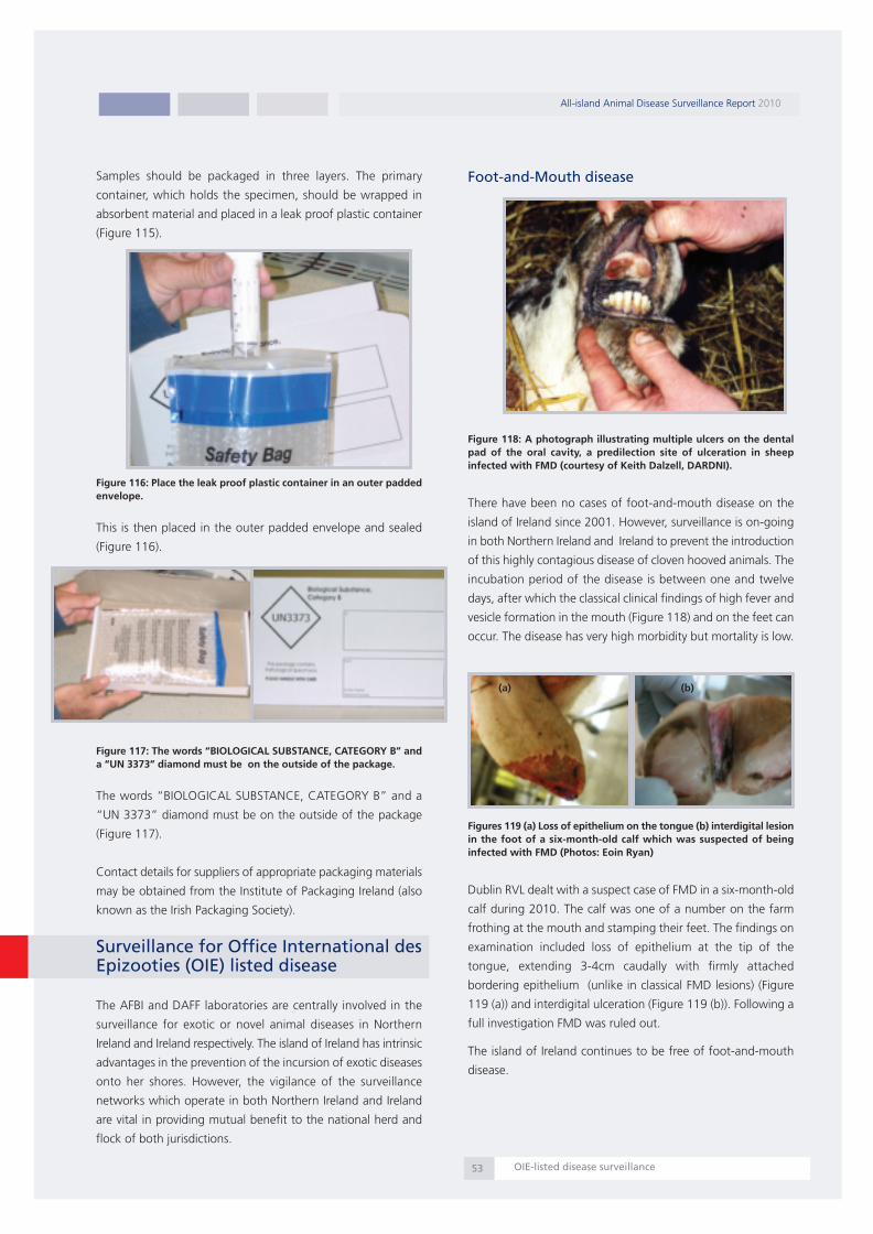
Samples should be packaged in three layers. The primary
container, which holds the specimen, should be wrapped in
absorbent material and placed in a leak proof plastic container
(Figure 115).
Figure 116: Place the leak proof plastic container in an outer paddedenvelope.
This is then placed in the outer padded envelope and sealed
(Figure 116).
Figure 117: The words “BIOLOGICAL SUBSTANCE, CATEGORY B” anda “UN 3373” diamond must be on the outside of the package.
The words “BIOLOGICAL SUBSTANCE, CATEGORY B” and a
“UN 3373” diamond must be on the outside of the package
(Figure 117).
Contact details for suppliers of appropriate packaging materials
may be obtained from the Institute of Packaging Ireland (also
known as the Irish Packaging Society).
Surveillance for Office International desEpizooties (OIE) listed disease
The AFBI and DAFF laboratories are centrally involved in the
surveillance for exotic or novel animal diseases in Northern
Ireland and Ireland respectively. The island of Ireland has intrinsic
advantages in the prevention of the incursion of exotic diseases
onto her shores. However, the vigilance of the surveillance
networks which operate in both Northern Ireland and Ireland
are vital in providing mutual benefit to the national herd and
flock of both jurisdictions.
Foot-and-Mouth disease
Figure 118: A photograph illustrating multiple ulcers on the dentalpad of the oral cavity, a predilection site of ulceration in sheepinfected with FMD (courtesy of Keith Dalzell, DARDNI).
There have been no cases of foot-and-mouth disease on the
island of Ireland since 2001. However, surveillance is on-going
in both Northern Ireland and Ireland to prevent the introduction
of this highly contagious disease of cloven hooved animals. The
incubation period of the disease is between one and twelve
days, after which the classical clinical findings of high fever and
vesicle formation in the mouth (Figure 118) and on the feet can
occur. The disease has very high morbidity but mortality is low.
Figures 119 (a) Loss of epithelium on the tongue (b) interdigital lesionin the foot of a six-month-old calf which was suspected of beinginfected with FMD (Photos: Eoin Ryan)
Dublin RVL dealt with a suspect case of FMD in a six-month-old
calf during 2010. The calf was one of a number on the farm
frothing at the mouth and stamping their feet. The findings on
examination included loss of epithelium at the tip of the
tongue, extending 3-4cm caudally with firmly attached
bordering epithelium (unlike in classical FMD lesions) (Figure
119 (a)) and interdigital ulceration (Figure 119 (b)). Following a
full investigation FMD was ruled out.
The island of Ireland continues to be free of foot-and-mouth
disease.
All-island Animal Disease Surveillance Report 2010
OIE-listed disease surveillance53
(a) (b)
Report 2010:Layout 1 07/09/2011 13:30 Page 54

Bluetongue
Surveillance for Bluetongue in Northern Ireland and Ireland is
ongoing and is conducted on all susceptible animals (cattle and
sheep) imported from countries that have not been declared
disease-free by the EU. A random sample of susceptible cattle
from Northern Ireland and Ireland is also tested.
In Northern Ireland during 2010, a total of three thousand nine
hundred and sixty eight serological tests (three thousand and
one NI random sample surveillance and nine hundred and sixty
seven post-importation samples) and seven thousand one
hundred and two PCR tests following importation was
performed by the Immunodiagnostic and Virology Branches of
Veterinary Sciences Division, AFBI. All were negative for BT virus.
In Ireland DAFF Virology Division in CVRL tested eight hundred
and thirty five cattle following importation for BTV antibody
with five hundred and fifty seven (67 per cent) proving antibody
positive while a further seven hundred and six cattle were tested
for BT virus, all of which were negative. A further four hundred
and eighty five sheep were tested for BT antibody following
importation with three hundred and seventy five (77 per cent)
proving antibody positive. Four hundred and forty one sheep
were tested for BT virus, all of which were negative. Eighteen
cattle and six sheep which were live clinical suspects and five
cattle and three sheep which were suspected on post-mortem
examination were identified by the Regional Veterinary
Laboratories, all of which were negative. Random surveys were
also conducted by DAFF, testing one thousand seven hundred
and eighty cattle sera from three hundred and fifty six herds
and seven hundred and six sheep sera from one hundred and
thirteen flocks with all proving negative for BTV antibody. There
were thirty seven sheep samples with inconclusive results on
the ELISA in 2010 which required a second confirmatory ELISA
test. All proved negative on this second BT ELISA.
The island of Ireland continues to be Bluetongue-free.
Avian influenza
As part of the annual avian influenza (AI) survey of poultry
premises across the UK, AFBI performs a serological survey of
poultry flocks in Northern Ireland for antibodies against AI H5
and H7 viruses. The survey includes fowl, turkeys, ducks and
geese. During 2010, nine hundred samples were tested under
this surveillance programme. All samples proved negative for
AIV antibodies. In addition, thirty seven samples from poultry
flocks and forty seven from pigeons and other birds were also
tested by real time RT-PCR for avian influenza virus. All samples
tested negative for the presence of AI virus.
An active programme of surveillance of wild birds for avian
influenza was suspended in Northern Ireland for 2010 but wild
bird ‘die-offs’ continued to be investigated. Carcases from wild
birds found dead and reported to DARD in Northern Ireland
were examined by AFBI pathologists and tested for AIV by real
time RT-PCR testing. Forty samples in total were tested, all of
which were negative.
In Ireland, a total of two hundred and eighty five specimens
from wild birds and three hundred and eighty four specimens
from commercial flocks were tested by DAFF using real time
RT-PCR, and / or virus isolation, for avian influenza virus and
reported “online” as required by the EU Commission. In
addition, twenty two thousand three hundred and sixty four
samples from commercial poultry flocks were serologically
examined for AI as part of two major national surveys and to
satisfy requirements for movement and trade. All tests were
negative.
Porcine influenza
In Northern Ireland, one hundred and fifty nine samples were
tested by the Indirect Flourescent Antibody Test (IFAT), of which
four were positive. The IFAT test is an pan-influenza test and all
samples that are positive for influenza by IFAT are subsequently
tested for the influenza pandemic strain by PCR tests. Fifty eight
samples were tested by RT-PCR, of which fifteen were positive for
the pandemic strain H1N1 2009.
In Ireland twenty eight carcases were tested by DAFF for porcine
influenza pandemic H1N1 2009 strain by PCR and five of these
were positive. All five animals were from the same farm. An
additional forty seven sera were examined by the
haemaglutination inhibition test (HAI). Of these eleven were
positive, one for H3N2 and ten for H1N1. Serology does not
differentiate the pandemic strain from the classical swine H1N1
strain.
Newcastle disease
In Ireland a total of two hundred and forty seven samples were
tested by DAFF using PCR and three hundred and eighty four
were tested using virus isolation for Newcastle disease. All results
were negative. In addition, serology for antibodies to Newcastle
disease virus was performed on one thousand two hundred and
ninety seven sera.
In 2010 in Northern Ireland a total of ninety two tissue samples
were tested by PCR for Newcastle disease virus. All test results
were negative.
In addition a total of eighteen thousand, three hundred and
thirty-two samples from commercial flocks were tested
serologically for Newcastle disease by AFBI.
All-island Animal Disease Surveillance Report 2010
OIE-listed disease surveillance54
Report 2010:Layout 1 07/09/2011 13:30 Page 55

Classical swine fever
Classical swine fever is caused by infection with classical swine
fever virus (CSFV) (previously called hog cholera virus). The
effect of different CSFV strains varies widely, leading to a wide
range of clinical signs. Clinical signs include fever,
immunosuppression, chronic diarrhoea, skin lesions,
convulsions, and in young animals, death may occur. The clinical
signs are indistinguishable from those of African swine fever.
Infected piglets born to subclinically infected sows help
maintain the disease within a population. There have been no
cases of classical swine fever in Ireland since 1958. AFBI
maintains a level of surveillance by testing tissues from pigs
submitted for post-mortem examination by immuno-
fluorescence test (IFAT). During 2010, two hundred and thirty
six samples were tested by IFAT in Northern Ireland and found
to be negative. In Ireland seven thousand nine hundred and
sixty five CSF ELISA tests and eight CSF PCR tests were
performed in 2010. All were negative.
Bovine Spongiform Encephalopathy (BSE)
Surveillance for BSE is both active and passive. Active
surveillance involves the routine sampling of fallen animals in
knackeries and the sampling of animals over a given age in
meat factories, while passive surveillance refers to the
notification of suspect clinical cases of BSE by veterinary
practitioners to the veterinary service.
In 2010 in Ireland, DAFF confirmed BSE in two animals, both of
which were identified by active surveillance and were confirmed
by the National Reference Laboratory (NRL). In Ireland the age
limit for the compulsory rapid test sampling of animals in meat
factories for BSE was increased to six years of age on the 1st July
2011, while the age limit for BSE rapid testing remains at four
years of age for all fallen animals.
Thirty five clinically suspect cases were also examined at regional
veterinary laboratories and samples were sent to the NRL in
Backweston for confirmatory diagnosis. None of these were
confirmed as positive for BSE. Table 11 shows a breakdown of
the histopathological diagnoses reached for each of these cases:
H&E Result Number of Cases
Listerial Encephalitis 5
Hepatic Encephalopathy 3
Neoplasia 2
Viral Encephalitis 1
Cerebrocortical Necrosis 1
No Specific Findings 23
Table 11: Histopathological Diagnoses for BSE Clinical Suspects inIreland's National Reference Laboratory (DAFF) in 2010.
AFBI conduct both rapid test surveillance and confirmatory
diagnosis (Histopathology, Immunohistochemistry, and Western
Blot) of BSE and Scrapie in Northern Ireland on behalf of DARD
in accordance with EU Regulation (EC) 999/2001 and the
Transmissible Spongiform Encephalopathies Regulations
(Northern Ireland) 2008.
Figure 120: A photomicrograph of BSE in a H&E section of a bovinebrain showing the typical vacuolation associated with BSE infection(Photo: AFBI).
In 2010 there were no BSE positives detected by AFBI as part of
the active surveillance programme. There were a total of six
clinical suspect submissions, all of which were confirmed as
negative for BSE.
Scrapie
In 2010 twenty four cases of scrapie were confirmed by the NRL
in Ireland. Of these twenty four, twenty two were classified as
classical scrapie, with the remaining two being classified as
atypical scrapie.
The twenty two confirmed cases of classical scrapie came from
nine separate flocks; seventeen were identified through active
surveillance and the remaining five were identified as clinical
suspects (passive surveillance).
Tissues from both atypical cases were sent to the NRL from
rapid testing laboratories i.e. they were detected through active
surveillance programmes.
DAFF identified seven new flocks with scrapie in Ireland in 2010;
five with classical scrapie and two with atypical scrapie. Co-
existence of classical and atypical scrapie was not detected in
any flock during 2010.
All samples tested by discriminatory western blotting (to
differentiate between scrapie and BSE in sheep) were reported
as “scrapie-like” in 2010.
All-island Animal Disease Surveillance Report 2010
OIE-listed disease surveillance55
Report 2010:Layout 1 07/09/2011 13:30 Page 56

Figure 121: Positive (brown) immunohistochemistry staining forscrapie in an ovine brain (photo: AFBI).
In Northern Ireland the annual Scrapie Active Surveillance
programme of the Department of Agriculture and Rural
Development for Northern Ireland (DARDNI) encompasses the
following :
� a pro rata (UK) human consumption sheep survey (minimum
of six hundred slaughtered animals over eighteen-months-
old)
� a pro rata (UK) sheep fallen stock survey (random sample of
approximately seven hundred sheep over eighteen-months-
old)
� a fallen goat survey (all animals over eighteen-months-old)
� a fallen sheep survey from Scrapie Monitored Flocks and
flocks restricted under the Compulsory Scrapie Flocks
Scheme (all animals over eighteen-months-old)
� an annual sample of cull sheep (end of productive life)
submitted for human consumption from Compulsory
Scrapie Flocks.
In 2010 there were no submissions in Northern Ireland for
confirmatory diagnosis for scrapie in either clinical suspects or
active surveillance samples.
A selection of farm investigations
The investigatory and advisory role of the veterinary laboratories
in cases of novel, exotic or unresolved disease syndromes is an
important part of their surveillance function, as well as
providing a valuable support to food animal production.
Veterinary pathologists of AFBI and DAFF are available to advise
on animal disease and production problems. On occasion,
laboratory pathologists may conduct on-farm visits to
investigate cases where exotic, zoonotic or novel diseases are
suspected. Presented below is a selection of summaries of farm
investigations conducted by AFBI and DAFF pathologists during
2010.
Dwarfism outbreaks in calves
In 2010, all the veterinary laboratories on the island of Ireland
reported an increased prevalence of calves born with congenital
chondrystrophy (disproportionate dwarfism) in recent years.
Kilkenny RVL investigated eight herd outbreaks involving fifty
dwarf calves. The mean prevalence of dwarfism was 13 per cent
of calvings in the affected herds, with a range 2.4 per cent to
20 per cent. One farm had seen cases in two previous years and
two other farms had recorded cases in only one other year. Forty
five per cent of dwarf calves were born to first calvers and 30
per cent to second calvers.
Figure 122: Shortened fore limbs on an affected Charolais calf (Photo:Dónal Toolan).
Cases were defined as disproportionate shortening of the limbs
(chondrodystrophy) with or without joint laxity (hocks that
appear sickle shaped in lateral view or hyper-extended fetlocks)
(Figure 122). However on visiting farms, it was found that all
eight farms had chondrodystrophic dwarf calves. Some herds
reported that some calves were born with severe joint laxity but
normal bone length.
All-island Animal Disease Surveillance Report 2010
A selection of farm investigations56
Report 2010:Layout 1 07/09/2011 13:30 Page 57

Such calves were often unable to stand unaided but, if assisted
to suckle for up to two or three weeks, they tended to
strengthen and their locomotion became normal. This suggests
a wide spectrum of clinical signs with this condition, making it
difficult to define the syndrome or to recognise all affected
animals by inspection. The following clinical signs were
reported on the eight farms visited (not necessarily in all
affected animals); hyperextension of fetlocks on seven farms;
carpus or hock almost on ground on five farms; sickle hocks on
three; bowing of forelimbs on three; rotation of limbs on three;
and undershot jaw on two.
Most herds were fed on grass silage only during the winter, with
one herd feeding 4 kg sugar beet plus correct balancing
minerals for the month of December; one farm fed hay in
March after silage ran out and another used some hay (no
details); at least one thin cow that had been fed 2-3 kg
concentrate from November 1st until February 1st went on to
deliver a dwarf calf. Two herds used baled silage only; two used
pit silage and four used a mixture. No herds analysed their
silage but none thought that quality was poorer than in other
years. The conventional advice given to control the disease is
to replace 30 per cent of the silage dry matter by concentrates
or hay. However despite this, some affected herds were feeding
cows solely on silage during the winter.
A similar syndrome has been reported worldwide over many
decades. Like previous reports, in this study males and females
were equally affected; suckler herds were more frequently
affected than dairy herds; affected calves tended to be born
towards the end of the spring calving period and often in a
cluster over a short period. Affected calves were not born
prematurely. Cases occurred with both natural service and
artificial insemination. A wide variety of beef breeds was
affected involved, with Charolais well represented, but affected
calves were also born to dairy breeds.
Mortality after acute disease in lambs
Sligo RVL investigated an outbreak of mortality in lambs
following acute illness. Acute Louping ill and pasteurellosis
(Mannhaemia haemolytica) (Figure 123), were diagnosed in
four lambs presented for post-mortem examination. The lambs
were deficient in cobalt and there were fulminant orf lesions
on the mouths, ears and limbs; some with secondary bacterial
infections. There were significant heavy infestations with
nematodes. There was evidence of tick infestation. All the
lambs had focal abscesses in the axillae where they were said to
have been injected with a clostridial vaccine. It was immediately
advised that cobalt be repeatedly given orally to all the affected
lambs. Systemic antimicrobial therapy was recommended for
lambs diagnosed with pneumonia, while topical treatment was
recommended for lambs with orf lesions.
During the course of the field investigation, it transpired the
affected lambs had been moved to a lush pasture on the
mountain before the disease outbreak. The group were blood
sampled on two occasions and faecal samples were analysed
for parasites.
Figure 123: Pneumonia lesion in a lamb from which Mannhaemiahaemolytica was cultured (Photo: Colm O’Muireagáin).
Two home-reared lambs that had never been on the mountain
pasture had no detectable antibodies to louping ill virus, which
led to the conclusion that this virus was present in the tick
population on the mountain pasture. In Ireland, it is thought
that there are two periods of major tick activity; spring and
autumn. Lambs being pastured on high risk ground should
ideally be vaccinated against louping ill virus. Another option
would be to treat the lambs with an acaricide to prevent tick
infestation before introducing them to the area. This would also
protect against other tick-related conditions and therefore was
recommended in addition to vaccination. Cobalt
supplementation was advised for all lambs, especially when
moved to mountain pasture. The cobalt deficiency was
suspected of adding to the severity of the infectious and
parasitic conditions identified. Preventative measures to address
the worm and potential fluke burdens were advised through
strategic dosing. Increased hygiene at clostridial vaccination was
advised to avoid the abscesses in the axilla (which is not a
recommended injection site in any event).
An investigation of milk drop in a dairy herd.
In October 2010 AFBI Stormont was asked to investigate a case
of milk drop in a spring calving dairy herd of sixty milking cows.
Cows presented with decreased milk yield, lethargy and pyrexia.
All-island Animal Disease Surveillance Report 2010
A selection of farm investigations57
Report 2010:Layout 1 07/09/2011 13:30 Page 58

Udders of affected animals appeared normal, were not swollen
or painful but there were occasional clots present in the milk.
Individual cases responded to broad spectrum antibiotics and
milk yield returned to normal ten to fourteen days after the
initial milk drop. Microbiological culture of milk samples and
leptospirosis serology on blood samples performed in another
laboratory had yielded negative results.
Initial investigations by AFBI involved testing of a small number
of blood samples from animals that had presented with clinical
signs in the previous three weeks for a variety of differential
diagnoses. One sample tested positive by Leptospira ELISA while
all others tested negative. Since there was no history of
vaccination for leptospirosis in the herd and in light of the
specific clinical signs observed, a microscopic agglutination test
(MAT) for Leptospira Hardjo was performed on these samples at
the OIE Leptospira reference laboratory in AFBI Stormont, in
which all samples tested positive. A positive MAT and negative
ELISA indicated that the animals tested were likely to be in the
acute stages of a leptospirosis infection. Results of further blood
samples indicated animals throughout the herd were at various
stages of the seroconversion process. Animals that had been
kept on an outfarm for the preceding three months tested
negative for leptospirosis antibodies indicating that the source
of the infection was likely to have been introduced in the
intervening period.
A recently purchased stock bull used to ‘mop up’ repeat
breeders in the main dairy herd had tested positive for
leptospirosis antibodies both by ELISA and MAT but could not
be definitively determined as the source of the infection, as his
vaccination status was unclear. However as no other animals
had been introduced to the herd in the recent past, and
seronegative animals on the outfarm had not had any access
to the bull following separation from the main herd, it was
determined that the bull was a potential source of infection.
Advice given by the laboratory was to immediately vaccinate all
cattle on the farm for leptospirosis. The vaccination strategy
was intended to reduce cow to cow transmission of the disease
by reducing the numbers of leptospires being shed by infected
animals and increasing overall herd immunity. The farmer was
advised that pregnant cows in the most at risk group i.e. greater
than 6 months pregnant could be treated with streptomycin to
minimise abortion risk. The abortion rate in this herd was
mitigated by the fact that it was a spring calving herd so that at
the time of initial outbreak most cows were in early-to-mid
gestation and that the referring vet and farmer were willing to
take immediate action to diagnose and treat the problem. The
farmer was made aware of the serious zoonotic risk posed by
such an outbreak and was advised that strenuous efforts should
be made to reduce risk of exposure to himself and farm staff.
This case and subsequent investigation highlights the important
co-operative roles played by farmer, submitting veterinary
practitioner and laboratory in diagnosing and controlling a
disease outbreak.
An investigation of recurring milk drop in adairy herd
Kilkenny RVL investigated a herd with a two year problem of
milk drop and respiratory signs in cows, with many individual
animals being affected more than once. Milk yield in some cows
recovered in a week or so, others took three weeks, while
others never recovered and went dry early. Many cows had
respiratory signs, with cough, nasal discharge and rapid
breathing. A diagnosis of IBR had been made clinically but
twice-yearly IBR vaccination did not control the problem.
Sero-conversion to IBR gE was demonstrated in paired sera from
a cow presented for examination at a farm visit. The cow had
an elevated temperature of 104ºF a few days earlier. This raised
suspicions of the involvement of wild IBR virus. Some animals
affected on earlier occasions had milk drop without obvious
respiratory signs, raising suspicions of the involvement of other
pathogens. As well as that, half of the blood samples taken in
spring of the previous year were hypocalcaemic and one third
were hypomagnesaemic. Salmonella Dublin was isolated a
couple of months before the laboratory visit from a maiden
heifer. A month after the visit, S. Dublin abortion was
confirmed in a number of cows. Many faecal samples contained
rumen fluke eggs. No evidence of ketosis or recent exposure to
leptospirosis was detected and body condition scores and grass
quantity and quality seemed adequate.
It was considered likely that the milk drop problem had more
than one cause and that this could explain why certain cows
had recurring problems. This emphasised the need for accurate
diagnosis to determine the cause of the milk drop in each case.
Careful clinical examination along with the following sampling
protocol was suggested for early cases: nasal swabs for
respiratory viruses PCR, Mycoplasma bovis and routine culture;
blood samples for IBR gE serology, calcium and magnesium and
faecal samples for Salmonella culture. The importance of
choosing animals in the earliest stage of disease for sampling
for PCR for respiratory viruses was stressed. Demonstration of
sero-conversion to IBR gE in paired blood samples is a useful
means of confirming IBR infection if virus isolation is negative
and can be used even in animals that have received IBR marker
vaccine. Nutritional advice to ensure that correct
calcium/magnesium status was maintained was recommended.
All-island Animal Disease Surveillance Report 2010
A selection of farm investigations58
Report 2010:Layout 1 07/09/2011 13:30 Page 59

The importance of biosecurity measures in reducing spread of
infection between animals was stressed. Clinically affected
animals were to be isolated (including those with milk drop,
even if respiratory signs were not obvious). It is important to
provide sufficient designated calving and isolation boxes.
This case study clearly showed that milk drop syndrome can
have a multi-factorial aetiology. It emphasises the need for a
holistic approach in dealing with the problem. The value of
good biosecurity, good husbandry and preventative medicine
are also highlighted.
Neonatal calf diarrhoea and forelimb paresis
Kilkenny RVL investigated an outbreak of neonatal diarrhoea
(and associated mortality) and forelimb paresis in the dairy
calves on an eighty five cow spring calving dairy farm. Twenty
five calves (29 per cent of calves born) died in the affected herd
between January and June 2010. All calves born developed
diarrhoea at seven to ten-days-old and approximately thirteen
of these calves died as a result of the diarrhoea. A further twelve
calves recovered from the diarrhoea and subsequently
developed forelimb paresis (Figure 125) and ataxia with
knuckling of the fetlocks at four to six-weeks-old. There was
100 per cent mortality in the calves that developed forelimb
paresis. The remaining calves recovered. In 2009 thirty to forty
calves developed neonatal diarrhoea at seven to ten-days-old
and approximately half the affected calves died.
Low ZST levels (Figure 124) were identified in number of calves
in spring 2010 submitted for post-mortem examination to
Kilkenny. In spring 2009 Salmonella Dublin was isolated from
cow faeces and rotavirus detected in samples of calf faeces.
Marginal/subnormal copper levels had been identified in this
herd in previous carcase and clinical sample submissions to the
RVL in 2009 and in 2010.
Figure 124: The zinc sulphate turbidity test (ZST) is used to determineimmunoglobulin levels in calves up to about ten days of age.
Figure 125: A recumbent calf with paresis on the affected farm(Photo: Denise Murphy).
The farm investigation revealed an inadequate number of
calving pens (one for eighty five cows) and poor bedding
hygiene. Calves were housed in a makeshift calf house. The
farmer was vaccinating for salmonellosis and neonatal enteritis.
Cows had access to mineral licks in the dry period and received
a selenium injection pre-calving. Young stock did not receive
mineral supplementation. The calves of all first calved heifers (n
= 15) died from diarrhoea/paresis.
Blood samples were collected from three calves, which were all
that were available as it was the end of the calving period, and
one cow. Significant Salmonella titres and low and marginal
blood copper levels were identified. As it was nearing the end
of calving period and there were no untreated diarrhoeic calves
available, a faecal sample was taken from a healthy three-day-
old calf; no enteric pathogens were detected. ZST levels were
adequate in the blood samples. A five-week-old calf with a
history of enteritis, and presenting with paresis, had a significant
Salmonella titre and was submitted for post-mortem
examination. A diagnosis of cervical vertebral osteomyelitis
caused by Salmonella Dublin was made.
It was concluded that Salmonella Dublin infection was active in
the herd and that there was inadequate colostral immunity, as
evidenced by low ZST levels and marginal serum copper levels.
The herdowner was advised to minimise the infection pressure
by improving hygiene (clean bedding etc); to ensure the
isolation and prompt treatment of sick animals; to avoid mixing
age groups; to ensure vaccinations are used correctly and
boosted at an appropriate interval pre-calving and that mineral
nutrition was addressed in a proactive way.
The farm veterinary practitioner has reported the neonatal
enteritis/paresis problems resolved in spring 2011.
All-island Animal Disease Surveillance Report 2010
A selection of farm investigations59
Report 2010:Layout 1 07/09/2011 13:30 Page 60

Pathological fractures in calves
A five-month-old Friesian-type calf was submitted to Kilkenny
RVL with a history of recumbency. Another four calves on the
same premises were recumbent and there was a history of a
calf with a similar history being euthanised and sent to a
knackery two weeks previously. At post-mortem examination
the calf was found to have bilateral distal comminuted femoral
fractures. Because the bone was relatively easy to cut with a
knife it was considered that the fractures were most likely to be
pathological in nature (due to an inherent weakness in the
bone) and it was decided to visit the farm of origin to
investigate further.
The calves were in a group of forty five being reared by a tillage
farmer. In addition to the four recumbent animals there was
another lame calf with a large swelling over the femur which
was judged clinically to be a healing fracture. Blood samples
were taken from the five affected calves and five randomly
selected unaffected animals. A detailed nutritional history was
also taken from the farmer. The four recumbent animals were
euthanised and brought to Kilkenny RVL for post-mortem
examination where each was found to have multiple fractures.
Multiple fractures in a group of animals without a history of
trauma are highly suggestive of an underlying metabolic bone
disease. There are various metabolic bone diseases which can
predispose to pathological fracture, but diagnosis of the specific
problem can be challenging. No biochemical abnormalities were
detected in the sampled animals (parathyroid hormone levels
were not tested). Bones from the animals were radiographed
and no abnormalities were detected in bone density or in the
growth plates. Histopathologically there were lesions
resembling fibrous osteodystrophy (replacement of bone with
fibrous tissue). Due to the limitations of standard
histopathological techniques it was not possible to determine if
there were lesions of osteoporosis. The most common causes of
metabolic bone disease in a group of farm animals are an
underlying nutritional or toxic problems. These calves had been
fed milk replacer up to eight-weeks of age. They were then fed
exclusively on a home-mixed ration of cereal and beans, plus
barley straw. No mineral supplements were being given.
Cereals and field beans are very low in calcium. The overall
calcium level in the diet was estimated by a nutritionist as being
approximately 0.12 per cent (should be 0.55 per cent or it
will predispose to osteoporosis). In addition the
calcium:phosphorosis ratio was estimated at 1:3.3 (which
should never be <1:1 or it will predispose to fibrous
osteodystrophy). Following mineral supplementation of the
calves’ rations, no further cases of fracture were reported.
Copper deficiency leading to scour andstunting in dairy calves
Sligo RVL investigated an outbreak of scour and stunting in a
fifty cow dairy herd. The herd was predominately spring calving.
Calves were retained and reared either as dairy replacement
heifers or for beef. There had been a history of scour and
consequential stunting in calves as they have gone to grass over
the previous two to three years in the herd. Morbidity was high
among both home-reared and purchased calves.
Young calves were fed milk, hay and approximately one
kilogram of concentrates while housed. These calves were
thriving well, with no evidence of scour. Calves were weaned
off milk before they went to grass, where they continued to be
fed conentrates. Within a few weeks of going to grass they
developed diarrhoea. Calves received various types of wormers,
with an initial apparent improvement following treatment on a
number of occasions, but within a week or two they were
scouring again.
Figure 126: Calves with the characteristic discolouration of the coatassociated with copper deficiency (Photo: John Moriarty).
At the farm visit, six calves which had recovered were in a
stunted state (Figure 126). Faecal samples were taken from
eight calves, all of which had loose faeces. Cryptosporidium
oocysts were detected in four of these. This was not considered
likely to be significant in the age group (three to six-month-old
calves). Five calves had temperatures of 103º-104ºF. Some
calves were coughing. The major finding on clinical pathology
was low serum copper concentration in the calves (range 5.2 to
12.1 micromoles per litre). The farmer reported an
improvement following supplementation with copper by
injection.
A nutritionist was consulted, and a forage analysis was
requested. Analysis of forage revealed very high levels of
molybdenum, sulphur, iron, and aluminium. All of these
antagonise copper. Forage levels of copper were regarded as
normal, but the antagonists would have reduced its availability.
The nutritionist recommended supplementation of copper to
calves through feed.
All-island Animal Disease Surveillance Report 2010
A selection of farm investigations60
Report 2010:Layout 1 07/09/2011 13:30 Page 61

Increased cell count and increased clinicalmastitis incidence
A laboratory-based investigation of mastitis pathogens causing
increased cell counts and an increased incidence of clinical
mastitis was performed on milk samples submitted to AFBI
Omagh from six herds in 2010. In one herd seventy two quarter
samples were submitted to investigate an increasing bulk milk
somatic cell count (SCC) which had reached 520,000 cells per
millilitre prior to investigation. Staph. aureus was isolated from
eleven individual quarter samples, coagulase-negative
staphylococci were isolated from eighteen individual quarter
samples and Corynebacterium bovis was isolated from sixteen
individual quarter samples. Advice was given on control
measures appropriate to contagious mastitis pathogens,
including culling chronic Staph. aureus-infected cows, the
treatment of new infections, and a review of the quantity,
quality and method of post-milking teat disinfection.
Streptococcus uberis was isolated from eight individual quarter
samples and Escherichia coli from seven individual quarter
samples. Advice was also given on control measures
appropriate to environmental pathogens including hygienic teat
preparation prior to milking and detailed improvements in
environmental hygiene in dry cow cubicles and calving boxes.
The prescribing veterinary surgeon advised on the appropriate
duration and type of antibiotic to use in the treatment of clinical
mastitis cases. An initial improvement in bulk milk SCC to
285,000 cells per ml was associated with the culling of
chronically-infected cows and drying-off some late lactation
high cell count cows. Improvements in parlour routine, parlour
maintenance and environmental hygiene over the next twelve
months resulted in a further decrease in bulk milk SCC to
178,000 cells/ml.
Periparturient neonatal mortality in a dairy herd
Sligo RVL investigated perinatal mortality in a hundred cow
dairy herd. The problem was confined to heifers. Affected
heifers were reported to be “slow about calving” with the calf
appearing to “just sit there” (uterine inertia). Any calf whose
delivery was assisted by traction survived. The afterbirth was
expelled immediately after calving (abnormally quickly). The
predominant breed of the calves which died was Aberdeen
Angus (AA), but this was coincidental with the breeding policy
of putting heifers in calf to AA bulls. The heifers went to an
outfarm as yearlings, where their diet before calving consisted
of baled silage; a 16 per cent protein concentrate ration was
fed up to one month pre-calving and pre-calving minerals were
given.
There was no evidence of a mineral deficiency or an active
infectious agent on clinical pathology testing. Mineral analysis
of second cut silage revealed high level of calcium, sodium, iron
and iodine. This was the silage the heifers were fed when the
problem was at its peak. Consultation with a nutritionist
indicated these were not likely to be of significance, especially
in the absence of such problems in mature cows. Laboratory
investigation did not reveal any infectious or mineral-related
cause for the problem.
The placenta was expelled almost immediately after the calf,
which is similar to a premature placental separation and
expulsion syndrome described by Mee (1991). The premature
separation leads to anoxia and foetal death. The only
association Mee found between the syndromes was with foetal
malposition. However, he could not determine if this was a
cause or consequential to the syndrome. Mee noted that the
prevalence of the condition was higher among AA calves than
Friesian Holsteins, which appears to be the experience in the
case herd, although it probably relates to the fact that more
heifers are in calf to AA bulls than to Holstein or other breeds.
Mee found no association with maternal parity, although other
authors he cites have found a greater prevalence among heifers.
The association between first parity and the condition in the
case herd did not appear to be documented in the literature,
although infections, hormonal and mineral imbalances have
been implicated as possible causes of similar syndromes.
While we have ruled out infectious and mineral issues in this
case we are not in a position to rule out hormonal imbalances.
The standard of calving supervision was already quite good. It
was suspected that any malposition was likely to be rather mild.
It was therefore recommended that calving heifers be handled
early in the course of labour to identify any malpositions and
correct them, without necessarily delivering the calf. This is
normally contraindicated and is not recommended as a general
practice.
Mee noted that a significant proportion of cases had
sanguineous (bloody) uterine fluid, so this should be looked out
for when heifers are calving.
Reference:
Mee J.F. (1991) Premature expulsion of the placenta and bovine
perinatal mortality. Veterinary Record 128:521-523.
All-island Animal Disease Surveillance Report 2010
A selection of farm investigations61
Report 2010:Layout 1 07/09/2011 13:30 Page 62

An outbreak of ill-thrift in a dairy herd
Sligo RVL investigated a chronic wasting syndrome, progressing
in some cases to downer cows, in an eighty cow spring calving
dairy herd. Rumen fluke eggs had been identified in faecal
samples, but despite treatment the cows were continuing to
waste. Eight cows had been affected, many of which were
among the oldest in the herd. Milk protein concentrations
dropped to 1.12 per cent at that stage. Quite a few cows were
also regurgitating the cud.
The body condition scores of the cows were considered quite
poor at the farm visit. The heifers had received a fluke dose
while still at pasture on September 25th, while the majority of
cows were dosed with a fluke dose on December 2nd. They
were again dosed with oxyclozanide on March 31st for rumen
fluke and adult liver fluke.
High creatine kinase (CK) levels were found in three cows,
which is indicative of muscle damage and / or muscle wastage.
Liver fluke eggs were detected in a faecal sample from one
heifer. BVD virus was detected in one purchased heifer, and
unsurprisingly all cohort heifers associated with this animal had
antibodies to BVD virus. The herd had been vaccinating against
BVD for some time. The herd had been fed poor quality hay for
the first six weeks of the dry period. During this time they lost
a considerable amount of body condition. The cows never
regained the condition lost during this period and were
considered to be too thin at the time of calving.
It became clear that the problem was due to poor nutrition
exacerbated by a burden of liver and rumen fluke. Therefore a
two-pronged approach was recommended to avoid a
recurrence. Firstly the farmer was told that such losses in body
condition were to be avoided at all costs. The value of
monitoring condition score at the time of drying-off was
emphasised. Cows that are particularly thin at that stage need
additional feeding.
The BVD virus detected in a heifer was not thought to be of
significance to the wasting syndrome observed in the cows, but
it was considered that it may have been of significance in some
of the heifers. A comrade of the BVDV PI animal had died
exhibiting clinical signs consistent with mucosal disease. A BVD
screen was recommended.
This case clearly showed the importance of dry cow nutrition
and the challenge that internal parasites can present to dairy
cows.
A selection of abstracts from publishedscientific papers
Occasionally the findings in the veterinary laboratories lead to
publications in the scientific literature. Equally, veterinary
laboratory staff may participate in research projects which
further our understanding of the risk factors which predispose
animals to disease, the progression of the disease process or
the diagnosis and control of specific animal diseases. Presented
below is a selection of scientific publications by AFBI and DAFF
laboratory staff during 2010.
Control of caseous lymphadenitis in six sheepflocks using clinical examination and regularELISA testing
Reprinted from Veterinary Record (2010) 166, 358-362 with
permission from BMJ Publishing Group Ltd.
G. J. Baird1, F. E. Malone2
1Scottish Agricultural College Veterinary Services, 5 Bertha Park
View, Perth PH1 3FZ
2Agri-Food and Biosciences Institute, Veterinary Sciences
Division, Stormont, Belfast BT4 35D
In an effort to control the spread of caseous lymphadenitis
(CLA) infection, flocks of affected sheep on six holdings were
tested serologically at regular intervals using an antibody ELISA
with a mean (sd) specificity of 99 (1) per cent and a sensitivity
of 79 (5) per cent. Western blot assays to detect antibodies to
the phospholipase D (PLD) exotoxin of Corynebacterium
pseudotuberculosis were used as a further test when ELISA
results were inconclusive. Owners were advised to remove from
the flock any sheep that demonstrated clinical signs of CLA or
tested positive for PLD by ELISA or western blot. Of the six trial
flocks, one was dispersed after only two blood tests, and in
another the recommendations for CLA control were not
followed and infected animals were retained within the flock.
In the remaining four flocks, the testing regimen and other
advice enabled the disease to be controlled to such an extent
that the appearance of new clinical cases of CLA was effectively
halted. This remained the case for up to five years after the end
of the trial. In two of these flocks, a small number of
seropositive animals were detected at the last flock test.
However, on the other two holdings all sheep were
seronegative in the final two flock tests, consistent with the
complete eradication of infection.
All-island Animal Disease Surveillance Report 2010
Abstracts from published papers62
Report 2010:Layout 1 07/09/2011 13:30 Page 63
Fasciola hepatica: Histological changes in thereproductive structures of triclabendazole(TCBZ)-sensitive and TCBZ-resistant flukes aftertreatment in vivo with TCBZ and the relatedbenzimidazole derivative, Compound Alpha
Reprinted from Veterinary Parasitology, 168 (2010) 240–254
with permission from Elsevier
R.E.B. Hanna1, H.W.J. Edgar 1, S. McConnell 1, E. Toner 2, M.
McConville 2, G.P. Brennan2, C. Devine 2, A. Flanagan 2, L.
Halferty 2, M. Meaney 2, L. Shaw 2, D. Moffett 1, M. McCoy 1, I.
Fairweather 2
1Veterinary Sciences Division, Agri-Food and Biosciences
Institute, Stormont, Dundonald, Belfast, Northern Ireland BT4
3SD, United Kingdom
2 School of Biological Sciences, Queen’s University, Belfast
BT71NN, United Kingdom
Twenty-four shed-reared lambs were each infected orally with
250 metacercariae of Fasciola hepatica, using either the
triclabendazole (TCBZ)-sensitive Cullompton isolate or the
TCBZ-resistant Sligo isolate. Twelve weeks after infection the
lambs were treated with TCBZ (10mg/kg) or with the
experimental fasciolicide, Compound Alpha (Cpd a), a
benzimidazole derivative of TCBZ (15 mg/kg). The lambs were
euthanised 48, 72 and 96 h after TCBZ treatment, or 24, 48
and 72 h after Cpd a treatment, and flukes were collected from
the liver and/or gall bladder of each animal. Untreated animals
harbouring 12-week infections were euthanised 24 h after
administration of anthelmintic to the treatment groups, and the
untreated flukes provided control material. A semi-quantitative
assessment of the degree of histological change induced by the
two drugs after different times of exposure was achieved by
scoring the intensity of three well-defined lesions that
developed in the testes and uteri of a representative sample of
flukes from each lamb. In general, it was found that in those
tissues where active meiosis and/or mitosis occurred (testis,
ovary, and vitelline follicles), there was progressive loss of cell
content due to apparent failure of cell division to keep pace
with expulsion of the mature or effete products. Further, actively
dividing cell types tended to become individualised, rounded
and condensed, characteristic of apoptotic cell death. Protein
synthetic activity was apparently inhibited in the Mehlis’
secretory cells. In the uterus, where successful formation of
shelled eggs represents the culmination of a complex sequence
of cytokinetic, cytological and synthetic activity involving the
vitelline follicles, the ovary and the Mehlis’ gland, histological
evidence indicating failure of ovigenesis was evident from 24 h
posttreatment onwards. The development of these lesions may
be related to the known antitubulin activity of the
benzimidazole class of anthelmintics, to the induction of
apoptosis in cells where mitosis or meiosis has aborted due to
failure of spindle formation, and to drug-induced inhibition of
protein synthesis. The semi-quantitative findings indicated that
Cpd a is slightly less efficacious than TCBZ itself in causing
histological damage to the reproductive structures of TCBZ-
sensitive flukes, and that, like TCBZ, it caused no histological
damage in flukes of the TCBZ-resistant isolate. This study
illustrates the potential utility of histological techniques for
conveniently screening representative samples of flukes in field
trials designed to validate instances of drug resistance or to test
the efficacy of new products against known drug-resistant and
drug-susceptible fluke isolates. It also provides reference criteria
for drug-induced histopathological changes in fluke
reproductive structures which may aid interpretation of TEM
findings.
Detection and quantification of Toxoplasmagondii in ovine maternal and foetal tissuesfrom experimentally infected pregnant ewesusing real-time PCR.
Reprinted from Veterinary Parasitology 2010 Aug 27; 172(1-2):
8-15 with permission from Elsevier
J. Gutierreza, , J. O’Donovanb, E. Williamsa, A. Proctora, C.
Bradyc, P.X. Marquesa, S. Worralla, J.E. Nallya, M. McElroyc, H.
Bassetta, D. Sammind, D. Buxtone, S. Maleye and B.K. Markeya
a School of Agriculture, Food Science and Veterinary Medicine,
University College Dublin, Belfield, Dublin 4, Ireland
b Regional Veterinary Laboratory, Department of Agriculture,
Fisheries and Food, Coosan, Athlone, Co. Westmeath, Ireland
c Central Veterinary Research Laboratory, Department of
Agriculture, Fisheries and Food, Backweston, Celbridge, Co.
Kildare, Ireland
d Regional Veterinary Laboratory, Department of Agriculture,
Fisheries and Food, Kilkenny, Co. Kilkenny, Ireland
e Moredun Research Institute, Edinburgh, Scotland, UK
All-island Animal Disease Surveillance Report 2010
Abstracts from published papers63
Report 2010:Layout 1 07/09/2011 13:30 Page 64

A real-time PCR (rt-PCR) targeting the 529-bp repeat element
(RE) of Toxoplasma gondii was used to detect and quantify the
parasite burden in maternal and foetal tissues in 18
seronegative ewes infected with 3000 Toxoplasma oocysts on
day 90 of pregnancy. The infected ewes were sacrificed in
groups of 4-6 at 21, 25, 33 and 35 days post-challenge. Ten
sham inoculated pregnant ewes were used as controls. T. gondii
was not detected in the control ewes or their foeti. The parasite
was only detected in the maternal tissues in a few of the
challenged ewes on a small number of occasions where it was
identified in spleen and uterine lymph nodes. T. gondii was
detected in the foetal spleen and liver at the early sacrifice times
but only sporadically thereafter. In the case of amniotic, allantoic
and foetal aqueous humor samples T. gondii was only detected
on a small number of occasions. However, it was found in the
majority of the foetal lung and placentome samples throughout
the study period, while placentomes and foetal brains
contained high levels of the parasite during the later stages.
Histopathological examination of placentome and brain tissue
from the foeti in the present study revealed a strong correlation
between histopathological lesions and quantities of the parasite
DNA detected. These results indicate that the cotyledonary
component of the foetal membranes is the sample of choice
for the diagnosis of T. gondii by rt-PCR, followed by foetal lung
and brain.
Identification of immunologically relevantproteins of Chlamydophila abortus using serafrom experimentally infected pregnant ewes.
Reprinted from Clinical Vaccine Immunology 2010 Aug;
17(8):1274-81. Reproduced with permission from the American
Society for Microbiology
P. X. Marques,1 Puneet Souda,5 J. O'Donovan,3 J. Gutierrez,1 E.
J. Williams,1 S. Worrall,1 M. McElroy,2 A. Proctor,1 C. Brady,2 D.
Sammin,4 H. F. Basset,1 Julian P. Whitelegge,5 B. E. Markey,1 and
J. E. Nally1*
1 UCD School of Agriculture Food Science & Veterinary
Medicine, UCD Conway Institute of Biomolecular and
Biomedical Research, College of Life Sciences, University
College Dublin, Belfield, Dublin 4,
2 Central Veterinary Research Laboratory, Department of
Agriculture and Food, Staccumny Lane, Backweston, Celbridge,
Co. Kildare,
3 Regional Veterinary Laboratory, Department of Agriculture,
Fisheries and Food, Coosan, Athlone, Co. Westmeath,4 Regional Veterinary Laboratory, Department of Agriculture,
Fisheries and Food, Kilkenny, Co. Kilkenny, Ireland,
5 The Pasarow Mass Spectrometry Laboratory, The Jane & Terry
Semel Institute for Neuroscience and Human Behavior, David
Geffen School of Medicine, University of California Los Angeles,
California
Chlamydophila abortus is an intracellular pathogen and the
etiological agent of enzootic abortion of ewes (EAE). C. abortus
has a biphasic development cycle; extracellular infectious
elementary bodies (EB) attach and penetrate host cells, where
they give rise to intracellular, metabolically active reticulate
bodies (RB). RB divide by binary fission and subsequently mature
to EB, which, on rupture of infected cells, are released to infect
new host cells. Pregnant ewes were challenged with 2 x 10(6)
inclusion forming units (IFU) of C. abortus cultured in yolk sac
(comprising both EB and RB). Serum samples were collected at
0, 7, 14, 21, 27, 30, 35, 40, and 43 days postinfection (dpi) and
used to identify antigens of C. abortus expressed during disease.
Additionally, sera from fetal lambs were collected at 30, 35, 40,
and 43 dpi. All serum samples collected from experimentally
infected pregnant ewes reacted specifically with several
antigens of EB as determined by one-dimensional (1-D) and 2-
D gel electrophoresis; reactive antigens identified by mass
spectrometry included the major outer membrane protein
(MOMP), polymorphic outer membrane protein (POMP), and
macrophage infectivity potentiator (MIP) lipoprotein.
All-island Animal Disease Surveillance Report 2010
Abstracts from published papers64
Report 2010:Layout 1 07/09/2011 13:30 Page 65


