Alperen Kutay YILDIRIM dhp930(001129-089) 1 TED ANKARA COLLEGE FOUNDATION HIGH SCHOOL IN VITRO EFFECTS OF LEAD OXIDE ON THE HUMAN ERYTHROCYTE ................................................................................................. Biology Extended Essay Super visor : Mualla Şirin GÜNTÜRKÜN Name of student : Alperen Kutay YILDIRIM Candidate number : dhp930(001129-089) Word Count : 3977
Lead has no biological function; however, low, and particularly, high levels of
exposure have a number of negative consequess for human healt. Despite the number of
reports about lead tocicity, very little information has been obtained regarding its effects on
erythrocytes. For this reason, the morphological effects of lead on human erythrociyte were
investigated.
Four lead and four no lead crystal glasses were bougth and divided two groups each
other. After completing the decontamination process, 200 ml of blood, or water were
poured into one of the four lead and no lead crystal glasses. We measured concentrations
of lead that leached into water and blood that were stored in lead and no lead crystal
glasses for 1-, 2-, ….and 10- day periods at room temperature. Lead concentrations in the
liquid matrix were measured using atomic absorption spectroscopy. Significant amounts of
lead leached into the liquid within one day; 724 µg/L in blood, 820 µg/L in water. Lead
continued to leach into both blood and water with the passage of time; 1832 µg/L in blood,
1653 µg/L in water (10th day results). Lead release was less in water than blood. Light
microscobic examination of peripheral blood smears from no lead glasses relevead no
morphologic changes over the erythrocytes but peripheral blood smears from lead crystal
glasses showed basophilic stippling. We found four basophilic stippling at 22 lead crystal
glass peripheral blood smears (18%). Significant lead contamination of water was detected
when it was left in lead crystal glass. If a liter of contaminated water was drunk daily, the
daily intake of lead could have been as high as 1653 µg. Such a high degree of
contamination could cause chronic lead poisoning and anemia. It is recommended that we
should avoid all lead based glassware.
Word Count: 293
Alperen Kutay YILDIRIM dhp930(001129-089)
3
Table of Contents
I. Introduction 4
II. Hypothesis 6
III. Method Development and plannning 8
a. Planning 8
b. Materials 11
c. Method 12
IV. Results 22
V. Discussion 28
VI. Conclusion 31
VII. Acknowledgement 32
VIII. Appendix 33
Appendix 1 33
Appendix 2 34
Appendix 3 36
Appendix 4 38
Appendix 5 39
Appendix 6 40
Appendix 7 41
Appendix 8 42
Bibliography 44
Alperen Kutay YILDIRIM dhp930(001129-089)
4
Introduction
Crystal glasses have very good shape and designs. I wonder the difference between
crystal glasses and the normal glasses. When I searched the distinction between them; I
learned that crystal glasses contain some percentage of lead. The reason for this was to
give shape. Lead is the most suitable heavy metal for giving shape. Therefore
manufacturers use lead to produce these beautiful crystal glasses. As we know that lead is
an heavy metal and have the potential for intoxication when it is taken over some cut-off
levels. With this point of view, I thought that whether drinking or storage of liquids in the
crystal glasses would cause any lead intoxication by leaching into the liquids. As a result I
decided to test this possibility.
Lead is an environmentally persistent metal that has been redistributed in the
environment as a result of human activities over thousands of years (1). Lead is present in
our environment in water, soil, dust and products manufactured with lead (2). It has been
used in construction, for decoration, and even as a food additive. It also has been a known
health risk for centuries (3). Hippocrates is thought to have written the first case report of
lead poisoning in 600 BC. The Romans also were aware of the toxicity of lead, with Pliny,
Paulus Aegineta, and Vesuvius all commenting on its effects (4). As a summary usage of
lead and lead glasses have history of nearly 5000 years in the history of humankind (5, 6).
Detailed information can be found in appendix 1.
………………………………………………………………………………………………………………………………………………………………. 1. Warniment C, Tsang K, Galazka SS. Lead poisoning in children. Am Fam Physician. 2010 Mar 15;81(6):751-7) 2. Patrick L. Lead toxicity, a review of the literature. Part 1: Exposure, evaluation, and treatment. Altern Med Rev. 2006 Mar;11(1):2-22. Review.) 3. Warniment C, Tsang K, Galazka SS. Lead poisoning in children. Am Fam Physician. 2010 Mar 15;81(6):751-7) 4. Aub JC, Fairhill LT, Minot AS, Reznikoff P, Hamilton A. Lead Poisoning. Medicine Monographs Volume 7. Baltimore, Md.: Williams & Wilkins; 1926. 5. http://en.wikipedia.org/wiki/Lead_glass 6. Benhima H, Chiban M, Sinan F, Seta P, Persin M. Removal of lead and cadmium ions from aqueous solution by adsorption onto micro-particles of dry plants. Colloids Surf B Biointerfaces. 2008 Jan 15;61(1):10-6. Epub 2007 Jun 30.
Alperen Kutay YILDIRIM dhp930(001129-089)
5
Lead glass contains typically 18–40 weight % lead oxide (7). Lead crystal ware
may release lead into the food and beverages when it comes in contact with. As well, any
container you drink from, including one made of lead crystal, that has an exterior
decorative pattern around the rim, such as a coating or glaze, may also release lead from
the coating or glaze. Lead can be harmful to your health (8).
Lead interferes with heme biosynthesis, and it affects formation and function of
erythrocytes. Lead, furthermore, interferes with iron utilization for heme formation, and
radio-iron studies showed that lead competes with iron for incorporation into erythrocytes.
If lead was indeed toxic to the hematopoietic system, one would expect the risk of aplastic
anemia might be associated with lead exposure (9).
According to the above information. I learned that lead is a toxic heavy metal that
can give harms to many biological systems in our body. In our daily life, we usually use
lead crystal glasses unconciously. Does the use of crystal lead glasses cause lead
intoxication by the leaching of lead into beverages and liquids that we drink from those
glasses? First of all, I tired to confirm the existency of lead in the fluids that are stored in
lead glasses. This was the first step of my study. Does this amount of leached lead cause
any damage on erythtrocytes and what are the types of these damages? The second part of
the study is occurred after confirming the availability of lead in the fluids that I studied. As
it is known lead may harm many biological systems. Blood is one of these systems. As a
general information, lead exposure may cause anemia by distortion of erythrocytes. As a
second part of my study, I tried to show toxic effects of lead on light microscobe (i.e.
basophilic stippling, toxic granulation etc.).
………………………………………………………………………………………………………………………………. 7. http://en.wikipedia.org/wiki/Lead_glass 8. Labbé RF. Lead poisoning mechanisms. Clin Chem. 1990; 36:1870 9. Emsley, John (2005). Elements of murder. Oxford University Press. ISBN 0192805991. http://books.google.com/?id=qBnfMimUoCYC&printsec=frontcover.)
Alperen Kutay YILDIRIM dhp930(001129-089)
6
Hypothesis
Lead was used for ceramic lead glazes. This material interdependence suggests a
close working relationship between potters, glassmakers, and metalworkers (10). Items
made of lead glass may leach lead into the food and beverages contained (11). Under
conditions of repeated use of the decanter, the lead leaching steeply decreases with
increasing use (12).
By the help of this information, it was hypothesized that the transition of lead will
occur from crystallized products. Since lead have some important inhibitor role in the
enzymatic production process of heme (Appendix 2); the leached lead in the liquids might
have some detrimental effects on the erythrocytes.
Since the synthesis of heme is an important factor in my hypothesis, I will
summarize the production of heme in short. Heme is the prosthetic group of hemoglobin,
myoglobin, and the cytochromes. Heme synthesis occurs partly in the mitochondria and
partly in the cytoplasm. The process begins in the mitochondria because one of the
precursors is found only there (13).
Heme synthesis begins with condensation of glycine & succinyl-CoA, with
decarboxylation, to form d-aminolevulinic acid (ALA). Then stops with the formation of
the heme as shown the appendix 3. Lead inhibits the heme pathway in several steps. These
are summarized in appendix 4 (14).
………………………………………………………………………………………………………………………………………………………………. 10. Lin; Tan, DT; Ho, HH; Yu, CC. "Environmental lead exposure and urate excretion in the general population.". The American journal of medicine. 2002, 113 (7): 563–8. doi:10.1016/S0002-9343(02)01296-2. PMID 12459402. 11. "Lead Crystalware and Your Health". It's Your Health. Health Canada. http://www.hc-sc.gc.ca/hl-vs/iyh-vsv/prod/crystal-cristal-eng.php. 12. Barbee SJ, Constantine LA. Release of lead from crystal decanters under conditions of normal use. Food Chem Toxicol. 1994 Mar;32(3):285-8. 13. Layer G, Reichelt J, Jahn D, Heinz DW. Structure and function of enzymes in heme biosynthesis.Protein Sci. 2010 Jun;19(6):1137-61 14. Layer G, Reichelt J, Jahn D, Heinz DW. Structure and function of enzymes in heme biosynthesis.Protein Sci. 2010 Jun;19(6):1137-61.
Alperen Kutay YILDIRIM dhp930(001129-089)
7
In view of this information, it was hypothesized that liquids that are stored and
dronk in crystal glassware contain lead. Amount of this lead increase in proportion to the
storage time. Consumption of lead containing liquids may cause toxic effect on many
organ systems (such as blood, brain etc.) following the absorption of lead from
gastrointestinal tract. It is clear that lead passing to blood have detrimental effects on
erythrocytes. In my opinion, if we perform peripheral blood smear to the lead containing
blood, we can hope to see basophilic stipplings on erythrocytes that are the sign of lead
intoxication.
The purpose of this investigation is to study the effects of low levels of Lead on
erythrocytes in an in vitro study design.
Alperen Kutay YILDIRIM dhp930(001129-089)
8
Method Development and Planning
Planning
I will plan to make this study in a two step design. In the first step, I will research
to detect any leaching from crystal glasses into drinking water and blood. In order to test
this existency, I will pour water and blood into the lead glasses and then measure the levels
of lead. In the second step; I will plan to investigate the potential detrimental effects of
leach lead over erythrocytes such as basophilic stippling and toxic granulation by light
microscobe.
I will perform this study in GATA Military Medical Faculty. I will identify people
who can help me. In order to do this work, I will have a meeting with these persons.
I will meet with Assoc. Prof. Oral Nevruz, MD from Department of Hematology
for hematological procedure. I will meet with Assoc. Prof. Ismail Avci, MD from
Department of Blood Bank. I will want two bags blood from him. I will meet with Asist.
Prof. Ayse Eken, MSci from Deparment of Pharmacology. I will want to help me with the
usage of atomic absorption spectroscopy and measure Lead concentration. I will meet with
Asist. Prof. Suat Doganci, MD from Department of Cardiovascular Surgery. I want to help
me decontamination procedure and statistical analyses. I will meet with Sati Uludogan
from Department of Radiology. I want to help me X-ray imaiging.
Alperen Kutay YILDIRIM dhp930(001129-089)
9
I will buy four lead crystal glasses and four no lead glasses representing one
manufacturer. The metal composition of lead crystal glasses will 24% metallic lead. All
compositions will be fabricated to hold approximately 200 ml of liquid.
For this study, drinking water and human blood will be used as the test liquid.
Human blood samples will obtained from Department of Blood Bank, Gulhane Military
Academy of Medicine, Ankara/Turkey. Water will be purchased from the market.
Before the beginning of the study period all of the eigth glasses will be undertaken
in a decontamination process. After completing the decontamination process, I will begin
the study protocol.
On day 0 of the study, 200 ml of blood, or water will be poured into one of the four
lead crystals and four no lead crystal glasses. Five milliliters of liquids will be removed
for lead measurement and periferal blood smear at before pouring and 1, 2, 3,…,10 days
after the initial day of the experiment. For this process, I will buy 88 blood tubes and 44
peripheral blood smear materials.
All incubations will be at room temperature. After 10 days, we will collected lead
leachate samples and measured lead levels with atomic absorption spectroscopy. Meassure
of lead levels will be performed at the department of of Pharmaceutical Toxicology with
the supervision of Asist. Prof. Ayse Eken, Msci.
Also, lead leaching into water and blood from crystal glasses will be tried to
confirm by radiological imaging. Radiologic examination will be performed in Department
Alperen Kutay YILDIRIM dhp930(001129-089)
10
of Radiology, Gülhane Military Medical Academy, Ankara, Turkey, with the supervision
of Sati Uludogan.
In the seceond step detrimental effects of leached lead over erythrocytes will be
detected. This will be performed with light microscope by investigation of peripheral blood
smears that will be prepared in 10 days period. Fourty-four peripheral blood smear will be
prepared this process. Morphologic abnormalities of blood cells will be discovered by
microscopic examination with the oil immersion lens of well-prepared films of peripheral
blood stained with Wright's stain. Inspection of erytrocytes will have been done in GATA
Hematology Deparment with the supervision of Assoc. Prof. Oral Nevruz, MD.
Data colection and Statistical analyses will be done by a SPSS (Chicago,IL, USA)
statistical program. These procedure will be performed in Department of Cardiovascular
Surgery, Gülhane Military Medical Academy, Ankara, Turkey. Asist. Prof. Suat Doganci,
MD help me to have these findings.
Alperen Kutay YILDIRIM dhp930(001129-089)
11
Materials
Four lead crystal glasses
Four no lead crystal glasses
800 ml blood
800 ml water
44 blood sample tubes
44 water sample tubes
44 peripheral blood smear materials
Atomic absorption spectroscopy
Roentgenogram cassette
Mobile X-ray machine
Non sterile gloves
Injectors
Light microscope
Alperen Kutay YILDIRIM dhp930(001129-089)
12
Method
I planned to make this study in a a two step design. In the first step, I researched to
detect any leaching from crystal glasses into drinking water and blood. In order to test this
existency, I will pour water and blood into the lead glasses and then measure the levels of
lead. In the second step; I planned to investigate the potential detrimental effects of leached
lead over erythrocytes such as basophilic stippling and toxic granulation by light
microscobe.
I thought to perform this study in GATA Military Medical Faculty. I have identified
people who can help me. In order to do this work, I had a meeting with these persons.
Asist. Prof. Suat Doganci, MD (Department of Cardiovascular Surgery), Assoc. Prof. Oral
Nevruz, MD (Department of Hematology), Asist. Prof. Ayse Eken, MSci (Deparment of
Pharmacology), Sati Uludogan (Department of Radiology), Assoc. Prof. Ismail Avci, MD
(Department of Blood Bank) have valuable contributions in every step of my study.
Alperen Kutay YILDIRIM dhp930(001129-089)
13
First Step Study Method:
Figure 1. Crystal glasses
Four lead crystal glasses and four no lead glasses representing one manufacturer
(Paşabahçe®, Turkey) were bought (Figure 1). The metal composition of lead crystal
glasses was 24% metallic lead. All compositions were fabricated to hold approximately
200 ml of liquid, and their dimensions were as fallows; upper diameter 11 cm, lower
diameter 8 cm and height 4 cm.
For this study, drinking water and human blood were used as the test liquid. Human
blood samples were obtained from Department of Blood Bank, Gulhane Military Academy
of Medicine, Ankara/Turkey (Assoc. Prof. Ismail Avci, MD). Water was purchased from
the market (Erikli®, Turkey).
Before the beginning of the study period all of the eigth glasses were undertaken in
a decontamination process. The aim of this process is to prevent any possible unwanted
material that can be on study glasses. Asist. Prof. Suat Doganci, MD adviced this
decontamination process and we together applied this process (Appendix 5). After
completing the decontamination process, I begin the study protocol.
Alperen Kutay YILDIRIM dhp930(001129-089)
14
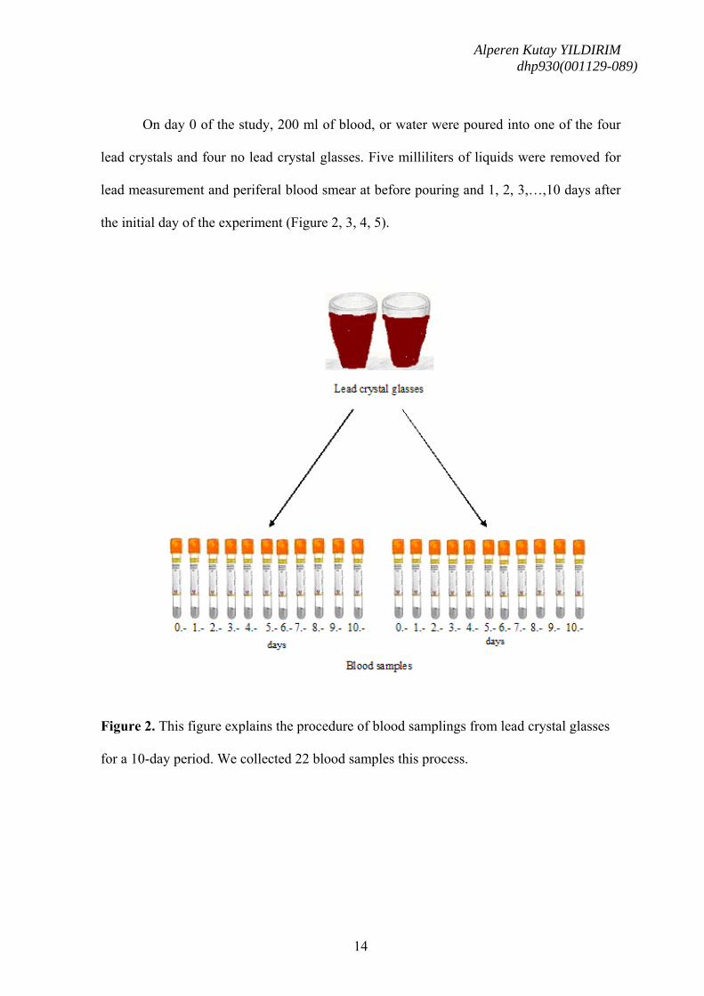
On day 0 of the study, 200 ml of blood, or water were poured into one of the four
lead crystals and four no lead crystal glasses. Five milliliters of liquids were removed for
lead measurement and periferal blood smear at before pouring and 1, 2, 3,…,10 days after
the initial day of the experiment (Figure 2, 3, 4, 5).
Figure 2. This figure explains the procedure of blood samplings from lead crystal glasses
for a 10-day period. We collected 22 blood samples this process.
Alperen Kutay YILDIRIM dhp930(001129-089)
15
Figure 3. This figure explains the procedure of blood samplings from no lead crystal
glasses for a 10-day period. We collected 22 blood samples this process.
Alperen Kutay YILDIRIM dhp930(001129-089)
16
Figure 4. This figure explains the procedure of water samplings from lead crystal glasses
for a 10-day period. We collected 22 water samples this process.
Alperen Kutay YILDIRIM dhp930(001129-089)
17
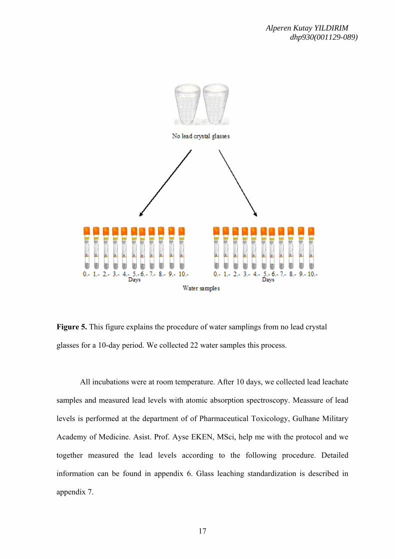
Figure 5. This figure explains the procedure of water samplings from no lead crystal
glasses for a 10-day period. We collected 22 water samples this process.
All incubations were at room temperature. After 10 days, we collected lead leachate
samples and measured lead levels with atomic absorption spectroscopy. Meassure of lead
levels is performed at the department of of Pharmaceutical Toxicology, Gulhane Military
Academy of Medicine. Asist. Prof. Ayse EKEN, MSci, help me with the protocol and we
together measured the lead levels according to the following procedure. Detailed
information can be found in appendix 6. Glass leaching standardization is described in
appendix 7.
Alperen Kutay YILDIRIM dhp930(001129-089)
18
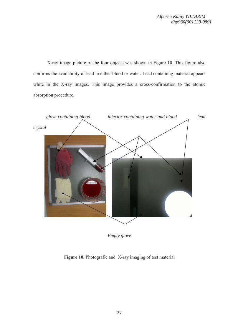
Also, lead leaching into water and blood from crystal glasses was tried to confirm
by radiological imaging (Mobile X-ray machine, Philips, USA). This was done by having
the X-ray image of four different objects (lead crystal glass, empty glove, glove containing
blood (after 10 days of incubation), injector that contain water (after 10 days of incubation)
over a roentgenogram cassette. Radiologic examination was performed in Department of
Radiology, Gülhane Military Medical Academy, Ankara, Turkey. Sati ULUDOGAN help
me to have the X-ray film.
Second Step Study Method:
In this step detrimental effects of leached lead over erythrocytes were detected.
This was performed with light microscope by investigation of peripheral blood smears that
were prepared in 10 days period (Figure 6, 7). Inspection of erytrocytes had been done in
GATA Hematology Deparment with the supervision of Assoc. Prof. Oral Nevruz, MD.
Detailed information can be found in appendix 8.
Alperen Kutay YILDIRIM dhp930(001129-089)
19
Figure 6. This figure explains the procedure of peripheral blood smears from lead crystal
glasses for a 10-day period. We collected 22 peripheral blood smears samples this process.
Alperen Kutay YILDIRIM dhp930(001129-089)
20
Figure 7. This figure explains the procedure of peripheral blood smears from no lead
crystal glasses for a 10-day period. We collected 22 peripheral blood smears samples this
process.
Alperen Kutay YILDIRIM dhp930(001129-089)
21
Data colection and Statistical analyses were done by a SPSS (Chicago,IL, USA)
statistical program. Measured data are expressed as mean values ± standart deviation.
Wilcoxon test was used for the comparison of measurements before and after the leaching
period and morphologic abnormalities. A p value < .05 was considered significant. These
procedure were performed in Department of Cardiovascular Surgery, Gülhane Military
Medical Academy, Ankara, Turkey. Asist. Prof. Suat Doganci, MD help me to have these
findings.
Alperen Kutay YILDIRIM dhp930(001129-089)
22
RESULTS
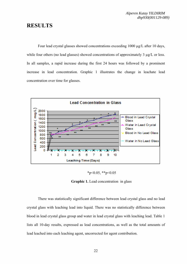
Four lead crystal glasses showed concentrations exceeding 1000 µg/L after 10 days,
while four others (no lead glasses) showed concentrations of approximately 3 µg/L or less.
In all samples, a rapid increase during the first 24 hours was followed by a prominent
increase in lead concentration. Graphic 1 illustrates the change in leachate lead
concentration over time for glasses.
*p<0.05, **p<0.05
Graphic 1. Lead concentration in glass
There was statistically significant difference between lead crystal glass and no lead
crystal glass with leaching lead into liquid. There was no statistically difference between
blood in lead crystal glass group and water in lead crystal glass with leaching lead. Table 1
lists all 10-day results, expressed as lead concentrations, as well as the total amounts of
lead leached into each leaching agent, uncorrected for agent contribution.
Alperen Kutay YILDIRIM dhp930(001129-089)
23
Table 1. Summary of 10-day lead and no lead crystal glass leaching results.
Leaching
agent
Glass aLeachate
concentration
(µg/L)
Total lead
(µg/Leachate)
Blood Lead crystal glasses 1832±7* 1272
Water Lead crystal glasses 1653±8** 1114
Blood No-Lead crystal glasses 3±0.3 1
Water No-Lead crystal glasses 0 0
*p<0.05, **p<0.05
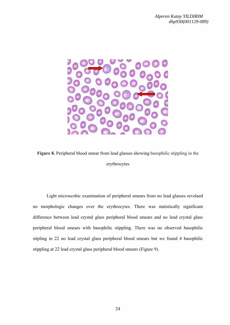
In the study of periferal blood smear, basophilic stipplings (Figure 8) were seen. (In
order to detect basophilic stipplings more efficiently, RBCs were packed with micro
haematocrit method and after Wright-Gimsa staining, these cells were seen more clearly).
Identification of basophilic stippling in peripheral blood smear and blood lead level of
1832 μg/dl were recorded with AAS at the 10th day.
Alperen Kutay YILDIRIM dhp930(001129-089)
24
Figure 8. Peripheral blood smear from lead glasses showing basophilic stippling in the
erythrocytes
Light microscobic examination of peripheral smears from no lead glasses revelaed
no morphologic changes over the erythrocytes. There was statistically significant
difference between lead crystal glass peripheral blood smears and no lead crystal glass
peripheral blood smears with basophilic stippling. There was no observed basophilic
stipling in 22 no lead crystal glass peripheral blood smears but we found 4 basophilic
stippling at 22 lead crystal glass peripheral blood smears (Figure 9).
Alperen Kutay YILDIRIM dhp930(001129-089)
25
*p<0.05
Figure 9. Morphologic abnormalities of blood cells at peripheral blood smears.
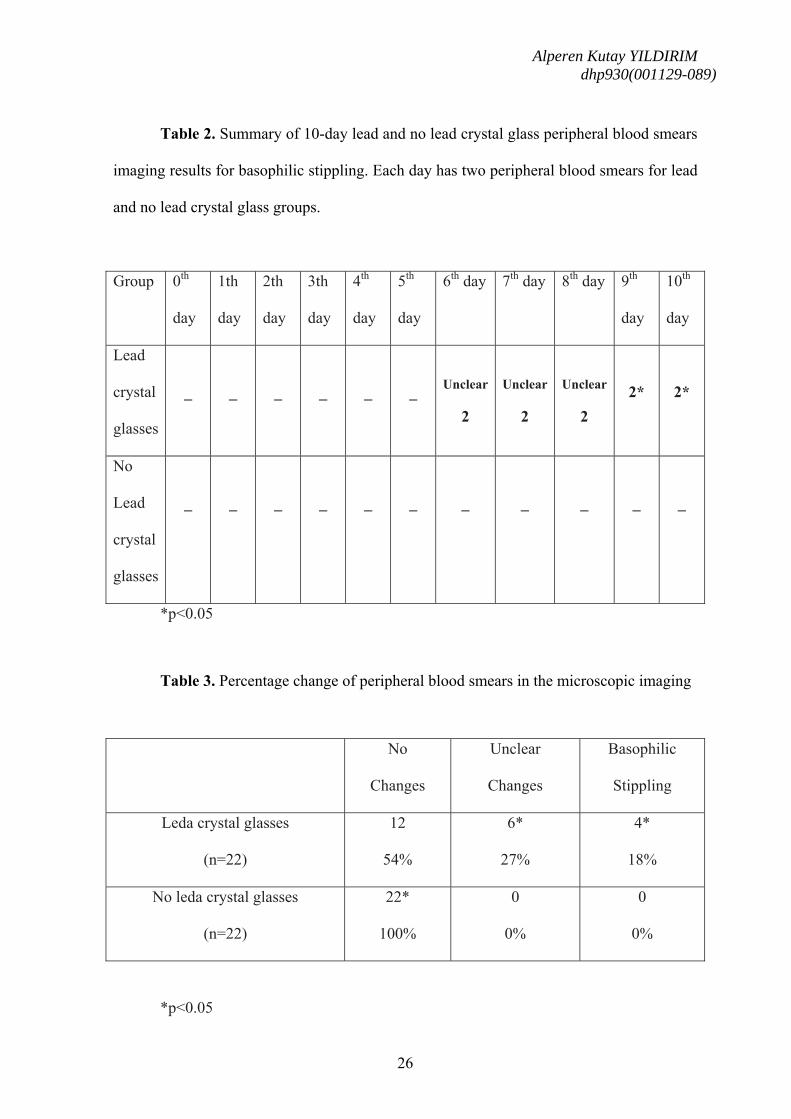
Four peripheral blood smear from lead crystal glasses at 9th, and 10th day revealed
basophilic stippling of red cells. These basophilic stipling was shown 18% of total
peripheral blood smear from lead crystal glasses. We investigated some changes at 6th, 7th,
and 8th days peripheral blood smear from lead crystal glasses, but these imaging changes
was unclear for basophilic stippling. I think, these imaging changes was polychromasia.
Therefore, we didn’t consider these imaging changes. There was statistically significant
difference between lead crystal glass and no lead crystal glass with basophilic stippling
(Table 2 and 3).
Alperen Kutay YILDIRIM dhp930(001129-089)
26
Table 2. Summary of 10-day lead and no lead crystal glass peripheral blood smears
imaging results for basophilic stippling. Each day has two peripheral blood smears for lead
and no lead crystal glass groups.
Group 0th
day
1th
day
2th
day
3th
day
4th
day
5th
day
6th day 7th day 8th day 9th
day
10th
day
Lead
crystal
glasses
_
_
_
_
_
_
Unclear
2
Unclear
2
Unclear
2
2*
2*
No
Lead
crystal
glasses
_
_
_
_
_
_
_
_
_
_
_
*p<0.05
Table 3. Percentage change of peripheral blood smears in the microscopic imaging
No
Changes
Unclear
Changes
Basophilic
Stippling
Leda crystal glasses
(n=22)
12
54%
6*
27%
4*
18%
No leda crystal glasses
(n=22)
22*
100%
0
0%
0
0%
*p<0.05
Alperen Kutay YILDIRIM dhp930(001129-089)
27
X-ray image picture of the four objects was shown in Figure 10. This figure also
confirms the availability of lead in either blood or water. Lead containing material appears
white in the X-ray images. This image provides a cross-confirmation to the atomic
absorption procedure.
glove containing blood injector containing water and blood lead
crystal
Empty glove
Figure 10. Photografic and X-ray imaging of test material
Alperen Kutay YILDIRIM dhp930(001129-089)
28
DISCUSSION
Our findings support the concentration that the lead crystal glass could have
suffered lead poisoning from liquid stored in lead crystal glasses. Blood and water stored
in the lead crystal glass used in this study reached concentrations as high as 1832 mcg/l
and 1653 mcg/l after 10 days of stroge, and in a used, no lead crystal glass reached as low
as inconspicuous level at 10 days (Table 1). These results confirm the hypothesis of lead
leaching into liquids that are stored in lead crystal glasses.
We prewash and decontamination the glass, residual surface powder from
manufacture could not have accounted for the higher rates of lead contamination seen in
the first versus the other following days.
We chose to store the liquids at room temperature. If we warmed our liquids, it
could have increased the extent of leaching. An experiment by Hoffmann, in which he
boiled various wines in leaden vessels according to ancient Roman instruction, found
levels of 390-781 mg lead per liter of wine (15).
The rate of lead leaching into liquid has previously been shown to be enhanced with
increasing acidity (16). Differences in rates of leaching by two liquids in this sutdy could
be explained in their pH, because blood (pH 7.4) had highest lead concentrations, whereas
water (pH 6.0) had an intermediate amount of lead leaching.
20, Graziano, P. "Lead exposure from lead crystal". The Lancet (1991). 337 (8734): 141-143. 21. De Leacy EA Lead-crystal decanters - a health risk? Med J Aust, 1987 147: 622. 22. Kutbi II, Ahmed M, & Saber A. Measurement of blood-lead levels in school children of Jeddah Saudi Arabia and assessment of sub-toxic levels of lead on some sensitive hematological parameters. J Environ, 1989, Sci Health, A24: 943-955.
Alperen Kutay YILDIRIM dhp930(001129-089)
31
CONCLUSION
The original test using blood and water leaching solution was developed to test for
lead migration from crystal containing added lead. This work clearly demonstrates that it is
also an excellent leaching agent for assessing the safety of crystal containing added lead.
The concentration of elements leached water used were all considerably less than the
concentrations leached by blood. Therefore, if the concentrations leached into blood are
within acceptable limits, it may be safely assumed that stemware manufactured from the
glass composition is safe for human use (23). But the study of peripheral blood smear
showed basophilic stipplings. Basophilic stippling is equal to anemia and anemia is a
harmful state for human beings.
Although we found that significant amount of lead leach into the blood and water
from lead crystal glasses, this is an experimental design and we usually never drink water
that were waited for 10 days from any glasses. With daily usage style of these glasses, we
usually do not expect to have lead intoxication. However we should be aware of the
danger. So while using lead crystal glasses, we should give care not keep beverages for
longer periods.
………………………………………………………………………………………………………………………………………………………………….. 23. Hynes MJ, Forde S, Jonson B. Element migration from glass compositions containing no added lead. Sci Total Environ. 2004 Feb 5;319(1-3):39-52.
Alperen Kutay YILDIRIM dhp930(001129-089)
32
Limitation of this is simply its’design. Although this study is performed over blood
as a biological material, this study is an in-vitro study. Therefore the evidence level of this
study is not so high. In order to increase this evidence level, a new study should be
designed in an in-vivo style (i.e. animal study). By this methodology the real biological
effect of the lead intoxication can be observed over living tissues. Furthermore, in order to
give a new perspective to this study the effect of lead over the stem cells can be researched.
This may be done in two spectrum. One part of this is to study the effect of lead over blood
stem cells. In my opinion the second and the most important part is to study over pregnant
animals and to see the effect over embrios as the most important stem cell.
Acknowledgement
I want to thank to Asist. Prof. Suat Doganci, MD (Department of Cardiovascular
Surgery), Assoc. Prof. Oral Nevruz, MD (Department of Hematology), Asist. Prof. Ayse
Eken, MSci (Deparment of Pharmacology) , Sati Uludogan (Department of Radiology),
Assoc. Prof. Ismail Avci, MD (Department of Blood Bank) for their valuable contribution
in every step of my study.
Alperen Kutay YILDIRIM dhp930(001129-089)
33
APPENDIX
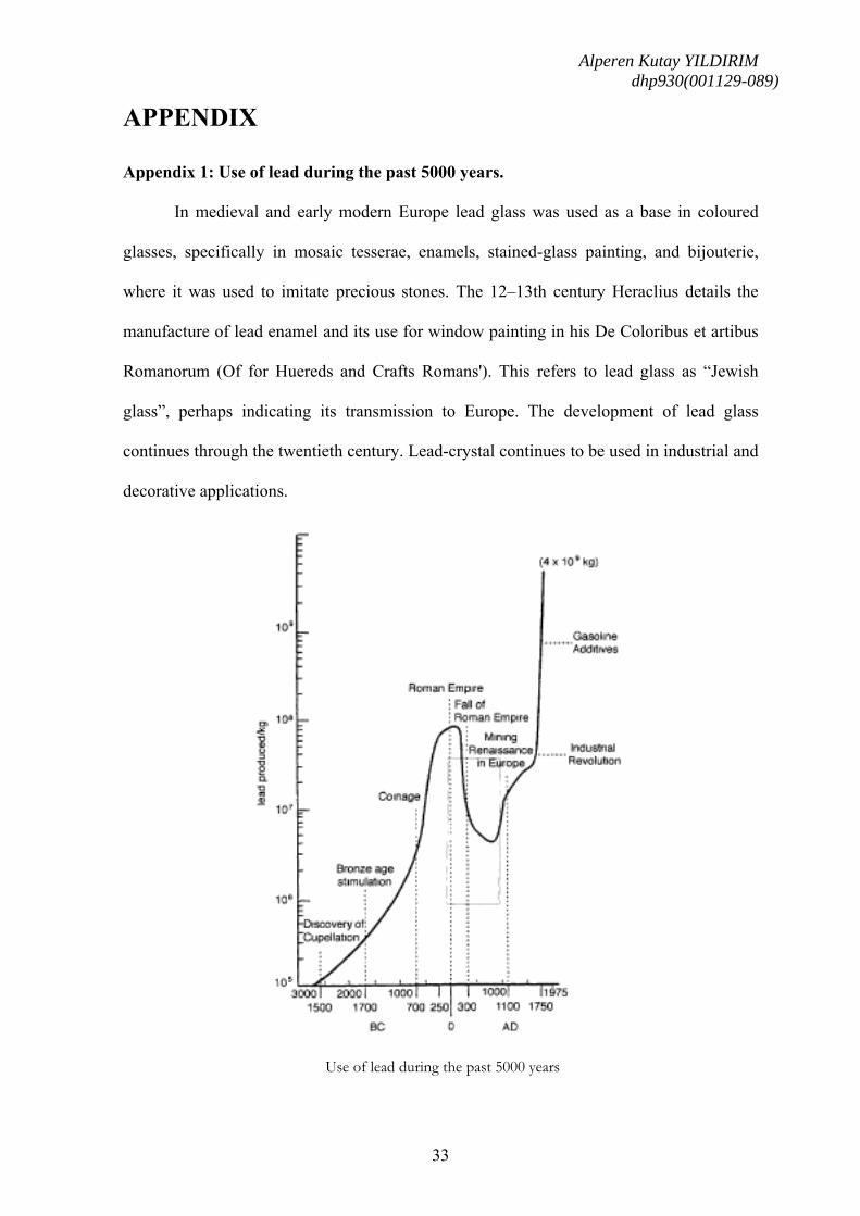
Appendix 1: Use of lead during the past 5000 years.
In medieval and early modern Europe lead glass was used as a base in coloured
glasses, specifically in mosaic tesserae, enamels, stained-glass painting, and bijouterie,
where it was used to imitate precious stones. The 12–13th century Heraclius details the
manufacture of lead enamel and its use for window painting in his De Coloribus et artibus
Romanorum (Of for Huereds and Crafts Romans'). This refers to lead glass as “Jewish
glass”, perhaps indicating its transmission to Europe. The development of lead glass
continues through the twentieth century. Lead-crystal continues to be used in industrial and
decorative applications.
Use of lead during the past 5000 years
Alperen Kutay YILDIRIM dhp930(001129-089)
34
Appendix 2: Lead interaction in the heme pathway
A.
Lead blocks enzymatic action of ALA-Dehydrogenase and ferrochelatase, halting
pathway heme synthesis pathway and leading to ALA secretion (in urine) from the body
Heme synthesis-some reactions occur in the cytoplasm and some in the
mitochondrion.
Alperen Kutay YILDIRIM dhp930(001129-089)
35
B.
Heme B biosynthesis patway and its modulators. Major enzyme deficiences are also
shown here
Alperen Kutay YILDIRIM dhp930(001129-089)
36
Appendix 3: Steps of heme biosynthesis.
A: The first cyclic tetrapyrrole uroporphyrinogen III is formed from the precursor 5-
aminolevulinic acid in three enzymatic steps via the intermediates porphobilinogen and
pre-uroporphyrinogen. Depending on the organism, ALA is either synthesized by
condensation of glycine with succinyl-CoA or from tRNA-bound glutamate via glutamate-
1-semialdehyde.
Alperen Kutay YILDIRIM dhp930(001129-089)
37
B: Uroporphyrinogen III is converted into heme in four consecutive enzymatic steps via
the intermediates coproporphyrinogen III, protoporphyrinogen IX, and protoporphyrin IX.
Structures of all heme biosynthesis enzymes have been determined with the exception of
oxygen-independent PPO (n.d., structure not determined).
Alperen Kutay YILDIRIM dhp930(001129-089)
38
Appendix 4: Inhibition points of heme by lead.
Lead interactions in heme pathway. ALAS, δ-aminolevunic acid synthase; CoA,
coenzyme A. The heme biosynthesis patway is represented. Several enzymes in patway
can be affected by lead; two of the most clinically important are ALAD and ferrochelatase.
Both these enzymes are inhibited by lead. Their activity can be measured directly or by the
measurement of accumulation of their respective subsrates. In the presence of lead, δ-
aminolevunic acid accumulates when ALAD is inhibited. Inhibition of ferrochelatase
results in the increased production of zinc protoporphyrin.
Alperen Kutay YILDIRIM dhp930(001129-089)
39
Appendix 5: Decontamination process
The glasses were kept in a climate chamber for 1 week at a temperature of 50oC and
with a relative humidity of 100%. The climate chamber was an isolated box
thermostatically controlled with a fan for air circulation. The glasses were placed upside
down with a slight tilt. The glasses were rearranged at regular intervals during the
treatment. Before and after the treatment, the glasses were washed once in a dishwasher.
These glasses were then leached in the same way as the untreated samples.
Alperen Kutay YILDIRIM dhp930(001129-089)
40
Appendix 6: Lead level testing
Lead concentration was measured by atomic absorption spectroscopy (AAS)
(atomic absorption spectrophotometer model A analyst 600 and model A analyst 800
equipped with graphite furnace, Zeeman background correction system, and lead hollow
cathode lamp; Perkin Elmer, Norwalk, CT, USA). Six micro liters of the sample mixture
prepared with matrix modifier (see below) was heated in a graphite furnace to 2,450°C.
During the process, the atomized lead sample was excited for absorbance at λ = 283.3 nm
for 2 s by a lead hollow cathode lamp. Matrix-specific standards containing 0,10, 50, 250,
and 500 µg/L of lead were prepared by spiking aqueous lead standard solution of 1,000
µg/mL (Perkin Elmer) into water, and blood, respectively. One hundred microliters of
standard was mixed with 200 µL lead matrix modifier (2 g ammonium phosphate
monobasic in 10 mL 10% Triton-X QS 200 mL H2O) prior to heating in the atomic
absorption spectrophotometer. Each set of standards (0, 10, 50, 250, and 500 µg/L) was
used to calibrate for the respective matrix (e.g., standards made in water were used to
generate the standard curve for water specimens). Three levels of QC prepared for routine
blood lead measurements (two purchased from BioRad, Hercules, CA, USA; one made in
lab) were run after calibrations to verify calibrations. The precision (coefficients of
variation) for these quality control levels at 80, 300, and 400 µg/L were 4.4, 3.3, and 3.7%
(n =200 each), respectively. Experimental specimens (water, and blood) taken at different
time points were diluted with the respective blank solution and mixed with lead measured
concentrations were within the reportable range of our assay (0–500 µg/L). The laboratory
at Department of Pharmaceutical Toxicology, Gulhane Military Academy of Medicine,
Ankara, Turkey
Alperen Kutay YILDIRIM dhp930(001129-089)
41
Appendix 7: Glass leaching standardization
The all glasses were tested to determine whether similar amounts of lead would be
leached from the vessel using the Center for Disease Control (CDC) standardized protocol
for lead contamination assessment. Each vessel was rinsed with de-ionized water three
times before the addition of 500 mL of acetic acid (4%, v/v). After 24 h, lead content was
measured using a similar method as outlined above except that standards were prepared
using acetic acid to generate the calibration curve.
Alperen Kutay YILDIRIM dhp930(001129-089)
42
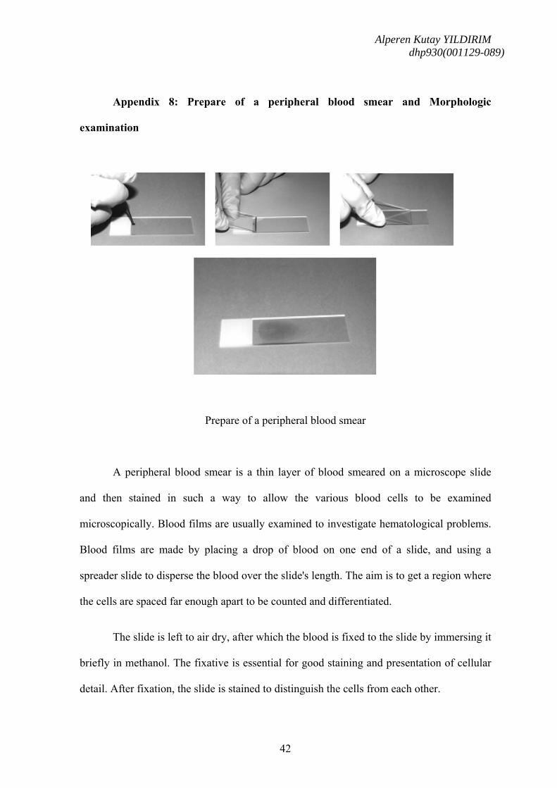
Appendix 8: Prepare of a peripheral blood smear and Morphologic
examination
Prepare of a peripheral blood smear
A peripheral blood smear is a thin layer of blood smeared on a microscope slide
and then stained in such a way to allow the various blood cells to be examined
microscopically. Blood films are usually examined to investigate hematological problems.
Blood films are made by placing a drop of blood on one end of a slide, and using a
spreader slide to disperse the blood over the slide's length. The aim is to get a region where
the cells are spaced far enough apart to be counted and differentiated.
The slide is left to air dry, after which the blood is fixed to the slide by immersing it
briefly in methanol. The fixative is essential for good staining and presentation of cellular
detail. After fixation, the slide is stained to distinguish the cells from each other.
Alperen Kutay YILDIRIM dhp930(001129-089)
43
Morphologic abnormalities of blood cells were discovered by microscopic
examination with the oil immersion lens of well-prepared films of peripheral blood stained
with Wright's stain. For appropriate interpretation of the morphology of erythrocytes, one
concentrates on areas of the slide where the red cells appear singly and have central pallor.
Examination of erythrocytes far out on the feathered edge discloses erythrocytes lacking
central pallor, whereas in thick areas of the slide the morphology of the erythrocytes was
distorted by contact between cells. Hematoloic examination was performed in Department
of Hematology, Gülhane Military Medical Academy, Ankara, Turkey.
Alperen Kutay YILDIRIM dhp930(001129-089)
44
Bibliography
1. Warniment C, Tsang K, Galazka SS. Lead poisoning in children. Am Fam
Physician. 2010 Mar 15;81(6):751-7.
2. Patrick L. Lead toxicity, a review of the literature. Part 1: Exposure, evaluation, and
treatment. Altern Med Rev. 2006 Mar;11(1):2-22. Review.
3. Aub JC, Fairhill LT, Minot AS, Reznikoff P, Hamilton A. Lead Poisoning.
Medicine Monographs Volume 7. Baltimore, Md.: Williams & Wilkins; 1926.
4. http://en.wikipedia.org/wiki/Lead_glass
5. Benhima H, Chiban M, Sinan F, Seta P, Persin M. Removal of lead and cadmium
ions from aqueous solution by adsorption onto micro-particles of dry plants.
Colloids Surf B Biointerfaces. 2008 Jan 15;61(1):10-6. Epub 2007 Jun 30.
6. Labbé RF. Lead poisoning mechanisms. Clin Chem. 1990; 36:1870.
7. Emsley, John (2005). Elements of murder. Oxford University Press. ISBN


![YILDIRIM Presentation PRIMEPhDConference[1]](https://static.documents.pub/doc/80x56/577d2f7b1a28ab4e1eb1d743/yildirim-presentation-primephdconference1.jpg)


