48
Alpha – 1 Antitrypsin Alpha – 1 Antitrypsin Deficiency Deficiency Jorge Mera, MD Jorge Mera, MD Presbyterian Hospital of Presbyterian Hospital of Dallas Dallas
| Date post: | 27-Dec-2015 |
| Category: |
Documents |
| Upload: | chrystal-boyd |
| View: | 215 times |
| Download: | 0 times |
Alpha – 1 Antitrypsin Alpha – 1 Antitrypsin DeficiencyDeficiency
Jorge Mera, MDJorge Mera, MDPresbyterian Hospital of DallasPresbyterian Hospital of Dallas

Alpha – 1 – Antitrypsin DeficiencyAlpha – 1 – Antitrypsin Deficiency
Mechanism of Alpha-1- Antitrypsin Deficiency Mechanism of Alpha-1- Antitrypsin Deficiency (AATD)(AATD) Clinical Case (Presentation)Clinical Case (Presentation)Lung DiseaseLung Disease– PathogenesisPathogenesis– Clinical PresentationClinical Presentation– TreatmentTreatment
Extra-Pulmonary DiseaseExtra-Pulmonary Disease– Hepatic DiseaseHepatic Disease
PathogenesisPathogenesisClinical PresentationClinical Presentation
– OtherOther
Clinical Case (Resolution)Clinical Case (Resolution)

Serpin Serpin
These are inhibitors of proteolytic enzymes with a serine These are inhibitors of proteolytic enzymes with a serine residue at the active siteresidue at the active site– AAT,AAT, Antithrombin, C1-inhibitor and alpha 1 antichymotrypsin Antithrombin, C1-inhibitor and alpha 1 antichymotrypsin
When they bind to its target proteinase it undergoes a When they bind to its target proteinase it undergoes a conformational changeconformational change
The The advantageadvantage– Is that the conformational change stabilizes the complexIs that the conformational change stabilizes the complex– It allows the modulation of inhibitory activityIt allows the modulation of inhibitory activity
The The disadvantagedisadvantage of conformational mobility is their of conformational mobility is their vulnerability to mutations which can :vulnerability to mutations which can :– Decrease its activityDecrease its activity– Allow inappropriate changes that lead to polymerizationsAllow inappropriate changes that lead to polymerizations

Protein Folding and FunctionProtein Folding and Function
AAT
Elastase
AAT

AATD is a Protein Folding DiseaseAATD is a Protein Folding Disease
Protein folding is the process by which an Protein folding is the process by which an unfolded polypeptide chain folds in to a unfolded polypeptide chain folds in to a specific native and functional structurespecific native and functional structure
Defective protein folding is an important Defective protein folding is an important mechanism underlying the pathogenesis mechanism underlying the pathogenesis of many diseasesof many diseases

Protein Folding and DiseaseProtein Folding and Disease
DiseaseDisease Protein AffectedProtein Affected Molecular DefectMolecular Defect
Cystic FibrosisCystic Fibrosis Cystic fibrosis Cystic fibrosis transmembrane regulator transmembrane regulator
(CFFTR)(CFFTR)
Misfolding and retention Misfolding and retention in the ER, leading to in the ER, leading to
degradationdegradation
Marfan SyndromeMarfan Syndrome FibrillinFibrillin MisfoldingMisfolding
Nephrogenic Diabetes Nephrogenic Diabetes InsipidusInsipidus
Vasopressin receptor or Vasopressin receptor or aquaporin water channelaquaporin water channel
Misfolding and retention Misfolding and retention in the ERin the ER
Alfa -1- Antitrypsin Alfa -1- Antitrypsin DeficiencyDeficiency
Alfa -1- AntitrypsinAlfa -1- Antitrypsin Misfolding and retention Misfolding and retention in the ER leading to in the ER leading to
aggregation in cells of aggregation in cells of synthesissynthesis
Creutzfeldt-Jakob Creutzfeldt-Jakob DiseaseDisease
Prion proteinPrion protein Aggregation in brain Aggregation in brain (after protein release)(after protein release)
Alzheimer’s DiseaseAlzheimer’s Disease Beta-amyloidBeta-amyloid Aggregation in brain Aggregation in brain (after protein release)(after protein release)
ER: Endoplasmic Reticulum

Abnormal Folding and Abnormal Folding and Polymerization of AATPolymerization of AAT
The most common and severe The most common and severe form of AAT deficiency is form of AAT deficiency is caused by e Z mutation, a caused by e Z mutation, a single base substitution (Glu-single base substitution (Glu-342-lys) in the AAT gene. 342-lys) in the AAT gene.
This slows the rate of protein This slows the rate of protein folding in the cell folding in the cell
Allowing the accumulation of Allowing the accumulation of an intermediate which an intermediate which polymerizes Impeeding its polymerizes Impeeding its releaserelease
Leading to plasma deficiencyLeading to plasma deficiency
AAT Polymer
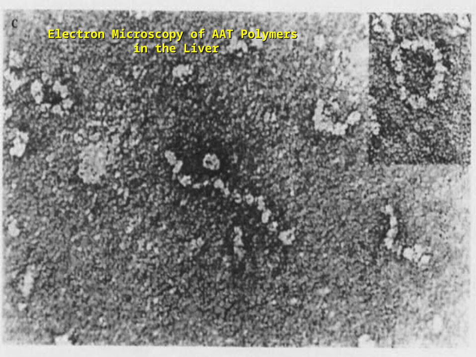
Electron Microscopy of AAT PolymersElectron Microscopy of AAT Polymers in the Liver in the Liver

Clinical CaseClinical Case
CC:CC: 45 yowm comes to your office with a CC of Dyspnea on mild 45 yowm comes to your office with a CC of Dyspnea on mild exercise.exercise.
PMH:PMH: Is unremarkable, and he never smoked Is unremarkable, and he never smoked
Family HxFamily Hx: His Father was a smoker and died of Emphysema at 43 : His Father was a smoker and died of Emphysema at 43 years of age and his mother is 73 yo and in good health. He has 2 years of age and his mother is 73 yo and in good health. He has 2 sons 19 and 21 years old, his older son has a 3 pack/year smoking sons 19 and 21 years old, his older son has a 3 pack/year smoking Hx and the 19 yo has IgA deficiencyHx and the 19 yo has IgA deficiency
PE: PE: Vital signs reveal BP 120/74 HR: 88 RR: 20/minVital signs reveal BP 120/74 HR: 88 RR: 20/min. . The only The only positive findings are diminished bilateral breath sounds and an positive findings are diminished bilateral breath sounds and an emphysematous type Chest wall.emphysematous type Chest wall.

Clinical CaseClinical Case
His Chest X ray shows His Chest X ray shows bullous images in both LL bullous images in both LL
His Chest CTHis Chest CT
His PFT reveal a FEV1 His PFT reveal a FEV1 48% of predicted with a 48% of predicted with a 35% increase on inhaled 35% increase on inhaled bronchodilators.bronchodilators.
CBC and Chem 14 are CBC and Chem 14 are normal. His AAT level is normal. His AAT level is 45 mg/dL.45 mg/dL.

Clinical CaseClinical Case
Does he have a AAT deficiency ?Does he have a AAT deficiency ?What other tests should you order?What other tests should you order?What is his prognosis?What is his prognosis?What information regarding treatment should you give What information regarding treatment should you give him?him?Is he a candidate for AAT augmentation therapy?Is he a candidate for AAT augmentation therapy?If so, what precautions should you take before starting If so, what precautions should you take before starting treatment?treatment?Should his siblings be tested for AATD and Phenotype? Should his siblings be tested for AATD and Phenotype? What will you do with the results if they are abnormal?What will you do with the results if they are abnormal?

AATDAATD
Described in 1963 by Laurell and EriksonDescribed in 1963 by Laurell and Erikson11
UnderrecognizedUnderrecognized Disorder that may affect Disorder that may affect– LungsLungs– LiverLiver– Skin (rarely)Skin (rarely)
AATAAT– Inhibitor of proteolytic enzyme Inhibitor of proteolytic enzyme elastaseelastase
– Member of the Serpins Family (Serine Protease Inhibitors)Member of the Serpins Family (Serine Protease Inhibitors)
– 90 Alleles Identified90 Alleles Identified
1. Laurell, C-B,Eriksson, A. Scand J Clin Lab Invest 1963: 15:32

AATD Lung DiseaseAATD Lung Disease

AAT PhenotypesAAT Phenotypes
Phenotype AAT Levels AAT Function
Normal MM Normal Normal.
Deficient ZZ (most common)
Under 35 % of normal level
Normal
Null Null Null 0% NA
Dysfunctional varies Normal Abnormal

What is the minimum Level of AAT What is the minimum Level of AAT necessary for lung protection?necessary for lung protection?
11 umol/L or 80 mg/dL(NV: 20-53 umol/L or 150 300 mg/dL)
Based on population studies

Pathogenesis of Lung Damage in AATDPathogenesis of Lung Damage in AATD
Clinical CaseClinical Case
AAT

EpidemiologyEpidemiology
USA:USA: 80,000 – 100,000 80,000 – 100,000
WorldwideWorldwide 3,000,000 3,000,000
Worldwide racial and ethnic distribution of alpha(1)-antitrypsin deficiency. Chest 2002;122:1818

PrevalencePrevalence
Based on a US population of 250 millionBased on a US population of 250 million
– COPD screeningCOPD screening11: 2 - 3 % of 965 COPD patients screened: 2 - 3 % of 965 COPD patients screened11
If in the USA there are 2.1 million patients with Emphysema, If in the USA there are 2.1 million patients with Emphysema, 40,000-60,000 would be expected to be AAT Deficient40,000-60,000 would be expected to be AAT Deficient
– Direct population screeningDirect population screening studies studies22
1:1575 – 1:5097 are positive 80,000 - 100,000 would be expected to 1:1575 – 1:5097 are positive 80,000 - 100,000 would be expected to be AAT Deficientbe AAT Deficient
1. Chest 1986;89:370
2. N Eng J Med 1976;294:1316

Why is AAT Deficiency Why is AAT Deficiency Underdetected ?Underdetected ?
Many patients are Many patients are asymptomaticasymptomatic despite despite severe deficiencysevere deficiency
Lack of recognitionLack of recognition of symptomatic patients by of symptomatic patients by physicians physicians – In a cohort of 304 AAT deficient patientsIn a cohort of 304 AAT deficient patients
Mean time to diagnosis was 7.2 yearsMean time to diagnosis was 7.2 years
Number of physicians seen before diagnosis was madeNumber of physicians seen before diagnosis was made– 3 (43% of the patients)3 (43% of the patients)
– 6 – 10 (12 % of the patients)6 – 10 (12 % of the patients)
Cleve Clin J Med 1994;61:461

Why is it important to detect AATDWhy is it important to detect AATD
Treatment is availableTreatment is available
Counseling Counseling – Of the patient to avoid other risk factorsOf the patient to avoid other risk factors– Of the siblings for screening andOf the siblings for screening and

Clinical PresentationClinical Presentation
EmphysemaEmphysema– Pathogenesis:
Imbalance between neutrophil elastase in the lung which destroys Imbalance between neutrophil elastase in the lung which destroys elaste and elastase inhibitor AAT which protects against proteolytic elaste and elastase inhibitor AAT which protects against proteolytic degradation of elastindegradation of elastin
– Risk factorsRisk factors::Phenotypes associated with a AAT levels below the “Protective Phenotypes associated with a AAT levels below the “Protective threshold” of 11umol/Lthreshold” of 11umol/LSmokingSmokingParental Hx of o COPDParental Hx of o COPD
Bronchiectasis ?Bronchiectasis ?
Asthma ?Asthma ?

Lung Related Clinical ManifestationsLung Related Clinical Manifestations
EmphysemaEmphysema– Presenting SymptomsPresenting Symptoms::
Dyspnea (most common symptom)Dyspnea (most common symptom)
Cough, phlegm production and wheezingCough, phlegm production and wheezing
Bronchodilator responsivenessBronchodilator responsiveness– Differences with patients w usual COPDDifferences with patients w usual COPD
Earlier AgeEarlier Age
Bullous changesBullous changes prominent prominent in lung basesin lung bases– > 90 % of ZZ phenotype have lung bases involved> 90 % of ZZ phenotype have lung bases involved– Limited to lung bases in 24 %Found exclusively inLimited to lung bases in 24 %Found exclusively in
Asthma and Bronchiectasis: Asthma and Bronchiectasis: – Relationship not provenRelationship not proven

DiagnosisDiagnosis
Measure AAT levelMeasure AAT level
Phenotype by isoelectric focusingPhenotype by isoelectric focusing
GenotypeGenotype

PhenotypePhenotype Risk for Risk for EmphysemaEmphysema
True Plasma True Plasma level (umol/L)level (umol/L)
Commercial Commercial Standard (mg/dL)Standard (mg/dL)
MMMM No increaseNo increase 20-5320-53 150-350150-350
MZMZ Possible mild Possible mild increaseincrease
12-3512-35 90-21090-210
SSSS No increaseNo increase 15-3015-30 100-140100-140
SZSZ Mild Increase Mild Increase
(20 -50%)(20 -50%)
8-198-19 75-12075-120
ZZZZ High Risk High Risk
(80 – 100%)(80 – 100%)
2.5-72.5-7 20-4520-45
NullNull High Risk High Risk
(100% by age 30)(100% by age 30)
00 00
Am Rev Respir Dis 1989;140:1494
AAT Phenotypes

Risk for developing lung diseaseRisk for developing lung disease
SmokingSmoking::– Age of onset of Dyspnea in AAT (ZZ) Deficient Non-smokers Vs Age of onset of Dyspnea in AAT (ZZ) Deficient Non-smokers Vs
SmokerSmoker11:: 32 - 40 vs 48 – 5432 - 40 vs 48 – 54– In heterozygous In heterozygous SZSZ phenotype, COPD rarely occurs unless phenotype, COPD rarely occurs unless smokingsmoking
is presentis present22
Family HistoryFamily History::– MZMZ phenotypes have increased risk of COPD only when they have a phenotypes have increased risk of COPD only when they have a
symptomatic first degreesymptomatic first degree relative relative33
Airway irritantsAirway irritants::– Risk of other irritants in disease progression is controversialRisk of other irritants in disease progression is controversial
1. Lancet 1985;1:152 2. Am J Respir Crit Care Med 1996;154:1718
3. Am J Respir Crit Care Med 2000;161:81

Survival in AAT according to FEV1Survival in AAT according to FEV1
0
0.1
0.2
0.3
0.4
0.5
0.6
Mortality rate
15 20 25 30 35 60
% of predicted FEV 1
2 year Mortality
Seersholm N et al. Eur Respir J 1994;7:1985

TreatmentTreatment
Augmentation TherapyAugmentation Therapy– Intravenous (only one FDA approved)Intravenous (only one FDA approved)– AerosolizedAerosolized
Enhancement of endogenous AATEnhancement of endogenous AAT
Gene TherapyGene Therapy

IV Augmentation TherapyIV Augmentation Therapy
FDA approved IV AATFDA approved IV AAT based on clinical studies that based on clinical studies that proved that the infusion:proved that the infusion:– Increase plasma and ELF levels of AATIncrease plasma and ELF levels of AAT– Increase Levels anti-neutrophil elastase activity in ELF Increase Levels anti-neutrophil elastase activity in ELF
recovered by BALrecovered by BAL– Is Safe and well toleratedIs Safe and well tolerated
There are There are no randomizedno randomized clinical trials that prove clinical trials that prove clinical efficacy in change in natural history of clinical efficacy in change in natural history of emphysemaemphysema
Indication of IV AAT is Indication of IV AAT is based on observational studiesbased on observational studies

IV Augmentation Therapy: ConcernsIV Augmentation Therapy: Concerns
The true protective threshold value (AAT level)The true protective threshold value (AAT level)– Is not availableIs not available– It is estimated from values that separate affected from It is estimated from values that separate affected from
unaffected individualsunaffected individuals
Some severely deficient patients have normal lung Some severely deficient patients have normal lung functionfunction– Plasma levels alone do not predict disease they only assign riskPlasma levels alone do not predict disease they only assign risk
The proportion of individuals with ZZ phenotype that do The proportion of individuals with ZZ phenotype that do not develop clinically significant emphysema is not not develop clinically significant emphysema is not knownknown

Observational StudiesObservational Studies
National Registry of Patients with Severe AATD National Registry of Patients with Severe AATD conducted a prospective cohort studyconducted a prospective cohort study11
– Survival was enhanced in recipients of augmentation therapySurvival was enhanced in recipients of augmentation therapy– The subset with FEV1 35 % – 49 % of predicted had a slower The subset with FEV1 35 % – 49 % of predicted had a slower
decline of FEV1 over timedecline of FEV1 over time
Study comparing Ex- German Smokers (198) with Study comparing Ex- German Smokers (198) with treatment (3.2 years) with Ex Danish smokers (98) treatment (3.2 years) with Ex Danish smokers (98) without treatmentwithout treatment22
– Lower FEV1 decline in treatment group (53ml vs 75ml per year, Lower FEV1 decline in treatment group (53ml vs 75ml per year, P= 02)P= 02)
Study evaluating 96 patients with severe AAT before and Study evaluating 96 patients with severe AAT before and after treatmentafter treatment33
– Showed a lower FEV1 only in those with mild airflow obstructionShowed a lower FEV1 only in those with mild airflow obstruction
1. Am J Respir Crit Care Med 1998;158:49. 2. Eur Respir J 1997;10:2260
3. Chest 2001;119:737

Selection Criteria for Selection Criteria for TreatmentTreatment
High – risk phenotype (ZZ or Null)High – risk phenotype (ZZ or Null)Plasma AAT level below 11 umol/LPlasma AAT level below 11 umol/LAirflow obstruction by Spirometry Airflow obstruction by Spirometry – American Thoracic Society: < 80 % of predictedAmerican Thoracic Society: < 80 % of predicted– Canadian Thoracic Society: 35% - 50 % of predictedCanadian Thoracic Society: 35% - 50 % of predicted
Patient compliance to treatmentPatient compliance to treatmentAge equal to or greater than 18Age equal to or greater than 18Nonsmoker or ex-smokerNonsmoker or ex-smoker

Selection Criteria for Selection Criteria for TreatmentTreatment
Not recommended for Not recommended for – Heterozygous PhenotypesHeterozygous Phenotypes– AAT > 11umol/LAAT > 11umol/L
UnknownUnknown– Fixed severe obstructionFixed severe obstruction– Normal airflow but radiographic evidence of Normal airflow but radiographic evidence of
EmphysemaEmphysema

Goals of IV InfusionGoals of IV Infusion
Maintain a through level above the Maintain a through level above the protective thresholdprotective threshold
Diffusion of AAT in lung tissue (ELF)Diffusion of AAT in lung tissue (ELF)
In vivo anti neutrophil elastase activity In vivo anti neutrophil elastase activity after infusionafter infusion

AAT Infusion: Side EffectsAAT Infusion: Side Effects
Low grade self limited feverLow grade self limited fever
Anaphylaxis with IgE antibody formation to Anaphylaxis with IgE antibody formation to AAT (rare)AAT (rare)
Syndrome of Syndrome of – Transient fever Transient fever – Chest and low back painChest and low back pain
Biological hazardBiological hazard
Anaphylaxis in IgA deficient patientsAnaphylaxis in IgA deficient patients

Weekly Infusions of AAT 60 mg/KgWeekly Infusions of AAT 60 mg/Kg
0
350
150
350
150
350
150
350
150
350
0
50
100
150
200
250
300
350
400
-7 0 2 7 9 14 16 21 23 28
Days
Co
nc
en
tra
tio
n o
f A
AT
in m
g/d
L
Am J Med 1988;84(supp; 6A):52.

Monthly Infusion of 250mg/Kg of AATMonthly Infusion of 250mg/Kg of AAT
ELF Antitrypsin Activity
0
0.5
1
1.5
2
2.5
3
3.5
0 1 2 3 4 5 6 7 8 9 10
Months
EL
F A
AT
Lev
el,
um
ol
Patient 1
Patient 2
Patient 3
JAMA 1988;260:1259
AAT protective level

Efficacy of aerosolized AATEfficacy of aerosolized AAT
0
1
2
3
4
5
6
1 2 3 4 5 6 7 8
Days
EL
F a
nti
ela
sta
se
ca
pa
cit
y,
mm
ol
Line 1
Hubbard, RC et al. Ann Intern Med 1989;111:206
Normal range

Management of Candidates for Management of Candidates for Augmentation TherapyAugmentation Therapy
Pre- Treatment TestingPre- Treatment Testing– Respiratory FunctionRespiratory Function::
SpirometrySpirometryDLCODLCO
– LaboratoryLaboratory::Hepatitis profileHepatitis profileLFT’sLFT’sHIV TiterHIV Titer
– ImmunizationImmunizationHepatitis B vaccineHepatitis B vaccineIVIG immunoglobulinIVIG immunoglobulin
Supportive TherapySupportive Therapy– CessationCessation
SmokingSmokingRespiratory irritantsRespiratory irritants
– Non-Specific TreatmentsNon-Specific TreatmentsBronchodilatorsBronchodilatorsPulmonary RehabPulmonary RehabOxygen TherapyOxygen TherapyEarly treatment of Early treatment of respiratory infectionsrespiratory infections
– VaccinesVaccines::Pneumococcal Pneumococcal InfluenzaInfluenza

Extra-pulmonary AAT DeficiencyExtra-pulmonary AAT Deficiency
Hepatic Disease (most frequent)Hepatic Disease (most frequent)Skin DiseaseSkin Disease– Panniculitis (1:1000 of AATD)Panniculitis (1:1000 of AATD)
More inflammatoryMore inflammatoryMore “oily discharge”More “oily discharge”More acute inflammation in histologyMore acute inflammation in histology
Vascular diseaseVascular disease– AneurysmsAneurysms– Fibromuscular displasiaFibromuscular displasia– AAT Pittsburg mimics effects of antithrombin IIIAAT Pittsburg mimics effects of antithrombin III
GlomerulonephritisGlomerulonephritis– Prolipherative GNProlipherative GN– IgA GNIgA GN
Inflammatory Bowel diseaseInflammatory Bowel disease– AATD patients have more severe ColitisAATD patients have more severe Colitis

Hepatic DiseaseHepatic Disease
Liver Diseases associated with AAT phenotypesLiver Diseases associated with AAT phenotypes
Neonatal hepatitisNeonatal hepatitis
Elevated transaminases in young adultsElevated transaminases in young adults
Cirrhosis in children and adultsCirrhosis in children and adults
Hepatocellular carcinomaHepatocellular carcinoma
““Null” PhenotypeNull” Phenotype has no risk of hepatic disease has no risk of hepatic disease and a High risk of Emphysemaand a High risk of Emphysema

Pathogenesis of Liver DiseasePathogenesis of Liver Disease
Intra-hepatocyte Polymerization of AAT variants (Z and M)
Intra-hepatocyte accumulation of AAT molecules
in the endoplasmic reticulum (ER)
Decreased degradation of the AAT polymers in the ER
Cell engorgement due to increase mass and release of lysosomal
enzymes
Increase risk of viral mediated hepatitis

Polymerization of AAT in the Polymerization of AAT in the HepatocyteHepatocyte
Intra-hepatocyte accumulation of AAT molecules in the endoplasmic reticulum (ER)
PAS positive granules
AAT polymers

Natural History of Hepatic Disease of Natural History of Hepatic Disease of ZZ PhenotypeZZ Phenotype
0% 20% 40% 60% 80% 100%
Neonatal Disease Adulthood Disease Free of Disease

Natural History of Hepatic DiseaseNatural History of Hepatic Disease
15 % neonatal hepatitis15 % neonatal hepatitis– 5% Cirrhosis in the 15% Cirrhosis in the 1stst year of life year of life– 10 %10 %
25% Resolution of hepatitis by ages 3 to 1025% Resolution of hepatitis by ages 3 to 1025% Cirrhosis between age 6 mo and 17 years 25% Cirrhosis between age 6 mo and 17 years 25% Histological evidence of cirrhosis with survival through 25% Histological evidence of cirrhosis with survival through the first decadethe first decade25% Elevated LFT’s without Cirrhosis25% Elevated LFT’s without Cirrhosis
85% Asymptomatic at childhood85% Asymptomatic at childhood– Cirrhosis in 11.8 %Cirrhosis in 11.8 %– Hepatocellular carcinoma in 3.3 %Hepatocellular carcinoma in 3.3 %– 85% No Disease85% No Disease

Clinical Case ResolutionClinical Case Resolution
Does he have a AAT deficiency ? Does he have a AAT deficiency ? – YESYES
What other tests should you order?What other tests should you order?– PHENOTYPEPHENOTYPE ZZ ZZ
What is his prognosis?What is his prognosis?– According to FEV1 15 % in 2 yearsAccording to FEV1 15 % in 2 years
What information regarding treatment should you give What information regarding treatment should you give him?him?– That he is a candidate for Augmentation therapy but that there That he is a candidate for Augmentation therapy but that there
are no clinical trials to assure him improvementare no clinical trials to assure him improvement
Is he a candidate for AAT augmentation therapy?Is he a candidate for AAT augmentation therapy?– Yes, his age, FEV1, AAT level and phenotype and non-smoker Yes, his age, FEV1, AAT level and phenotype and non-smoker
status making him a good candidatestatus making him a good candidate

Clinical Case ResolutionClinical Case Resolution
If so, what precautions should you take before starting If so, what precautions should you take before starting treatment?treatment?– Hep B vaccination, HIV testing, Influenza and Pneumococcal Hep B vaccination, HIV testing, Influenza and Pneumococcal
vaccinesvaccines
Should his siblings be tested for AATD and Phenotype? Should his siblings be tested for AATD and Phenotype? – YesYes
What will you do with the results if they are abnormal?What will you do with the results if they are abnormal?– His 21 year old son is ZZ phenotype, FEV1 is normalHis 21 year old son is ZZ phenotype, FEV1 is normal
Stop smoking and control of FEV1Stop smoking and control of FEV1– His 19 year old son is ZM phenotype (probably like his mother) His 19 year old son is ZM phenotype (probably like his mother)
and FEV1 is also normaland FEV1 is also normalAvoid smoking Avoid smoking No treatment warranted since AAT infusion can cause anaphylaxis No treatment warranted since AAT infusion can cause anaphylaxis in IgA deficiency in IgA deficiency

Situations to Suspect Severe Situations to Suspect Severe Deficiency of AATDeficiency of AAT
Emphysema in a young individual Emphysema in a young individual (less than 45 years old)(less than 45 years old)
Emphysema in a non smokerEmphysema in a non smoker
Emphysema characterized by predominant basilar Emphysema characterized by predominant basilar changes on the chest x-raychanges on the chest x-ray
Family History of Emphysema and/or liver disease Family History of Emphysema and/or liver disease (unexplained cirrhosis or hepatoma)(unexplained cirrhosis or hepatoma)
Clinical findings or history of panniculitisClinical findings or history of panniculitis
Clinical findings or history of unexplained chronic liver Clinical findings or history of unexplained chronic liver diseasedisease

THANK YOUTHANK YOU


