University of Groningen Altered Energetics of Exercise Explain Risk of Rhabdomyolysis in Very Long-Chain Acyl-CoA Dehydrogenase Deficiency Diekman, E. F.; Visser, G.; Schmitz, J. P. J.; Nievelstein, R. A. J.; de Sain-van der Velden, M.; Wardrop, M.; Van der Pol, W. L.; Houten, S. M.; van Riel, N. A. W.; Takken, T. Published in: PLoS ONE DOI: 10.1371/journal.pone.0147818 IMPORTANT NOTE: You are advised to consult the publisher's version (publisher's PDF) if you wish to cite from it. Please check the document version below. Document Version Publisher's PDF, also known as Version of record Publication date: 2016 Link to publication in University of Groningen/UMCG research database Citation for published version (APA): Diekman, E. F., Visser, G., Schmitz, J. P. J., Nievelstein, R. A. J., de Sain-van der Velden, M., Wardrop, M., Van der Pol, W. L., Houten, S. M., van Riel, N. A. W., Takken, T., & Jeneson, J. A. L. (2016). Altered Energetics of Exercise Explain Risk of Rhabdomyolysis in Very Long-Chain Acyl-CoA Dehydrogenase Deficiency. PLoS ONE, 11(2), [e0147818]. https://doi.org/10.1371/journal.pone.0147818 Copyright Other than for strictly personal use, it is not permitted to download or to forward/distribute the text or part of it without the consent of the author(s) and/or copyright holder(s), unless the work is under an open content license (like Creative Commons). The publication may also be distributed here under the terms of Article 25fa of the Dutch Copyright Act, indicated by the “Taverne” license. More information can be found on the University of Groningen website: https://www.rug.nl/library/open-access/self-archiving-pure/taverne- amendment. Take-down policy If you believe that this document breaches copyright please contact us providing details, and we will remove access to the work immediately and investigate your claim. Downloaded from the University of Groningen/UMCG research database (Pure): http://www.rug.nl/research/portal. For technical reasons the number of authors shown on this cover page is limited to 10 maximum.
Transcript
University of Groningen
Altered Energetics of Exercise Explain Risk of Rhabdomyolysis in Very Long-Chain Acyl-CoADehydrogenase DeficiencyDiekman, E. F.; Visser, G.; Schmitz, J. P. J.; Nievelstein, R. A. J.; de Sain-van der Velden, M.;Wardrop, M.; Van der Pol, W. L.; Houten, S. M.; van Riel, N. A. W.; Takken, T.Published in:PLoS ONE
DOI:10.1371/journal.pone.0147818
IMPORTANT NOTE: You are advised to consult the publisher's version (publisher's PDF) if you wish to cite fromit. Please check the document version below.
Document VersionPublisher's PDF, also known as Version of record
Publication date:2016
Link to publication in University of Groningen/UMCG research database
Citation for published version (APA):Diekman, E. F., Visser, G., Schmitz, J. P. J., Nievelstein, R. A. J., de Sain-van der Velden, M., Wardrop,M., Van der Pol, W. L., Houten, S. M., van Riel, N. A. W., Takken, T., & Jeneson, J. A. L. (2016). AlteredEnergetics of Exercise Explain Risk of Rhabdomyolysis in Very Long-Chain Acyl-CoA DehydrogenaseDeficiency. PLoS ONE, 11(2), [e0147818]. https://doi.org/10.1371/journal.pone.0147818
CopyrightOther than for strictly personal use, it is not permitted to download or to forward/distribute the text or part of it without the consent of theauthor(s) and/or copyright holder(s), unless the work is under an open content license (like Creative Commons).
The publication may also be distributed here under the terms of Article 25fa of the Dutch Copyright Act, indicated by the “Taverne” license.More information can be found on the University of Groningen website: https://www.rug.nl/library/open-access/self-archiving-pure/taverne-amendment.
Take-down policyIf you believe that this document breaches copyright please contact us providing details, and we will remove access to the work immediatelyand investigate your claim.
Downloaded from the University of Groningen/UMCG research database (Pure): http://www.rug.nl/research/portal. For technical reasons thenumber of authors shown on this cover page is limited to 10 maximum.
Altered Energetics of Exercise Explain Risk ofRhabdomyolysis in Very Long-Chain Acyl-CoADehydrogenase DeficiencyE. F. Diekman1,2, G. Visser1,2, J. P. J. Schmitz3,4, R. A. J. Nievelstein5, M. de Sain-van derVelden1, M. Wardrop6, W. L. Van der Pol7, S. M. Houten2, N. A. W. van Riel4, T. Takken6, J.A. L. Jeneson6,8¤*
1 Department of Metabolic Diseases, Wilhelmina Children’s Hospital, University Medical Center Utrecht,Utrecht, the Netherlands, 2 Laboratory of Genetic Metabolic Diseases, Departments of Clinical Chemistryand Pediatrics, Emma Children’s Hospital, Academic Medical Center, University of Amsterdam, Amsterdam,the Netherlands, 3 Computational Biology Group, Department of Biomedical Technology, EindhovenUniversity of Technology, Eindhoven, the Netherlands, 4 Systems Bioinformatics, Department of Life andEarth Sciences, Vrije Universiteit, Amsterdam, the Netherlands, 5 Department of Radiology, UniversityMedical Center Utrecht, Utrecht, the Netherlands, 6 Child Development & Exercise Center, WilhelminaChildren’s Hospital, University Medical Center Utrecht, Utrecht, the Netherlands, 7 Rudolf Magnus Instituteof Neuroscience, Spieren voor Spieren Children’s Center, Department of Neurology and Neurosurgery,University Medical Center Utrecht, Utrecht, the Netherlands, 8 Laboratory of Liver, Digestive and MetabolicDiseases, Department of Pediatrics, University Medical Center Groningen, Groningen, the Netherlands
¤ Current address: Neuroimaging Center, Division of Neuroscience, University Medical Center Groningen,Groningen, the Netherlands* [email protected]
AbstractRhabdomyolysis is common in very long-chain acyl-CoA dehydrogenase deficiency
(VLCADD) and other metabolic myopathies, but its pathogenic basis is poorly understood.
Here, we show that prolonged bicycling exercise against a standardized moderate workload
in VLCADD patients is associated with threefold bigger changes in phosphocreatine (PCr)
and inorganic phosphate (Pi) concentrations in quadriceps muscle and twofold lower
changes in plasma acetyl-carnitine levels than in healthy subjects. This result is consistent
with the hypothesis that muscle ATP homeostasis during exercise is compromised in
VLCADD. However, the measured rates of PCr and Pi recovery post-exercise showed that
the mitochondrial capacity for ATP synthesis in VLCADDmuscle was normal. Mathematical
modeling of oxidative ATP metabolism in muscle composed of three different fiber types
indicated that the observed altered energy balance during submaximal exercise in VLCADD
patients may be explained by a slow-to-fast shift in quadriceps fiber-type composition corre-
sponding to 30% of the slow-twitch fiber-type pool in healthy quadriceps muscle. This study
demonstrates for the first time that quadriceps energy balance during exercise in VLCADD
patients is altered but not because of failing mitochondrial function. Our findings provide
new clues to understanding the risk of rhabdomyolysis following exercise in human
VLCADD.
PLOSONE | DOI:10.1371/journal.pone.0147818 February 16, 2016 1 / 19
OPEN ACCESS
Citation: Diekman EF, Visser G, Schmitz JPJ,Nievelstein RAJ, de Sain-van der Velden M, WardropM, et al. (2016) Altered Energetics of ExerciseExplain Risk of Rhabdomyolysis in Very Long-ChainAcyl-CoA Dehydrogenase Deficiency. PLoS ONE 11(2): e0147818. doi:10.1371/journal.pone.0147818
IntroductionThe mitochondrial enzyme very-long chain Acyl-CoA dehydrogenase (VLCAD, OMIM201475) is the first enzyme in the fatty acid oxidation cycle and, as such, a key enzyme in thispathway for mitochondrial energy transduction from fatty acids [1,2]. The clinical presentationof patients with VLCAD deficiency (VLCADD) varies from death in early childhood, associ-ated with fasting intolerance and failing glucose homeostasis [1,3], to exercise intolerance withepisodic rhabdomyolysis and myalgia in (early-)childhood and adult life, to asymptomaticindividuals [3–5]. In the past decade VLCADD has been included in newborn screening pro-grams all over the world [6]. Treatment consists mainly of dietary advices aimed at preventionof catabolism [7]. However, patients still suffer from exercise intolerance and myalgia, withrisk of episodic rhabdomyolysis [8].
Rhabdomyolysis is a grave complication in VLCADD that may lead to kidney damage andeven renal failure [4] and can be triggered by prolonged or intense exercise, prolonged fastingand fever or illness. Post-exercise rhabdomyolysis has also been described in other metabolicmyopathies including glycogen storage disease [9,10] and statin-induced myopathy (18). Thepathophysiological basis of rhabdomyolysis following exercise in these diseases is, however,incompletely understood. A first and longstanding hypothesis invokes myocellular ATP deple-tion during exercise followed by irreversible myocellular calcium overload [9,10]. In VLCADD,failing myocellular ATP homeostasis during prolonged or intense exercise may result frominhibition of mitochondrial respiration by accumulated incompletely oxidized long-chain fattyacids. Evidence for such an inhibitory mechanism has been found in vitro [11]. However, acase study of VLCADD in a patient with a history of rhabdomyolysis following prolonged exer-cise reported normal mitochondrial ATP synthetic function in vivo [12]. Any consistent associ-ation between impaired mitochondrial oxidative metabolism and rhabdomyolysis is, in fact,lacking. For example, rhabdomyolysis is a common complication in mitochondrial fatty acidoxidation defects [13–16], but rare in primary defects in mitochondrial oxidative phosphoryla-tion [17,18]. An alternative hypothesis specifically for defects in fat oxidation invokes directdamage to the myocellular membrane by high concentrations of incompletely oxidized acyl-carnitines that accumulate during prolonged or intense exercise [18]. Supportive evidence forthis hypothesis has likewise been found in vitro [19–21]. However, the absence of any severecardiomyopathic phenotype in adult VLCADD patients (thesis E.F. Diekman 2015) may argueagainst the relevance of such a pathophysiological mechanism in vivo.
Here, we investigated if muscle ATP homeostasis during prolonged stationary exercise at astandardized workload corresponding to maximal fat oxidation (FATMAX) in healthy subjectsis compromised in VLCADD patients. Five VLCADD patients and five healthy controls per-formed a maximal cardiopulmonary exercise test (CPET) to determine the workload corre-sponding to their individual maximal rate of fat oxidation. During a second visit to thehospital, subjects then performed 45 minutes of bicycling exercise at their individual FATMAXworkload. Blood samples were drawn at various timepoints during the protocol for selectivemetabolite profiling. The last five minutes of the exercise task were performed inside a clinicalMRI scanner for continuous in vivo phosphorus nuclear magnetic resonance spectroscopy (31PMRS) recordings of the concentrations of ATP, phosphocreatine (PCr) and inorganic phos-phate (Pi) as well as intramuscular pH in the quadriceps muscle during bicycling exercise atFATMAX and subsequent recovery. The time course data of post-exercise PCr and Pi recoveryto basal levels were applied to a linear model of muscle respiration[22] to determine the mito-chondrial capacity for ATP synthesis of quadriceps muscle after 45 min of exercise at FAT-MAX. The steady-state quadriceps concentrations of ATP, PCr and Pi during FATMAXexercise were applied to a computational model of oxidative ATP metabolism in a skeletal
Rhabdomyolysis in VLCADD
PLOSONE | DOI:10.1371/journal.pone.0147818 February 16, 2016 2 / 19
muscle composed of slow-twitch oxidative (SO), fast-twitch oxidative glycolytic (FOG) andfast-twitch glycolytic (FG), respectively, to identify combinations of muscle fiber type composi-tion and motor unit recruitment scenarios in healthy subjects and VLCADD patients that wereconsistent with the 31P MRS results. Our results show that prolonged bicycle exercise at FAT-MAX workload is associated with threefold larger changes in quadriceps PCr and Pi concentra-tion and twofold lower changes in acetyl-carnitine blood levels in VLCADD patients comparedto healthy subjects. Yet the rate of PCr and Pi recovery post-exercise in VLCADD patients wasnormal. Model simulations showed that the observations in VLCADD patients were bestexplained by a slow-to-fast shift in quadriceps fiber-type composition corresponding to 30% ofthe slow-twitch fiber-type pool.
Methods
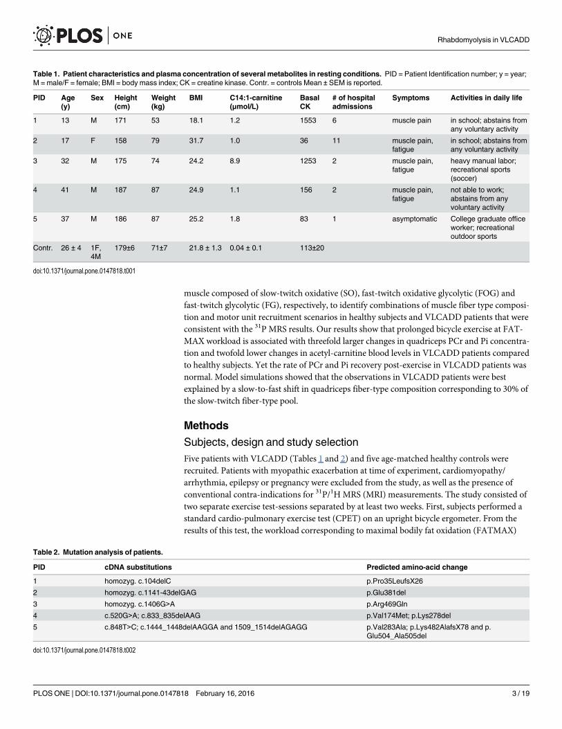
Subjects, design and study selectionFive patients with VLCADD (Tables 1 and 2) and five age-matched healthy controls wererecruited. Patients with myopathic exacerbation at time of experiment, cardiomyopathy/arrhythmia, epilepsy or pregnancy were excluded from the study, as well as the presence ofconventional contra-indications for 31P/1H MRS (MRI) measurements. The study consisted oftwo separate exercise test-sessions separated by at least two weeks. First, subjects performed astandard cardio-pulmonary exercise test (CPET) on an upright bicycle ergometer. From theresults of this test, the workload corresponding to maximal bodily fat oxidation (FATMAX)
Table 1. Patient characteristics and plasma concentration of several metabolites in resting conditions. PID = Patient Identification number; y = year;M = male/F = female; BMI = body mass index; CK = creatine kinase. Contr. = controls Mean ± SEM is reported.
PID Age(y)
Sex Height(cm)
Weight(kg)
BMI C14:1-carnitine(μmol/L)
BasalCK
# of hospitaladmissions
Symptoms Activities in daily life
1 13 M 171 53 18.1 1.2 1553 6 muscle pain in school; abstains fromany voluntary activity
2 17 F 158 79 31.7 1.0 36 11 muscle pain,fatigue
in school; abstains fromany voluntary activity
3 32 M 175 74 24.2 8.9 1253 2 muscle pain,fatigue
heavy manual labor;recreational sports(soccer)
4 41 M 187 87 24.9 1.1 156 2 muscle pain,fatigue
not able to work;abstains from anyvoluntary activity
5 37 M 186 87 25.2 1.8 83 1 asymptomatic College graduate officeworker; recreationaloutdoor sports
5 c.848T>C; c.1444_1448delAAGGA and 1509_1514delAGAGG p.Val283Ala; p.Lys482AlafsX78 and p.Glu504_Ala505del
doi:10.1371/journal.pone.0147818.t002
Rhabdomyolysis in VLCADD
PLOSONE | DOI:10.1371/journal.pone.0147818 February 16, 2016 3 / 19
was determined for each subject. Two or more weeks later, subjects performed 45 minutes ofbicycling exercise at their individual FATMAX workload, of which the final five minutes wereperformed supine inside the MR scanner using a MR-compatible bicycle ergometer [23]. Bloodsamples were taken at three timepoints: before, 5 and 180 minutes after the FATMAX exercisetest, respectively. The study was approved by the medical ethics committee of the UniversityMedical Centre Utrecht (METC 12-211/K). All patients provided written informed consent forparticipation in this study.
Dietary standardizationPatients and healthy controls were asked to keep a food record the three days preceding thetest day. A light meal was allowed before the test. An extensive dietary analysis based on thethree-day food diary and a subsequent interview by a nutritionist, was performed in all patientsprior to the second exercise test (NEVO-table 2011 (Dutch Food Composition Table), RIVM/Voedingscentrum, Den Haag 2011).
Exercise testingI. Cardio-pulmonary exercise test (CPET). Maximal exercise capacity was measured dur-
ing the baseline graded CPET[24]. The participant performed exercise on a standard uprightbicycle ergometer under increasing load until exhaustion (duration +/- 10min). By using therespiratory gas exchange parameters, the intensity of maximal fat oxidation (FATMAX) couldbe determined [25]. Subjects sat placed on an upright cycle ergometer and fitted with a 12-leadECG, a pulse oximeter on the index finger or forehead, an automatic blood pressure cuff, and asmall face mask attached to a breath-by-breath gas analysis system. Subjects performed a pul-monary function test at rest to determine the forced expiratory volume in 1 sec (FEV1) andforced vital capacity (FVC). After this measurement, subjects began the test with 5 minutes ofresting measurements while sitting on the cycle ergometer. A modified protocol for determin-ing the maximal fat oxidation (FATMAX) as recommended for testing on a cycle ergometer(Lode Corival, Lode BV, Groningen, the Netherlands) was used[26]. The subjects wereinstructed to cycle at an increasing load of 35 watts every 3 minutes, starting with 0 watts andending at voluntary exhaustion. During this time several measurements were taken: a full elec-trocardiogram (ECG; Cardioperfect, Accuramed BVBA, Lummen, Belgium), oxygen (O2) satu-ration (Masimo, Rad 8, Masimo BV, Tilburg, the Netherlands), heart rate (HR), blood pressure(BP) (Suntech Tango, Suntech Med, Morrisville, NC, USA), minute ventilation (VE), oxygenuptake (VO2), carbon dioxide output (VCO2), respiratory exchange ratio (RER), workload(W), using a calibrated metabolic cart (ZAN 600, Accuramed BVBA, Lummen, Belgium). Sub-jects were asked to keep their cadence per minute between 60–80, and were given verbalencouragement to continue if they fell below that range. Immediately after exhaustion, the loadwas decreased to 0 watts and subjects were asked to cool down for 5 minutes. FATMAX work-load was calculated according to the formula proposed by Péronnet and Massicotte for calcu-lating fat oxidation, as it is both accurate in estimating fat oxidation and does not require anestimate of protein degradation during exercise (Eq (1);[27]:
Fat oxidation ¼ 1:695 � VO2 � 1:701 � VCO2 eqnð1Þwhere fat oxidation is in mg�min-1, and VO2 and VCO2 are both In mL�min-1. Workload, theindependent variable for the FATMAX curve, was taken in terms of HR, Watt, and percentageof VO2peak. The final 60 seconds of exercise for each workload interval were averaged to achievesteady-state values. Each set of steady-state FATMAX values were fit along a second orderpolynomial curve.
Rhabdomyolysis in VLCADD
PLOSONE | DOI:10.1371/journal.pone.0147818 February 16, 2016 4 / 19
II. Endurance exercise test. Subjects were asked to exercise at their individual FATMAXworkload for a total of 45 minutes. Of these, 40 minutes were performed on an upright stan-dard bicycle ergometer (Lode, Groningen, the Netherlands) in a room adjacent to the MRIscanner. Next, subjects were moved to the MR scanner and performed the final five minutes ofexercise task in supine position inside the MR scanner at a workload equivalent to FATMAX.Hereto, the braking force on the MR-compatible bicycle ergometer was gravimetricallyadjusted to the appropriate amount using 30 N as empirical reference for maximal sustainablebraking force in healthy subjects (data not shown).
31P-Magnetic Resonance SpectroscopySubjects were positioned feet-forward in a supine position in a 1.5T Philips MR Achieva scan-ner (Philips Healthcare, Best, the Netherlands). The upper body was supported by a wedge-shaped cushion to facilitate supine bicycling. A 6-cm diameter single-turn 31P surface coil sup-plied by the manufacturer was fastened over the m. vastus lateralis of the right leg. Subjectsthen performed a short bout of unloaded bicycling supervised by an on-site coach to familiarizethem with the supine exercise task. The desired pedalling frequency (target setting: 75 rpm)was set by a metronome audible over the in-magnet speaker system.[24] This training exercisebout was typically performed within five min after the conclusion of the upright FATMAXbicycling test. Next, a series of scout MR images was acquired to evaluate correct positioning ofthe subject and the coil and image-based shimming was performed. 31P NMR spectra wereacquired from the m. vastus lateralis at rest, during exercise and recovery using an adiabatichalf-passage excitation pulse as described elsewhere [23]. First, a resting spectrum was acquiredunder fully-relaxed conditions (repetition time (TR) 20 s). During exercise and recovery, fourand two free induction decays (FIDs), respectively, were acquired with a TR of 3s and averaged,yielding time resolution of 12 and 6 seconds, respectively, in each dynamic phase. [24]
31P-MRS data processingFIDs were processed and analyzed in the time domain using the AMARES algorithm in thepublic jMRUI software environment (version 3.0) as described elsewhere[23]. Kramer-Raobounds of the AMARES Lorentzian model fitting were used as statistical information on accu-racy of peak area estimation. Absolute PCr and Pi concentrations were calculated after correc-tion for signal saturation assuming total adenylate nucleotide and creatine pool sizes of 8.2 and42.7 mM, respectively, as previously described[28]. Intramuscular pH was determined fromthe resonance frequency of Pi using standard methods[28]. Steady-state PCr and Pi concentra-tions during exercise were determined from summed FIDs acquired 60s after onset of bicy-cling. Datasets were analysed in a blinded fashion.
Computational ModelingThis study employed a multi-fiber computational model of human muscle oxidative ATPmetabolism to analyze the in vivo 31P MRS data obtained during exercise. This approach wasnecessary to address two intrinsic sources of microscopic metabolic heterogeneity in exercisingquadriceps muscle. First of all, human skeletal muscles are composed of SO, FOG and FG fibertypes organized in motor units of uniform fiber type that vary in properties including size, con-tractile speed, ATP cost of contraction and relaxation, power-output, capacity for fat and car-bohydrate metabolism, adenine nucleotide and creatine pool sizes, mitochondrial density andvascularisation [29]. Secondly, during voluntary exercise motor units are neurally recruited ina stepwise order according to the size principle–i.e., SO< FOG< FG [30]. As a result, any 31PNMR spectrum of human muscle engaged in a submaximal voluntary exercise task reflects the
Rhabdomyolysis in VLCADD
PLOSONE | DOI:10.1371/journal.pone.0147818 February 16, 2016 5 / 19
average of the particular concentrations of PCr, Pi and ATP and intramuscular pH in SO, FOGand FG fibers, respectively, including active and inactive fibers within the sampled tissue vol-ume. Investigations of metabolic changes in individual fiber types employing dissection ofbiopsy specimens have previously demonstrated profound differences in PCr content and gly-cogen depletion between slow and fast fibers[31]. A multi-fiber kinetic model of oxidative ATPmetabolism in human skeletal muscle was developed to investigate if, and if so, what magni-tude of fiber type composition changes in VLCADD skeletal muscle may contribute to anymeasured average change in stationary states of energy balance at FATMAX exercise inVLCADD versus healthy muscle. A kinetic model of oxidative ATP metabolism described indetail elsewhere [32] was used as core model in each fiber type submodel. Fiber-type specificsubmodel parameter values with regard to oxidative ATP metabolism and default healthyquadriceps composition are given in Table A in S1 File; other model parameters were assumedto be uniform across fiber types. The relation between measured average Pi and PCr concentra-tions in the quadriceps muscle mass sampled by the 31P surface coil and fiber-type-specificconcentrations is described by (Eq (2))
whereMSO,MFOG,MFG denote the fiber-type specific concentration of metabolite PCr or Piaccording to the model, and XSO, XFOG, XFG denote the fraction of quadriceps muscle com-posed of SO, FOG and FG fibers, respectively. The default model parameterization for XtSO,XFOG, XFG was 0.5, 0.35 and 0.15, respectively [33]. For the simulations, it was assumed thatthe maximal mechanical output that muscle fibers can sustain without any significant fatigue isassociated with a cellular oxygen consumption rate equal to 80% of maximal rate [34,35]. Themodel was implemented in Matlab 7.5.0 (The Mathworks, Natick, MA, USA). Ordinary differ-ential equations were solved numerically by using ODE15s with absolute and relative toler-ances set to 10−8.
Analysis of blood samplesPlasma of blood samples were profiled for glucose, lactate, acylcarnitines and creatine kinase.Plasma was stored at -20°C. Plasma acylcarnitines were measured as described previously [36]using internal standards. Blood for measurement of lactate and pyruvate was treated immedi-ately after withdrawal with an equal volume of 1M perchloric acid and subsequently stored at-20°C. Plasma was stored at -20°C. Plasma glucose and CK were measured using standardenzymatic assays [37]. Blood lactate and pyruvate, were measured using tandem mass spec-trometry[38]. All samples were analysed in a blinded fashion.
Statistics. Two-tailed MannWhitney U-tests were used to determine significant differ-ences between control and patients at p<0.05. Data are presented as mean ± standard error ofmean (SEM) unless specified otherwise. Time courses of variables were analysed and character-ized kinetically using non-linear curve-fitting (Origin 6.0, Caltech Pasadena, US). Wheneverpossible, statistical information on data accuracy was incorporated in the curve-fitting bymeans of statistical weighting.
Results
Baseline characteristics of the patientsFive VLCADD patients (Table 1) and five healthy subjects were included in the study. Diagno-sis of VLCADD was confirmed by mutation analysis of the ACADVL gene (Table 2). Fourpatients had exercise intolerance, myalgia and fatigue while one patient (PID#5) was
Rhabdomyolysis in VLCADD
PLOSONE | DOI:10.1371/journal.pone.0147818 February 16, 2016 6 / 19
asymptomatic. Three patients had a residual VLCAD enzymatic activity of�5% in fibroblasts(PID#1–3), and in two patients residual VLCAD activity was�13% (compared to referencecontrol fibroblasts). The symptomatic patients had been admitted to the hospital at least twicein their lives (range 2–11) (Table 1). Plasma creatine kinase level was increased mildly in twoof the patients. C14:1-carnitine was significantly increased in VLCADD patients compared tocontrols (p = 0.02). Age, height, weight and BMI were comparable between controls andVLCADD patients (Table 1). 31P metabolite concentrations and intramuscular pH in restingquadriceps muscle were not different between VLCADD patients and healthy controls (Datanot shown). Dietary intake of the last three days before the second test did not differ signifi-cantly between controls and VLCADD patients (Table B in S1 File).
Maximal exercise capacity is reduced in VLCADD patientsThe maximal sustained workload during CPET in the four symptomatic VLCADD patientswas 46 ± 6% reduced compared to healthy controls (Table 3 and Fig 1A). In contrast, the maxi-mal workload attained by the asymptomatic patient was well within the normal range(Table 3). Two patients (PID #2 and #3) had a RERpeak comparable to controls, while the otherthree patients (including the asymptomatic patient) had an elevated RERpeak between 0.90 and1.04. VO2_peak was 55% decreased in the four myopathic patients (Table 3 and Fig 1B). Theestimated peak rate of fat oxidation per kg of bodymass was 46% lower in myopathic patientscompared to controls (Fig 1C).
To investigate if ATP homeostasis in muscle of VLCADD patients is compromised duringprolonged stationary exercise at a normalized workload corresponding to maximal fat oxida-tion in healthy subjects (FATMAX), subjects reported to the MRI Center two weeks or moreafter CPET to perform a second exercise test consisting of 45 min of bicycling at their individ-ual FATMAX workload. The first 40 min of the test involved conventional upright bicyclingexercise. Immediately afterwards, subjects were transferred to the MRI scanner room andmounted on a MR-compatible bicycle ergometer for in vivo 31P MR spectroscopic measure-ment of intramuscular energy balance and pH in the quadriceps during the remaining fiveminutes of the exercise test. All subjects were able to complete the test except patient PID#4who did manage to complete 43 of the requested 45 minutes of exercise at FATMAX.
Metabolic profiling of blood samples shows abnormal dynamics ofplasma acetyl-carnitine levels in exercising VLCADD patientsGlucose levels remained unchanged (Fig 2A). Lactate levels were not different between controlsand myopathic VLCADD patients (Fig 2B). In the asymptomatic patient lactate levelsincreased fourfold from 1.3 to 5 mmol/L during exercise at FATMAX (Fig 2B). Basal CK levels
Table 3. Graded CPET exercise test characteristics of controls and patients. HRpeak = peak heart rate; RER = respiratory exchange ratio.Mean ± SEM is reported.
PLOSONE | DOI:10.1371/journal.pone.0147818 February 16, 2016 7 / 19
Fig 2. (A) Glucose, (B) lactate and(C) acetylcarnitine at t = 0 (rest), t = 1 (directly after exercise) and t = 2 (3 hours after exercise) in symptomatic VLCADDpatients and controls. Error bars indicate mean ± SEM, *P<0.05.
doi:10.1371/journal.pone.0147818.g002
Fig 1. (A) Maximal workload of symptomatic (black squares) and asymptomatic (open squares) VLCADD patients and controls. (B) in symptomatic (blacksquares) and asymptomatic (open squares) VLCADD patients. (C) Fatty acid oxidation in mg/kg body mass/min in VLCADD patients and controls. Error barsindicate ± SEM. *P<0.05 for polynominal curve.
doi:10.1371/journal.pone.0147818.g001
Rhabdomyolysis in VLCADD
PLOSONE | DOI:10.1371/journal.pone.0147818 February 16, 2016 8 / 19
were normal (i.e., (<250U/L) in all controls and three VLCADD patients (Table 1). In responseto the FATMAX exercise test, CK levels in two myopathic patients slightly increased (PID #2and #4; 400 and 576 U/L, respectively) but still remained low. Resting plasma C14:1-acyl-carni-tine levels were significantly higher in VLCADD patients than healthy controls (Table 1).Plasma acetyl-carnitine (C2-carnitine) levels in resting state were similar between patients andhealthy controls (Fig 2C). In the time span between onset of exercise and 3 hours post-exercise,plasma acetyl-carnitine levels in healthy controls nearly doubled compared to resting levels,with the majority of the rise occurring over the 40 min exercise test (Fig 2C). In VLCADDpatients, plasma acetyl-carnitine levels remained constant.
In vivo 31P MRS identifies altered quadriceps energy balance inVLCADD during exercise at FATMAX workloadFig 3A shows a time series of 31P NMR spectra recorded sequentially from the vastus lateralismuscle of a healthy control (upper trace) and a VLCADD patient (lower trace) during supinebicycling exercise at FATMAX workload. Fig 3B shows the corresponding 31P NMR spectra ofthe summed FIDs recorded after 60s into the exercise for these subjects. In all healthy controlsubjects, only minor changes in steady-state Pi and PCr concentrations from resting valueswere observed during exercise at FATMAX (ΔPCr: -2.0±0.7 mM; ΔPi: +2.6±1.0 mM (mean+ SE) (Fig 3C). In contrast, in both myopathic as well as the asymptomatic VLCADD patients,pronounced changes in quadriceps Pi and PCr concentrations from resting values wereobserved during exercise at FATMAX (ΔPCr: -7.9 ± 1.0 mM; ΔPi: +8.8 ± 0.8 mM (mean + SE)(Fig 3C). Quadriceps pH during exercise did not fall below 7.0 in any healthy control subject(Fig 4A) but intramuscular pH changes during exercise in VLCADD patients were more het-erogeneous. Specifically, in patient PID#1 quadriceps pH did not fall below 7.0 during exercisesimilar to healthy controls (Fig 4B). In patients PID#02, #04 and #05 intramuscular pH pro-gressively dropped during exercise to values as low as 6.7 (example shown in Fig 4C). In patient#03, intramuscular pH immediately dropped 0.1 units to 6.95 at the onset of exercise andremained stable at this value during the remainder of the exercise (Fig 4D).
Normal kinetics of post-exercise metabolic recovery rule out impairedmitochondrial ATP synthetic function in VLCADDTo evaluate the capacity of oxidative ATP synthesis in quadriceps muscle of the patients after 45 min-utes of exercise at FATMAX, the rate of metabolic recovery to resting state was measured immedi-ately after exercise. In all VLCADD patients, normal kinetics were found for the recovery of PCr andPi concentrations to basal levels post-exercise compared to typical literature values for human quadri-ceps muscle (Table 4 and Fig 5; Fig A in S1 File). Nomajor abnormalities were found in the post-exercise dynamics of quadriceps pH in any of the patients compared to healthy controls (Fig 4). Theancillary drop in intramuscular pH at the onset of recovery followed by a slow recovery to resting val-ues typically observed in healthy control subjects (Fig 4A) was likewise observed in patients PID#01and #03 (Fig 4B and 4D). In the patients PID#02, PID#04 and PID#05 intramuscular pH recoveredwithout any further drop of pH at the onset of recovery (Fig 4C).
Metabolic model simulations indicate a fast-to-slow shift in fiber typecomposition of VLCADDmuscle explains altered energy balance duringexerciseOur findings in the VLCADD patients of altered quadriceps energy balance during exercise butintact capacity for oxidative ATP production after 45 minutes of exercise prompted us to
Rhabdomyolysis in VLCADD
PLOSONE | DOI:10.1371/journal.pone.0147818 February 16, 2016 9 / 19
Fig 3. (A) 31P NMR spectra acquired from the lateral head of the quadriceps muscle of the right leg of a VLCAD deficient patient versus a healthy controlsubject during 5 min of bicycling exercise at a workload equivalent to FATMAX in each subject. (B) Each spectrum represents the sum of the FIDs collectedafter 60 s of exercise. FIDs were apodized in the time domain using a 10-Hz low-pass filter prior to Fourier transform and phasing. Peak assignments: Piinorganic phosphate, PCr phosphocreatine, ATP adenosine triphosphate (gamma, alpha and beta resonances, respectively). (C) Average change in PCrand Pi during exercise (in mM) in healthy control subjects versus symptomatic and asymptomatic VLCAD deficient patients. Error bars indicate mean ± SEM,*P<0.05.
doi:10.1371/journal.pone.0147818.g003
Rhabdomyolysis in VLCADD
PLOSONE | DOI:10.1371/journal.pone.0147818 February 16, 2016 10 / 19
Fig 4. pH dynamics during exercise and first minutes of recovery. The error bars show the variance in the data from AMARES fitting of the MR spectra(see Methods section). (A) Healthy controls. (B-D) VLCADD patients. Solid red lines show the fit of linear or monoexponential functions to the data; blue linesshow the 95% confidence interval of the fit.
doi:10.1371/journal.pone.0147818.g004
Table 4. PCr and Pi recovery rates.
PID tau PCr (s) tau Pi (s)
1 21 ± 9 12 ± 7
3 ND 12 ± 5
2 16 ± 9 21 ± 13
4 28 ± 13 19 ± 14
5 ND 9 ± 5
Controls 24 ± 5 1 24 ± 52
1 value for vastus lateralis muscle of healthy human subjects (22)2 Pi recovery kinetics are similar although not quite identical to PCr recovery[65]. ND = not determined.
doi:10.1371/journal.pone.0147818.t004
Rhabdomyolysis in VLCADD
PLOSONE | DOI:10.1371/journal.pone.0147818 February 16, 2016 11 / 19
conduct numerical simulations of oxidative ATP metabolism in skeletal muscle composed ofthree fiber types and compare each outcome to the 31P MRS results. In the control subjects, themeasured concentrations of PCr and Pi during stationary exercise at FATMAX workload wereconsistent with recruitment of 70% of the total pool of SO fibers of the quadriceps during thevoluntary exercise task (Table 5). If no change was made to the model with respect to quadri-ceps fiber type composition, the PCr and Pi concentrations measured during exercise in thepatients could only be explained by recruitment of 100% of the SO muscle fiber pool (Table 5).Alternatively, if the absolute number of SO fibers in the model was reduced by 30% andreplaced by FOG fibers, the 31P MRS observations in the patients could be explained by recruit-ment of 100% of the reduced SO fiber pool plus 35% of the augmented FOG muscle fiber pool(Table 5).
Fig 5. Pi dynamics during exercise and the first minutes of recovery rates. The error bars show the variance in the data from AMARES fitting of the MRspectra (see Methods section). (A) Healthy controls. (B-D) VLCADD patients. Solid red lines show the fit of a monoexponential function to the data; blue linesshow the 95% confidence interval of the fit.
doi:10.1371/journal.pone.0147818.g005
Rhabdomyolysis in VLCADD
PLOSONE | DOI:10.1371/journal.pone.0147818 February 16, 2016 12 / 19
DiscussionIn this study, we demonstrate that prolonged stationary bicycling exercise against a standard-ized moderate workload in VLCADD patients is associated with threefold bigger changes inPCr and Pi concentrations in quadriceps muscle and twofold lower changes in plasma acetyl-carnitine levels than in healthy subjects. This result is consistent with the hypothesis that mus-cle ATP homeostasis during prolonged exercise in VLCADD is compromised. However,kinetic measurements of the recovery of ATP metabolism to resting state immediately afterexercise, showed that the mitochondrial capacity for ATP synthesis in VLCADD quadricepsmuscle after 45 min of exercise was not impaired. Mathematical model-based simulations ofoxidative ATP metabolism in skeletal muscle composed of SO, FOG and FG fiber types showedthat these findings may be explained by a slow-to-fast shift in VLCADD quadriceps fiber typecomposition.
All five VLCADD patients that participated in this study had highly elevated C14:1-carni-tine levels in diagnostic studies in fibroblasts consistent with the molecular defect. Here, plasmaacetyl-carnitine (C2-carnitine) levels remained constant over the time span between onset ofexercise and 3 hours after exercise in the VLCADD patients whereas in the control subjectsthese metabolite levels almost doubled. The latter finding is consistent with previous findingsin healthy subjects [39]. Acetyl-carnitine concentrations in blood are assumed to reflect con-centrations of mitochondrial acetyl-CoAs [39]. As such, the observed absence of any increaseof blood acetyl-carnitine concentrations in the patients suggests that mitochondrial acetyl-CoAconcentrations did not increase under the test conditions of prolonged exercise at FATMAX.This inability to increase acetyl-CoA, could have caused the inability to provide enough ATPto replenish PCr levels. Blood lactate levels did not increase in response to exercise in neitherhealthy subjects nor patients, except for patient PID#5. This result was consistent with theintended oxidative nature of the exercise task. Indeed, all subjects were able to complete thetask with the exception of patient PID#4 who, however, still managed to complete 43 of the 45min of exercise.
The main result of the present study was the finding of threefold bigger change in quadri-ceps PCr and Pi concentrations during stationary exercise at FATMAX in VLCADD patientsthan in healthy control subjects. This result indicates that homeostasis of the concentrations ofthe ATP hydrolysis products ADP and Pi is less tight in VLCADDmuscle than in healthy mus-cle. The latter is significant because it is thought that control of these concentrations is criticalfor proper function of myocellular cation pumps that are involved in fiber relaxation [40–42].Control of the concentrations of ADP and Pi in muscle resides in the network of myocellularenzymes involved in the ATP hydrolysis-driven fiber contraction and relaxation [42,43].
Table 5. results of model simulations of motor unit recruitment (in % of total pool size) during volun-tary exercise at FATMAX in healthy control subjects and VLCADD patients.
Scenario I: X type I = 0.5 X type IIA = 0.35 X type IIX = 0.15
SO FOG FG
Controls 70% 0% 0%
VLCADD 100% 0% 0%
Scenario II: X type I = 0.35 X type IIA = 0.5 X type IIX = 0.15
SO FOG FG
VLCADD 100% 35% 0%
Xi: fraction of fiber type i in composition of quadriceps muscle; three fiber types; sum of all fractions = 1.0.
SO: slow oxidative fiber
doi:10.1371/journal.pone.0147818.t005
Rhabdomyolysis in VLCADD
PLOSONE | DOI:10.1371/journal.pone.0147818 February 16, 2016 13 / 19
Therefore, our observation raised the question of which network components and/or theirproperties are changed in VLCADD muscle compared to healthy muscle.
Analogous elevated changes in PCr and Pi levels during stationary exercise against a stan-dardized submaximal workload have previously been described in patients with failing mito-chondria due to a molecular defect in the respiratory chain [44,45]. Therefore, a first possibleexplanation would be that the mitochondrial capacity for ATP production during exercise at aworkload associated with maximal rates of fat oxidation and low rates of carbohydrate utiliza-tion is lower in VLCADDmuscle than in healthy muscle. Inhibition of mitochondrial oxidativeADP phosphorylation by high concentrations of incompletely oxidized long chain acyl-carni-tines that may accumulate during prolonged exercise has been demonstrated in vitro [11].However, we found no evidence in the kinetic measurements of post-exercise metabolic recov-ery that a total of 45 minutes of exercise at FATMAX workload produced any cellular conditionin VLCADD muscle that interfered with mitochondrial ATP synthesis in vivo.
An alternative mitochondrial function-related explanation of the result could be that mito-chondria in VLCADD and healthy muscle are not oxidizing the same mix of fat and carbohy-drates during stationary exercise at FATMAX workload. It has been well documented ex vivoin both skeletal and cardiac muscle that changing the oxidative fuel source without changingthe work (and thereby ATP turnover) rate, may cause changes in myocellular PCr and Pi levels[46,47]. If this would be the case here, it may be expected that VLCADDmuscle would be oxi-dizing more carbohydrate, not fat, than healthy muscle. It has, however, been shown that ashift in substrate utilization towards fat, not carbohydrates, is associated with higher Pi andlower PCr levels at the same work rate [46,47]. This only leaves open the third alternativeexplanation that the elevated changes in quadriceps PCr and Pi concentrations in the patientswere simply the result of a higher ATP turnover rate during exercise at FATMAX workloadthan in healthy muscle. In skeletal muscle, this metabolic flux is determined first and foremostby the myosin isoform composition of the muscle and, as such, by its fiber type composition[42]. Therefore, we investigated what change in fiber type composition of the quadriceps mus-cle may explain the result.
The mathematical model of oxidative ATP metabolism in human quadriceps muscle thatwas used to simulate oxidative ATP metabolism in human quadriceps muscle, provides a fairrepresentation of the quantitative knowledge base on human muscle anatomy and physiologyas well as the biochemistry of oxidative energy transduction. Amongst others, parameterizationof the core kinetic model of myofiber oxidative ATP metabolism included model fitting to invivo kinetic studies of ATP metabolism in human quadriceps muscle [32,48]. However, theresults of the model simulations also depended on variables that we did not measure includingthe default fiber type composition of quadriceps muscle in our particular control group ofhealthy subjects, thereby introducing a degree of uncertainty in the model predictions. There-fore, the specific objective of the numerical studies was to generate rational hypotheses explain-ing the observed differences in quadriceps energy balance during FATMAX exercise betweenVLCADD patients and healthy subjects rather than specifically predict absolute values of anyparticular metabolic variable such as fiber type specific respiration rates during exercise atFATMAX.
A first explanation of the main result of this study proposed by the simulations was thatVLCADD patients recruited a larger fraction of their total pool of SO fibers of the quadricepsthan healthy subjects to maintain the workload corresponding to individual FATMAX(Table 5 and Table C in S1 File, scenario I). A corollary of this particular numerical solution isthat VLCADD SO fibers produce 1.3-foldless force per contraction than in healthy controls.However, no evidence for muscle weakness has been documented in VLCADD patients [49].Therefore, this solution was rejected. The next simplest numerical solution was that the
Rhabdomyolysis in VLCADD
PLOSONE | DOI:10.1371/journal.pone.0147818 February 16, 2016 14 / 19
absolute number of SO fibers in VLCADD quadriceps muscle was 30% reduced and replacedby FOG fibers (Table 5 and Table C in S1 File, scenario II). Independent data from two alterna-tive transgenic mouse models of a long chain fatty acid oxidation defect -VLCADD knockout[50] and Steroid Receptor Coactivator-3 (SRC-3) knockout, respectively [51]- are both qualita-tively and quantitatively consistent with this model prediction [50]. Specifically, in theVLCADD knockout model a slow-to-fast shift in quadriceps fiber type composition was foundcorresponding to 20% of the SO fibers in VLCAD knockout mice compared to wild type (WT)littermates [50]. Likewise, in mice lacking SRC-3 a trend towards an increase in fast twitchfiber type compared to WT was found which was further supported by qPCR analysis of genemarkers for different fiber types[51]. Importantly, this slow-to-fast shift in skeletal muscle fibertype composition in response to either VLCADD or SRC-3 gene ablation was found withoutany employment of exercise or nutritional intervention [50], suggesting the adaptation may begeneric to an impaired muscular capacity for long chain fatty acid oxidation[51]. In this light,it is of interest to note that in the present study, the magnitude of the change in PCr and Pi con-centrations in exercising quadriceps muscle was near-identical in all five patients giving rise tothe model-based prediction of a uniform change in fiber type composition of the quadricepsmusle in all patients. Due to the limited number of patients that were enrolled in the investiga-tion, however, it cannot be ruled out that this uniformity was serendipitous. Yet, it was remark-able considering the highly non-uniform clinical manifestation of the VLCADD acrosspatients, ranging from an asymptomatic adult phenotype to severe myopathic phenotypes withelevated basal CK levels.
To our knowledge, the fiber type composition of skeletal muscle in human VLCADD hasonly been reported in a single case study [12]. In that particular patient, a fast-to-slow shift infiber type composition was found [12]. However, two additional findings in that particular casereport raise doubts about a strict monogenic (i.e., VLCADD) origin of the clinical phenotypeof this particular patient. Firstly, it was reported that the amount of exercise required to triggera rhabdomyolytic attack in this patient decreased with age [12]. Such a progressive time courseof the disease has not been observed in any of the 45 VLCADD patients seen in our clinic; how-ever, others have observed progressive disease with lower amounts of exercise inducing rhab-domyolysis as patients with VLCAD age (unpublished observations). Secondly, an abnormal,alkaline pH in resting muscle was found in this VLCADD case report (pH 7.30; [12]). Thisstriking abnormality was not found in any of the five patient studied here and, in fact, moreconsistent with previous findings in patients with dystrophinopathies [52–54]. Muscular type Ifiber predominance is likewise a common finding in dystrophin myopathies [55–57]. There-fore, we concluded that this single case report of a SO fiber type predominance in humanVLCADD provided insufficient ground to reject our model-based hypothesis of a slow-to-fastfiber type transformation. Future studies of the fiber type composition of skeletal muscle inVLCADD patients are needed to rigorously test the hypothesis.
The result of this study fit the longstanding hypothesis of an energetic cause of post-exerciserhabdomyolysis in metabolic myopathies including VLCADD. Healthy active muscle derivesthe majority of its substrate for oxidative ATP production during exercise from intramyocellu-lar lipid and glycogen stores [58]. Absence of any significant capability for fat oxidation such asin VLCADD renders the myofibers dependent on intracellular glycogen stores to produce ATPduring exercise[51]. The capacity of these stores to produce glycosyl units for oxidative glycoly-sis is, however, finite [59]. As such, a molecular defect of VLCADD in and by itself puts themyofibers at risk of an energy crisis during prolonged exercise. Any slow-to-fast phenotypicadaptation of VLCADD skeletal muscle would aggravate this problem due to the threefoldlower energetic contractile efficiency in combination with the twofold higher glycogenolytic
Rhabdomyolysis in VLCADD
PLOSONE | DOI:10.1371/journal.pone.0147818 February 16, 2016 15 / 19
capacity of FOG fibers compared to SO fibers [60]. The fact that specifically fast-twitch fibersare lost in rhabdomyolysis [61] is in support of this hypothesis.
Finally, the results of this study may provide a basis for rational therapeutic approaches tominimize risk of exertional rhabdomyolysis in myopathic VLCADD patients. Currently, noeffective therapy has been developed to prevent rhabdomyolysis in VLCADD [4]. Instead,patients are typically advised to avoid any intense or prolonged exercise[17,18,62]. The long-term health outcome of lifelong restricted physical activity is, however, likewise a concern[63].Our findings suggest that any therapeutic approach resorting in glycogen sparing during exer-cise should be considered for clinical testing. For one, exercise training programs to reverse anyslow-to-fast phenotypic adaptation of skeletal muscle in VLCADD patients may be considered.However, such a training program should be carefully monitored and tailored to individualphenotypic traits. Secondly, novel strategies to supply VLCADD patients with alternative oxi-dative fuel sources during physical work should be pursued. In this light, the recent break-through synthesis of an ingestible ketone ester as a vehicle for establishing acute nutritionalketosis in human subjects to provide muscles with ketones as oxidative fuel source is especiallypromising [64]. In vivo 31P MRS offers a suitable, non-invasive platform to both map such per-sonalized phenotypic traits in individual patients as well as monitor the efficacy of personalizedexercise training programs.
Supporting InformationS1 File. Fiber-type-specific submodel parameterization (Table`A). Results of 3-day diary priorto second test. Mean ± SEM’s are reported (Table B). Time constants of PCr recovery for vari-ous fiber type compositions of quadriceps muscle (Table C). Timecourse of phosphocreatine(PCr; arbitrary units (AU)) level in quadriceps muscle of patient ID#01 immediately followingexercise. The error bars show the variance in the data from AMARES fitting of the MR spectra(see Methods section). The solid red line shows the fit of a monoexponential function to thedata; the dashed blue lines shows the 95% confidence interval of the fit (Fig A).(DOCX)
Author ContributionsConceived and designed the experiments: EFD GV JALJ. Performed the experiments: EFDMWMSV JALJ. Analyzed the data: EFD GV JPJS MSV MWWLVP SMH NAWR TT JALJ.Contributed reagents/materials/analysis tools: MSV JPJS NAWR JALJ. Wrote the paper: EFDGV RAJNMSVWLVP SMH TT JALJ.
References1. Strauss AW, Powell CK, Hale DE, Anderson MM, Ahuja A, Brackett JC, et al. Molecular basis of human
mitochondrial very-long-chain acyl-CoA dehydrogenase deficiency causing cardiomyopathy and sud-den death in childhood. Proc Natl Acad Sci USA. 1995; 92: 10496–10500. PMID: 7479827
2. Houten SM, Wanders RJA. A general introduction to the biochemistry of mitochondrial fatty acid β-oxi-dation. J Inherit Metab Dis. 2010; 33: 469–477. doi: 10.1007/s10545-010-9061-2 PMID: 20195903
3. Vianey-Saban C, Divry P, Brivet M, Nada M, Zabot MT, Mathieu M, et al. Mitochondrial very-long-chainacyl-coenzyme A dehydrogenase deficiency: clinical characteristics and diagnostic considerations in30 patients. Clin Chim Acta. 1998; 269: 43–62. PMID: 9498103
4. Laforêt P, Acquaviva-Bourdain C, Rigal O, Brivet M, Penisson-Besnier I, Chabrol B, et al. Diagnosticassessment and long-term follow-up of 13 patients with Very Long-Chain Acyl-Coenzyme A dehydro-genase (VLCAD) deficiency. Neuromuscul Disord. 2009; 19: 324–329. doi: 10.1016/j.nmd.2009.02.007 PMID: 19327992
Rhabdomyolysis in VLCADD
PLOSONE | DOI:10.1371/journal.pone.0147818 February 16, 2016 16 / 19
5. Baruteau J, Sachs P, Broué P, Brivet M, Abdoul H, Vianey-Saban C, et al. Clinical and biological fea-tures at diagnosis in mitochondrial fatty acid beta-oxidation defects: a French pediatric study of 187patients. J Inherit Metab Dis. 2012. doi: 10.1007/s10545-012-9542-6
6. Lindner M, Hoffmann GF, Matern D. Newborn screening for disorders of fatty-acid oxidation: experi-ence and recommendations from an expert meeting. J Inherit Metab Dis. 2010; 33: 521–526. doi: 10.1007/s10545-010-9076-8 PMID: 20373143
7. Arnold GL, VanHove J, Freedenberg D, Strauss AW, Longo N, Burton B, et al. A Delphi clinical practiceprotocol for the management of very long chain acyl-CoA dehydrogenase deficiency. Molecular Genet-ics and Metabolism. Elsevier Inc; 2009; 96: 85–90. doi: 10.1016/j.ymgme.2008.09.008 PMID:19157942
8. Orngreen MC, Madsen KL, Preisler N, Andersen G, Vissing J, Laforêt P. Bezafibrate in skeletal musclefatty acid oxidation disorders: A randomized clinical trial. Neurology. 2014. doi: 10.1212/WNL.0000000000000118
10. Visweswaran P, Guntupalli J. Rhabdomyolysis. 1999; 15: 415–28, ix–x.
11. Ventura FV, Ruiter JP, Ijlst L, Almeida IT, Wanders RJ. Inhibition of oxidative phosphorylation by palmi-toyl-CoA in digitonin permeabilized fibroblasts: implications for long-chain fatty acid beta-oxidation dis-orders. Biochim Biophys Acta. 1995; 1272: 14–20. PMID: 7662716
12. Scholte HR, Van Coster RN, de Jonge PC, Poorthuis BJ, Jeneson JA, Andresen BS, et al. Myopathy invery-long-chain acyl-CoA dehydrogenase deficiency: clinical and biochemical differences with the fatalcardiac phenotype. Neuromuscul Disord. 1999; 9: 313–319. PMID: 10407852
13. Boer den MEJ, Wanders RJA, Morris AAM, IJLst L, Heymans HSA, Wijburg FA. Long-chain 3-hydro-xyacyl-CoA dehydrogenase deficiency: clinical presentation and follow-up of 50 patients. Pediatrics.2002; 109: 99–104. PMID: 11773547
14. Boer den MEJ, Dionisi-Vici C, Chakrapani A, van Thuijl AOJ, Wanders RJA, Wijburg FA. Mitochondrialtrifunctional protein deficiency: a severe fatty acid oxidation disorder with cardiac and neurologicinvolvement. J Pediatr. 2003; 142: 684–689. doi: 10.1067/mpd.2003.231 PMID: 12838198
15. Bonnefont JP, Demaugre F, Prip-Buus C, Saudubray JM, Brivet M, Abadi N, et al. Carnitine palmitoyl-transferase deficiencies. Molecular Genetics and Metabolism. 1999; 68: 424–440. doi: 10.1006/mgme.1999.2938 PMID: 10607472
18. Vissing CR, DunøM, Olesen JH, Rafiq J, Risom L, Christensen E, et al. Recurrent myoglobinuria andderanged acylcarnitines due to a mutation in the mtDNAMT-CO2 gene. Neurology. 2013; 80: 1908–1910. doi: 10.1212/WNL.0b013e3182929fb2 PMID: 23616164
19. Piper MH, Sezer O, Schwartz P, Hütter JF, Schweickhardt C, Spieckermann PG. Acyl-carnitine effectson isolated cardiac mitochondria and erythrocytes. Basic Res Cardiol. 1984; 79: 186–198. PMID:6743188
20. Watanabe H, Kobayashi A, Hayashi H. Effects of long-chain acyl carnitine on membrane fluidity ofhuman erythrocytes. Biochimica et Biophysica . . .. 1989. doi: 10.1016/0005-2736(89)90318-0
21. Kahle M, Schäfer A, Seelig A, Schultheiß J, Wu M, Aichler M, et al. High fat diet-induced modificationsin membrane lipid and mitochondrial-membrane protein signatures precede the development of hepaticinsulin resistance in mice. Molecular Metabolism. 2015; 4: 39–50. doi: 10.1016/j.molmet.2014.11.004PMID: 25685688
22. Meyer RA. A linear model of muscle respiration explains monoexponential phosphocreatine changes.Am J Physiol. 1988; 254: C548–53. PMID: 3354652
23. Jeneson JAL, Schmitz JPJ, Hilbers PAJ, Nicolay K. An MR-compatible bicycle ergometer for in-magnetwhole-body human exercise testing. Magn Reson Med. 2010; 63: 257–261. doi: 10.1002/mrm.22179PMID: 19918886
24. van Brussel M, van Oorschot JWM, Schmitz JPJ, Nicolay K, van Royen-Kerkhof A, Takken T, et al.Muscle Metabolic Responses During Dynamic In-Magnet Exercise Testing: A Pilot Study in Childrenwith an Idiopathic Inflammatory Myopathy. Acad Radiol. Elsevier; 2015; 0: 1443–1448. doi: 10.1016/j.acra.2015.06.013
25. Zakrzewski J, Tolfrey K. Exercise protocols to estimate Fatmax and maximal fat oxidation in children.Pediatr Exerc Sci. 2011; 23: 122–135. PMID: 21467596
Rhabdomyolysis in VLCADD
PLOSONE | DOI:10.1371/journal.pone.0147818 February 16, 2016 17 / 19
26. Achten J, Gleeson M, Jeukendrup AE. Determination of the exercise intensity that elicits maximal fatoxidation. Med Sci Sports Exerc. 2002; 34: 92–97. PMID: 11782653
27. Péronnet F, Massicotte D. Table of nonprotein respiratory quotient: an update. Can J Sport Science.1991; 16: 23–29.
28. Jeneson JA, Wiseman RW, Kushmerick MJ. Non-invasive quantitative 31P MRS assay of mitochon-drial function in skeletal muscle in situ. Mol Cell Biochem. 1997; 174: 17–22. PMID: 9309660
29. Bottinelli R, Reggiani C. Human skeletal muscle fibres: molecular and functional diversity. Prog BiophysMol Biol. 2000; 73: 195–262. PMID: 10958931
30. Rowell LB, Shepherd JT. Handbook of Physiology: Exercise: regulation and integration of multiplesystems.
32. van Oorschot JWM, Schmitz JPJ, Webb A, Nicolay K, Jeneson JAL, Kan HE. 31P MR spectroscopyand computational modeling identify a direct relation between Pi content of an alkaline compartment inresting muscle and phosphocreatine resynthesis kinetics in active muscle in humans. PLoS ONE.2013; 8: e76628. doi: 10.1371/journal.pone.0076628 PMID: 24098796
33. Staron RS, Hagerman FC, Hikida RS, Murray TF, Hostler DP, Crill MT, et al. Fiber type composition ofthe vastus lateralis muscle of young men and women. J Histochem Cytochem. 2000; 48: 623–629. doi:10.1177/002215540004800506 PMID: 10769046
34. Veld ter F, Nicolay K, Jeneson JAL. Increased resistance to fatigue in creatine kinase deficient muscleis not due to improved contractile economy. Pflugers Arch. 2006; 452: 342–348. doi: 10.1007/s00424-005-0041-6 PMID: 16491397
35. Jeneson JAL, Veld ter F, Schmitz JPJ, Meyer RA, Hilbers PAJ, Nicolay K. Similar mitochondrial activa-tion kinetics in wild-type and creatine kinase-deficient fast-twitch muscle indicate significant Pi controlof respiration. Am J Physiol Regul Integr Comp Physiol. 2011; 300: R1316–25. doi: 10.1152/ajpregu.00204.2010 PMID: 21451138
36. de Sain-van der Velden MGM, Diekman EF, Jans JJ, van der HamM, Prinsen BHCMT, Visser G, et al.Differences between acylcarnitine profiles in plasma and bloodspots. Molecular Genetics and Metabo-lism. 2013. doi: 10.1016/j.ymgme.2013.04.008
37. Bergmeyer HU. Methods of Enzymatic Analysis. Wiley-VCH; 1986.
38. Chuang C-K, Wang T-J, Yeung C-Y, Lin D-S, Lin H-Y, Liu H-L, et al. A method for lactate and pyruvatedetermination in filter-paper dried blood spots. J Chromatogr A. 2009; 1216: 8947–8952. doi: 10.1016/j.chroma.2009.10.074 PMID: 19913794
39. Violante S, IJLst L, van Lenthe H, de Almeida IT, WANDERS RJ, Ventura FV. Carnitine palmitoyltrans-ferase 2: New insights on the substrate specificity and implications for acylcarnitine profiling. BiochimBiophys Acta. 2010; 1802: 728–732. doi: 10.1016/j.bbadis.2010.06.002 PMID: 20538056
40. Dawson MJ, Gadian DG, Wilkie DR. Muscular fatigue investigated by phosphorus nuclear magneticresonance. Nature. 1978. doi: 10.1038/274861a0
41. Allen DG, Lamb GD, Westerblad H. Skeletal muscle fatigue: cellular mechanisms. PhysiologicalReviews. 2008; 88: 287–332. doi: 10.1152/physrev.00015.2007 PMID: 18195089
42. Jeneson JA, Westerhoff HV, Kushmerick MJ. A metabolic control analysis of kinetic controls in ATPfree energy metabolism in contracting skeletal muscle. Am J Physiol, Cell Physiol. 2000; 279: C813–32. PMID: 10942732
44. Arnold DL, Taylor DJ, Radda GK. Investigation of humanmitochondrial myopathies by phosphorusmagnetic resonance spectroscopy. Ann Neurol. 1985; 18: 189–196. doi: 10.1002/ana.410180205PMID: 4037759
45. Wu F, Jeneson JAL, Beard DA. Oxidative ATP synthesis in skeletal muscle is controlled by substratefeedback. Am J Physiol, Cell Physiol. 2007; 292: C115–24. doi: 10.1152/ajpcell.00237.2006 PMID:16837647
46. From AH, Petein MA, Michurski SP, Zimmer SD, Uğurbil K. 31P-NMR studies of respiratory regulationin the intact myocardium. FEBS Lett. 1986; 206: 257–261. PMID: 3530811
47. Kim DK, Heineman FW, Balaban RS. Effects of beta-hydroxybutyrate on oxidative metabolism andphosphorylation potential in canine heart in vivo. Am J Physiol. 1991; 260: H1767–73. PMID: 2058715
48. Jeneson JAL, de Snoo MW, Verlinden NAT, Joosten BJLJ, Doornenbal A, Schot A, et al. Treadmill butnot wheel running improves fatigue resistance of isolated extensor digitorum longus muscle in mice.Acta Physiol (Oxf). 2007; 190: 151–161. doi: 10.1111/j.1748-1716.2007.01680.x
Rhabdomyolysis in VLCADD
PLOSONE | DOI:10.1371/journal.pone.0147818 February 16, 2016 18 / 19
49. Diekman EF, Pol WL, Nievelstein RAJ, Houten SM, Wijburg FA, Visser G. Muscle MRI in patients withlong-chain fatty acid oxidation disorders. J Inherit Metab Dis. 2013. doi: 10.1007/s10545-013-9666-3
50. Tucci S, Herebian D, Sturm M, Seibt A, Spiekerkoetter U. Tissue-Specific Strategies of the Very-LongChain Acyl-CoA Dehydrogenase-Deficient (VLCAD−/−) Mouse to Compensate a Defective Fatty Acidβ-Oxidation. Guerrero-Hernandez A, editor. PLoS ONE. 2012; 7: e45429. doi: 10.1371/journal.pone.0045429.t002 PMID: 23024820
51. York B, Reineke EL, Sagen JV, Nikolai BC, Zhou S, Louet J-F, et al. Ablation of steroid receptor coacti-vator-3 resembles the human CACTmetabolic myopathy. Cell Metabolism. 2012; 15: 752–763. doi: 10.1016/j.cmet.2012.03.020 PMID: 22560224
52. Dunn JF, Frostick S, Brown G, Radda GK. Energy status of cells lacking dystrophin: an in vivo/in vitrostudy of mdx mouse skeletal muscle. Biochim Biophys Acta. 1991; 1096: 115–120. doi: 10.1016/0925-4439(91)90048-E PMID: 2001426
53. Barbiroli B, McCully KK, Iotti S, Lodi R, Zaniol P, Chance B. Further impairment of muscle phosphatekinetics by lengthening exercise in DMD/BMD carriers. J Neurol Sci. 1993; 119: 65–73. doi: 10.1016/0022-510X(93)90192-2 PMID: 8246012
54. Tosetti M, Linsalata S, Battini R, Volpi L, Cini C, Presciutti O, et al. Muscle metabolic alterationsassessed by 31-phosphorus magnetic resonance spectroscopy in mild Becker muscular dystrophy.Muscle Nerve. 2011; 44: 816–819. doi: 10.1002/mus.22181 PMID: 21952990
55. Dennett X, Shield LK, Clingan LJ, Woolley DA. Becker and Duchenne muscular dystrophy: a compara-tive morphological study. Aust Paediatr J. 1988; 24 Suppl 1: 15–20. PMID: 3202735
56. Kihira S, Nonaka I. Congenital muscular dystrophy. A histochemical study with morphometric analysison biopsied muscles. J Neurol Sci. 1985; 70: 139–149. PMID: 4056819
57. Webster C, Silberstein L, Hays AP, Blau HM. Fast muscle fibers are preferentially affected in Duchennemuscular dystrophy. Cell. 1988; 52: 503–513. doi: 10.1016/0092-8674(88)90463-1 PMID: 3342447
58. Weibel ER, Taylor CR, Weber JM, Vock R, Roberts TJ, Hoppeler H. Design of the oxygen and sub-strate pathways. VII. Different structural limits for oxygen and substrate supply to muscle mitochondria.J Exp Biol. 1996; 199: 1699–1709. PMID: 8708577
59. McBride A, Ghilagaber S, Nikolaev A, Hardie DG. The glycogen-binding domain on the AMPK betasubunit allows the kinase to act as a glycogen sensor. Cell Metabolism. 2009; 9: 23–34. doi: 10.1016/j.cmet.2008.11.008 PMID: 19117544
60. Kushmerick MJ, Meyer RA. Chemical changes in rat leg muscle by phosphorus nuclear magnetic reso-nance. American Journal of Physiology- . . .. 1985.
61. Brumback RA. Iodoacetate inhibition of glyceraldehyde-3-phosphate dehydrogenase as a model ofhuman myophosphorylase deficiency (McArdle's disease) and . . .. J Neurol Sci. 1980; 48: 383–398.doi: 10.1016/0022-510X(80)90110-0 PMID: 6449564
62. Milone M, Wong L-J. Diagnosis of mitochondrial myopathies. Molecular Genetics and Metabolism.2013; 110: 35–41. doi: 10.1016/j.ymgme.2013.07.007 PMID: 23911206
63. Roberts LD, Boström P, O’Sullivan JF, Schinzel RT, Lewis GD, Dejam A, et al. β-Aminoisobutyric AcidInduces Browning of White Fat and Hepatic β-Oxidation and Is Inversely Correlated with Cardiometa-bolic Risk Factors. Cell Metabolism. 2014; 19: 96–108. doi: 10.1016/j.cmet.2013.12.003 PMID:24411942
64. Veech RL, Chance B, Kashiwaya Y, Lardy HA, Cahill GF. Ketone Bodies, Potential Therapeutic Uses.IUBMB Life. Informa Healthcare; 2001; 51: 241–247. doi: 10.1080/152165401753311780 PMID:11569918
65. Westerhoff HV, van Echteld CJ, Jeneson JA. On the expected relationship between Gibbs energy ofATP hydrolysis and muscle performance. Biophys Chem. 1995; 54: 137–142. PMID: 7756565
Rhabdomyolysis in VLCADD
PLOSONE | DOI:10.1371/journal.pone.0147818 February 16, 2016 19 / 19