j ourna l homepage: www.e lsev ie r .com/ locate /yn ic l
Altered resting state neuromotor connectivity in men with chronicprostatitis/chronic pelvic pain syndrome: A MAPP
Research Network Neuroimaging Study
Jason J. Kutcha,*, Moheb S. Yania, Skulpan Asavasoponb, Daniel J. Kiragesa, Manku Ranaa, Louise Cosandc,Jennifer S. Labusd, Lisa A. Kilpatrickd, Cody Ashe-McNalleyd, Melissa A. Farmere, Kevin A. Johnsonf,Timothy J. Nessg, Georg Deutschg, Richard E. Harrish, A. Vania Apkariane, Daniel J. Clauwh, Sean C. Mackeyf,Chris Mullinsi, Emeran A. Mayerd
aDivision of Biokinesiology and Physical Therapy, University of Southern California, Los Angeles, CA, USAbPhysical Therapy Department, Loma Linda University, Loma Linda, CA, USAcDepartment of Psychology, University of Southern California, Los Angeles, CA, USAdOppenheimer Center for Neurobiology of Stress, Pain and Interoception Network (PAIN), David Geffen School of Medicine at UCLA, Los Angeles, CA, USAeDepartment of Physiology, Northwestern University, Feinberg School of Medicine, Chicago, IL, USAfDepartment of Anesthesiology, Perioperative and Pain Medicine, Stanford University Medical Center, Division of Pain Medicine, Stanford, CA, USAgDepartments of Radiology and Anesthesiology, BirminghamMedical Center, University of Alabama, Birmingham, AL, USAhDepartment of Anesthesiology, Chronic Pain and Fatigue Research Center, University of Michigan, Ann Arbor, MI, USAiNational Institute of Diabetes and Digestive and Kidney Diseases, NIH, Bethesda, MD, USA
* Corresponding author at: University of SouthernCalifo155, Los Angeles, CA 90033, USA.
Article history:Received 19 February 2015Received in revised form 25 April 2015Accepted 29 May 2015Available online 5 June 2015
Brain network activity associated with altered motor control in individuals with chronic pain is not well under-stood. Chronic Prostatitis/Chronic Pelvic Pain Syndrome (CP/CPPS) is a debilitating condition in which previousstudies have revealed altered resting pelvic floor muscle activity in menwith CP/CPPS compared to healthy con-trols. We hypothesized that the brain networks controlling pelvic floor muscles would also show altered restingstate function inmenwith CP/CPPS. Herewe describe the results of thefirst test of this hypothesis focusing on themotor cortical regions, termed pelvic-motor, that can directly activate pelvic floor muscles. A group of men withCP/CPPS (N=28), as well as group of age-matched healthy male controls (N=27), had resting state functionalmagnetic resonance imaging scans as part of the Multidisciplinary Approach to the Study of Chronic Pelvic Pain(MAPP) Research Network study. Brain maps of the functional connectivity of pelvic-motor were compared be-tween groups. A significant group difference was observed in the functional connectivity between pelvic-motorand the right posterior insula. The effect size of this group difference was among the largest effect sizes in func-tional connectivity between all pairs of 165 anatomically-defined subregions of the brain. Interestingly, many ofthe atlas region pairs with large effect sizes also involved other subregions of the insular cortices. We concludethat functional connectivity between motor cortex and the posterior insula may be among the most importantmarkers of altered brain function in men with CP/CPPS, and may represent changes in the integration ofviscerosensory and motor processing.
A large body of literature suggests that pain affects muscle activity.Alteredmuscle activity in regions affected by chronic pain have been re-ported for patientswith awide range of chronic pain conditions, includ-ing low back pain (Arendt-Nielsen et al., 1996; Leinonen et al., 2001),
rnia, 1540 E. Alcazar Street, CHP
. This is an open access article under
temporomandibular joint disorder (Castroflorio et al., 2012), and chron-ic pelvic pain (Hetrick et al., 2006; Jantos, 2008). Recent evidence sug-gests that altered muscle activity in chronic pain may be related tochanges in motor cortical structure and function (Tsao et al., 2008;Jacobs et al., 2010; Seminowicz et al., 2011; Wand et al., 2011; Balikiet al., 2012). However, large-scale brain networks contributing to al-tered motor cortical function in individuals with chronic pain remainpoorly understood.
A number of previous studies have identified abnormalities in pelvicfloormuscle activity inmenwith Chronic Prostatitis/Chronic Pelvic Pain
the CC BY-NC-ND license (http://creativecommons.org/licenses/by-nc-nd/4.0/).
Syndrome (CP/CPPS), even during supine resting (Hellstrom et al.,1987; Anderson et al., 2005; Cornel et al., 2005; Hetrick et al., 2006;FitzGerald et al., 2009; Davis et al., 2011). CP/CPPS is a debilitating con-dition affectingmen inwhich sufferers report persistent pain associatedwith many fundamental activities of daily living — including bladderfunction, sitting, and sexual activity — and report lower quality-of-lifecompared to other prevalent chronic conditions (Allsop et al., 2011). Anumber of studies have indicated that CP/CPPS has a worldwide preva-lence of 2–10% (Collins et al., 1998; Krieger et al., 2002; Bartoletti et al.,2007; Marszalek et al., 2009), indicating that it is a major healthcareproblemwith significant economic and social cost. CP/CPPS is currentlya symptom-based diagnosis, and the etiology of the disorder remainspoorly understood. To further study the benign urologic conditions ofCP/CPPS and interstitial cystitis/bladder pain syndrome (IC/BPS), theNational Institute of Diabetes and Digestive and Kidney Diseases(NIDDK) of the U.S. National Institutes of Health (NIH) initiated theMultidisciplinary Approach to the Study of Chronic Pelvic Pain (MAPP)Research Network in 2008 (Clemens et al., 2014; Landis et al., 2014).The MAPP Research Network collected clinical, biomarker, and neuro-imaging data from a large cohort of individuals with chronic pelvicpain — results of an analysis of neuroimaging data from the MAPP Net-work are described in this manuscript.
We hypothesized that men with CP/CPPS may have altered brainnetwork function related to pelvic floor control. Based on previous stud-ies relating pelvic floormuscle activity to CP/CPPS pain intensity (Cornelet al., 2005; Davis et al., 2011), we further hypothesized that functionin the brain network of pelvic floor control might further change withCP/CPPS pain intensity. Functional connectivity among brain regions,as derived from functional magnetic resonance imaging (fMRI) data ofparticipants at rest, has emerged as an important tool to quantify the in-teraction among different brain regions in health and disease, includingchronic urogenital pain conditions (Kilpatrick et al., 2014). We first ad-dressed our hypothesis in this manuscript by comparing the functionalconnectivity of amotor cortical region— that directly controls the pelvicfloor— betweenmenwith CP/CPPS and healthy controls (HC).We thensought to define the importance of motor cortical changes by determin-ing if group differences of effect sizes larger than motor cortical func-tional connectivity could be found by a global screen of functionalconnectivity between pairs of 165 anatomically-defined brain regions.The results to be described below show that chronic pain can affectthe interaction between motor cortical areas controlling painful bodyregions and distant non-motor cortical brain regions.
2. Methods
2.1. Participants
fMRI and questionnaire data from 69 men were analyzed in thisstudy. A cohort of HC (n= 14), recruited outside of the MAPP Networkat the University of Southern California (the USC cohort), performed atask-based neuroimaging procedure to localize nodes in the normalbrain network of pelvic floor control. Resting state neuroimagingprocedures (and no task-based procedures) were performed in menwith CP/CPPS (n = 28) and HC (n = 27) as part of the MAPP ResearchNetwork study (the MAPP cohort). Two MAPP recruiting sites, North-western University (NU) and the University of California, Los Angeles(UCLA), recruited and performed neuroimaging on a significant numberof men with CP/CPPS and were included in the analysis for this manu-script. For eligibility, CP/CPPS patients had to report an unpleasant sen-sation of pain, pressure, or discomfort perceived to be related to thebladder and/or pelvic region formost of the time during themost recent3months. HCwere recruited by community advertisements and report-ed an absence of current pain problems and nohistory of chronic pain inthe pelvic or bladder region. At each site, the Institutional Review Boardapproved the study. All participants provided informed consent.
2.2. Questionnaire data
All participants in the MAPP cohort completed the validated Genito-urinary Pain Index (GUPI), which measures the intensity of CP/CPPSsymptoms in domains of urinary function, pain, and quality-of-life(Clemens et al., 2009). To assess pain localization, MAPP cohort partici-pants also completed the Brief Pain Inventory (BPI) painmap indicatingyes or no to the presence of pain in 45 pre-defined body regions(Cleeland and Ryan, 1994). To assess recent history of urologic andnon-urologic pain in MAPP cohort participants, each participant ratedthe severity of their urologic or pelvic pain symptoms over the past2 weeks on a 0–10 scale as well as the severity of non-urologic or pelvicpain symptoms (e.g. back pain, headache) over the past 2 weeks on a0–10 scale.
2.3. Overview of rationale
BPI pain map data from the MAPP cohort indicated that men withCP/CPPS report pain focused in the pelvic floor region, and not in the ex-tremities such as the hand (see Results section and Fig. 1). The rationalefor our analysis of MAPP cohort neuroimaging data were therefore tocompare the whole-brain functional connectivity of two regions-of-in-terest (ROI): motor cortex more associated with a painful body region(pelvic floor) and motor cortex more associated with a non-painfulbody region (e.g. hand). However, to our knowledge, there is no existingdata set in which the same set of men on the same scanner performedpelvic floor and hand muscle contractions. We obtained the neededROI by re-analyzing recently published fMRI data from our laboratoryon pelvic floor muscle control in healthy men (Asavasopon et al.,2014), with the methods of our re-analysis described below.
2.4. Pelvic floor motor cortical representation localization task procedure
The task-based fMRI procedure to localize brain regions associatedwith pelvic floor muscle and right hand muscle contractions havebeen described previously (Asavasopon et al., 2014). Briefly, using avideo projection screen, we cued participants to voluntarily contractthe pelvic floor and the first dorsal interosseous (FDI) muscle of thehand (both to approximately 20% effort) in separate runs consisting ofsix 30 s blocks of 10 repeated contractions interspersedwith 30 s blocksof rest.
2.5. MAPP resting state procedures
MAPP resting state neuroimaging procedures have been describedpreviously (Kilpatrick et al., 2014). Briefly, before entering the scanner,subjects were asked to empty their bladder. During the 10 minute rest-ing state scan, participants in the MAPP cohort were asked to rest witheyes closed without going to sleep.
2.6. fMRI acquisition and preprocessing
TheUSC cohortwas imaged using a 3 Tesla scanner (GE Signa Excite)with an 8-channel head coil (Asavasopon et al., 2014). We positionedparticipants supine viewing a fixation crosshair, and placed foam padsto limit head motion. As in previous fMRI studies of pelvic floor musclecontraction (Schrumet al., 2011),we collected T2-weighted echo planarimage volumes with blood oxygen level dependent (BOLD) contrast(echo time 34.5 ms, flip angle 90°, field of view 220 mm, pixel size3.43 mm) continually every 2.5 s during 3 imaging runs. Each volumeconsisted of 37 axial slices (3 mm slice thickness, 0.5 mm interslicegaps) that covered the brain fromvertex to cerebellum.We additionallyacquired a T1-weighted anatomical image from each participant.
The MAPP cohort was imaged using a 3 Tesla scanners (SiemensTrio) at NU and UCLA according to the following procedures. A highresolution structural image was acquired from each subject with a
0
100
50
% R
epor
ting
Pai
n
nem72=N:slortnoCyhtlaeHnem82=N:SPPC/PC
26 of 28
1 of 28
0 of 27
0 of 27
Fig. 1. Spatial distribution of CP/CPPS pain in theMAPP cohort. Bodymaps of pain for theMAPP patients with CP/CPPS andMAPP healthy controls (HC) used in the neuroimaging analysis.Each participant filled out a bodymap indicating yes or no to pain in the 45 body regions define by the image above. Color maps indicate the percentage of participants indicating pain ineach region in each cohort. 26 of 28menwith CP/CPPS indicated pain in the pubic/perineal region, but only 1 of 28menwith CP/CPPS indicated pain in the right hand. By contrast, none of27 HC indicated either pubic/perineal pain or pain in the right hand.
495J.J. Kutch et al. / NeuroImage: Clinical 8 (2015) 493–502
magnetization-prepared rapid gradient-echo (MP-RAGE) sequence,with repetition time (TR) = 2200 ms, echo time (TE) = 3.26 ms, slicethickness = 1 mm, 176 slices, 256 × 256 voxel matrices, and 13 mmvoxel size. Resting state scans were acquired while subjects restedwith eyes closed for 10 min in 40-slice whole brain volumes, withslice thickness = 4 mm, TR = 2000 ms, TE = 28 ms, and flip angle =77°. MAPP neuroimaging data were collected, quality controlled (inde-pendently of the authors) and archived according to multi-site imagingprocedures (PAINrepository.org).
USC cohort andMAPP cohort fMRI data were preprocessed using theFMRIB Expert Analysis Tool (FEAT, http://www.fmrib.ox.ac.uk)(Jenkinson et al., 2012), which included skull extraction using thebrain extraction tool (BET) in FSL, slice timing correction,motion correc-tion, spatial smoothing using a Gaussian kernel with full-width half-maximum of 5 mm and nonlinear high-pass temporal filtering (100 s).
2.7. Task-based analysis to determine regions of interest
We used a general linear model (GLM) to examine the changes inBOLD signal associated with muscle contraction for the pelvic floorand the FDI (right hand). We performed participant-level whole-brainGLManalyses of individual runs in each participant to determine the re-gression coefficients during the muscle contraction blocks compared tothe rest blocks for both the pelvic floor and FDI. We then performed agroup-level mixed-effect (FLAME 1 in FSL) analysis to identify voxelsin standardMontreal Neurological Institute (MNI) coordinateswith sig-nificant differences in regression coefficients between the two tasks:voxels that were more associated with pelvic floor contraction (pelvicfloor N right hand) and voxels that were more associated with rightFDI contraction (right hand N pelvic floor). We thresholded group-level images with cluster-based correction for multiple comparisonswith Z N 2.3 and p b 0.05. To extract motor cortical ROI from the activa-tion maps we first extracted the probabilistic maps of precentral andpostcentral gyri from Harvard–Oxford Cortical Structural Atlas withinFSL, and then definedmotor cortical voxels as those for which the prob-ability of belonging to precentral gyrus exceeded the probability of be-longing to postcentral gyrus. ROI were then derived within motorcortex as the centroid of the cluster where pelvic floor N right handwas significant (termed pelvic-motor) and the centroid of the clusterwhere right hand N pelvic floor (termed hand-motor).
2.8. Functional connectivity analysis of resting state data
Weperformed functional connectivity analyses onMAPP resting statedata as a multi-level GLM using FSL according to established pre-processing methods used previously (Roy et al., 2009; Baliki et al.,2012). Briefly, resting state data fromeach participant in theMAPP cohortwas preprocessed according to the following procedures: slice time cor-rection, motion correction, spatial smoothing using a Gaussian kernel offull-width half-maximum of 5 mm and nonlinear high-pass temporal fil-tering (150 s). At the individual-participant level, we performed a motorconnectivity analysis by contrasting the whole-brain connectivity of sig-nals derived from 10 mm-spheres centered at the pelvic-motor andhand-motor ROI. We controlled for several sources of noise by includingthe following covariates in the participant-level functional connectivityanalysis: six parameters obtained by rigid body correction of headmotion,the whole-brain signal averaged over all voxels of the brain, a signal froma ventricular ROI, and a signal from a white matter ROI. We performedgroup-level analyses using FLAME 1, contrasting the participant-levelcontrasts between HC and men with CP/CPPS. We assessed significancewith cluster-based corrections for multiple comparisons using Gaussianrandom field theory using typical thresholds (Z N 2.3; cluster significance:p b 0.05, corrected). All participants in the MAPP cohort, from both scan-ning sites, were included in the group-level analysis.
Regression coefficients were averaged across significant clusters todefine a single outcome variable for each participant for each significantcluster.
2.9. Statistical analysis of behavioral variables
Amultiple linear regressionmodel was used to determine, in the CP/CPPS cohort, the effect of symptom scores in different domains (painsymptoms, urinary symptoms, and quality-of-life) from the GUPI ques-tionnaire on functional connectivity outcome variable described in theprevious section. Symptom scores in each domain were converted intostandard scores within the CP/CPPS cohort prior to regression.
analyses. First, to screen for non-motor regions of potentially importantfunctional connectivity changes in men with CP/CPPS, and comparethem to altered pelvic-motor connectivity, we performed a whole-brain analysis by extracting signals of interest from 165 anatomically-defined ROI within the Destrieux structural atlas (Destrieux et al.,2010; Irimia et al., 2012). This atlas has been used previously to extractfunctional signals from anatomical ROI (Han et al., 2014; Hong et al.,2014). Resting state preprocessing in the atlas-based functional connec-tivity analysis was identical to that described above. Each of the 165regions, defined in standard MNI coordinates, were mapped intoparticipant-specific functional space by the same linear transformationused to map the pelvic-motor and hand-motor ROI in the seed-basedfunctional connectivity analysis described above. Signals from all func-tional voxels within each of the 165 regions were averaged to create atemporal activity profile for each region. A GLMwas then constructed—using the same confounds of motion parameters, global signal, whitematter signal, and ventricular signal described above — to assess thestrength of functional connectivity in the temporal activity profileamong pairs of regions. For all pairs of 165 regions, the effect size (and95% confidence interval) was calculated for the group difference in re-gression coefficients relating the activity in the pair of regions. Effectsize was quantified using Hedges3 g formula, implemented in the Mea-sures of Effect Size Toolbox in MATLAB (Hentschke and Stüttgen,2011). Connectivity between regional pairs was visualized using theCircos software package (Krzywinski et al., 2009).
Second, to assess stability of altered functional connectivity of motorcortex in CP/CPPS, we perturbed the pelvic-motor ROI in CP/CPPS pa-tients (not in HC) by 10mm in the anterior, posterior, left, and right di-rections. 10mm shifts were chosen because a previous study has foundapproximately 1 cm changes in cortical representation of trunkmusclesin patients with chronic low back pain (Tsao et al., 2008).
3. Results
3.1. Participant characteristics
Table 1 lists the characteristics for the participants in theMAPP Net-work Study. Men with CP/CPPS in the MAPP cohort expressed height-ened indications of pain in specific regions on a body map (Fig. 1).Men with CP/CPPS were very likely to indicate pain in the pubic/perineal region (26/28, 93%), with a lower likelihood of indicating ab-dominal (9/28, 32%) or low back (12/28, 43%) pain. By contrast, menwith CP/CPPS were unlikely to indicate more widespread pain acrossthe rest of the body, with a maximum of 4/28 (14%) reporting pain ina region outside of the pelvic/low back/abdominal/gluteal group of re-gions— these non-pelvic regions included the upper thigh and shoulderarea. For the purposes of our fMRI analysis to be described next, we no-ticed that only 1/28 (3%) ofmenwith CP/CPPS indicated right handpain.None of the men in the healthy control group of the MAPP cohort re-ported either pubic/perineal pain or right hand pain. Most men in theHC group did not report any non-urologic pain symptoms in the2 weeks prior to scan (22/27), and importantly no men in the HC
group reported non-urologic symptoms as intense as the average inten-sity of urologic symptoms in the CP/CPPS cohort (3.85/10). Men in theUSC cohort did not report any history of pain in any body region, butwere younger on average (mean age ± SE of 32.7 ± 1.5) than the HCin the MAPP cohort. We therefore tested for age-dependence withinthe pelvic floor motor cortical representation localization results: wefound that regression coefficients for the contrast (pelvic floor N rest)within the pelvic-motor region did not depend on age (p = 0.21), andthe regression coefficients for the contrast (right hand N rest) withinthe hand-motor region similarly did not depend on age (p = 0.68).
3.2. Distinct regions-of-interest for pelvic floor muscle and hand musclecontractions
Following the pain localization results described above, we next lo-calized regions of motor cortex (precentral gyrus) associated with pel-vic floor muscle contractions and hand muscle contractions in the USCcohort of healthy men. Brain activity during right handmuscle contrac-tions (compared to pelvic floor contractions) was localized in the leftmotor cortex (Fig. 2A). Brain activity during pelvic floormuscle contrac-tions (compared to right hand muscle contractions) were localized inmoremedial aspects of motor cortex (Fig. 2B). Coordinates correspond-ing to the centroid of activation within motor cortex for the hand mus-cles and pelvic floor muscles were derived from motor cortex: pelvic-motor (4, −26, 66 mm), hand-motor (−34, −20, 62 mm) (Fig. 2C).The pelvic-motor representation was found to be slightly right of mid-line; previous literature has noted lateralization in the motor represen-tation of the pelvic floor (Turnbull et al., 1999).
3.3. Altered connectivity between pelvic-motor and right posterior insula
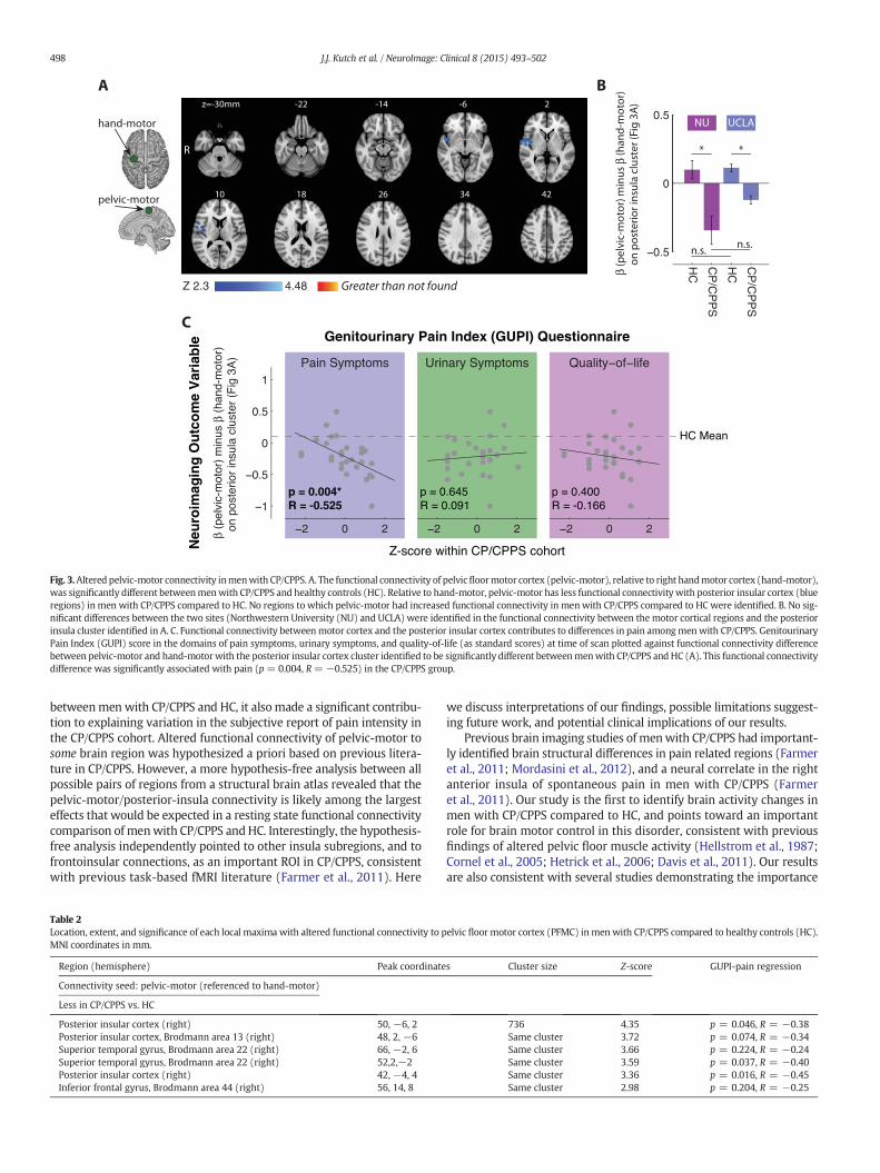
The resting state functional connectivity of pelvic-motor (painfulbody region) was altered relative to the functional connectivity ofhand-motor (non-painful body region) in men with CP/CPPS comparedto HC. This alteration in the difference of functional connectivity regres-sion coefficients (β) occurred in awell-confined cluster in the right pos-terior insular cortex (Fig. 3A). The cluster of altered functionalconnectivity had 736 voxels and a peak z-score of 4.35 (Table 2). The re-gion of altered functional connectivity also included regions of thesuperior temporal gyrus (BA 22) and inferior frontal gyrus (BA 44).The difference in regression coefficients, β(pelvic-motor) minusβ(hand-motor), were averaged across all 736 voxels in the right poste-rior insular cortex cluster to derive an outcome measure of pelvic-motor/posterior-insula functional connectivity for each participant. AnANOVA analysis of this outcome measure using patient status andneuroimaging site as factors (Fig. 3B) revealed a significant main effectof patient status (p b 0.0001), no significant main effect of site(p N 0.05), and no significant interaction between patient status andsite (p N 0.05). A post-hoc multiple comparison test (with Kramer–Tukey correction for multiple comparisons) revealed that pelvic-motor/posterior-insula functional connectivity was significantly less(p b 0.05) inmenwith CP/CPPS at both theNorthwesternUniversity im-aging site and the UCLA imaging site, while there were no significantdifference between the sites in either HC or men with CP/CPPS(Fig. 3B). The difference in functional connectivity regression coeffi-cients was positive for HC (p = 0.007, two-sided t-test) and negativefor men with CP/CPPS (p = 0.0005, two-sided t-test).
We analyzed the functional connectivity regression coefficients forpelvic-motor and hand-motor to the right posterior insula cluster iden-tified to explore the contribution of the separate ROI to the differencebetween HC and men with CP/CPPS. Within the right posterior insulacluster, we performed an ANOVA of the functional connectivity regres-sion coefficient using ROI (pelvic-motor or hand-motor) and cohort(CP/CPPS or HC) as factors. Therewas no significantmain effect of eitherROI or cohort, but there was a significant interaction of ROI and cohort(p = 0.0001). A post-hoc test, with Kramer–Tukey correction for
right hand > pelvic floor
Regions of Interest
pelvic-motor
hand-motor
A B Cpelvic floor > right hand
Fig. 2. Selection of ROI (regions-of-interest) for analysis ofMAPP resting state fMRI data 14healthymen (USC cohort)were recruited to localize brain regions preferentially associatedwithvoluntary hand muscle contraction, and preferentially associated with voluntary pelvic floor muscle contraction. A. Right hand N pelvic floor shows brain regions where there was signif-icantlymore activity during right index fingermuscle contraction compared to pelvicfloormuscle contraction. B. Pelvic floor N right hand shows brain regionswhere therewas significantlymore activity during pelvic floor muscle contraction compared to right index finger muscle contraction. C. 10-mm radius spherical ROI, represented as green circles, were derived withinmotor cortex (precentral gyrus) to form the pelvic-motor ROI and the hand-motor ROI.
497J.J. Kutch et al. / NeuroImage: Clinical 8 (2015) 493–502
multiple comparisons, revealed that there were significant differences(p b 0.05) in right posterior insula functional connectivity regression co-efficient for the pelvic-motor ROI (average of 0.04 for HC and−0.03 formen with CP/CPPS) and for the hand-motor ROI (average of−0.004 forHC and 0.08 for men with CP/CPPS).
3.4. Pelvic-motor connectivity to the right posterior insula is related to painintensity in CP/CPPS
We found that pelvic-motor/posterior-insula functional connectivitydescribed in the previous section, while identified simply by comparingHC and men with CP/CPPS, also made a significant contribution to vari-ance in subjective pain across the cohort of men with CP/CPPS (Fig. 4).Multiple linear regression indicated that only pain symptoms, and noturinary symptoms or quality-of-life, had a significant (p b 0.05) impacton the altered functional connectivity between motor and insularcortices in the CP/CPPS cohort, such this connectivity deviated morefrom the healthy control average in men with more intense pain symp-toms (p=0.004, R=−0.525). Pelvic-motor/posterior-insula function-al connectivity was also analyzed at local maxima within the 736 voxelcluster for evidence of pain-dependence, and all local maxima sug-gested a decrease in functional connectivity with increasing score onthe GUPI pain subscale (Table 2).
We found that HC men (Fig. 4A) and men with CP/CPPS (Fig. 4B)displayed an approximately similar pattern of functional connectivityamong pairs of regions defined by the Destrieux anatomical atlas. How-ever, the comparison of HC and CP/CPPS suggested possible group dif-ferences (Fig. 4C). The largest effect size magnitudes of all possiblepairs of regions was 1.01 (left medial orbital sulcus to right caudate)and−1.2 (left vertical ramus of the anterior segment of the lateral sul-cus to right putamen). Of the 7 region pairs with connectivity effect
sizes for the difference between CP/CPPS and HC greater than 1 inmag-nitude, 3 pairs involved frontoinsular connectivity, 3 involved the rightbasal ganglia connectivity, and 1 involved bilateral temporal cortex con-nectivity (Fig. 4C). The effect size of the functional connectivity changein CP/CPPS patients between pelvic-motor and the right posterior insula(Hedges3 g = −0.87) was among the top 6% of significant (p b 0.05)pair-wise effect sizes in the change in functional connectivity amongatlas regions (Fig. 4D). The sizes of pelvic-motor (520 voxels) and theright posterior insula cluster (736 voxels) were not widely differentcompared to sizes of regions in the atlas, suggesting that the results ofthe hypothesis-based seed approach and the hypothesis-free atlas areapproximately comparable (Fig. 4E).
3.6. Robustness of right posterior insula finding
Moving the pelvic-motor seed in the CP/CPPS group of theMAPP co-hort demonstrated that the right posterior insula cortex cluster of al-tered functional connectivity displayed robustness to perturbations ofthe seed region (Fig. 5A). We found a nearly identical cluster of alteredconnectivity in the right posterior insula if the samepelvic-motor regionwas used in CP/CPPS and HC (Fig. 5B), if the pelvic-motor region wasshifted posteriorly in the CP/CPPS group (Fig. 5C), or if the pelvic-motor region was shifted left in the CP/CPPS group (Fig. 5D). The rightposterior insula cortex connectivity cluster did not appear whenpelvic-motor was shifted anteriorly (Fig. 5E) or right (Fig. 5F) in theCP/CPPS group (see Discussion section).
4. Discussion
To our knowledge, this is the first report of abnormalities in restingstate brain activity measures inmenwith CP/CPPS compared to healthymale controls. Our results reveal disease related alterations in thefunctional connectivity between the pelvic part of motor cortex(pelvic-motor) and the right posterior insula. Strikingly, not only wasthe pelvic-motor/posterior-insula functional connectivity different
Fig. 3.Altered pelvic-motor connectivity inmenwith CP/CPPS. A. The functional connectivity of pelvicfloormotor cortex (pelvic-motor), relative to right handmotor cortex (hand-motor),was significantly different betweenmenwith CP/CPPS and healthy controls (HC). Relative to hand-motor, pelvic-motor has less functional connectivity with posterior insular cortex (blueregions) in men with CP/CPPS compared to HC. No regions to which pelvic-motor had increased functional connectivity in menwith CP/CPPS compared to HC were identified. B. No sig-nificant differences between the two sites (Northwestern University (NU) and UCLA) were identified in the functional connectivity between the motor cortical regions and the posteriorinsula cluster identified in A. C. Functional connectivity betweenmotor cortex and the posterior insular cortex contributes to differences in pain amongmenwith CP/CPPS. GenitourinaryPain Index (GUPI) score in the domains of pain symptoms, urinary symptoms, and quality-of-life (as standard scores) at time of scan plotted against functional connectivity differencebetween pelvic-motor and hand-motorwith the posterior insular cortex cluster identified to be significantly different betweenmenwith CP/CPPS and HC (A). This functional connectivitydifference was significantly associated with pain (p = 0.004, R = −0.525) in the CP/CPPS group.
betweenmen with CP/CPPS and HC, it also made a significant contribu-tion to explaining variation in the subjective report of pain intensity inthe CP/CPPS cohort. Altered functional connectivity of pelvic-motor tosome brain region was hypothesized a priori based on previous litera-ture in CP/CPPS. However, a more hypothesis-free analysis between allpossible pairs of regions from a structural brain atlas revealed that thepelvic-motor/posterior-insula connectivity is likely among the largesteffects that would be expected in a resting state functional connectivitycomparison of menwith CP/CPPS andHC. Interestingly, the hypothesis-free analysis independently pointed to other insula subregions, and tofrontoinsular connections, as an important ROI in CP/CPPS, consistentwith previous task-based fMRI literature (Farmer et al., 2011). Here
Table 2Location, extent, and significance of each local maxima with altered functional connectivity to pMNI coordinates in mm.
Region (hemisphere) Peak coordinate
Connectivity seed: pelvic-motor (referenced to hand-motor)
we discuss interpretations of our findings, possible limitations suggest-ing future work, and potential clinical implications of our results.
Previous brain imaging studies of menwith CP/CPPS had important-ly identified brain structural differences in pain related regions (Farmeret al., 2011; Mordasini et al., 2012), and a neural correlate in the rightanterior insula of spontaneous pain in men with CP/CPPS (Farmeret al., 2011). Our study is the first to identify brain activity changes inmen with CP/CPPS compared to HC, and points toward an importantrole for brain motor control in this disorder, consistent with previousfindings of altered pelvic floor muscle activity (Hellstrom et al., 1987;Cornel et al., 2005; Hetrick et al., 2006; Davis et al., 2011). Our resultsare also consistent with several studies demonstrating the importance
elvic floor motor cortex (PFMC) inmen with CP/CPPS compared to healthy controls (HC).
s Cluster size Z-score GUPI-pain regression
736 4.35 p = 0.046, R = −0.38Same cluster 3.72 p = 0.074, R = −0.34Same cluster 3.66 p = 0.224, R = −0.24Same cluster 3.59 p = 0.037, R = −0.40Same cluster 3.36 p = 0.016, R = −0.45Same cluster 2.98 p = 0.204, R = −0.25
Fig. 4.Whole-brain connectivity among all regions in theDestrieux anatomical atlas inmenwith CP/CPPS and healthy controls (HC). A. Connectivity in theHC group. B. Connectivity in theCP/CPPS group. For clarity, only the strongest 1% of functional connectivity pairs are shown in A and B. C. Connections between atlas region pairs forwhich the functional connectivitymaybe affected by group (CP/CPPS vs. HC). Regional pairs with effect size greater than functional connectivity change between pelvic-motor and the right posterior insula (−0.87) in magni-tude are shown, with pairs for which effect size was greater than 1 emphasized with stronger line weight. D. The functional connectivity between pelvic-motor and the right posteriorinsula (Fig. 3A) had an associated effect size greater than the vast majority of atlas-based region pairs. E. Confirmation that the pelvic-motor and right posterior insula cluster had com-parable sizes to regions in the structural atlas.
499J.J. Kutch et al. / NeuroImage: Clinical 8 (2015) 493–502
Fig. 5. Sensitivity analysis of right posterior insula result. A. The location of pelvic-motor was moved several different locations for the CP/CPPS cohort, and the comparison with healthycontrols (HC) of Fig. 3 was repeated. B. Pelvic-motor HC shows the connectivity assuming the same pelvic-motor ROI in bothmenwith CP/CPPS andHC, and is identical to cluster shown inFig. 3A. Altered connectivity between motor cortex and the right posterior insula cluster persisted when the pelvic-motor was moved 10 mm posterior (C) or 10 mm left (D), but disap-peared when pelvic-motor was moved 10 mm anterior (E) or 10 mm right (F).
of primary motor cortex for pain processing. For example, primarymotor cortex appears to process nociceptive signals in parallel with pri-mary sensory cortex (Frot et al., 2013), and there is emerging evidencethat repetitive transcranial magnetic stimulation over motor cortex hasanalgesic effects (Lefaucheur et al., 2014), including in patients withchronic pelvic pain (Louppe et al., 2013).
One hypothesis thatwould explain our findings is that functional con-nectivity is altered by changes in direct neural communication betweenright posterior insular cortex andmotor cortex. The insula is currently un-derstood to have a posterior-to-mid-to-anterior integration of interocep-tive information (Craig, 2011). Primary interoceptive representations ofviscerosensory information are believed to be concentrated in the poste-rior insula, with progressive integration with cognitive and affectiveaspects and resultant subjective awareness of this interoceptive informa-tion developing in the anterior insula. Insular cortex plays a more impor-tant role during voluntary pelvic floor muscle contraction compared toother lower limbmuscles (Schrumet al., 2011), with posterior insula pos-sibly relaying necessary sensory information to motor cortical structuresfor control of muscles associated with the viscera (Deen et al., 2011;Cauda et al., 2011; Levinthal & Strick, 2012). The existence of an importantfunctional connection between pelvic-motor cortex and the posteriorinsula is further supported by our finding that HC participants in theMAPP cohort had significantly greater functional connectivity betweenpelvic-motor and the posterior insula compared to hand-motor, suggest-ing that this connection is important in the healthy human male brain. Ifthis hypothesis is correct, the altered functional connectivity betweenposterior insular cortex and motor cortex controlling the pelvic floor inCP/CPPS patientsmay relate to a change in the brain connectionsmediat-ing the sensorimotor communication between these areas.
Screening a large number of functional connections between 165 re-gions in the Destrieux structural atlas for changes in men with CP/CPPSalso indirectly pointed to insular cortex as an important contributor tothe difference between the CP/CPPS and HC groups. Specifically, asnoted in the results, 3 of 7 pairs with group difference effect size greaterthan 1.0 involved involved frontoinsular connectivity, all of which werefocused on the right insula. Even though the reason for the observedgroup differences in these frontoinsular connections remains to be de-termined, it is of interest that the frontoinsular region of the brain, inparticular in the right hemisphere, has been shown to be associatedwith sympathetic autonomic control (Allman et al., 2011). Since chang-es in autonomic function have been identified in men with CP/CPPS(Anderson et al., 2008; Dimitrakov et al., 2008; Anderson et al., 2009),future research can examine the association between frontoinsular con-nectivity and measures of altered autonomic function.
Our current study has some limitations. As a single-observationstudy, we focused our analysis on overall group differences rather
than controlling for the effects of other factors, such as treatment, age,and chronicity of symptoms. Another limitation is that the MAPP Re-search Network studywas exploratory in nature and designed to collectgeneral brain anatomical and functional data as well as participant de-mographic, symptom, and biomarker data— nomeasures of muscle ac-tivity were collected which could have defined a possible peripheralcorrelate of altered functional connectivity of the motor cortex in menwith CP/CPPS. Furthermore, participants in theMAPP study did not per-form pelvic floor contractions (or contractions of other muscles) in thescanner, so we were unable to use patient-specific ROI. We compensat-ed for this limitation by carefully localizing the normal cortical repre-sentation of pelvic floor and hand contractions in a new sample ofhealthymen, and using relatively large ROI (2 cm in diameter) to devel-op a first-ordermap of neuromotor pathology inmenwith CP/CPPS. It issurprising that group differences emerged at this level of spatial resolu-tion, and suggest that future studies with better localization of sensori-motor regions in men with CP/CPPS may even further increase thespecificity of our findings. We have recently demonstrated, in concertwith previous studies, that the pelvic-motor region likely producescorticospinal motor projections as evidenced by very short latencymotor evoked potentials (MEP) (Asavasopon et al., 2014). The corticalMEP intensity map of particular muscles can change in individualswith chronic pain (Tsao et al., 2008); thus, future studies of individualswith CP/CPPS must determine if the cortical MEP intensity map of thepelvic floor is changing with respect to HC, and if such changes couldcontribute to the altered functional connectivity effects observed inthis study. As a preliminary analysis of possible changes in motor corti-cal representation of the pelvic floor in CP/CPPS, we performed a sensi-tivity analysis by perturbing the pelvic-motor ROI in the CP/CPPS group.Since altered connectivity with the right posterior insula did disappearfor some pelvic-motor perturbations in the CP/CPPS group (anteriorand right), it is possible that our results might be explained by shiftsof the normalmotor representation of the pelvic floor in CP/CPPS. None-theless, since we identified that motor cortical functional connectivitychanges in CP/CPPS have relatively large effect sizes, motor cortex islikely an important region of interest in studying CP/CPPS despite thefact that it remains to be determined if motor representation or funda-mental communicationwith the posterior insula is driving the observedchanges.
Several critical lines of futurework emerge from this study. First, it isimportant to determine if effective treatment for CP/CPPS functions tonormalize motor cortical/insula connectivity in men with CP/CPPSwho improve. Second, it is important to determine if baseline levels ofmotor cortical/insula connectivity could predict response to treatment,evenwhen controlled for baseline levels of pain intensity. Third, it is im-portant to create amore expansivemap of brain functional connectivity
501J.J. Kutch et al. / NeuroImage: Clinical 8 (2015) 493–502
alterations in men with CP/CPPS, to determine the relative prominenceof motor cortical changes, and to relate this map to changes in grayand white matter structure. Finally, defining the pelvic floor muscle ac-tivity correlates of brain functional connectivity changes that accompa-ny CP/CPPS symptoms is important for a more complete understandingof how functional connectivity changes contribute to the experience ofpain in men with CP/CPPS.
While the current results are limited to men with CP/CPPS, it is pos-sible that our approach and findings have implications for other chronicpain conditions in which motor control is altered. It has been hypothe-sized that the threat of pain/injury triggers changes in themotor systemthat leads to a redistribution of activity within and between muscles(Hodges and Tucker, 2011). However, the location of these neuralchanges have not yet been localized. Our results suggest that areasthat do not generate direct corticospinal projections, such as the poste-rior insula, could play a critical role inmodulatingmotor cortical activityin individuals with chronic pain. Further research is necessary to deter-mine if the identity of these non-motor cortical regions related to chron-ic pain are general or are specific to different chronic pain conditions.
Acknowledgements
We thank all of the volunteers who participated in the study. Wewould like to thank Nina Bradley, Bruce Naliboff, and Kirsten Tillischfor helpful discussions. Funding for the MAPP Research Network wasobtained under a cooperative agreement fromNational Institute of Dia-betes and Digestive and Kidney Diseases (NIDDK), National Institutes ofHealth (NIH) (DK82370, DK82342, DK82315, DK82344, DK82325,DK82345, DK82333, and DK82316). This work was also supported, inpart, by the USC Division of Biokinesiology and Physical Therapyunder award number USCBKN/PT-2013A, the Loma Linda UniversityPhysical Therapy Department under award number LLU-647525-2007,and National Center for Medical Rehabilitation Research of the NationalInstitutes of Health under award number T32 HD064578. We declarethe following interests: financial interest and/or other relationshipwith Pfizer, Cerephex, Lilly, Merck, Nuvo, Furest, Tonix, Purdue,Therauance and Johnson & Johnson (DJC), financial interest and/orother relationship with National Institutes of Health and Medtronic(TJN), financial interest and/or other relationship with National Insti-tutes of Health (CM).
Appendix A. Supplementary data
Supplementary data to this article can be found online at http://dx.doi.org/10.1016/j.nicl.2015.05.013.
References
Allman, J.M., Tetreault, N.A., Hakeem, A.Y., Manaye, K.F., Semendeferi, K., Erwin, J.M., Park,S., Goubert, V., Hof, P.R., 2011. The von Economo neurons in the frontoinsular and an-terior cingulate cortex. Ann. New York Acad. Sci. 1225, 59–71. http://dx.doi.org/10.1111/j.1749-6632.2011.06011.x21534993.
Allsop, S.A., Erstad, D.J., Brook, K., Bhai, S.F., Cohen, J.M., Dimitrakoff, J.D., 2011. TheDABBEC phenotyping system: towards a mechanistic understanding of CP/CPPS.Nat. Rev. Urol. 8 (2), 107–113. http://dx.doi.org/10.1038/nrurol.2010.22721243018.
Anderson, R.U., Orenberg, E.K., Chan, C.A., Morey, A., Flores, V., 2008. Psychometric profilesand hypothalamic–pituitary–adrenal axis function in men with chronic prostatitis/chronic pelvic pain syndrome. J. Urol. 179 (3), 956–960. http://dx.doi.org/10.1016/j.juro.2007.10.08418207189.
Anderson, R.U., Orenberg, E.K., Morey, A., Chavez, N., Chan, C.A., 2009. Stress induced hy-pothalamus–pituitary–adrenal axis responses and disturbances in psychological pro-files in men with chronic prostatitis/chronic pelvic pain syndrome. J. Urol. 182 (5),2319–2324. http://dx.doi.org/10.1016/j.juro.2009.07.04219762053.
Anderson, R.U., Wise, D., Sawyer, T., Chan, C., 2005. Integration of myofascial triggerpoint release and paradoxical relaxation training treatment of chronic pelvic painin men. J. Urol. 174 (1), 155–160. http://dx.doi.org/10.1097/01.ju.0000161609.31185.d515947608.
Arendt-Nielsen, L., Graven-Nielsen, T., Svarrer, H., Svensson, P., 1996. The influence of lowback pain on muscle activity and coordination during gait: a clinical and experimen-tal study. Pain 64 (2), 231–240. http://dx.doi.org/10.1016/0304-3959(95)00115-88740599.
Asavasopon, S., Rana, M., Kirages, D.J., Yani, M.S., Fisher, B.E., Hwang, D.H., Lohman, E.B.,Berk, L.S., Kutch, J.J., 2014. Cortical activation associated with muscle synergies ofthe human male pelvic floor. J. Neurosci. 34 (41), 13811–13818. http://dx.doi.org/10.1523/JNEUROSCI.2073-14.201425297107.
Bartoletti, R., Cai, T., Mondaini, N., Dinelli, N., Pinzi, N., Pavone, C., Gontero, P.,Gavazzi, A., Giubilei, G., Prezioso, D., Mazzoli, S., Boddi, V., Naber, K.G., ItalianProstatitis Study Group, 2007. Prevalence, incidence estimation, risk factorsand characterization of chronic prostatitis/chronic pelvic pain syndrome in uro-logical hospital outpatients in Italy: results of a multicenter case–control obser-vational study. J. Urol. 178 (6), 2411–2415. http://dx.doi.org/10.1016/j.juro.2007.08.04617937946.
Castroflorio, T., Falla, D., Tartaglia, G.M., Sforza, C., Deregibus, A., 2012. Myoelectric mani-festations of jaw elevator muscle fatigue and recovery in healthy and TMD subjects.J. Oral Rehabil. 39 (9), 648–658. http://dx.doi.org/10.1111/j.1365-2842.2012.02309.x22490056.
Cauda, F., D3Agata, F., Sacco, K., Duca, S., Geminiani, G., Vercelli, A., 2011. Functional con-nectivity of the insula in the resting brain. Neuroimage 55 (1), 8–23. http://dx.doi.org/10.1016/j.neuroimage.2010.11.04921111053.
Cleeland, C.S., Ryan, K.M., 1994. Pain assessment: global use of the Brief Pain Inventory.Ann. Acad. Med. Singapore 23 (2), 129–1388080219.
Clemens, J.Q., Calhoun, E.A., Litwin, M.S., McNaughton-Collins, M., Kusek, J.W., Crowley,E.M., Landis, J.R., Urologic Pelvic Pain Collaborative Research Network, 2009. Valida-tion of a modified National Institutes of Health chronic prostatitis symptom indexto assess genitourinary pain in both men and women. Urology 74 (5), 983–987.http://dx.doi.org/10.1016/j.urology.2009.06.07819800663.
Clemens, J.Q., Mullins, C., Kusek, J.W., Kirkali, Z., Mayer, E.A., Rodríguez, L.V., Klumpp, D.J.,Schaeffer, A.J., Kreder, K.J., Buchwald, D., Andriole, G.L., Lucia, M.S., Landis, J.R., Clauw,D.J., MAPP Research Network Study Group, 2014. The MAPP research network: anovel study of urologic chronic pelvic pain syndromes. B.M.C. Urol. 14, 57. http://dx.doi.org/10.1186/1471-2490-14-5725085007.
Collins, M.M., Stafford, R.S., O3Leary, M.P., Barry, M.J., 1998. How common is prostatitis? Anational survey of physician visits. J. urol. 159 (4), 1224–1228. http://dx.doi.org/10.1016/S0022-5347(01)63564-X9507840.
Cornel, E.B., van Haarst, E.P., Schaarsberg, R.W.M., Geels, J., 2005. The effect of biofeedbackphysical therapy in men with chronic pelvic pain syndrome type III. Eur. Urol. 47 (5),607–611. http://dx.doi.org/10.1016/j.eururo.2004.12.01415826751.
Craig, A.D., 2011. Significance of the insula for the evolution of human awareness of feel-ings from the body. Ann. New York Acad. Sci. 1225, 72–82. http://dx.doi.org/10.1111/j.1749-6632.2011.05990.x21534994.
Davis, S.N., Morin, M., Binik, Y.M., Khalife, S., Carrier, S., 2011. Use of pelvic floor ultra-sound to assess pelvic floor muscle function in urological chronic pelvic pain syn-drome in men. J. Sex. Med. 8 (11), 3173–3180. http://dx.doi.org/10.1111/j.1743-6109.2011.02452.x21883952.
Deen, B., Pitskel, N.B., Pelphrey, K.A., 2011. Three systems of insular functional connectiv-ity identified with cluster analysis. Cereb. Cortex 21 (7), 1498–1506. http://dx.doi.org/10.1093/cercor/bhq18621097516.
Destrieux, C., Fischl, B., Dale, A., Halgren, E., 2010. Automatic parcellation of human corti-cal gyri and sulci using standard anatomical nomenclature. Neuroimage 53 (1), 1–15.http://dx.doi.org/10.1016/j.neuroimage.2010.06.01020547229.
Dimitrakov, J., Joffe, H.V., Soldin, S.J., Bolus, R., Buffington, C.A., Nickel, J.C., 2008. Adreno-cortical hormone abnormalities in men with chronic prostatitis/chronic pelvic painsyndrome. Urology 71 (2), 261–266. http://dx.doi.org/10.1016/j.urology.2007.09.02518308097.
Farmer, M.A., Chanda, M.L., Parks, E.L., Baliki, M.N., Apkarian, A.V., Schaeffer, A.J., 2011.Brain functional and anatomical changes in chronic prostatitis/chronic pelvic painsyndrome. J. urol. 186 (1), 117–124. http://dx.doi.org/10.1016/j.juro.2011.03.02721571326.
FitzGerald, M.P., Anderson, R.U., Potts, J., Payne, C.K., Peters, K.M., Clemens, J.Q., Kotarinos,R., Fraser, L., Cosby, A., Fortman, C., Neville, C., Badillo, S., Odabachian, L., Sanfield, A.,O3Dougherty, B., Halle-Podell, R., Cen, L., Chuai, S., Landis, J.R., Mickelberg, K., Barrell,T., Kusek, J.W., Nyberg, L.M., Urological Pelvic Pain Collaborative Research Network,2009. Randomized multicenter feasibility trial of myofascial physical therapy forthe treatment of urological chronic pelvic pain syndromes. J. urol. 182 (2),570–580. http://dx.doi.org/10.1016/j.juro.2009.04.02219535099.
Frot, M., Magnin, M., Mauguière, F., Garcia-Larrea, L., 2013. Cortical representation of painin primary sensory-motor areas (S1/M1)—a study using intracortical recordings inhumans. Hum. Brain Mapp. 34 (10), 2655–2668. http://dx.doi.org/10.1002/hbm.2209722706963.
Han, K., Mac Donald, C.L., Johnson, A.M., Barnes, Y., Wierzechowski, L., Zonies, D., Oh, J.,Flaherty, S., Fang, R., Raichle, M.E., Brody, D.L., 2014. Disrupted modular organizationof resting-state cortical functional connectivity in U.S. military personnel followingconcussive ‘mild. Neuroimage 84, 76–96. http://dx.doi.org/10.1016/j.neuroimage.2013.08.01723968735.
Hentschke, H., Stüttgen, M.C., 2011. Computation of measures of effect size for neurosci-ence data sets. Eur. J. Neurosci. 34 (12), 1887–1894. http://dx.doi.org/10.1111/j.1460-9568.2011.07902.x22082031.
Hetrick, D.C., Glazer, H., Liu, Y.W., Turner, J.A., Frest, M., Berger, R.E., 2006. Pelvic floor elec-tromyography in men with chronic pelvic pain syndrome: a case–control study.Neurourol. urodyn. 25 (1), 46–49. http://dx.doi.org/10.1002/nau.2016216167354.
Hodges, P.W., Tucker, K., 2011. Moving differently in pain: a new theory to explain the ad-aptation to pain. Pain 152 (3 Suppl), S90–S98. http://dx.doi.org/10.1016/j.pain.2010.10.02021087823.
Hong, J.Y., Kilpatrick, L.A., Labus, J.S., Gupta, A., Katibian, D., Ashe-McNalley, C., Stains, J.,Heendeniya, N., Smith, S.R., Tillisch, K., Naliboff, B., Mayer, E.A., 2014. Sex anddisease-related alterations of anterior insula functional connectivity in chronic ab-dominal pain. J. Neurosci. 34 (43), 14252–14259. http://dx.doi.org/10.1523/JNEUROSCI.1683-14.201425339739.
Irimia, A., Chambers, M.C., Torgerson, C.M., Van Horn, J.D., 2012. Circular representation ofhuman cortical networks for subject and population-level connectomic visualization.Neuroimage 60 (2), 1340–1351. http://dx.doi.org/10.1016/j.neuroimage.2012.01.10722305988.
Jacobs, J.V., Henry, S.M., Nagle, K.J., 2010. Low back pain associates with altered activityof the cerebral cortex prior to arm movements that require postural adjustment.Clin. Neurophysiol. 121 (3), 431–440. http://dx.doi.org/10.1016/j.clinph.2009.11.07620071225.
Jantos, M., 2008. Vulvodynia: a psychophysiological profile based on electromyographicassessment. Appl. Psychophysiol. Biofeedback 33 (1), 29–38. http://dx.doi.org/10.1007/s10484-008-9049-y18214669.
Kilpatrick, L.A., Kutch, J.J., Tillisch, K., Naliboff, B.D., Labus, J.S., Jiang, Z., Farmer, M.A.,Apkarian, A.V., Mackey, S.C., Martucci, K.T., Clauw, D.J., Harris, R.E., Deutsch, G.,Ness, T.J., Yang, C.C., Maravilla, K., Mullins, C., Mayer, E.A., 2014. Alterations in restingstate oscillations and connectivity in sensory and motor networks in womenwith in-terstitial cystitis/painful bladder syndrome. J. urol. 192 (3), 947–955. http://dx.doi.org/10.1016/j.juro.2014.03.09324681331.
Krieger, J., Ross, S., Riley, D., 2002. Chronic prostatitis: epidemiology and role of infection.Urology 60 (6 Suppl), 8–12. http://dx.doi.org/10.1016/S0090-4295(02)02294-X12521579.
Krzywinski, M., Schein, J., Birol, I., Connors, J., Gascoyne, R., Horsman, D., Jones, S.J., Marra,M.A., 2009. Circos: an information aesthetic for comparative genomics. Genome Res.19 (9), 1639–1645. http://dx.doi.org/10.1101/gr.092759.10919541911.
Lefaucheur, J.P., André-Obadia, N., Antal, A., Ayache, S.S., Baeken, C., Benninger, D.H.,Cantello, R.M., Cincotta, M., de Carvalho, M., de Ridder, D., Devanne, H., Di Lazzaro,V., Filipović, S.R., Hummel, F.C., Jääskeläinen, S.K., Kimiskidis, V.K., Koch, G.,Langguth, B., Nyffeler, T., Oliviero, A., 2014. Evidence-based guidelines on the thera-peutic use of repetitive transcranial magnetic stimulation (rTMS). Clin. Neurophysiol.125 (11), 2150–2206. http://dx.doi.org/10.1016/j.clinph.2014.05.02125034472.
Leinonen, V., Kankaanpää, M., Luukkonen, M., Hänninen, O., Airaksinen, O., Taimela, S.,2001. Disc herniation-related back pain impairs feed-forward control of paraspinalmuscles. Spine (Phila. Pa. 1976) 26 (16), E367–E372. http://dx.doi.org/10.1097/00007632-200108150-0001411493866.
Levinthal, D.J., Strick, P.L., 2012. The motor cortex communicates with the kidney.J. Neurosci. 32 (19), 6726–6731. http://dx.doi.org/10.1523/JNEUROSCI.0406-12.201222573695.
Louppe, J.-M., Nguyen, J.-P., Robert, R., Buffenoir, K., de Chauvigny, E., Riant, T., Péréon, Y.,Labat, J.J., Nizard, J., 2013. Motor cortex stimulation in refractory pelvic andperineal pain: report of two successful cases. Neurourol. Urodyn. 32 (1), 53–57.http://dx.doi.org/10.1002/nau.2226922674567.
Marszalek, M., Wehrberger, C., Temml, C., Ponholzer, A., Berger, I., Madersbacher, S., 2009.Chronic pelvic pain and lower urinary tract symptoms in both sexes: analysis of2749 participants of an Urban health screening project. Eur. Urol. 55 (2), 499–507.http://dx.doi.org/10.1016/j.eururo.2008.03.07318395963.
Mordasini, L., Weisstanner, C., Rummel, C., Thalmann, G.N., Verma, R.K., Wiest, R., Kessler,T.M., 2012. Chronic pelvic pain syndrome in men is associated with reduction ofrelative gray matter volume in the anterior cingulate cortex compared to healthycontrols. J. Urol. 188 (6), 2233–2237. http://dx.doi.org/10.1016/j.juro.2012.08.04323083652.
Roy, A.K., Shehzad, Z., Margulies, D.S., Kelly, A.M., Uddin, L.Q., Gotimer, K., Biswal, B.B.,Castellanos, F.X., Milham, M.P., 2009. Functional connectivity of the human amygdalausing resting state fMRI. Neuroimage 45 (2), 614–626. http://dx.doi.org/10.1016/j.neuroimage.2008.11.03019110061.
Schrum, A., Wolff, S., van der Horst, C., Kuhtz-Buschbeck, J.P., 2011. Motor cortical repre-sentation of the pelvic floor muscles. J. urol. 186 (1), 185–190. http://dx.doi.org/10.1016/j.juro.2011.03.00121575960.
Tsao, H., Galea, M.P., Hodges, P.W., 2008. Reorganization of the motor cortex is associatedwith postural control deficits in recurrent low back pain. Brain 131 (8), 2161–2171.http://dx.doi.org/10.1093/brain/awn15418669505.
Turnbull, G.K., Hamdy, S., Aziz, Q., Singh, K.D., Thompson, D.G., 1999. The cortical topog-raphy of human anorectal musculature. Gastroenterology 117 (1), 32–39. http://dx.doi.org/10.1016/S0016-5085(99)70547-010381907.
Wand, B.M., Parkitny, L., O3Connell, N.E., Luomajoki, H., McAuley, J.H., Thacker, M.,Moseley, G.L., 2011. Cortical changes in chronic low back pain: current state of theart and implications for clinical practice. Man. Ther. 16 (1), 15–20. http://dx.doi.org/10.1016/j.math.2010.06.00820655796.