Alveolar Macrophages Play a Key Role in Cockroach- Induced Allergic Inflammation via TNF-a Pathway Joo Young Kim 1,4 , Jung Ho Sohn 2,3 , Je-Min Choi 2 , Jae-Hyun Lee 3,5 , Chein-Soo Hong 3,5 , Joo-Shil Lee 6 , Jung-Won Park 3,4,5 * 1 Ewha Womans University College of Pharmacy, Research Institute of Pharmaceutical Sciences, Seoul, South Korea, 2 Department of Life Science, Hanyang University, Seoul, South Korea, 3 Department of Internal Medicine, Yonsei University College of Medicine, Seoul, South Korea, 4 Brain Korea 21 Project for Medical Science, Yonsei University College of Medicine, Seoul, South Korea, 5 Institute of Allergy, Yonsei University College of Medicine, Seoul, South Korea, 6 Center for Immunology and Pathology, Korea National Institute of Health, Osong, South Korea Abstract The activity of the serine protease in the German cockroach allergen is important to the development of allergic disease. The protease-activated receptor (PAR)-2, which is expressed in numerous cell types in lung tissue, is known to mediate the cellular events caused by inhaled serine protease. Alveolar macrophages express PAR-2 and produce considerable amounts of tumor necrosis factor (TNF)-a. We determined whether the serine protease in German cockroach extract (GCE) enhances TNF-a production by alveolar macrophages through the PAR-2 pathway and whether the TNF-a production affects GCE- induced pulmonary inflammation. Effects of GCE on alveolar macrophages and TNF-a production were evaluated using in vitro MH-S and RAW264.6 cells and in vivo GCE-induced asthma models of BALB/c mice. GCE contained a large amount of serine protease. In the MH-S and RAW264.7 cells, GCE activated PAR-2 and thereby produced TNF-a. In the GCE-induced asthma model, intranasal administration of GCE increased airway hyperresponsiveness (AHR), inflammatory cell infiltration, productions of serum immunoglobulin E, interleukin (IL)-5, IL-13 and TNF-a production in alveolar macrophages. Blockade of serine proteases prevented the development of GCE induced allergic pathologies. TNF-a blockade also prevented the development of such asthma-like lesions. Depletion of alveolar macrophages reduced AHR and intracellular TNF-a level in pulmonary cell populations in the GCE-induced asthma model. These results suggest that serine protease from GCE affects asthma through an alveolar macrophage and TNF-a dependent manner, reflecting the close relation of innate and adaptive immune response in allergic asthma model. Citation: Kim JY, Sohn JH, Choi J-M, Lee J-H, Hong C-S, et al. (2012) Alveolar Macrophages Play a Key Role in Cockroach-Induced Allergic Inflammation via TNF-a Pathway. PLoS ONE 7(10): e47971. doi:10.1371/journal.pone.0047971 Editor: Stephania Ann Cormier, Louisiana State University Health Sciences Center, United States of America Received May 25, 2012; Accepted September 18, 2012; Published October 19, 2012 Copyright: ß 2012 Kim et al. This is an open-access article distributed under the terms of the Creative Commons Attribution License, which permits unrestricted use, distribution, and reproduction in any medium, provided the original author and source are credited. Funding: This study was supported by the grant of the Korea Healthcare Technology R&D Project, Ministry for Health, Welfare & Family Affairs, Republic of Korea (A092076). The funders had no role in study design, data collection and analysis, decision to publish, or preparation of the manuscript. Competing Interests: The authors have declared that no competing interests exist. * E-mail: [email protected]Introduction German cockroaches are a well-known causative allergen for allergic asthma [1]. It contains several major allergens and proteases. Classically, allergens induce immune responses that lead to Th2 lymphocyte differentiation, production of IgE, and mast cell activation; however, prolonged administration of allergen may induce regulatory T cells and tolerance to the allergens [2]. Recently, another type of allergens were classified as type II allergens, and these allergens bypass normal tolerogenic mecha- nisms and directly induce allergic diseases by sensitization of local routes [3,4]. One example of such type II allergens includes the active proteases derived from cockroach, house dust mite, and fungal extracts. German cockroach extract (GCE) was reported to contain active serine proteases [5,6,7]. Serine protease affects the development of inflammation and allergic immune responses through specific receptor systems, such as the protease-activated receptor (PAR)-2 in a variety of cell types [4]. PAR-2, a member of the G protein-coupled receptor family [8], is activated by various serine proteases such as mast cell tryptase [9], trypsin-like enzymes [10], and certain allergens from house dust mites [11] or cockroaches [8,12]. Serine proteases stimulate the N-terminal exodomain of the receptor and cleave the receptor at this site [13]. Alteration of PAR-2 results in coupling and activation of G proteins, triggers a cascade of signaling events, and thereby leads to intracellular Ca 2+ influx [14] and tumor necrosis factor (TNF) production. These events contribute to the development of eosinophilic inflammation and airway hyperresponsiveness (AHR) in asthma [4]. Others, however, have reported that PAR-2 may be protective against bronchoconstriction or AHR [15]. Thus, the role of PAR-2 in asthma remains controversial. PAR-2-expressing cells, such as alveolar macrophages [13], epithelial cells, mast cells, and fibroblasts are located throughout the airways and encounter inhaled allergens or particles that contain serine protease activity. Alveolar macrophages are able to produce large amounts of TNF-a [4,16]. Recent studies indicated that depletion of alveolar macrophages [17] or blockade of TNF-a [18] prevents AHR and progressive inflammatory injuries in an ovalbumin-induced asthma model. TNF-a blockade also amelio- rates AHR, impairment of lung function, and quality of life in patients with severe asthma [19,20]. These findings suggest that TNF-a expression by alveolar macrophages may play a key role in PLOS ONE | www.plosone.org 1 October 2012 | Volume 7 | Issue 10 | e47971
Transcript
Alveolar Macrophages Play a Key Role in Cockroach-Induced Allergic Inflammation via TNF-a PathwayJoo Young Kim1,4, Jung Ho Sohn2,3, Je-Min Choi2, Jae-Hyun Lee3,5, Chein-Soo Hong3,5, Joo-Shil Lee6,
Jung-Won Park3,4,5*
1 Ewha Womans University College of Pharmacy, Research Institute of Pharmaceutical Sciences, Seoul, South Korea, 2 Department of Life Science, Hanyang University,
Seoul, South Korea, 3 Department of Internal Medicine, Yonsei University College of Medicine, Seoul, South Korea, 4 Brain Korea 21 Project for Medical Science, Yonsei
University College of Medicine, Seoul, South Korea, 5 Institute of Allergy, Yonsei University College of Medicine, Seoul, South Korea, 6 Center for Immunology and
Pathology, Korea National Institute of Health, Osong, South Korea
Abstract
The activity of the serine protease in the German cockroach allergen is important to the development of allergic disease.The protease-activated receptor (PAR)-2, which is expressed in numerous cell types in lung tissue, is known to mediate thecellular events caused by inhaled serine protease. Alveolar macrophages express PAR-2 and produce considerable amountsof tumor necrosis factor (TNF)-a. We determined whether the serine protease in German cockroach extract (GCE) enhancesTNF-a production by alveolar macrophages through the PAR-2 pathway and whether the TNF-a production affects GCE-induced pulmonary inflammation. Effects of GCE on alveolar macrophages and TNF-a production were evaluated using invitro MH-S and RAW264.6 cells and in vivo GCE-induced asthma models of BALB/c mice. GCE contained a large amount ofserine protease. In the MH-S and RAW264.7 cells, GCE activated PAR-2 and thereby produced TNF-a. In the GCE-inducedasthma model, intranasal administration of GCE increased airway hyperresponsiveness (AHR), inflammatory cell infiltration,productions of serum immunoglobulin E, interleukin (IL)-5, IL-13 and TNF-a production in alveolar macrophages. Blockadeof serine proteases prevented the development of GCE induced allergic pathologies. TNF-a blockade also prevented thedevelopment of such asthma-like lesions. Depletion of alveolar macrophages reduced AHR and intracellular TNF-a level inpulmonary cell populations in the GCE-induced asthma model. These results suggest that serine protease from GCE affectsasthma through an alveolar macrophage and TNF-a dependent manner, reflecting the close relation of innate and adaptiveimmune response in allergic asthma model.
Citation: Kim JY, Sohn JH, Choi J-M, Lee J-H, Hong C-S, et al. (2012) Alveolar Macrophages Play a Key Role in Cockroach-Induced Allergic Inflammation via TNF-aPathway. PLoS ONE 7(10): e47971. doi:10.1371/journal.pone.0047971
Editor: Stephania Ann Cormier, Louisiana State University Health Sciences Center, United States of America
Received May 25, 2012; Accepted September 18, 2012; Published October 19, 2012
Copyright: � 2012 Kim et al. This is an open-access article distributed under the terms of the Creative Commons Attribution License, which permits unrestricteduse, distribution, and reproduction in any medium, provided the original author and source are credited.
Funding: This study was supported by the grant of the Korea Healthcare Technology R&D Project, Ministry for Health, Welfare & Family Affairs, Republic of Korea(A092076). The funders had no role in study design, data collection and analysis, decision to publish, or preparation of the manuscript.
Competing Interests: The authors have declared that no competing interests exist.
(flexiVent 5.1H; SCIREQ, Montreal, Canada) and challenged with a
saline aerosol followed by increasing concentrations of methacho-
line (MCh; Sigma-Aldrich). Aerosols were generated with an
ultrasonic nebulizer (Omron Healthcare, Kyoto, Japan) and
delivered to the inspiratory line of the flexiVent using a bias flow
of medical air.
BAL fluidBronchoalveolar lavage (BAL) fluid was obtained as previously
described [23]. To collect BAL fluid, the lungs were lavaged with
1 mL Hank’s balanced salt solution (HBSS) via the tracheostomy
tube. Total cell numbers were counted with a hemocytometer.
After the procedure, BAL fluid was centrifuged at 1,5006g for 3
minutes at 4uC, and then smears of BAL cells were prepared by
cytocentrifugation (Cytospin3, Thermo, Billerica, MA) at
1,000 rpm for 3 minutes. BAL cells were stained with Hemacolor
Staining Kit (Merck, Darmstadt, Germany) counted, and classified
as neutrophils, eosinophils, lymphocytes, or macrophages.
Lung homogenateFor assessment of cytokine levels, lung tissues were homoge-
nized in 20 mL/g tissue protein extraction reagent (Thermo
Fisher Scientific Inc., Rockford, IL) using a tissue homogenizer
(Biospec Products, Bartlesville, OK). Homogenates were incubated
at 4uC for 30 min and then centrifuged at 1,0006g for 10 min.
Supernatants were collected, passed through a 0.45-micron filter
(Gelman Sciences, Ann Arbor, MI), and then stored at 270uC for
assessment of cytokine levels.
ImmunocytohistochemistryImmunofluorescence staining of PAR-2 and TNF-a in Raw
264.7 cells and lung tissues was examined by confocal laser
scanning microscopy (LSM700, Carl Zeiss, Jena, Germany).
Cytospin-fixed RAW 264.7 cells or formalin-fixed, paraffin-
embedded lung tissues were stained with FITC anti-mouse
PAR-2 (SAM11, Santa Cruz Biotechnology, Santa Cruz, CA),
phycoerythrin (PE) anti-mouse CD11b, and allophycocyanin
(APC) anti-mouse TNF-a (BD PharmingenTM, San Jose, CA) at
4uC for 30 minutes. After staining, the samples were washed and
observed under confocal laser scanning microscopy with excitation
Figure 1. Serine protease of GCE activates PAR-2. (a) Standard curve of trypsin at concentrations ranging from 0.16 to 20 mg/mL. (b) Serineprotease activities on FITC-casein. * indicates statistical significance compared with ‘‘GCE’’ (n = 3, p,0.05). (c) PAR-2 internalization followingactivation with GCE and/or aprotinin in MH-S cells. Internalization of the PAR-2 is visualized by confocal imaging of intracellular staining. Intracellularexpressions of (d) PAR-2 and (f) TNF-a in MH-S cells incubated with GCE or GCE+ENMD. (e) and (g) are relative MFI ration in ‘‘d’’ and ‘‘f’’ panels,respectively. * indicates statistical significance between ‘‘Control’’ and ‘‘GCE’’ (n = 3, p,0.05), # indicates statistical significance between ‘‘GCE’’ and‘‘GCE+ENMD’’ (n = 3, p,0.05). All data are representative of three independent experiments. ENMD, ENMD-1068.doi:10.1371/journal.pone.0047971.g001
The Role of Macrophage in Cockroach-Induced Asthma
PLOS ONE | www.plosone.org 3 October 2012 | Volume 7 | Issue 10 | e47971
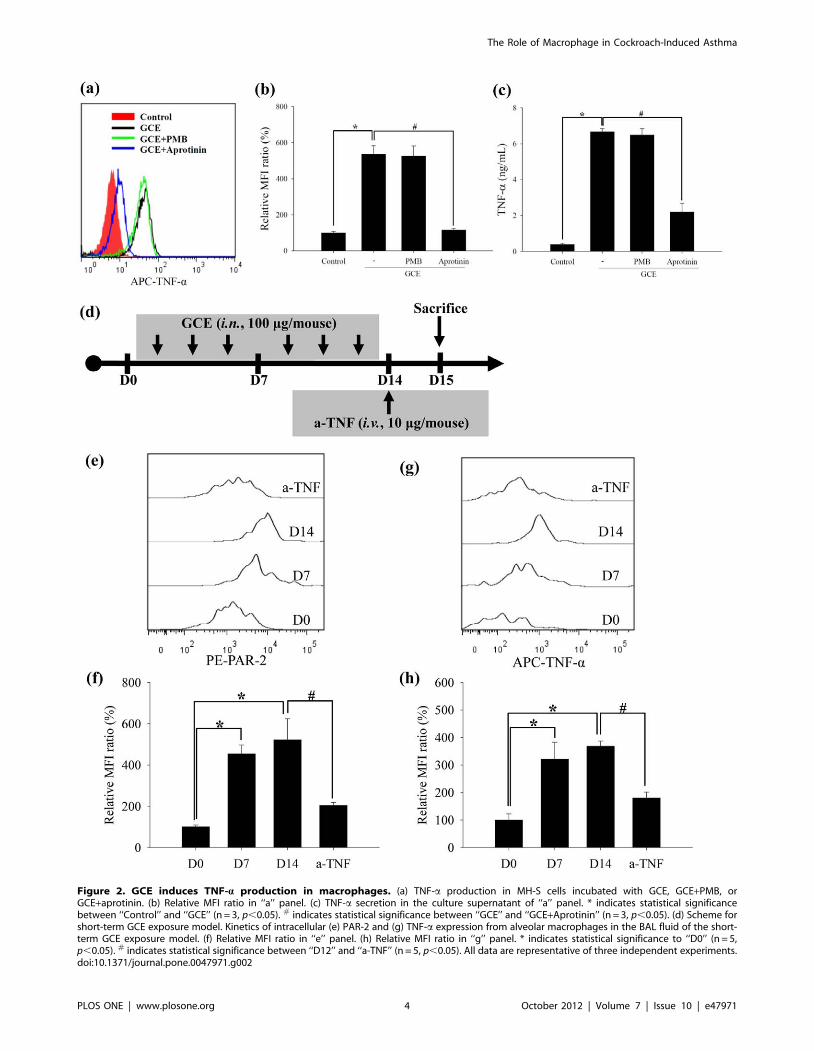
Figure 2. GCE induces TNF-a production in macrophages. (a) TNF-a production in MH-S cells incubated with GCE, GCE+PMB, orGCE+aprotinin. (b) Relative MFI ratio in ‘‘a’’ panel. (c) TNF-a secretion in the culture supernatant of ‘‘a’’ panel. * indicates statistical significancebetween ‘‘Control’’ and ‘‘GCE’’ (n = 3, p,0.05). # indicates statistical significance between ‘‘GCE’’ and ‘‘GCE+Aprotinin’’ (n = 3, p,0.05). (d) Scheme forshort-term GCE exposure model. Kinetics of intracellular (e) PAR-2 and (g) TNF-a expression from alveolar macrophages in the BAL fluid of the short-term GCE exposure model. (f) Relative MFI ratio in ‘‘e’’ panel. (h) Relative MFI ratio in ‘‘g’’ panel. * indicates statistical significance to ‘‘D0’’ (n = 5,p,0.05). # indicates statistical significance between ‘‘D12’’ and ‘‘a-TNF’’ (n = 5, p,0.05). All data are representative of three independent experiments.doi:10.1371/journal.pone.0047971.g002
The Role of Macrophage in Cockroach-Induced Asthma
PLOS ONE | www.plosone.org 4 October 2012 | Volume 7 | Issue 10 | e47971
Figure 3. GCE promotes pulmonary inflammation through the TNF-a pathway. (a) Scheme for TNF-a neutralization of GCE-induced asthmamodel. a-TNF, anti-TNF-a mAb; goat, goat IgG. (b) AHR and (c) BAL cell count. (d) TNF-a-producing macrophage population in the lung tissue. (e)Quantitative analysis of CD11b+TNF-a+ cell population from ‘‘d’’ panel. (f) TNF-a levels in lung homogenates. * indicates statistical significancebetween ‘‘Sham’’ and ‘‘GCE’’ (n = 5, p,0.05). # indicates statistical significance between ‘‘GCE’’ and ‘‘a-TNF’’ (n = 5, p,0.05). All data are representativeof three independent experiments. RL, pulmonary resistance; Mac, macrophage; Lym, lymphocyte; Eos, eosinophil; Neu, neutrophil.doi:10.1371/journal.pone.0047971.g003
The Role of Macrophage in Cockroach-Induced Asthma
PLOS ONE | www.plosone.org 5 October 2012 | Volume 7 | Issue 10 | e47971
The Role of Macrophage in Cockroach-Induced Asthma
PLOS ONE | www.plosone.org 6 October 2012 | Volume 7 | Issue 10 | e47971
wavelengths of 493, 565, and 645 nm and emission wavelengths of
525, 575, and 660 nm, respectively.
Intracellular cytokine stainingIntracellular cytokine staining was performed using a Cytofix/
Cytoperm kit (BD Biosciences, San Diego, CA) according to the
supplier’s recommendations. The cells were stained with PE/Cy7-
(PMSF)-a serine/cysteine protease inhibitor, and soybean trypsin
inhibitor (STI)-a trypsin inhibitor, each protease activities were
decreased. In addition, one of these inhibitors, aprotinin, markedly
reduced protease activity in the GCE (Figure 1B).
To determine PAR-2 immunolocalization following GCE
stimulation in the mouse macrophages, alveolar macrophage cell-
lines (MH-S cells) and peritoneal macrophage cell-lines (RAW264.7
cells) were incubated with either GCE or a combination of GCE and
aprotinin. GCE stimulation led to internalization of the PAR-2 in
the MH-S cells, while the cells incubated with a combination of GCE
and aprotinin revealed similar patterns of control, as visualized by
confocal imaging of intracellular staining (Figure 1C) and cell
surface staining samples (Figure S1). In the RAW264.7 cells, PAR-2
expression of the cell surface decreased following GCE stimulation,
but the cells incubated with a combination of GCE and aprotinin
revealed similar to the control (Figure S2)
To confirm a specific interaction between the GCE and the
PAR-2, MH-S cells were incubated with an ENMD-1068, a novel
selective PAR-2 antagonist [24,25,26], and then stimulated with
GCE. The intracellular expression of PAR-2 and TNF-a was
markedly inhibited by ENMD-1068 (Figure 1D–G).
GCE induces TNF-a production in macrophagesTo examine whether PAR-2 activation by serine proteases
within GCE induces inflammation via macrophages, we studied
TNF-a production and secretion in the MH-S and RAW264.7
cells. Intracellular TNF-a levels were significantly increased in
GCE- stimulated MH-S cells, but these levels were not increased
when GCE protease activity was inhibited by aprotinin. In
GCE+PMB-stimulated condition, the endotoxin level less than 0.1
EU/mL in GCE had no effect in GCE-stimulated cells (Figure 2A
and 2B). These results showed that serine protease but not
endotoxin in GCE is critical for TNF-a production. The culture
supernatants from cells grown under each condition revealed
similar patterns of TNF-a production (Figure 2C). The results of
RAW264.7 cells revealed similar to the MH-S cells (Figure S3).
To identify the kinetics of PAR-2 and TNF-a expression during
GCE stimulation process, GCE was administered intranasally to
BALB/c mice 3 times per week for 2 weeks (short-term GCE
exposure model; Figure 2D). Intracellular PAR-2 and TNF-alevels of alveolar macrophages (CD11c+ and F4/80+ cells) were
increased continuously for up to 2 weeks (Figure 2E–2G).
According to the immunohistochemistric analysis, TNF-aaccumulation of macrophages were increased in the lung tissues of
long-term GCE exposure model (3 times per week for 4 weeks;
Figure S4).
GCE promotes pulmonary inflammation through therelease of TNF-a
To determine whether GCE promotes allergic asthma-like
symptoms, we used a long-term GCE exposure model (Figure 3A).
In an AHR assay, mice that received GCE developed pulmonary
resistance based on the requirement for increasing doses of MCh
inhalation (Figure 3B). The mice receiving GCE exhibited
Figure 4. GCE promotes allergic phenotype through the TNF-a pathway. Lung homogenates were harvested and used for measuring (a) IL-5, (b) IL-13 and (c) IFN-c. (d) The level of serum IgE in the blood. Lung tissues were stained with (e) PAS and (f) Masson’s Trichrome. (g) PAS-positivecells in peri-bronchial regions and (h) total collagen deposition in the lung tissue were quantitatively calculated. * indicates statistical significancebetween ‘‘Sham’’ and ‘‘GCE’’ (n = 5, p,0.05). # indicates statistical significance between ‘‘GCE’’ and ‘‘a-TNF’’ (n = 5, p,0.05). All data are representativeof three independent experiments.doi:10.1371/journal.pone.0047971.g004
The Role of Macrophage in Cockroach-Induced Asthma
PLOS ONE | www.plosone.org 7 October 2012 | Volume 7 | Issue 10 | e47971
Figure 5. Serine protease in GCE develops pulmonary inflammation, alveolar macrophage infiltration and TNF-a expression. (a)Scheme for the inhibition of GCE protease activity of GCE-induced asthma model. (b) AHR and (c) BAL cell count. (d) Alveolar macrophage, interstitialmacrophage, and dendritic cell population in the lung tissue. (e) Quantitative analysis of alveolar macrophage from ‘‘d’’ panel. (f) Intracellular
The Role of Macrophage in Cockroach-Induced Asthma
PLOS ONE | www.plosone.org 8 October 2012 | Volume 7 | Issue 10 | e47971
inflammatory cell infiltration in the BAL fluid, and the majority of
the cells in the BAL fluid were macrophages, lymphocytes, and
neutrophils (Figure 3C). Intracellular TNF-a production and
secretion of the macrophages were increased in the long-term
GCE exposure model (Figure 3D–3F).
To confirm the helper T cell differentiation during GCE-
stimulation, intracellular and secreted levels of different cytokines
expressions of TNF-a in the alveolar macrophages of the GCE-induced asthma experiment. (g) Relative MFI ratio in ‘‘f’’ panel. * indicates statisticalsignificance between ‘‘Sham’’ and ‘‘GCE’’ (n = 5, p,0.05). # indicates statistical significance between ‘‘GCE’’ and ‘‘GCE+Aprotinin’’ (n = 5, p,0.05). Alldata are representative of three independent experiments. AMs, alveolar macrophages; IMs, interstitial macrophages; DCs, dendritic cells.doi:10.1371/journal.pone.0047971.g005
Figure 6. Alveolar macrophages are the major source of TNF-a in the GCE-induced asthma model. (a) Scheme for the alveolarmacrophage depletion of GCE-induced asthma model. Cl2MDP, Cl2MDP-containing liposomes; Liposome, control liposomes. (b) Macrophages fromBAL fluid and (c) AHR in alveolar macrophage-depleted animals. (d) Intracellular TNF-a production in lung tissue. (e) Relative MFI ratio in ‘‘d’’ panel. *indicates statistical significance between ‘‘Liposome/sham’’ and ‘‘Liposome/GCE’’ (n = 5, p,0.05). # indicates statistical significance between‘‘Liposome/GCE’’ and ‘‘Cl2MDP/GCE’’ (n = 5, p,0.05). All data are representative of three independent experiments. Liposome/sham, control liposome-treated group; Liposome/GCE, control liposome-treated GCE-treated group; Cl2MDP/sham, Cl2MDP-containing liposome-treated sham group; Cl2MDP/GCE, Cl2MDP-containing liposome-treated GCE-treated group.doi:10.1371/journal.pone.0047971.g006
The Role of Macrophage in Cockroach-Induced Asthma
PLOS ONE | www.plosone.org 9 October 2012 | Volume 7 | Issue 10 | e47971
in CD4+ T cells were measured. The intracellular levels of IFN-c,
IL-5 and IL-17 were increased in the CD4+ T cells of the long-
term GCE exposure mice (Figure S5A and S5B).IFN-c, IL-5, IL-
10, IL-13 and IL-17 production in lung, and serum IgE levels were
also increased in this model (Figure 4A–4D; Figure S5C).
Histologically, goblet cell hyperplasia and collagen deposition in
the peri-bronchiolar area were exacerbated in the long-term GCE
exposure model as compared to controls (Figure 4E–4H).
To confirm the role of TNF-a in the long-term GCE exposure
model, we blocked TNF-a in these animals. We intravenously
Figure S7 Quantitative analysis of (a) interstitial mac-rophage and (b) dendritic cell population from GCE-induced asthma model. All data are representative of three
Conceived and designed the experiments: JYK JWP. Performed the
experiments: JYK JHS. Analyzed the data: JYK JWP CSH. Contributed
reagents/materials/analysis tools: JHS JHL JSL JMC. Wrote the paper:
JYK.
The Role of Macrophage in Cockroach-Induced Asthma
PLOS ONE | www.plosone.org 11 October 2012 | Volume 7 | Issue 10 | e47971
References
1. Rosenstreich DL, Eggleston P, Kattan M, Baker D, Slavin RG, et al. (1997) Therole of cockroach allergy and exposure to cockroach allergen in causing
morbidity among inner-city children with asthma. N Engl J Med 336: 1356–
1363.
2. Huang H, Dawicki W, Zhang X, Town J, Gordon JR (2010) Tolerogenicdendritic cells induce CD4+CD25hiFoxp3+ regulatory T cell differentiation
from CD4+CD25-/loFoxp3- effector T cells. J Immunol 185: 5003–5010.
3. Kheradmand F, Kiss A, Xu J, Lee S, Kolattukudy PE, et al. (2002) A protease-
activated pathway underlying Th cell type 2 activation and allergic lung disease.J Immunol 169: 5904–5911.
4. Ebeling C, Lam T, Gordon JR, Hollenberg MD, Vliagoftis H (2007) Proteinase-
activated receptor-2 promotes allergic sensitization to an inhaled antigenthrough a TNF-mediated pathway. J Immunol 179: 2910–2917.
expression in human bronchial epithelial cells via activation of protease-activated
receptor (PAR)-2 and extracellular-signal-regulated kinase. J Allergy ClinImmunol 112: 1112–1118.
6. Lee KE, Kim JW, Jeong KY, Kim KE, Yong TS, et al. (2007) Regulation of
German cockroach extract-induced IL-8 expression in human airway epithelialcells. Clin Exp Allergy 37: 1364–1373.
7. Hong JH, Lee S, Kim K, Yong T, Seo JT, et al. (2004) German cockroachextract activates protease-activated receptor 2 in human airway epithelial cells.
J Allergy Clin Immunol 113: 315–319.
8. Ebeling C, Forsythe P, Ng J, Gordon JR, Hollenberg M, et al. (2005) Proteinase-activated receptor 2 activation in the airways enhances antigen-mediated airway
inflammation and airway hyperresponsiveness through different pathways.
J Allergy Clin Immunol 115: 623–630.
9. Compton SJ, Renaux B, Wijesuriya SJ, Hollenberg MD (2001) Glycosylationand the activation of proteinase-activated receptor 2 (PAR(2)) by human mast
11. Sun G, Stacey MA, Schmidt M, Mori L, Mattoli S (2001) Interaction of miteallergens Der p3 and Der p9 with protease-activated receptor-2 expressed by
lung epithelial cells. J Immunol 167: 1014–1021.
12. Poms A, Chapman MD, Vailes LD, Blundell TL, Dhanaraj V (2002) Cockroach
allergen Bla g 2: structure, function, and implications for allergic sensitization.Am J Respir Crit Care Med 165: 391–397.
13. Colognato R, Slupsky JR, Jendrach M, Burysek L, Syrovets T, et al. (2003)
Differential expression and regulation of protease-activated receptors in humanperipheral monocytes and monocyte-derived antigen-presenting cells. Blood
102: 2645–2652.
14. Kiselyov K, Shin DM, Muallem S (2003) Signalling specificity in GPCR-
dependent Ca2+ signalling. Cell Signal 15: 243–253.
15. Cocks TM, Fong B, Chow JM, Aderson GP, Frauman AG, et al. (1999) Aprotective role for protease-activated receptors in the airways. Nature 398: 156–
160.
16. Zhang-Hoover J, Sutton A, van Rooijen N, Stein-Streilein J (2000) A critical role
for alveolar macrophages in elicitation of pulmonary immune fibrosis.Immunology 101: 501–511.
interstitial macrophages alter dendritic cell functions to prevent airway allergy inmice. J Clin Invest 119: 3723–3738.
18. Brightling C, Berry M, Amrani Y (2008) Targeting TNF-alpha: a noveltherapeutic approach for asthma. J Allergy Clin Immunol 121: 5–10.
19. Howarth PH, Babu KS, Arshad HS, Lau L, Buckley M, et al. (2005) Tumour
necrosis factor (TNFalpha) as a novel therapeutic target in symptomaticcorticosteroid dependent asthma. Thorax 60: 1012–1018.
20. Berry MA, Hargadon B, Shelley M, Parker D, Shaw DE, et al. (2006) Evidence
of a role of tumor necrosis factor alpha in refractory asthma. N Engl J Med 354:
697–708.
21. Twining SS (1984) Fluorescein isothiocyanate-labeled casein assay for proteolyticenzymes. Anal Biochem 143: 30–34.
22. Thepen T, Van Rooijen N, Kraal G (1989) Alveolar macrophage elimination in
vivo is associated with an increase in pulmonary immune response in mice. J Exp
Med 170: 499–509.
23. Park J, Taube C, Joetham A, Takeda K, Kodama T, et al. (2004) Complementactivation is critical to airway hyperresponsiveness after acute ozone exposure.
Am J Respir Crit Care Med 169: 726–732.
24. Kelso EB, Ferrell WR, Lockhart JC, Elias Jones I, Hembrough T, et al. (2007)
Expression and proinflammatory role of proteinase-activated receptor 2 inrheumatoid synovium: ex vivo studies using a novel proteinase-activated
28. Dry O, Corvera CU, Steinhoff M, Bunnett NW (1998) Proteinase-activatedreceptors: novel mechanisms of signaling by serine proteases. Am J physiol 274:
C1429–C1452.
29. Chapman MD, Pomes A, Breitneder H, Ferreira F (2007) Nomenclature andstructural biology of allergens. J Allergy Clin Immunol 119: 414–420.
30. Douwes J, Gibson P, Pekkanen J, Pearce N (2002) Non-eosinophilic asthma:importance and possible mechanisms. Thorax 57: 643–648.
31. Di Valentin E, Crahay C, Garbacki N, Hennuy B, Guders M, et al. (2009) New
asthma biomarkers: lessons from murine models of acute and chronic asthma.Am J Physiol Lung Cell Mol Physiol 296: L185–L197.
32. Arizmendi NG, Abel M, Puttagunta L, Asaduzzaman M, Davidson C, et al.(2011) Mucosal exposure to cockroach extract induces allergic sensitization and
allergen exposure can induce persistent tolerance. Am J Respir Cell Mol Biol 36:
573–584.34. Goplen N, Karim MZ, Liang Q, Gorska MM, Rozario S, et al. (2009)
Combined sensitization of mice to extracts of dust mite, ragweed, andAspergillus species breaks through tolerance and establishes chronic features of
asthma. J Allergy Clin Immunol 123: 925–932 e911.
35. Wang Q, Li H, Yao Y, Xia D, Zhou J (2010) The overexpression of heparin-binding epidermal growth factor is responsible for Th17-induced airway
remodeling in an experimental asthma model. J Immunol 185: 834–841.36. Arizmendi NG, Abel M, Mihara K, Davidson C, Polley D, et al. (2011) Mucosal
Allergic Sensitization to Cockroach Allergens Is Dependent on ProteinaseActivity and Proteinase-Activated Receptor-2 Activation. J Immunol 186: 3164–
3172.
37. Johnson JR, Wiley RE, Fattouh R, Swirski FK, Gajewska BU, et al. (2004)Continuous exposure to house dust mite elicits chronic airway inflammation and
structural remodeling. Am J Respir Crit Care Med 169: 378–385.38. Chapman MD, Wnschmann S, Poms A (2007) Proteases as Th2 adjuvants. Curr
Allergy Asthma Rep 7: 363–367.
39. Tua W, Lai S (2006) Induction of cysteine proteinase in the encapsulation ofHymenolepis diminuta eggs in the American cockroach, Periplaneta americana.
sensitization to German cockroach involves protease-activated receptor-2.Respir Res 11: 62.
44. Bhat RK, Page K, Tan A, Hershenson MB (2003) German cockroach extractincreases bronchial epithelial cell interleukin-8 expression. Clin Exp Allergy 33:
35–42.
45. Gordon S, Taylor PR (2005) Monocyte and macrophage heterogeneity. Nat RevImmunol 5: 953–964.
46. Kim EY, Battaile JT, Patel AC, You Y, Agapov E, et al. (2008) persistentactivation of an innate immune repsonse translates respiratory viral infection
into chronic lung disease. Nature Med 14: 633–640.
47. Feola DJ, Garvy BA, Cory TJ, Birket SE, Hoy H, et al. (2010) Azithromycinalters macrophage phenotype and pulmonary compartmentalization during lung
infection with Pseudomonas. Antimicrob Agents Chemother 54: 2437–2447.48. Wynn TA (2004) Fibrotic disease and the T(H)1/T(H)2 paradigm. Nat Rev
Immunol 4: 583–594.
49. Day SB, Zhou P, Ledford JR, Page K (2010) German cockroach frass proteasesmodulate the innate immune response via activation of protease-activated
receptor-2. J Innate immun 2: 495–504.50. Berger P, Perng DW, Thabrew H, Compton SJ, Cairns JA (2001) Tryptase and
agonists of PAR-2 induce the proliferation of human airway smooth muscle cells.J Appl Physiol 91: 1372–1379.
51. Peters T, Mann TS, Henry PJ (2010) Inhibitory influence of protease-activated
receptor 2 and E-prostanoid receptor stimulants in lipopolysaccharide models ofacute airway inflammation. J Pharmacol Exp Ther 335: 424–433.
53. Van Rooijen N (1989) The liposome-mediated macrophage ’suicide’ technique.J Immunol Methods 124: 1–6.
54. Roscic-Mrkic B, Schwendener RA, Odermatt B, Zuniga A, Pavlovic J, et al.(2001) Roles of macrophages in measles virus infection of genetically modified
mice. J Virol 75: 3343–3351.
The Role of Macrophage in Cockroach-Induced Asthma
PLOS ONE | www.plosone.org 12 October 2012 | Volume 7 | Issue 10 | e47971