19
Alzheimer’s Disease Neuroimaging Initiative PET Technical Procedures Manual Supplemental Imaging Protocol Using Pittsburgh Compound B (PIB) Version 1.2 May 10, 2007

Alzheimer’s Disease Neuroimaging Initiative PET Technical Procedures Manual
Supplemental Imaging Protocol Using Pittsburgh Compound B (PIB)
Version 1.2 May 10, 2007

ADNI PET Technical Procedures Manual PIB Protocol Page 2 of 19 V1.1 May 1, 2007
Table of Contents General Information ..................................................................................................................................................................... 3 Contact Information...................................................................................................................................................................... 3 Site Qualification ........................................................................................................................................................................... 4 Continued Quality Monitoring During Execution Phase........................................................................................................ 4 PET Pre-Scan Procedures/General Information...................................................................................................................... 4
Participants Pre-screening.......................................................................................................................................................4 Subject Preparation .................................................................................................................................................................5 Participant Positioning ............................................................................................................................................................5 Ambient Conditions ................................................................................................................................................................5 Image File Identification.........................................................................................................................................................5 Documentation ........................................................................................................................................................................6 Sample PIB Metadata Form....................................................................................................................................................7
PET Imaging Protocol................................................................................................................................................................. 11 Appendix A – Additional Examples of PIB / FDG Protocols ............................................................................................... 16
Example 1: 70 min Extended Dynamic PIB and Standard 5 x 300 sec frame dynamic FDG.........................................16 Example 2: 90 min Extended Dynamic PIB and Standard 5 x 300 sec frame dynamic FDG..........................................16 Example 3: Standard 4 x 300 s dynamic acquisition PIB Only (Single Day) ...................................................................17 Example 4: 70 min Extended Dynamic PIB Only (Single Day) ........................................................................................17 Example 5: 90 min Extended Dynamic PIB Only (Single Day) ........................................................................................18 Example 6: Standard 4 x 300 s acquisition PIB and Extended Dynamic (Quantitative) FDG ........................................18 Example7: 70 min Extended Dynamic PIB and Extended Dynamic (Quantitative) FDG ...............................................19 Example8: 90 min Extended Dynamic PIB and Extended Dynamic (Quantitative) FDG ...............................................19
ADNI PET Technical Procedures Manual PIB Protocol Page 3 of 19 V1.1 May 1, 2007
General Information The purpose of this manual is to further explain the supplemental PET imaging component to assess the amyloid plaque burden using [C-11] Pittsburgh Compound B (PIB) in a subset of ADNI subjects that have also been randomized to FDG PET imaging. Standard procedures are needed to ensure consistency of data collection in this longitudinal study. PIB PET scans will be conducted in these groups. MCI Group (n=48)
Entering subjects with no previous FDG PET will be scanned at 0, 12, 24, 36 months Subjects with baseline and 6 month FDG PET will be scanned at 12, 24 and 36 months
AD Group (n=24)
Entering subjects with no previous FDG PET will be scanned at 0, 12, 24 months Subjects with baseline and 6 month FDG PET will be scanned at 12 and 24 months
Control Group (n=24)
Entering subjects with no previous FDG PET will be scanned at 0, 12, 24, 36 months Subjects with baseline and 6 month FDG PET will be scanned at 12, 24 and 36 months
Contact Information If you have any questions or concerns regarding PIB or FDG PET imaging study please contact
If you have any questions or concerns regarding individual participants please contact the study coordinator at your referral site. If you have question regarding the scan uploading to the LONI website please contact
ADNI PET Technical Procedures Manual PIB Protocol Page 4 of 19 V1.1 May 1, 2007
Site Qualification Your institution must obtain a separate IRB approval, Radiation Safety Committee (RSC) or Radioactive Drug Research Committee (RDRC) approval prior to enrolling patients for the PIB protocol. In some cases your RDRC may indicate the necessity for an IND. All regulatory documents must be submitted to Kristin Woods ([email protected]) at the ADCS prior to enrolling subjects. No PIB slots will be released until all regulatory approval has been obtained. Sites which have passed the initial certification for FDG by scanning a Hoffman 3D brain phantom may begin PIB PET provided they have obtained the additional IRB and RSC/RDRC approvals. There are no additional phantom scans which need to be performed unless there is a change or upgrade in the hardware/software of the imaging system. Continued Quality Monitoring During Execution Phase To ensure scanner/ancillary equipment stability and quality throughout the project, each site is required to perform ongoing quality control procedures. The details for PET and PET/CT scanners and ancillary equipment are detailed in the PET Technologists Manual used for the FDG imaging. PET Pre-Scan Procedures / General Information
Participants Pre-screening
All participants should have been screened by the study coordinator for the following contraindications
Inability to cooperate/claustrophobia (sedation is not offered for this protocol) Inability to lie on the scanner bed for two 30-40 minute scan sessions for PIB and
FDG imaging. There will be a short break between the two imaging sessions.
Inability to achieve venous access sufficient for tracer administration (and venous blood sampling for the dynamic FDG protocol).
ADNI PET Technical Procedures Manual PIB Protocol Page 5 of 19 V1.1 May 1, 2007
Subject Preparation
Subjects to be imaged in the morning are asked to omit all food and fluids (except water) from midnight the night before the scan until after the imaging is completed. Subjects scanned later in the day are asked to omit food and fluids (except water) for at least 4 hours prior to the imaging session.
Participant Positioning
Proper patient positioning is a key aspect of the successful completion of the PET exam. Guidelines for patient positioning are described in detail in the PET Technologist Manual used for FDG imaging.
Ambient Conditions
Although the PIB uptake into the brain is independent of the ambient conditions, the patient should be allowed to rest quietly in a controlled environment similar to the FDG study.
Image File Identification
File identification will be conducted in a manner identical to that used for FDG PET imaging. The file ID will be assigned by the Clinical Study Coordinator at the clinical site prior to the PET visit. The naming convention is SSS_C_#### where SSS is the three digit site ID, C is either S (subject) or P (phantom), and #### is the unique four digit number assigned by the site. For example, 129_S_0012 is the 12th subject enrolled in ADNI across all sites, from Banner Good Samaritan.
IMPORTANT: It is preferred that the PIB and FDG PET scans are performed on the same day. If this is not possible, the two studies may be performed within 28 days of each other and should take place within 2 weeks of the in-clinic visit.
ADNI PET Technical Procedures Manual PIB Protocol Page 6 of 19 V1.1 May 1, 2007
Documentation
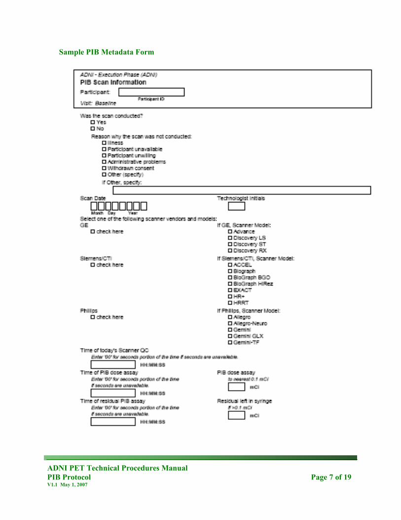
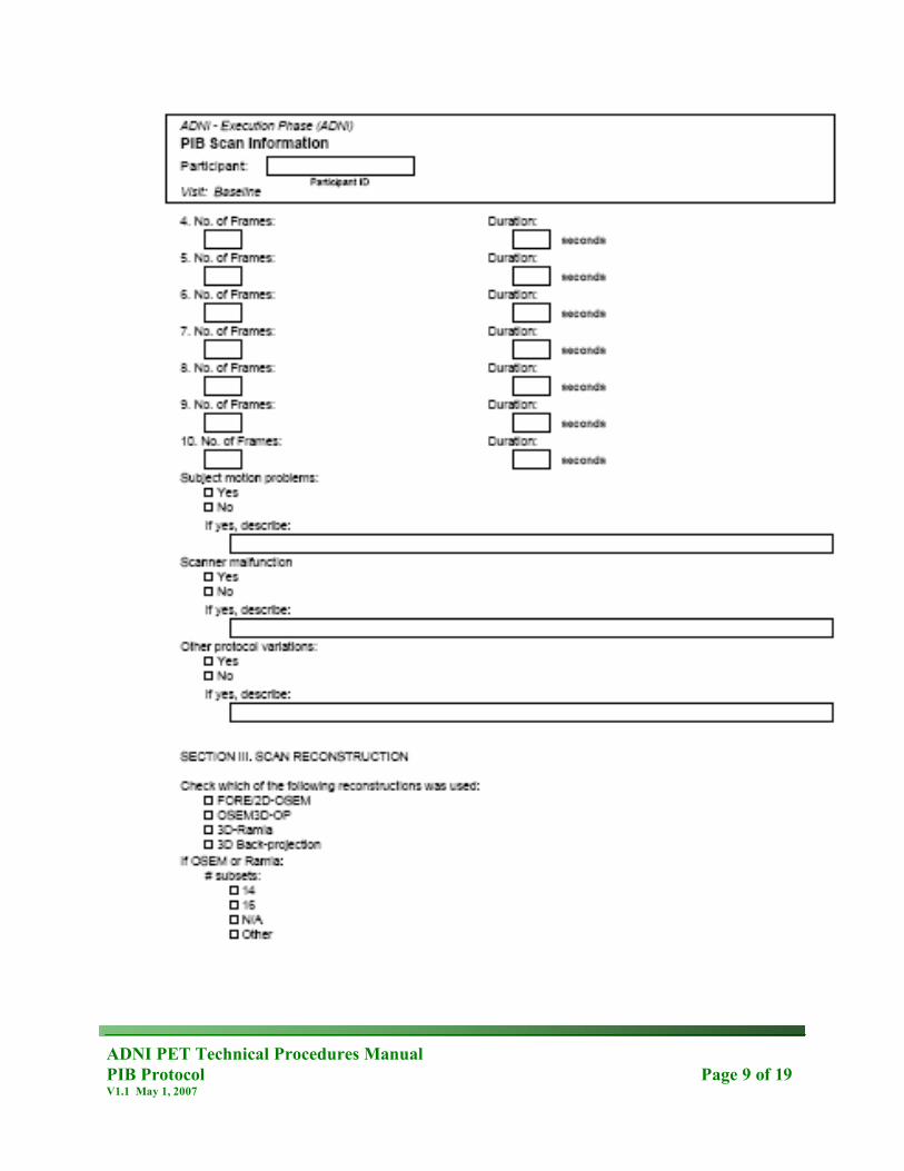
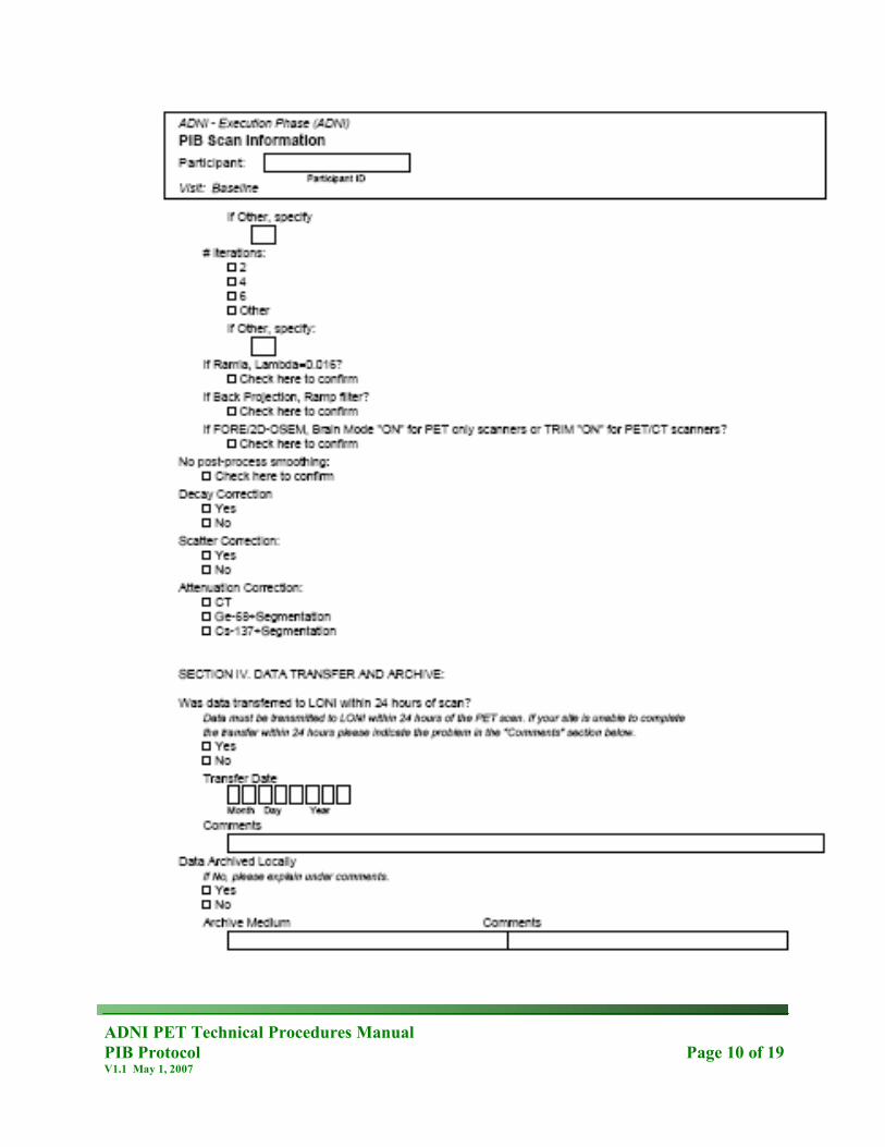
Be sure to complete the metadata sheet as the study is being acquired. The PET scan information form must be provided by the study coordinator prior to the scan.
An example of the PIB metadata sheet has also been included on pages 7-10 of this manual.
IMPORTANT: There is a different metadata sheet specifically for PIB PET imaging. Regardless of whether both PIB and FDG imaging are being conducted in the same imaging session, both forms need to be completed for all studies.
ADNI PET Technical Procedures Manual PIB Protocol Page 7 of 19 V1.1 May 1, 2007
Sample PIB Metadata Form

ADNI PET Technical Procedures Manual PIB Protocol Page 8 of 19 V1.1 May 1, 2007
ADNI PET Technical Procedures Manual PIB Protocol Page 9 of 19 V1.1 May 1, 2007
ADNI PET Technical Procedures Manual PIB Protocol Page 10 of 19 V1.1 May 1, 2007

ADNI PET Technical Procedures Manual PIB Protocol Page 11 of 19 V1.1 May 1, 2007
PET Imaging Protocol
Standard 4 x 300 sec frame dynamic PIB acquisition / Standard 5 x 300 sec frame dynamic FDG acquisition Upon arrival to the imaging center, compliance to the dietary requirements should be
confirmed and the blood glucose level checked.
Have the patient use the restroom and empty their bladder. Allow them to lie comfortably in a bed or reclining chair in a room in which the ambient noise
is minimal and the degree of lighting can be controlled and minimized as previously described. Supply them with blankets/pillows as needed to maximize their comfort.
Obtain intravenous access using either a small butterfly needle or angiocath. Obtain baseline
blood glucose level if not already performed.
Draw 15 + 1.5 mCi (555 MBq) of PIB and assay with a dose calibrator. Record the assayed dose (to the nearest 0.1 mCi) and assay time to the nearest minute. In the event of difficulties with radiochemical yields, the scan should not be performed if <10 mCi are available for injection. In this case the scan should be rescheduled.
Inject the PIB over 10-20 seconds. Rinse the syringe and flush the line with at least 10 cc of
normal saline. Record the injection time to the nearest minute. Do NOT discontinue the IV line at this time as it will be used for the FDG scan as well.
Re-assay the dose syringe and record the residual activity and time of assay.
Allow the subject to rest comfortably in the room for 40 minutes for the incorporation of PIB into the brain.
IMPORTANT: There are acceptable variations of the framing rate for PIB PET imaging. The following section details the basic PIB protocol. Additional protocols which detail potential variations are also detailed in Appendix A. However, it is required that your framing rate include 4 x 300 sec frames from 50 – 70 minutes post PIB injection. If your site would like to perform an imaging method not documented in this manual, please contact a member of the ADNI PET core prior to proceeding ([email protected]). Please also be sure to create separate acquisition protocols for PIB and FDG to ensure proper labeling.
ADNI PET Technical Procedures Manual PIB Protocol Page 12 of 19 V1.1 May 1, 2007
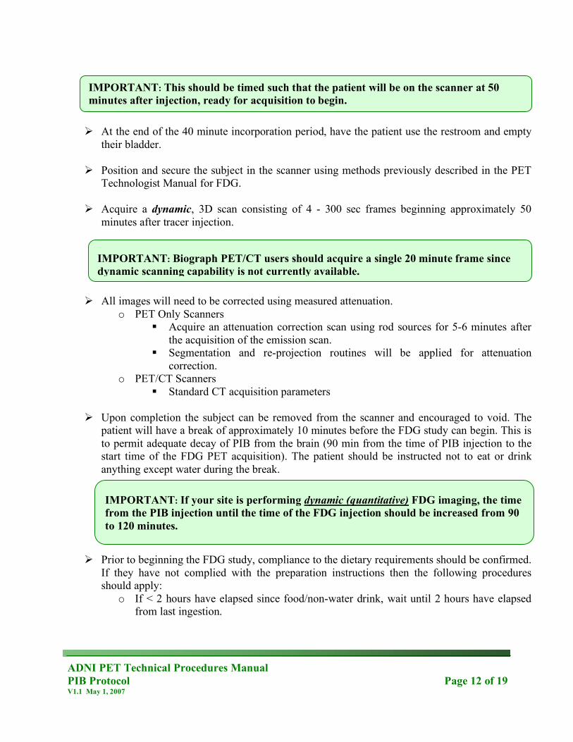
At the end of the 40 minute incorporation period, have the patient use the restroom and empty
their bladder. Position and secure the subject in the scanner using methods previously described in the PET
Technologist Manual for FDG.
Acquire a dynamic, 3D scan consisting of 4 - 300 sec frames beginning approximately 50 minutes after tracer injection.
All images will need to be corrected using measured attenuation.
o PET Only Scanners Acquire an attenuation correction scan using rod sources for 5-6 minutes after
the acquisition of the emission scan. Segmentation and re-projection routines will be applied for attenuation
correction. o PET/CT Scanners
Standard CT acquisition parameters
Upon completion the subject can be removed from the scanner and encouraged to void. The patient will have a break of approximately 10 minutes before the FDG study can begin. This is to permit adequate decay of PIB from the brain (90 min from the time of PIB injection to the start time of the FDG PET acquisition). The patient should be instructed not to eat or drink anything except water during the break.
Prior to beginning the FDG study, compliance to the dietary requirements should be confirmed.
If they have not complied with the preparation instructions then the following procedures should apply:
o If < 2 hours have elapsed since food/non-water drink, wait until 2 hours have elapsed from last ingestion.
IMPORTANT: This should be timed such that the patient will be on the scanner at 50 minutes after injection, ready for acquisition to begin.
IMPORTANT: Biograph PET/CT users should acquire a single 20 minute frame since dynamic scanning capability is not currently available.
IMPORTANT: If your site is performing dynamic (quantitative) FDG imaging, the time from the PIB injection until the time of the FDG injection should be increased from 90 to 120 minutes.

ADNI PET Technical Procedures Manual PIB Protocol Page 13 of 19 V1.1 May 1, 2007
o Once >2 hour have elapsed since last ingestion, measure the blood glucose levels. If the blood glucose level is <180 mg/dL (9.9 mmol/L) then proceed with the scan. If not, the subject will need to wait an additional amount of time until the blood glucose levels meet the above criteria or reschedule.
Have the patient use the restroom and empty their bladder. Allow them to lie comfortably in a bed or reclining chair in a room in which the ambient noise
is minimal and the degree of lighting can be controlled and minimized as previously described. Supply them with blankets/pillows as needed to maximize their comfort.
Verify the IV is patent and obtain baseline blood glucose level if not already performed. Draw 5 + 0.5 mCi (185 MBq) of FDG and assay with a dose calibrator. Record the assayed
dose and assay time to the nearest minute.
Inject the FDG. Rinse the syringe and flush the line with at least 10 cc of normal saline. Record the injection time to the nearest minute. The IV line can be discontinued at this time.
Re-assay the dose syringe. If the residual activity is 0.1 mCi or greater, record the amount and
correct the amount of the injected dose for the residual activity.
Allow the subject to rest comfortably in the room for 20 minutes for the incorporation of FDG into the brain. During the incorporation period, the patient’s eyes should be open and the ears should remain un-occluded.
At the end of the 20 minute incorporation period, have the patient use the restroom and empty
their bladder.
Position and secure the subject in the scanner using methods previously described.
Acquire a dynamic, 3D scan consisting of 6 - 300 sec frames.
IMPORTANT: This should be timed such that the patient will be on the scanner at 30 minutes after injection, ready for acquisition to begin.

ADNI PET Technical Procedures Manual PIB Protocol Page 14 of 19 V1.1 May 1, 2007
All images will need to be corrected using measured attenuation.
o PET Only Scanners Acquire an attenuation correction scan using rod sources for 5-6 minutes after
the acquisition of the emission scan. Segmentation and re-projection routines will be applied for attenuation
correction. o PET/CT Scanners
Standard CT acquisition parameters
Upon completion the subject can be removed from the scanner and encouraged to void. The subject should also be instructed to drink plenty of fluids and void frequently throughout the day to help reduce radiation exposure.
Below is a schematic representation to assist with the timing constraints of the procedure.
Reconstruct images using parameters specific to the system used for scanning. PIB and FDG
data sets will be reconstructed with the same parameters. (See Appendix A in the PET Technologists Manual for details).
Upon completion of the reconstruction, review all the images to assess for artifacts and motion.
Archive ALL raw and processed study data including copies of the normalization and blank
scans. It is necessary to archive and store raw and processed data at the imaging site for the duration of the ADNI project (approximately 5 years).
IMPORTANT: Biograph PET/CT users should acquire a single 30 minute frame since dynamic scanning capability is not currently available.
ADNI PET Technical Procedures Manual PIB Protocol Page 15 of 19 V1.1 May 1, 2007
Transfer both PIB and FDG image data to the Laboratory of Neuroimaging (LONI) at UCLA
using the procedure detailed in Appendix B. These should be performed as separate uploads.
IMPORTANT: Data uploads to LONI should be performed as soon as the images have been acquired & reconstructed as it will be important to promptly QC the data to identify if the scan needs to be repeated.

ADNI PET Technical Procedures Manual PIB Protocol Page 16 of 19 V1.1 May 1, 2007
Appendix A – Additional Examples of PIB / FDG Protocols Example 1: 70 min Extended Dynamic PIB and Standard 5 x 300 sec frame dynamic FDG
PIB: (4 x 15s)(8 x 30s)(9 x 60s)(2 x 180s)(10 x 300s) frames starting at time of PIB
injection FDG: (5 x 300s) frames starting 30 min post FDG injection.
Example 2: 90 min Extended Dynamic PIB and Standard 5 x 300 sec frame dynamic FDG
Framing Sequence PIB: (4 x 15s)(8 x 30s)(9 x 60s)(2 x 180s)(14 x 300s) frames starting at time of PIB
injection FDG: (5 x 300s) frames starting 30 min post FDG injection.

ADNI PET Technical Procedures Manual PIB Protocol Page 17 of 19 V1.1 May 1, 2007
Example 3: Standard 4 x 300 s dynamic acquisition PIB Only (Single Day) Framing Sequence PIB: 4 x 300 second frames starting at 50 minutes post PIB injection
Example 4: 70 min Extended Dynamic PIB Only (Single Day)
Framing Sequence PIB: (4 x 15s)(8 x 30s)(9 x 60s)(2 x 180s)(10 x 300s) frames starting at time of PIB
injection
ADNI PET Technical Procedures Manual PIB Protocol Page 18 of 19 V1.1 May 1, 2007
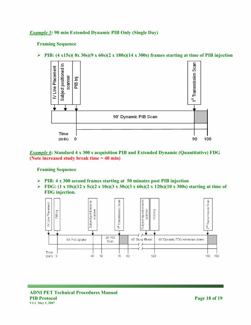
Example 5: 90 min Extended Dynamic PIB Only (Single Day)
Framing Sequence PIB: (4 x15s)( 8x 30s)(9 x 60s)(2 x 180s)(14 x 300s) frames starting at time of PIB injection
Example 6: Standard 4 x 300 s acquisition PIB and Extended Dynamic (Quantitative) FDG (Note increased study break time = 40 min)
Framing Sequence PIB: 4 x 300 second frames starting at 50 minutes post PIB injection FDG: (1 x 10s)(12 x 5s)(2 x 10s)(3 x 30s)(3 x 60s)(2 x 120s)(10 x 300s) starting at time of
FDG injection.
ADNI PET Technical Procedures Manual PIB Protocol Page 19 of 19 V1.1 May 1, 2007
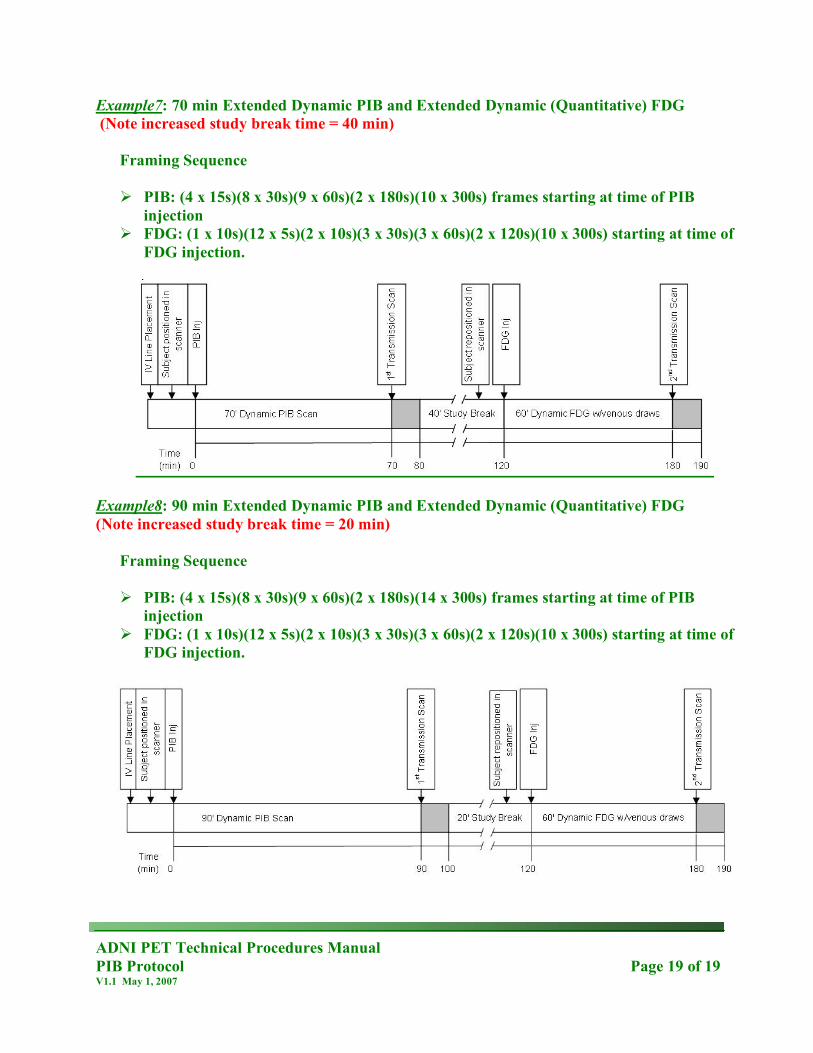
Example7: 70 min Extended Dynamic PIB and Extended Dynamic (Quantitative) FDG (Note increased study break time = 40 min)
Framing Sequence PIB: (4 x 15s)(8 x 30s)(9 x 60s)(2 x 180s)(10 x 300s) frames starting at time of PIB
injection FDG: (1 x 10s)(12 x 5s)(2 x 10s)(3 x 30s)(3 x 60s)(2 x 120s)(10 x 300s) starting at time of
FDG injection.
Example8: 90 min Extended Dynamic PIB and Extended Dynamic (Quantitative) FDG (Note increased study break time = 20 min)
Framing Sequence PIB: (4 x 15s)(8 x 30s)(9 x 60s)(2 x 180s)(14 x 300s) frames starting at time of PIB
injection FDG: (1 x 10s)(12 x 5s)(2 x 10s)(3 x 30s)(3 x 60s)(2 x 120s)(10 x 300s) starting at time of
FDG injection.


![7KLVHOHFWURQLFWKHVLVRU ......Novel [11C]CO2 radiolabelling methodologies for PET neuroimaging Abdul Karim Haji Dheere A thesis submitted in partial fulfilment of the requirements for](https://static.documents.pub/doc/80x56/5e4db790ed253e17d960e032/7klvhohfwurqlfwkhvlvru-novel-11cco2-radiolabelling-methodologies-for-pet.jpg)


