ARTICLE Received 9 Dec 2014 | Accepted 24 Apr 2015 | Published 15 Jun 2015 Ambient solid-state mechano-chemical reactions between functionalized carbon nanotubes Mohamad A. Kabbani 1 , Chandra Sekhar Tiwary 1, *, Pedro A.S. Autreto 2, *, Gustavo Brunetto 2, *, Anirban Som 3 , K.R. Krishnadas 3 , Sehmus Ozden 1 , Ken P. Hackenberg 1 , Yongi Gong 4 , Douglas S. Galvao 2 , Robert Vajtai 1 , Ahmad T. Kabbani 1,5 , Thalappil Pradeep 3 & Pulickel M. Ajayan 1,3,4 Carbon nanotubes can be chemically modified by attaching various functionalities to their surfaces, although harsh chemical treatments can lead to their break-up into graphene nanostructures. On the other hand, direct coupling between functionalities bound on individual nanotubes could lead to, as yet unexplored, spontaneous chemical reactions. Here we report an ambient mechano-chemical reaction between two varieties of nanotubes, carrying predominantly carboxyl and hydroxyl functionalities, respectively, facilitated by simple mechanical grinding of the reactants. The purely solid-state reaction between the chemically differentiated nanotube species produces condensation products and unzipping of nanotubes due to local energy release, as confirmed by spectroscopic measurements, ther- mal analysis and molecular dynamic simulations. DOI: 10.1038/ncomms8291 OPEN 1 Department of Materials Science and NanoEngineering, Rice University, Houston, Texas 77005, USA. 2 Department of Applied Physics, State University of Campinas, Campinas-SP 13083-959, Brazil. 3 DST Unit of Nanoscience and Thematic Unit of Excellence, Department of Chemistry, Indian Institute of Technology Madras, Chennai 600 036, India. 4 Department of Chemistry Rice University, Houston, Texas 77005, USA. 5 Department of Natural Science, Lebanese American University, P.O. Box 13-5053 Chouran, Beirut 1102 2801, Lebanon. *These authors contributed equally to this work. Correspondence and requests for materials should be addressed to M.A.K. (email: [email protected]) or to T.P. (email: [email protected]) or to P.M.A. (email: [email protected]). NATURE COMMUNICATIONS | 6:7291 | DOI: 10.1038/ncomms8291 | www.nature.com/naturecommunications 1 & 2015 Macmillan Publishers Limited. All rights reserved.
Transcript
ARTICLE
Received 9 Dec 2014 | Accepted 24 Apr 2015 | Published 15 Jun 2015
Ambient solid-state mechano-chemical reactionsbetween functionalized carbon nanotubesMohamad A. Kabbani1, Chandra Sekhar Tiwary1,*, Pedro A.S. Autreto2,*, Gustavo Brunetto2,*, Anirban Som3,
K.R. Krishnadas3, Sehmus Ozden1, Ken P. Hackenberg1, Yongi Gong4, Douglas S. Galvao2, Robert Vajtai1,
Ahmad T. Kabbani1,5, Thalappil Pradeep3 & Pulickel M. Ajayan1,3,4
Carbon nanotubes can be chemically modified by attaching various functionalities to their
surfaces, although harsh chemical treatments can lead to their break-up into graphene
nanostructures. On the other hand, direct coupling between functionalities bound on
individual nanotubes could lead to, as yet unexplored, spontaneous chemical reactions.
Here we report an ambient mechano-chemical reaction between two varieties of nanotubes,
carrying predominantly carboxyl and hydroxyl functionalities, respectively, facilitated by
simple mechanical grinding of the reactants. The purely solid-state reaction between the
chemically differentiated nanotube species produces condensation products and unzipping of
nanotubes due to local energy release, as confirmed by spectroscopic measurements, ther-
mal analysis and molecular dynamic simulations.
DOI: 10.1038/ncomms8291 OPEN
1 Department of Materials Science and NanoEngineering, Rice University, Houston, Texas 77005, USA. 2 Department of Applied Physics, State University ofCampinas, Campinas-SP 13083-959, Brazil. 3 DST Unit of Nanoscience and Thematic Unit of Excellence, Department of Chemistry, Indian Institute ofTechnology Madras, Chennai 600 036, India. 4 Department of Chemistry Rice University, Houston, Texas 77005, USA. 5 Department of Natural Science,Lebanese American University, P.O. Box 13-5053 Chouran, Beirut 1102 2801, Lebanon. * These authors contributed equally to this work. Correspondence andrequests for materials should be addressed to M.A.K. (email: [email protected]) or to T.P. (email: [email protected]) or to P.M.A. (email: [email protected]).
Chemical functionalization of nanoparticles can lead totheir surface decoration with a variety of covalentlyattached functionalities to serve different goals such as
drug delivery, cancer therapy, diagnostics and electronicdevices1–6. Carbon nanotubes (CNTs) have been the subject ofmore than two decades of intense research7–10. Variousapproaches have been used to modify their surfaces viacovalent and non-covalent attachments to change both physicaland chemical properties. Activation of CNTs by the incorporationof COOH on the exterior surfaces by treating them withconcentrated nitric acid has been widely used11,12. CNT-COOHcan further be activated by acylation to form CNT-COCl, whichcan in turn be amidated or esterified13,14. CNTs carryinghydroxyl groups (CNT-OH) on their surfaces have also beensynthesized by alkaline hydrothermal treatment of pristinenanotubes in alkaline medium15. Despite the remarkable CNTmechanical and electronic properties, their large use has beenprecluded by poor solubility in water or organic solvents, whichfavours bundle formation, thus limiting their chemical reactivity.Mechano-chemical reactions (MCRs) can be used to overcomesuch difficulties16,17. MCRs have been extensively used assynthetic protocols to obtain fullerene derivatives. Examples ofthese methods are fullerene dimers (C120), trimers (C180), crossdimers (C60-C70) and other fullerene derivatives obtained by thesolid-state reactions with potassium salts such as KCN, K2CO3,reducing metals such as Mg and Al and solid aromatic aminesunder the high-speed vibration milling conditions18–21. MCRsof CNT functionalizations with other molecules such as C60,nitrenes, diazoinium compounds and metallic hydroxides undervigorous milling conditions have also been reported22–25.Another way of reacting fullerenes has been through theirencapsulation into CNTs26. On the other hand, many methodshave been used to produce graphene27–31, including theunzipping of nanotubes to make graphene nanoribbons. Atypical chemical unzipping of CNTs makes use of oxidativetechniques31 in concentrated acid (H2SO4) and post treatmentswith harsh reagents such as highly concentrated potassiumpermanganate (KMnO4). These processes involve harshconditions to get to the final product (graphene), which cancontain broken up or unzipped CNT. What has not been done isthe use of nanotubes as solid-state reaction templates with specificchemical surface functionalities to induce direct coupling betweenthe functional groups and concomitant breakdown of thecylindrical structure.
Here, we report the demonstration of unzipping of CNTs via asolid-state room temperature reaction between multiwalled CNTs(MWCNTs) containing different reactive functionalities ofCOOH and OH groups. The reaction is mechano-chemicallyinduced, initiated at room temperature in ambient air, facilitatedby the simple grinding of two chemically variant CNT reactantsand leading to the unzipping of the nanotube (shown in Fig. 1a).By grinding equal weights of MWCNT-COOH and MWCNT-OH decorated with 1.41% and 0.46% by weight of COOH andOH (see experimental details of grinding and SupplementaryTable 1), respectively11,15, a sheet-like lustrous material is formedspontaneously (Fig. 2a). Characterization of the material usingdifferent microscopic and spectroscopic methods, described laterin the manuscript, suggests that the product consistspredominantly of graphene or partially opened CNTs, possiblyformed via the unzipping of the MWCNT reactants. Theunzipping reaction may be represented by equation (1)
MWCNT�COOHþMWCNT�OH!!GþG; þH2OþCO2 ð1Þ
Where G and G0 represent the graphenes originating from thecarboxylic and hydroxyl MWCNT (functionalized MWCNTs).
ResultsCharacterization. Attenuated total reflectance-Infrared Spectro-scopy (ATR-IR) of the solid-state reaction product reveals almostcomplete absence of the COOH/O-H stretch in the region 3,600–2,800 cm� 1 (Fig. 2a), in agreement with water formationduring the reaction. Also, the intensity of the carbonyl band dueto either carboxylic group or keto-enol tautomer in the CNT-OHdiminishes significantly with the appearance of the adsorbedasymmetric CO2 mode at 2,345 cm� 1. Compatible with theseconclusions is the large decrease in the bending infrared mode ofthe CNTs at 868 cm� 1 in the graphene product. The residualintensity is attributed to the unreacted CNTs. These results werefurther confirmed using X-ray photoelectron spectroscopicmeasurements.
In the C1s X-ray photoemission spectroscopy (XPS) of theMWCNTs, the signal at 289.2 eV corresponds to the carboxylgroup, whereas the shoulders at 286.1 and 285.6 eV correspond tothe C–O peak in MWCNT-COOH and MWCNT-OH, respec-tively15,31. Upon grinding, these features diminish in intensityand the most dominant peak becomes that of C¼C at 284.8 eV,as seen in Fig. 2b. This is a strong evidence in favour of acondensation reaction taking place between the COOH and theOH. In addition, according to XPS, oxygen content drops from0.715% in the unreacted mixture to 0.280% in the observed solidproduct (XPS data, Supplementary Table 1). The water formedcomes from the OH of the carboxylic acid and constitutes half theoxygen of the carboxylic group. The fact that the loss of oxygen(0.715–0.280¼ 0.435%) is appreciably larger than half theamount of oxygen of the carboxylic group or oxygen of thehydroxyl group in the unreactive mixture is in agreement with agraphene, H2O and CO2 reaction, the yield of which is B61%(Supplementary Table 1). This is comparable with the infrareddata given before as well as with the simulation data presentedlater. To provide further evidence in favour of the grapheneproduct from the solid-state reaction, we performed Ramanspectroscopy of the reactant MWCNTs and that of the solid-statereaction product (Fig. 2c). All the bands in the product areupshifted relative to the reacting MWCNTs, whereas thesecond-order Raman band (2D) appearing at 2,705 cm� 1 isdownshifted as compared with the graphite band at 2,714 cm� 1
(Fig. 2c). On the other hand, the 2D band in the product was wellfitted by a sharp and symmetric Lorentzian in agreement with afew layer graphene-like product (Fig. 2c). The observations oflower 2D peak position relative to graphite, smaller ID/IG ratio(0.201) and larger I2D/IG ratio (1.21) for our reaction product areall in support of the formation of few layer graphenematerials32,33.
Water formation during the solid-state reaction between theMWCNTs was also established through an in-situ mass spectro-metric study of the reaction products. This was performed asdescribed in Supplementary Note 1. Briefly, the experimentinvolved conducting the solid-state reaction in an enclosedmortar and pestle and sampling of the gases formed directly witha quadruple mass spectrometer, in the mass range of 1–300 amu.A blank measurement was done first without any CNTs in orderto estimate the contribution from atmospheric gases andmoisture. MWCNT-COOH and MWCNT-OH (1:1 ratio byweight) were then taken in the mortar and ground using a pestle.The gases in the reaction vessel were then allowed into the massspectrometer inlet by opening a valve (Supplementary Fig. 1a).Intensity of the peak at m/z 18 (due to H2Oþ ) increasedsignificantly than in the blank experiment (SupplementaryFig. 1d). It is to be noted that there was no increase in intensityof nitrogen and oxygen ion currents. Increase in the H2Oþ peakintensity with no increase in Nþ intensity (derived from N2)clearly shows that this enhancement is due to the water resulting
from solid-state condensation reaction between MWCNTs.No leak of atmospheric air would explain the increase inH2Oþ as that would have resulted in an increase in Nþ intensityas well. Corresponding mass spectral (intensity versus m/z) dataare given in Supplementary Fig. 1b, which also show that onlyH2Oþ intensity increased after the reaction while Nþ and Oþ
intensities remained the same. To check whether this increase isdue to the desorption of water vapour that was adsorbed onMWCNTs, a control experiment was carried out (SupplementaryFig. 1c). Initially, a blank was measured without MWCNTs.MWCNTs were then kept in the mortar without grinding for2 min and gases inside were sampled (after the evacuation step).Intensities of Nþ , Oþ and H2Oþ were almost the same as thatof the blank. After the mixture of MWCNTs was ground for20 min, mass spectral measurement clearly showed an increase inintensity of only H2Oþ . The OH and COOH functionalizedMWCNTs were separately ground and no increase in H2Oþ wasdetected in those cases. This shows that H2O desorption fromMWNTs is not the reason for increased H2Oþ intensity. Noincrease in CO2 intensity was seen as it appears to be adsorbedeffectively on the resulting graphene (see above).
Reproducibility of the results was confirmed by repeating theseexperiments several times with various ratios of the two MWCNTvarieties. Furthermore, the decrease in the oxygen content,revealed from the XPS measurements, is supported by our massspectrometric detection of water. Hence, the in-situ massspectrometric experiments unambiguously give evidence for thesolid-state condensation reaction between -COOH and -OHgroups of the functionalized MWCNTs.
This was further supported with differential thermal analysis atdifferent temperatures, which gave two distinct peaks, the moreintense one occurs at B50 �C, whereas the less intense one occursat B110 �C, shown in Supplementary Fig. 2. The peak at 110 �Cis due to the desorption of water. As the energy evolved at lowertemperature is appreciably higher than that due to desorption ofwater and in light of the strong CO2/graphene adsorption, theintense peak is assigned to the desorption of CO2. Thisconclusion is compatible with the infrared and XPS datapresented before.
The proposed reaction from above spectroscopic and thermalmeasurements is further verified using imaging techniques such
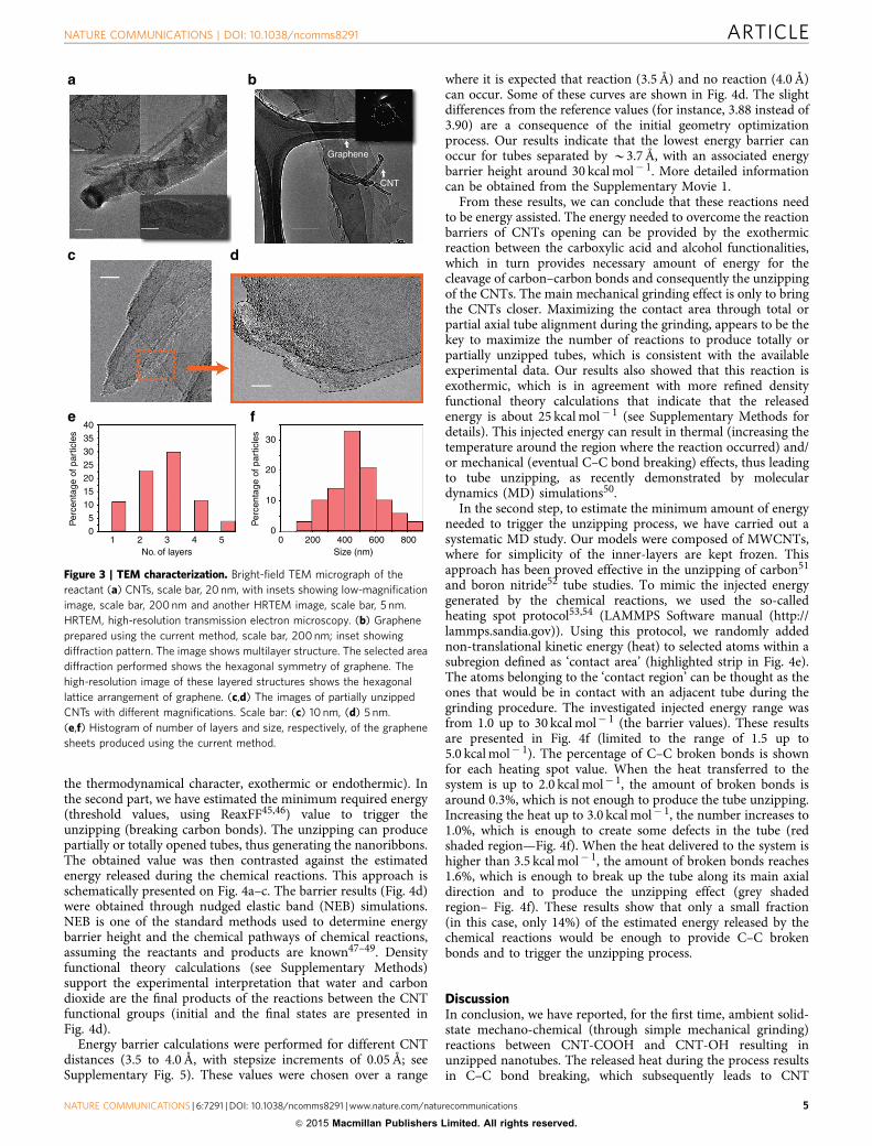
as scanning (SEM) and transmission electron microscopy (TEM).Figure 2e,f shows low and higher magnification SEM imagesrevealing the structure of CNTs for the two functionalizedraw materials. The amount of graphene-like material isnegligible in these samples. On the other hand, the productpowder shows predominantly sheet-like structure along withresidues of partially reacted nanotubes (Fig. 2g,h). The imageanalyses of SEM images from different regions are used forcalculating the amount of 2D sheets present and the residue ofCNTs. The sheets are randomly distributed with a range of sizeswith approximately 20% being unreacted CNT. To furtherconfirm the opening of CNTs and the quality of the graphene-like product, TEM imaging was performed. Figure 3a showsbright-field image of the functionalized CNTs. It clearly showsCNTs to be multiwalled with an average diameter of 20 nm. Thesurfaces of the nanotubes appear disordered, due to heavyfunctionalization. Figure 3b shows large sheets of graphene-likematerial with smooth edges. The image shows multilayerstructure. It is possible that the graphene flakes formed duringthe reaction have coalesced to form larger multilayer graphiticsheets. For further confirmation of the structure, selected areadiffraction was performed. The result shows the hexagonal latticeof graphene stacks (shown as inset). Several images were taken fordifferent samples of the product at different regions. Figure 3c,dshows products in the intermediate steps of the reaction. SeveralTEM images from different regions have been used fordetermining the number of layers in the multilayer sheets andthe size of these graphene sheets. Histograms showing the sizeand number of layers formed due to the reaction are shown inFig. 3e,f.
Kinetics of the reaction (and product formation) wasmonitored by measuring the intensity of the 2D Raman bandof the graphene product at different temperatures30,34–36
(Supplementary Fig. 3). Arrhenius plot (detailed data given inSupplementary Table 2) of ln k versus 1/T (SupplementaryFig. 3) gives an activation energy of 16.63 kJ mol� 1
(3.97 kcal mol� 1), a value compatible with the activationenergies reported for solid-state hydrogen bond-mediatedproton-transfer reactions between many organic compoundssuch as carboxylic acid/phenol and carboxylic acid/aminecombinations37–44.
CO
OO
H
H
Step B
Exothermic reaction
Δ
CO
OHO
H
Step A
CNT-COOH CNT-OH
Step C
C–C breaking bonds
OH
H
C OO
Water
Carbon dioxide
–OH
Functionalization
–COOH
FunctionalizationM
WC
NT
Unzipped nanotube
+
Figure 1 | General scheme of the current work. (a) Solid-state synthetic unzipping scheme. (b) Hydrogen bond-mediated proton transfer unzipping
mechanism: (Step A) hydrogen-bond formation and (Step B) fast proton-transfer, are followed by (Step C) water and CO2 as the products of the
exothermic reaction. The released heat can induce the breaking of carbon-carbon bonds (highlighted yellow region) leading to unzipped tubes.
CNT unzipping reaction. In light of the above, we suggest thatthe CNT unzipping reaction consists of a slow step that brings theCNTs together through mechanical grinding allowing the COOHand OH groups to react (Step A in Fig. 1b). Accordingly, this stepis followed by fast proton transfer (Step B in Fig. 1c) from thecarboxylic group to the hydroxyl group to form [MWCNT-OH2]þ and [MWCNT-COO]� , whose exothermic reactions
produce water and CO2 (Step C in Fig. 1d). The energy releasedcan induce carbon-carbon bond breaking (highlighted yellowregion) leading to unzipped structures.
Owing to the complexity of this process, simulations aredivided into two parts. The first one is related to the estimationof the energy barriers associated with the chemical reactionsbetween the CNT functional groups (value and determination of
a
GrapheneCNT-OH CNT-COOH Graphene
CNT-COOH
OH
C=0CO2
CNTbending C=C
OH
500 1,000 1,500 2,000 2,500 3,000 3,500 4,000
Tra
nsm
itanc
e
Wavenumber (cm−1)
CNT-OH
Graphene
CNT-COOH
CNT-OH
Graphene
Inte
nsity
(ar
b. u
nits
)
0
10
20
30
40
50
Binding energy (eV)282 284 286 288 290 292
1,500 2,000 2,500 3,0000
5
10
15
20
Inte
nsity
(ar
b. u
nits
)
D
G
D+D”
2D
D+D’CNT-OH
CNT-COOH
Graphene
0
5
10
15
20
2,500 2,600 2,700 2,800Raman Shift (cm−1)
Inte
nsity
(ar
b. u
nits
)
CNT-OH
CNT-COOH
Graphene
2D
Raman Shift (cm−1)
0.75
1.00
1.25
1.50
3.25
3.50
0 500 1,000 1,500 2,000 2,500
H 2O+
N+
O+
Time (s)
Ion
curr
ent (
10−
7 A)
Blankwith
CN Ts
Pumping on Pumping stopped Sample line open CNTs ground
b
c d
e f g h
0
5
10
15
20
2,600 2,800Raman Shift
(cm−1)In
tens
ity (
arb.
uni
ts)
Figure 2 | Materials characterization. (a) ATR-IR spectroscopy of the solid-state reaction graphene product after grinding (red) as compared with
MWCNTs starting material MWCNT-COOH (blue) and MWCNT-OH (black). Formation of water in the unzipping process is confirmed by the absence of
the COOH/OH stretch band in the 3,600–2,800 cm� 1 region of the graphene product. The inset shows an image of the product of the solid-state reaction
of MWCNT-COOH and MWCNT-OH. The image shows the thin lustrous sheets of the graphene product formed due to the unzipping of the MWCNTs.
These sheets are covered and surrounded with some traces of the unreacted CNTs. (b) High-resolution C1s XPS spectrum of the solid-state reaction
graphene product obtained after grinding (red) as compared with MWCNTs starting material (blue and green). (c) Raman spectroscopy of the solid-state
reaction mixture after grinding (red) as compared with MWCNTs starting material (green and blue). Insets a 2D-band spectrum of the product as
compared with those in the CNTs starting material, and a single-Lorentzian fit of the 2D band in the product. (d) Ion current versus time plots for
Nþ , Oþ and H2Oþ obtained using in-situ mass spectrometric measurements during the solid-state condensation reaction between MWCNTs. SEM image
of the two reactants (e) CNT-COOH, scale bar, 2 mm; and (f) CNT-OH, scale bar, 2 mm. (g,h) The graphene nanosheet product along with residue of CNTs
at two magnifications, scale bar: (g) 5mm, (h) 1mm.
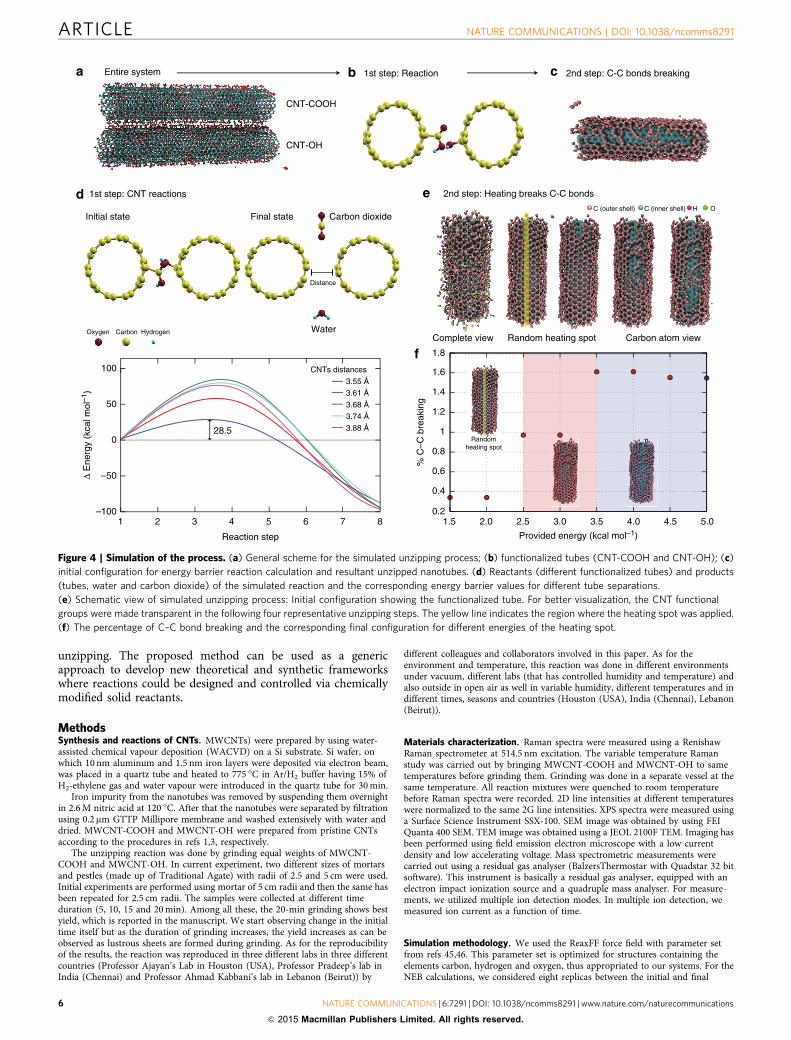
the thermodynamical character, exothermic or endothermic). Inthe second part, we have estimated the minimum required energy(threshold values, using ReaxFF45,46) value to trigger theunzipping (breaking carbon bonds). The unzipping can producepartially or totally opened tubes, thus generating the nanoribbons.The obtained value was then contrasted against the estimatedenergy released during the chemical reactions. This approach isschematically presented on Fig. 4a–c. The barrier results (Fig. 4d)were obtained through nudged elastic band (NEB) simulations.NEB is one of the standard methods used to determine energybarrier height and the chemical pathways of chemical reactions,assuming the reactants and products are known47–49. Densityfunctional theory calculations (see Supplementary Methods)support the experimental interpretation that water and carbondioxide are the final products of the reactions between the CNTfunctional groups (initial and the final states are presented inFig. 4d).
Energy barrier calculations were performed for different CNTdistances (3.5 to 4.0 Å, with stepsize increments of 0.05 Å; seeSupplementary Fig. 5). These values were chosen over a range
where it is expected that reaction (3.5 Å) and no reaction (4.0 Å)can occur. Some of these curves are shown in Fig. 4d. The slightdifferences from the reference values (for instance, 3.88 instead of3.90) are a consequence of the initial geometry optimizationprocess. Our results indicate that the lowest energy barrier canoccur for tubes separated by B3.7 Å, with an associated energybarrier height around 30 kcal mol� 1. More detailed informationcan be obtained from the Supplementary Movie 1.
From these results, we can conclude that these reactions needto be energy assisted. The energy needed to overcome the reactionbarriers of CNTs opening can be provided by the exothermicreaction between the carboxylic acid and alcohol functionalities,which in turn provides necessary amount of energy for thecleavage of carbon–carbon bonds and consequently the unzippingof the CNTs. The main mechanical grinding effect is only to bringthe CNTs closer. Maximizing the contact area through total orpartial axial tube alignment during the grinding, appears to be thekey to maximize the number of reactions to produce totally orpartially unzipped tubes, which is consistent with the availableexperimental data. Our results also showed that this reaction isexothermic, which is in agreement with more refined densityfunctional theory calculations that indicate that the releasedenergy is about 25 kcal mol� 1 (see Supplementary Methods fordetails). This injected energy can result in thermal (increasing thetemperature around the region where the reaction occurred) and/or mechanical (eventual C–C bond breaking) effects, thus leadingto tube unzipping, as recently demonstrated by moleculardynamics (MD) simulations50.
In the second step, to estimate the minimum amount of energyneeded to trigger the unzipping process, we have carried out asystematic MD study. Our models were composed of MWCNTs,where for simplicity of the inner-layers are kept frozen. Thisapproach has been proved effective in the unzipping of carbon51
and boron nitride52 tube studies. To mimic the injected energygenerated by the chemical reactions, we used the so-calledheating spot protocol53,54 (LAMMPS Software manual (http://lammps.sandia.gov)). Using this protocol, we randomly addednon-translational kinetic energy (heat) to selected atoms within asubregion defined as ‘contact area’ (highlighted strip in Fig. 4e).The atoms belonging to the ‘contact region’ can be thought as theones that would be in contact with an adjacent tube during thegrinding procedure. The investigated injected energy range wasfrom 1.0 up to 30 kcal mol� 1 (the barrier values). These resultsare presented in Fig. 4f (limited to the range of 1.5 up to5.0 kcal mol� 1). The percentage of C–C broken bonds is shownfor each heating spot value. When the heat transferred to thesystem is up to 2.0 kcal mol� 1, the amount of broken bonds isaround 0.3%, which is not enough to produce the tube unzipping.Increasing the heat up to 3.0 kcal mol� 1, the number increases to1.0%, which is enough to create some defects in the tube (redshaded region—Fig. 4f). When the heat delivered to the system ishigher than 3.5 kcal mol� 1, the amount of broken bonds reaches1.6%, which is enough to break up the tube along its main axialdirection and to produce the unzipping effect (grey shadedregion– Fig. 4f). These results show that only a small fraction(in this case, only 14%) of the estimated energy released by thechemical reactions would be enough to provide C–C brokenbonds and to trigger the unzipping process.
DiscussionIn conclusion, we have reported, for the first time, ambient solid-state mechano-chemical (through simple mechanical grinding)reactions between CNT-COOH and CNT-OH resulting inunzipped nanotubes. The released heat during the process resultsin C–C bond breaking, which subsequently leads to CNT
Graphene
CNT
Per
cent
age
of p
artic
les
Per
cent
age
of p
artic
les
4035302520151050
No. of layers Size (nm)0 200 400 600 8001 2 3 4 5
30
20
10
0
Figure 3 | TEM characterization. Bright-field TEM micrograph of the
reactant (a) CNTs, scale bar, 20 nm, with insets showing low-magnification
image, scale bar, 200 nm and another HRTEM image, scale bar, 5 nm.
HRTEM, high-resolution transmission electron microscopy. (b) Graphene
prepared using the current method, scale bar, 200 nm; inset showing
diffraction pattern. The image shows multilayer structure. The selected area
diffraction performed shows the hexagonal symmetry of graphene. The
high-resolution image of these layered structures shows the hexagonal
lattice arrangement of graphene. (c,d) The images of partially unzipped
CNTs with different magnifications. Scale bar: (c) 10 nm, (d) 5 nm.
(e,f) Histogram of number of layers and size, respectively, of the graphene
unzipping. The proposed method can be used as a genericapproach to develop new theoretical and synthetic frameworkswhere reactions could be designed and controlled via chemicallymodified solid reactants.
MethodsSynthesis and reactions of CNTs. MWCNTs) were prepared by using water-assisted chemical vapour deposition (WACVD) on a Si substrate. Si wafer, onwhich 10 nm aluminum and 1.5 nm iron layers were deposited via electron beam,was placed in a quartz tube and heated to 775 �C in Ar/H2 buffer having 15% ofH2-ethylene gas and water vapour were introduced in the quartz tube for 30 min.
Iron impurity from the nanotubes was removed by suspending them overnightin 2.6 M nitric acid at 120 �C. After that the nanotubes were separated by filtrationusing 0.2 mm GTTP Millipore membrane and washed extensively with water anddried. MWCNT-COOH and MWCNT-OH were prepared from pristine CNTsaccording to the procedures in refs 1,3, respectively.
The unzipping reaction was done by grinding equal weights of MWCNT-COOH and MWCNT-OH. In current experiment, two different sizes of mortarsand pestles (made up of Traditional Agate) with radii of 2.5 and 5 cm were used.Initial experiments are performed using mortar of 5 cm radii and then the same hasbeen repeated for 2.5 cm radii. The samples were collected at different timeduration (5, 10, 15 and 20 min). Among all these, the 20-min grinding shows bestyield, which is reported in the manuscript. We start observing change in the initialtime itself but as the duration of grinding increases, the yield increases as can beobserved as lustrous sheets are formed during grinding. As for the reproducibilityof the results, the reaction was reproduced in three different labs in three differentcountries (Professor Ajayan’s Lab in Houston (USA), Professor Pradeep’s lab inIndia (Chennai) and Professor Ahmad Kabbani’s lab in Lebanon (Beirut)) by
different colleagues and collaborators involved in this paper. As for theenvironment and temperature, this reaction was done in different environmentsunder vacuum, different labs (that has controlled humidity and temperature) andalso outside in open air as well in variable humidity, different temperatures and indifferent times, seasons and countries (Houston (USA), India (Chennai), Lebanon(Beirut)).
Materials characterization. Raman spectra were measured using a RenishawRaman spectrometer at 514.5 nm excitation. The variable temperature Ramanstudy was carried out by bringing MWCNT-COOH and MWCNT-OH to sametemperatures before grinding them. Grinding was done in a separate vessel at thesame temperature. All reaction mixtures were quenched to room temperaturebefore Raman spectra were recorded. 2D line intensities at different temperatureswere normalized to the same 2G line intensities. XPS spectra were measured usinga Surface Science Instrument SSX-100. SEM image was obtained by using FEIQuanta 400 SEM. TEM image was obtained using a JEOL 2100F TEM. Imaging hasbeen performed using field emission electron microscope with a low currentdensity and low accelerating voltage. Mass spectrometric measurements werecarried out using a residual gas analyser (BalzersThermostar with Quadstar 32 bitsoftware). This instrument is basically a residual gas analyser, equipped with anelectron impact ionization source and a quadruple mass analyser. For measure-ments, we utilized multiple ion detection modes. In multiple ion detection, wemeasured ion current as a function of time.
Simulation methodology. We used the ReaxFF force field with parameter setfrom refs 45,46. This parameter set is optimized for structures containing theelements carbon, hydrogen and oxygen, thus appropriated to our systems. For theNEB calculations, we considered eight replicas between the initial and final
CNTs distances
28.5
Complete view Carbon atom viewRandom heating spot
C (outer shell) C (inner shell) H O
0.2
0.4
0.6
0.8
1
1.2
1.4
1.6
1.8
1.5 2.0 2.5 3.0 3.5 4.0 4.5
Randomheating spot
0
50
Δ E
nerg
y (k
cal m
ol–1
)
3.55 Å
3.61 Å
3.68 Å
3.74 Å
3.88 Å
–50
–1001 2 3 4 5 6 7 8
100
Initial state Final state
Water
Carbon dioxide
Distance
5.0
% C
–C b
reak
ing
Provided energy (kcal mol–1)
Oxygen Carbon Hydrogen
CNT-COOH
CNT-OH
1st step: CNT reactions
Entire system
2nd step: Heating breaks C-C bonds
1st step: Reaction 2nd step: C-C bonds breaking
Reaction step
Figure 4 | Simulation of the process. (a) General scheme for the simulated unzipping process; (b) functionalized tubes (CNT-COOH and CNT-OH); (c)
initial configuration for energy barrier reaction calculation and resultant unzipped nanotubes. (d) Reactants (different functionalized tubes) and products
(tubes, water and carbon dioxide) of the simulated reaction and the corresponding energy barrier values for different tube separations.
(e) Schematic view of simulated unzipping process: Initial configuration showing the functionalized tube. For better visualization, the CNT functional
groups were made transparent in the following four representative unzipping steps. The yellow line indicates the region where the heating spot was applied.
(f) The percentage of C–C bond breaking and the corresponding final configuration for different energies of the heating spot.
configurations. The ‘spring’ that connects each replica has a constant force of10 kcal /mol� 1 Å� 1. The adopted criterion for NEB convergence was energydifferences below 1.0� 10� 4 kcal mol� 1. All the calculations using ReaxFF and/orNEB were carried out using the LAMMPS code52,53. For the MD simulations of theCNT unzipping processes, the addition of heat (through the heat spot protocol)into the system is performed every 1.0 ps during 50 ps. The heat is added to theatoms belonging to a circular (4 Å radius value). We also tested different circle radiivalues, ranging from 3 to 6 Å, but no significant differences in the results wereobserved.
References1. Schulz-Dobrick, M., Sarathy, K. V. & Jansen, M. Surfactant-free synthesis and
functionalization of gold nanoparticles. J. Am. Chem. Soc. 127, 12816–12817(2005).
2. Zatas, M., Katz, E., Baron, R. & Willner, I. Reconstitution of Apo-Glucosedehydrogenase on pyrroloquinoline quinine-functionalized Au nanoparticlesyields an electrically contacted biocatalyst. J. Am. Chem. Soc. 127, 12400–12406(2005).
3. Wang, L., Wang, L., Zhu, C., Wei, X. & Kan, X. Preparation and application offunctionalized nanoparticles of CdS as a fluorescence probe. Anal. Chim. Acta.468, 35–41 (2005).
4. Shenhar, R., Norsten, T. B. & Rotello, V. M. Polymer-mediated nanoparticleassembly: Structural control and applications. Adv. Mater. 17, 657–669 (2005).
5. Liu, H. & Alivisatos, A. P. Preparation of asymmetric nanostructures throughsite selective modification of tetrapods. Nano Lett. 4, 2397–2401 (2004).
6. Tomalia, D. A. Birth of a new macromolecular architecture: dendrimers asquantized building blocks for nanoscale synthetic polymer chemistry. Prog.Polym. Sci. 30, 294–324 (2005).
7. Iijima, S. Helical microtubules of graphitic carbon. Nature 354, 56–58 (1991).8. Jariwala, D., Sangwan, V. K., Lauhon, L. J., Marks, T. J. & Hersam, M. C.
9. Jeng, E. S., Moll, A. E., Roy, A. C., Gastala, J. B. & Strano, M. S. Detectionof DNA hybridization using the near-infrared band-gap fluorescence ofsingle-walled carbon nanotubes. Nano Lett. 6, 371–375 (2006).
10. Kovtyukhova, N. I., Mallouk, T. E., Pan, L. & Dickey, E. C. Individualsingle-walled nanotubes and hydrogels made by oxidative exfoliation of carbonnanotubes ropes. J. Am. Chem. Soc. 125, 9761–9769 (2003).
11. Liu, J. et al. Fullerene pipes. Science 280, 1253–1256 (1998).12. Worsley, K., Kondrat, R., Pal, S., Kalinina, I. & Haddon, R. Isolation and
identification of low molecular weight carboxylated carbons derived from thenitric acid treatment of single-walled carbon nanotubes. Carbon 49, 4982–4986(2011).
13. Niyogi, S. et al. Chemistry of single-walled carbon nanotubes. Acc. Chem. Res.35, 1105–1113 (2002).
14. Liu, Z. et al. Organizing single-walled carbon nanotubes on gold using chemicalself-assembling techniques. Langmuir 16, 3569–3573 (2000).
15. Yang, D., Guo, G., Hu, J., Wang, C. & Jiang, D. Hydrothermal treatment toprepare hydroxyl group modified multi-walled carbon nanotubes. J. Mater.Chem. 18, 350–354 (2008).
16. Zhu, S. E., Fei, F. L. & Wang, G. W. Mechanochemistry of fullerene and relatedmaterials. Chem. Soc. Rev. 42, 7535–7570 (2013).
17. Drexler, K. E. Nanosystems: Molecular Machinery, Manufacturing, andComputation (John Wiley and Sons, 1992).
18. Wang, G. W., Komatsu, K., Murata, Y. & Shiro, M. Synthesis and X-raystructure of dumb-bell-shaped C120. Nature 387, 583–584 (1997).
19. Komatsu, K., Fujiwara, K., Tanaka, T. & Murata, Y. The fullerene dimer C120
and related carbon allotropes. Carbon 38, 1529–1532 (2000).20. Kunitake, M. et al. First structural analysis of C60 trimers by direct observation
with STM. Angew. Chem. Int. Ed. 41, 969–972 (2002).21. Komatsu, K., Fujiwara, K. & Murata, Y. The fullerene cross dimer C130:
Synthesis and properties. Chem.Commun. 2000, 1583–1584 (2000).22. Li, X. et al. C60 modified single-walled carbon nanotubes. Chem. Phys. Lett.
377, 32–36 (2003).23. Holizinger, M. et al. Functionalization of single-walled carbon nanotubes with
(R-) oxycarbonylnitrenes. J. Am.Chem.Soc. 125, 8566–8568 (2003).24. Dyke, C. A. & Tour, J. Solvent-free functionalization of carbon nanotubes.
J.Am.Chem.Soc. 125, 1156–1158 (2003).25. Pan, H. et al. Carbon nanotubes from mechanochemical reaction. Nano Lett. 3,
29–32 (2003).26. Smith, B. M., Monthiooux, M. & Luzzi, D. L. Encapsulated C60 in carbon
nanotubes. Nature 396, 323–325 (1998).27. Novoselov, K. S. et al. Electric field effect in atomically thin carbon films.
Science 306, 666–669 (2004).28. Coleman, J. N. et al. Two-dimensional nanosheets produced by liquid
exfoliation of layered materials. Science 331, 568–571 (2011).29. Hersam, M. C. et al. Chemically resolved interface structure of epitaxial
graphene on SiC (0001). Phys. Rev. Lett. 111, 215501 (2013).
30. Jiao, L., Wang, X., Diankov, G. & Dai, H. Narrow graphene nanoribbons fromcarbon nanotubes. Nature 458, 877–880 (2009).
31. Kosynkin, D. V. et al. Longitudinal unzipping of carbon nanotubes to formgraphene nanoribbons. Nature 458, 872–876 (2009).
32. Graf, D. et al. Spatially resolved Raman spectroscopy of single and few layergraphene. Nano Lett 7, 238–242 (2007).
33. Ferrari, A. C. & Basko, D. M. Raman spectroscopy as a versatile tool forstudying the properties of graphene. Nat. Nanotechnol. 8, 235–246 (2013).
34. He, R. et al. Observation of low energy raman modes in twisted bilayergraphene. NanoLett 13, 3594–3601 (2013).
35. Ruoff, R. et al. Selective-area fluorination of graphene with fluoropolymer andlaser irradiation. Nano Lett. 12, 2374–2378 (2012).
36. Ferrari, A. C. et al. Raman spectrum of graphene and graphene layers. Phys.Rev. lett 97, 187401 (2006).
37. Stevens, J. et al. Proton transfer and hydrogen bonding in the organic solidstate. Phys. Chem 16, 1150–1160 (2014).
38. Koeppe, B., Tolsey, P. & Limbach, H. Reaction pathways of proton transferin hydrogen-bonded phenol-carboxylate complexes. J. Am. Chem Soc. 133,7897–7908 (2011).
39. Hynes, J., Klinman, J., Limbach, H. & Schowen, R. Hydrogen Transfer Reactions1–4 (WILEY-vch, 2007).
40. Bertran, J. F., Alvarez, J. & Reguera, E. Proton transfer in the solid state:Reactions of organic acids and amines. Solid State Ionics 106, 129–135 (1998).
41. Fernandez-Bertran, J. & Reguera, E. Proton transfer in the solid state:mechanochemical reactions of fluorides with acidic substances. Solid StateIonics 112, 351–354 (1998).
42. Vinogradov, S. N. & Linnel, R. H. Hydrogen bonding (Von Nostrand ReinholdCompany, 1971).
43. Steiner, T. & Saenger, W. Lengthening of the covalent O--H bond in O--H...Ohydrogen bonds. reexamination from low-temperature neutron diffraction dataof organic compounds. ActaCrystallogr B50, 348–357 (1994).
44. Limbach, H. H., Deniisov, G. S. & Golubev, N. S. Hydrogen Bond Isotope EffectsStudied by NMR, in isotopes effects in the biological and chemical sciences. (edsKohen, A. & Limbach, H. H.) Ch 7 (Taylor and Francis, 2005).
45. VanDuin, A. C. T., Dasgupta, S., Lorant, F. & Goddard, W. A. ReaxFF: areactive force field for hydrocarbons. J. Phys. Chem A 105, 9396–9409 (2001).
46. Chenoweth, K., van Duin, A. C. T. & Goddard, W. A. ReaxFF reactive forcefield for molecular dynamics simulations of hydrocarbon oxidation. J. Phys.Chem. A 112, 1040–1053 (2008).
47. Henkelman, G. & Jonsson, H. Improved tangent estimate in the nudged elasticband method for finding minimum energy paths and saddle points. J. Chem.Phys. 113, 9978–9985 (2000).
48. Henkelman, G., Uberuaga, B. P. & Jonsson, H. A climbing image nudged elasticband method for finding saddle points and minimum energy paths. J. Chem.Phys. 113, 9901–9904 (2000).
49. Nakano, A. A space–time-ensemble parallel nudged elastic band algorithm formolecular kinetics simulation. Comp. Phys. Commun 178, 280–289 (2008).
50. Santos, dos, R. P. B., Perim, E., Autreto, P. A. S., Brunetto, G. & Galvao, D. S.On the unzipping of multiwalled carbon nanotubes. Nanotechnology 23,465702 (2012).
51. Perim, E., Autreto, P. A. S., Paupitz, R. & Galvao, D. S. Dynamical aspects of theunzipping of multiwalled boron nitride nanotubes. Phys. Chem. Chem. Phys.15, 19147 (2013).
52. Plimpton, S. Fast parallel algorithms for short-range molecular dynamics.J. Comput. Phys. 117, 1–19 (1995).
53. Jund, P. & Jullien, R. Molecular-dynamics calculation of the thermalconductivity of vitreous silica. Phys. Rev. B 59, 13707–13711 (1999).
54. Chantrenne, P. & Barrat, J.-L. Finite size effects in determination of thermalconductivities: comparing molecular dynamics results with simple models.J. Heat Transfer 126, 577–585 (2004).
AcknowledgementsThis work has been supported by US Department of Defense: US Air Force of ScientificResearch for the Project MURI: ‘Synthesis and Characterization of 3-D Carbon NanotubeSolid Networks’ Award No. FA9550-12-1-0035. P.A.S.A. acknowledge financial supportfrom the Brazilian Agencies CNPq, CAPES and FAPESP and also thanks the Center forComputational Engineering and Sciences at Unicamp for financial support through theFAPESP/CEPID Grant #2013/08293-7. Work at IIT Madras was supported by a grantthrough the Nano Mission, Government of India. Part of work was done while MAK wasa visiting student at IIT Madras. P.M.A thanks IIT Madras for a Distinguished VisitingProfessorship.
Author contributionsM.A.K. and A.T.K. proposed the project. M.A.K. and C.S.T. designed and conductedexperiments. P.A.S.A., G.B. performed and D.S.G. supervised the MD simulation.A.S., K.R.K., S.O., K.H., Y.G. helped in characterization. A.S., K.R.K. performed andT.P. supervised the mass spectral measurements. T.P. proposed the mechano-chemical
name for the reaction. M.A.K., C.S.T., R.V., D.S.G., T.P., A.T.K. and P.M.A. analysed thedata and wrote the paper. All authors discussed and revised the final manuscript.
Additional informationSupplementary Information accompanies this paper at http://www.nature.com/naturecommunications
Competing financial interests: The authors declare no competing financial interests.
Reprints and permission information is available online at http://npg.nature.com/reprintsandpermissions/
How to cite this article: Kabbani, M. A. et al. Ambient solid-state mechano-chemicalreactions between functionalized carbon nanotubes. Nat. Commun. 6:7291doi: 10.1038/ncomms8291 (2015).
This work is licensed under a Creative Commons Attribution 4.0International License. The images or other third party material in this
article are included in the article’s Creative Commons license, unless indicated otherwisein the credit line; if the material is not included under the Creative Commons license,users will need to obtain permission from the license holder to reproduce the material.To view a copy of this license, visit http://creativecommons.org/licenses/by/4.0/