AMERICAN ASSOCIATION OF CLINICAL ENDOCRINOLOGISTS, AMERICAN COLLEGE OF ENDOCRINOLOGY, AND
ASSOCIAZIONE MEDICI ENDOCRINOLOGI MEDICAL GUIDELINES FOR CLINICAL PRACTICE FOR THE DIAGNOSIS AND MANAGEMENT OF
THYROID NODULES – 2016 UPDATEEXECUTIVE SUMMARY OF RECOMMENDATIONS
Complete guidelines are available at https://www.aace.com/publications/guidelines
Hossein Gharib, MD, MACP, MACE1, Co-Chair; Enrico Papini, MD, FACE2, Co-Chair; Jeffrey R. Garber, MD, FACP, FACE3; Daniel S. Duick, MD, FACP, FACE4;
R. Mack Harrell, MD, FACP, FACE, ECNU5; Laszlo Hegedüs, MD6; Ralf Paschke, MD7; Roberto Valcavi, MD, FACE8; Paolo Vitti, MD9;
on behalf of the AACE/ACE/AME Task Force on Thyroid Nodules*
American Association of Clinical Endocrinologists (AACE), American College of Endocrinology (ACE) and Associazione Medici Endocrinologi (AME) Medical Guidelines for Clinical Practice for the Diagnosis and Management of Thyroid Nodules are systematically developed statements to assist health care professionals in medical decision making for specific clinical conditions. Most of the content herein is based on literature reviews. In areas of uncertainty, professional judgment was applied. The first edition of the AACE/ACE/AME Guidelines for the Diagnosis and Management of Thyroid Nodules was published in 2006 after extensive review of the literature by representatives of endocrinologists, endocrine surgeons, and thyroid pathologists and with accurate external refereeing. These guidelines were updated in 2010 by a task-force group representing experts from the same scientific societies and from the European Thyroid Association on the basis of advances in diagnosis and management of thyroid nodules. The Task Force now editing this third edition of the guidelines on behalf of AACE/ACE/AME includes new contributors and referees. This updated edition incorporates recent scientific evidence, includes the use of new diagnostic tools and treatments, and addresses avoiding unneces-sary diagnostic procedures and risk of medical or surgical overtreatment. The importance of patient information and participation in clinical decision making and the role of a multidisciplinary approach to thyroid nodular disease are fully considered. These guidelines are a working document that reflects the state of the field at the time of publication. Because rapid changes in this area are expected, periodic revision is inevitable. We encourage medical professionals to use this information in conjunction with their best clinical judgment. Any decision by practitioners to apply these guidelines must be made in light of local resources and individual patient circumstances and preference.
AACE = American Association of Clinical Endocrinologists; ACE = American College of Endocrinology; AME = Associazione Medici Endocrinologi; BEL = best evidence level; CNB = core-needle biopsy; CT = computed tomography; FNA = fine-needle aspiration; FT3 = free triiodothy-ronine; FT4 = free thyroxine; LT4 = levothyroxine; MeSH = Medical Subject Headings; MNG = multi-nodular goiter; MRI = magnetic resonance imaging; PEI = percutaneous ethanol injection; PET = positron emission tomography; Tg = thyroglobulin; TIRADS = Thyroid Imaging Reporting and Data System; TPOAb = antithyroid peroxidase antibody; TSH = thyrotropin (thyroid-stimulating hormone); US = ultrasonography, ultrasonographic.
ABSTRACT
Thyroid nodules are detected in up to 50 to 60% of healthy subjects. Most nodules do not cause clinically sig-nificant symptoms, and as a result, the main challenge in their management is to rule out malignancy, with ultraso-nography (US) and fine-needle aspiration (FNA) biopsy serving as diagnostic cornerstones. The key issues dis-cussed in these guidelines are as follows: (1) US-based categorization of the malignancy risk and indications for US-guided FNA (henceforth, FNA), (2) cytologic
classification of FNA samples, (3) the roles of immunocy-tochemistry and molecular testing applied to thyroid FNA, (4) therapeutic options, and (5) follow-up strategy. Thyroid nodule management during pregnancy and in children are also addressed. On the basis of US features, thyroid nod-ules may be categorized into 3 groups: low-, intermediate- and high-malignancy risk. FNA should be considered for nodules ≤10 mm diameter only when suspicious US signs are present, while nodules ≤5 mm should be monitored rather than biopsied. A classification scheme of 5 catego-ries (nondiagnostic, benign, indeterminate, suspicious for malignancy, or malignant) is recommended for the cyto-logic report. Indeterminate lesions are further subdivided into 2 subclasses to more accurately stratify the risk of malignancy. At present, no single cytochemical or genetic marker can definitely rule out malignancy in indeterminate nodules. Nevertheless, these tools should be considered together with clinical data, US signs, elastographic pat-tern, or results of other imaging techniques to improve the management of these lesions. Most thyroid nodules do not require any treatment, and levothyroxine (LT4) suppres-sive therapy is not recommended. Percutaneous ethanol injection (PEI) should be the first-line treatment option for relapsing, benign cystic lesions, while US-guided thermal ablation treatments may be considered for solid or mixed symptomatic benign thyroid nodules. Surgery remains the treatment of choice for malignant or suspicious nod-ules. The present document updates previous guidelines released in 2006 and 2010 by the American Association of Clinical Endocrinologists (AACE), American College of Endocrinology (ACE) and Associazione Medici Endocrinologi (AME). (Endocr Pract. 2016;22:622-639)
I. INTRODUCTION
This document was prepared as a collabora-tive effort between the American Association of Clinical Endocrinologists (AACE), American College of Endocrinology (ACE) and Associazione Medici Endocrinologi (AME), Associazione Medici Endocrinologi). These guidelines cover diagnostic and therapeutic aspects of thyroid nodular disease but not thy-roid cancer management. Suggestions for thyroid nodule management during pregnancy and childhood are also pre-sented herein. The AACE protocol for standardized pro-duction of clinical practice guidelines was followed to rate the evidence level of each reference and to link the guide-lines to the strength of recommendations (see Methods). The basis of thyroid nodule management is the use of high-resolution ultrasonography (US), sensitive thy-rotropin (TSH, formerly thyroid-stimulating hormone) assay, and fine-needle aspiration (FNA) biopsy, together with clinical findings. Thyroid scintigraphy is not nec-essary for diagnosis in most cases; however, it may be warranted in patients with a low serum TSH value or a
multinodular gland to detect functional autonomy, most common in iodine-deficient areas. Measurement of serum TSH is the best initial laboratory test of thyroid function and should be followed by measurement of free thyroxine (FT4) and free triiodothyronine (FT3) when the TSH value is decreased, and measurement of thyroid peroxidase anti-bodies (TPOAbs) and FT4 when the TSH value is above the reference range. A single, nonstimulated serum calci-tonin measurement should be performed only when med-ullary thyroid carcinoma (MTC) is suspected due to FNA results or history. Thyroid nodules are a common finding because they are detected in up to 50 to 60% of healthy people. In most cases, they appear in euthyroid persons and cause neither compressive symptoms nor cosmetic concerns. Accordingly, the main clinical challenge in the treatment of these patients is to rule out malignancy. Most patients with thyroid nodules are asymptomatic, but the absence of symptoms does not rule out malignancy. Thus, clinical and US risk factors for malignant disease should always be reviewed. All patients with a palpable thyroid nodule or clinical risk factors should undergo US examination. Thyroid FNA should always be performed under US guidance because it makes the procedure safer, more reli-able, and more accurate. In light of the low clinical risk, nodules <5 mm should always be monitored with US rather than biopsied. FNA should be considered for nod-ules with a major diameter ≤5-10 mm only when suspi-cious US signs are present (high US risk thyroid lesions) in association with pathologic lymph nodes or extrathyroidal spread. FNA is also appropriate in cases where the patient has a personal or family history of thyroid cancer or of coexistent suspicious clinical or imaging findings. FNA should be performed on nodules >10 mm that are devoid of suspicious US and/or clinical findings yet do not show a definite benign appearance (intermediate US risk thyroid lesions). Finally, FNA should be performed on spongiform, isohyperechoic, or predominantly (>50%) cystic nodules in the absence of suspicious US findings (low US risk thy-roid lesions) only when nodules are ≥20 mm or progres-sively increasing in size. Nodules that are functioning on scintigraphy and devoid of suspicious US features can be excluded from FNA. A classification scheme of 5 cytologic diagnostic cat-egories and 2 subcategories is recommended for the cyto-logic report: nondiagnostic, benign, indeterminate, suspi-cious for malignancy, or malignant. Indeterminate lesions are further subdivided into 2 subclasses with significantly different estimated risks of cancer, to better stratify the risk of malignancy associated with the “indeterminate” nodules. Nondiagnostic aspirates composed of pure colloid and obtained from a nodule that is completely cystic on US should be labeled as compatible with a colloid cyst and require clinical and US follow-up. Solid, persistently nondiagnostic nodules may be considered for US-guided
core-needle biopsy (CNB) for microhistologic assessment. Alternatively, those with clearly favorable clinical and US findings can be monitored with close surveillance, whereas suspicious lesions should be surgically resected. Nodules with benign cytologic characteristics should undergo clinical and US follow-up. A repeat FNA should be performed in the case of suspicious clinical and/or US findings or with substantial and progressive nodule enlarge-ment, defined as a volume increase >50% (greater than the interobserver coefficient of variation). Most patients with benign thyroid nodules do not require any treatment; levo-thyroxine (LT4) suppressive therapy is not recommended in euthyroid patients. In iodine-deficient geographic regions, iodine supplementation is recommended, and a trial of non-TSH-suppressive treatment with LT4 may be considered in young patients with a small nodular goiter. Symptomatic goiters, whether euthyroid or hyperthyroid, may be treated surgically or with radioiodine. Percutaneous ethanol injec-tion (PEI) is the first-line treatment of relapsing benign cystic thyroid lesions. In patients with solid or complex, symptomatic or progressively enlarging benign thyroid nodules, US-guided thermal ablation treatments may effec-tively control nodule growth and local symptoms. Malignant or suspicious nodules should be treated surgically. Preoperative evaluation with US, FNA, and, if needed, further imaging techniques, is recommended for appropriate surgical planning. In nodules with indeterminate cytologic results, no sin-gle cytochemical or genetic marker is specific or sensitive enough to rule out malignancy with certainty. However, the use of immunohistochemical and molecular markers may be considered together with the cytologic subcategories and data from US, elastography, or other imaging tech-niques to obtain additional information for management of these patients. In selected cases (e.g., neck masses suspicious for lymph node metastasis from thyroid cancer, enlarged para-thyroid glands), hormone measurement in the needle wash-outs increases FNA diagnostic accuracy. These guidelines provide information to improve the management of patients with thyroid nodules. These rec-ommendations must always take into account available local expertise, the clinical setting, and patient preference.
II. METHODS
Development and Use of the Guidelines: Methods of Bibliographic Research
We searched for primary evidence to support the cur-rent guidelines by using a “clinical question” method. Each topic covered by the guidelines was translated to a related question. Accordingly, the bibliographic research was con-ducted by selecting studies able to yield a methodologi-cally reliable answer to each question. The first step was to select pertinent published reports. The U.S. National
Library of Medicine Medical Subject Headings (MeSH) database was used as a terminologic filter. Appropriate MeSH terms were identified, and care was taken to select them on a sensitive rather than a specific basis. The MeSH terms and their proper combination enabled us to retrieve the reports pertinent to a specific issue. The second step was to select relevant published stud-ies. Beginning with the pertinent reports indexed with the appropriate MeSH terminologic filters, we applied the PubMed clinical queries methodologic filters. The clinical queries were grouped into 4 categories: diagnosis, etiol-ogy, prognosis, and therapy. For each clinical question, a proper complex search string is available (1 [EL 4], 2 [EL 4]). From the combination of terminologic (MeSH terms) and methodologic filters (clinical queries), we selected the relevant studies that provided a reliable answer to the question. After the relevant published studies had been retrieved, the bibliographic research continued by looking for fur-ther evidence cited in the bibliography of each report and by following the Related Articles link listed next to each item in MEDLINE. Meta-analyses were searched both in MEDLINE and the Cochrane Library. Three methods were used to search for meta-analyses in MEDLINE:
• Selection of “Meta-Analysis” from the “Publication Type” menu on the “Limits” tab of the PubMed main page.
• Application of function “Find Systematic Reviews” on the “Clinical Queries” PubMed page.
• Use of Hunt and McKibbon’s complex string for systematic reviews (3 [EL 3]): AND (meta-anal-ysis [pt] OR meta-anal* [tw] OR metaanal* [tw]) OR (quantitative* review* [tw] OR quantitative* overview* [tw]) OR (systematic* review* [tw] OR systematic* overview* [tw]) OR (methodologic* review* [tw] OR methodologic* overview* [tw]) OR (review [pt] AND medline [tw]).
The Cochrane Library was browsed by entering free terms in the search window. Guidelines were searched in MEDLINE and several guidelines databases. Two methods were used to search for guidelines in MEDLINE:
• Selection of “Practice Guidelines” from the “Publication Type” menu on the “Limits” tab of the PubMed main page.
• Use of the following GIMBE-Gruppo Italiano Medicina Basata sulle Evidenze complex string for the guidelines: “guideline” [pt] OR “practice guideline” [pt] OR “health planning guidelines” [mh] OR “consensus development conference” [pt] OR “consensus development conference, nih” [pt] OR “consensus development conferences” [mh] OR “consensus development conferences, nih” [mh] OR “guidelines” [mh] OR “practice guidelines” [mh] OR (consensus [ti] AND state-ment [ti]).
Guidelines were searched in the following databases: National Guideline Clearinghouse (U.S.); Agency for Healthcare Research and Quality (U.S.); Canadian Medical Association–Clinical Practice Guidelines; Canadian Task Force on Preventive Health Care; National Institutes of Health–National Heart, Lung, and Blood Institute (U.S.); National Health Service Research and Development Health Technology Assessment Programme (UK); National Institute of Clinical Excellence (UK); New Zealand Guidelines Group; PRODIGY Guidance–National Health Service (UK); and the Scottish Intercollegiate Guidelines Network.
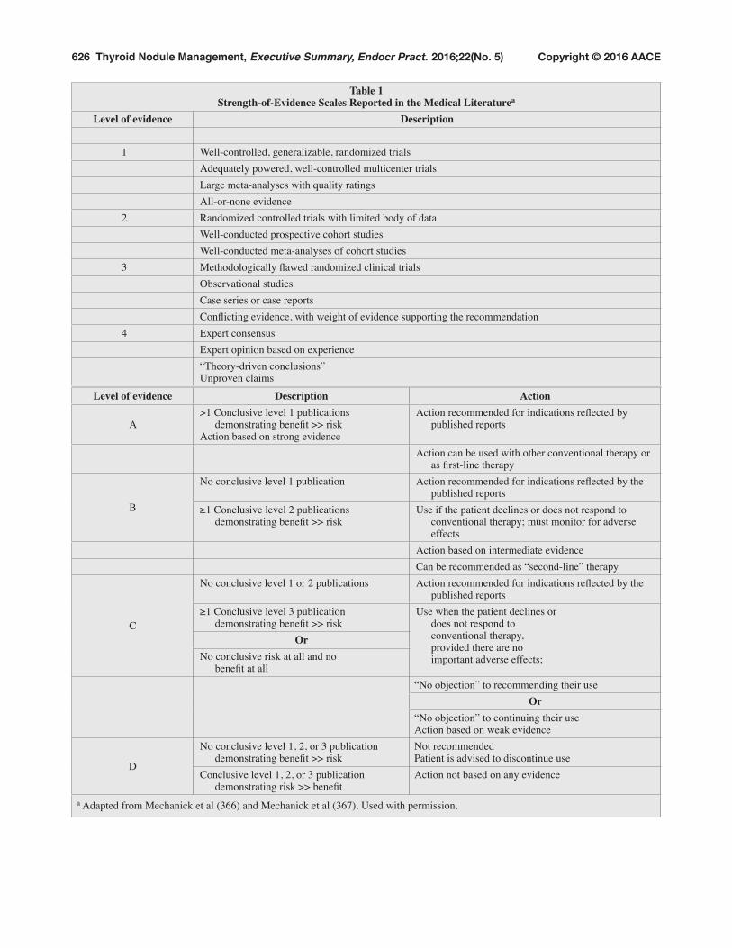
Levels of Evidence and Grading of Recommendations
The AACE protocol for standardized production of clinical practice guidelines was followed to rate the evi-dence level of each reference on a scale of 1 to 4 and to link the guidelines to the strength of recommendations on the basis of grade designations A (action based on strong evidence) through D (action not based on any evidence or not recommended) (Table 1) (4 [EL 4-guidelines]). The BEL, corresponding to the best conclusive evidence found, accompanies the recommendation grade (5 [EL 4]). All recommendations resulted from a consensus among the AACE and AME primary writers and were influenced by input from the Task Force members and reviewers. Some recommendations were upgraded or down-graded on the basis of expert opinion. In these cases, sub-jective factors such as clinical experience, cost, risk, and regional availability of specific technologies and expertise took priority over the reported BEL.
III. EXECUTIVE SUMMARY
A. SECTIONS
To guide readers, the 2016 Clinical Practice Guidelines for the Diagnosis and Management of Thyroid Nodules are divided into the following sections:
• The Scope of the Problem• Clinical Evaluation and Diagnosis• Thyroid Ultrasonography and Other Diagnostic
Imaging Studies • Thyroid Biopsy• Laboratory Evaluation• Radionuclide Scanning• Management and Therapy• Thyroid Nodules during Pregnancy• Management of Thyroid Nodules in Children
Readers are referred to the Appendix for more detail and supporting evidence for each section.
2 Randomized controlled trials with limited body of dataWell-conducted prospective cohort studiesWell-conducted meta-analyses of cohort studies
3 Methodologically flawed randomized clinical trialsObservational studiesCase series or case reportsConflicting evidence, with weight of evidence supporting the recommendation
4 Expert consensusExpert opinion based on experience“Theory-driven conclusions”Unproven claims
Level of evidence Description Action
A>1 Conclusive level 1 publications
demonstrating benefit >> riskAction based on strong evidence
Action recommended for indications reflected by published reports
Action can be used with other conventional therapy or as first-line therapy
B
No conclusive level 1 publication Action recommended for indications reflected by the published reports
1. Thyroid Nodules: The Scope of the Problem • These guidelines are designed to optimize the
current clinical practice in the diagnosis and management of thyroid nodules and nodular goiters.
• The recommendations should always be applied considering clinical setting, local medi-cal expertise, available technical resources, and patient preferences [BEL 3, GRADE B].
2. Clinical Evaluation and Diagnosis 2.1. History
• We recommend that the following data be recorded:AgePersonal or family history of thyroid disease or cancerPrevious head or neck irradiationRate of neck mass growthAnterior neck painDysphonia, dysphagia, or dyspneaSymptoms of hyper- or hypothyroidismUse of iodine-containing drugs or supplements [BEL 2, GRADE A]
• Most nodules are asymptomatic and benign, but the absence of symptoms does not rule out
• We recommend a careful, focused examination of the thyroid gland and cervical lymph nodes [BEL 3, GRADE B].
• Record the following data:Thyroid volume and consistency
Location, consistency, size, and number of nodule(s)
Neck tenderness or painCervical adenopathy [BEL 3, GRADE B]
• The risk of cancer is not substantially different in patients with a solitary nodule versus patients with a multinodular goiter (MNG) [BEL 2, GRADE B].
3. Thyroid Ultrasonography and Other Diagnostic Imaging Studies 3.1. When to Perform Thyroid US
• US evaluation is recommended for patients who are at risk for thyroid malignancy (see Table 3), have palpable thyroid nodules or goiter, or have neck lymphadenopathy suggestive of a malig-nant lesion [BEL 2, GRADE A].
• US evaluation is not recommended as a screen-ing test for the general population or patients with a normal thyroid on palpation and a low clinical risk of thyroid disease [BEL 4, GRADE C].
3.2. How to Describe US FindingsWe recommend the following approach to describe US findings:
• Focus the US report on stratification for risk of malignancy
• Describe position, size, shape, margins, con-tent, echogenic pattern, and vascular features of the nodule(s)
• For multiple nodules, detail the nodule(s) bearing the US characteristics associated with malignancy rather than describing the largest (dominant) nodule.
Table 2Comparison of the Italian AME Consensus, BSRTC, and UK-RCPath Systems for
Classification and Reporting of Thyroid Cytologic Results
Italian Consensus, 2014 BSRTC UK-RCPath
TIR 1. Nondiagnostic I. Nondiagnostic Thy 1. NondiagnosticTIR 1c. Nondiagnostic cystic I. Cystic fluid only Thy 1c. Unsatisfactory, consistent with cystTIR 2. Nonmalignant II. Benign Thy 2/Thy 2c. NonneoplasticTIR 3A. Low-risk indeterminate lesion III. AUS/FLUS atypia or follicular lesion
of undetermined significanceThy 3a. Neoplasm possible: atypia/
nondiagnosticTIR 3B. High-risk indeterminate lesion IV. Follicular neoplasm or suspicious for
follicular neoplasmThy 3f. Neoplasm possible: suggestive of
follicular neoplasmTIR 4. Suspicious for malignancy V. Suspicious for malignancy Thy 4. Suspicious for malignancyTIR 5. Malignant VI. Malignant Thy 5. Malignant
Abbreviations: AME = Associazione Medici Endocrinologi; AUS/FLUS = follicular lesion/atypia of undetermined significance; BSRTC = Bethesda System for Reporting Thyroid Cytopathology; TIR = Thyroid Imaging Reporting; Thy = thyroid; UK-RCPath = UK Royal College of Pathologists.
• For suspicious regional neck lymph nodes, describe the cervical compartment, number, shape, size, margins, content, echogenic pat-tern, presence of hilum, and vascular features [BEL 2, GRADE A].
3.3. US Rating of the Risk of Malignancy− In thyroid US reports, add to the description
of US features a rating that stratifies thyroid lesions on the basis of their risk of malignancy to reliably communicate the expected risk of cancer (see Table 4) [BEL 3, GRADE B].
3.4. US Classification SystemsThe following US rating system of the risk of malig-nancy is suggested (see Table 5 and Fig. 1 and 2):
• Class 1. Low-risk thyroid lesion. − Mostly cystic (>50%) nodules with rever-
berating artifacts that are not associated with suspicious US signs
− Isoechoic spongiform nodules confluent or with regular halo
ing thyroid tissue) and isoechoic nodules with ovoid-to-round shape and smooth or ill-defined margins.
− Intranodular vascularization, elevated stiff-ness at elastography, macro- or continuous rim calcifications, or hyperechoic spots of uncertain significance may be present.
− The expected risk of malignancy is 5 to 15%.
• Class 3. High-risk thyroid lesion. Nodules with at least 1 of the following suspicious features: − Marked hypoechogenicity (cf. prethyroid
muscles)− Spiculated or microlobulated margins − Microcalcifications − Taller-than-wide shape
− Evidence of extrathyroidal growth or patho-logic adenopathy
− The expected risk of malignancy is 50 to 90% in accordance with the presence of 1 or more suspicious findings [BEL 4, GRADE C].
3.5. Indications for US-Guided Fine-Needle Aspiration 3.5.1. How to select nodule(s) for US-guided FNA
• In the selection of nodules for US-guided FNA, consider a balance between the risk of a poten-tially delayed diagnosis and that of superfluous diagnostic procedures or surgery (see Fig. 1) [BEL 4, GRADE C].
• In light of the low clinical risk, nodules with a major diameter <5 mm should be monitored with US rather than biopsied, irrespective of their sonographic appearance [BEL 3, GRADE B].
• In nodules with a major diameter 5-10 mm that are associated with suspicious US signs (high US risk thyroid lesions), consider either FNA sampling or watchful waiting on the basis of the clinical setting and patient preference [BEL 3, GRADE B]. Specifically, US-guided FNA is recommended for the following nodules:¡ Subcapsular or paratracheal lesions ¡ Suspicious lymph nodes or extrathyroid
spread ¡ Positive personal or family history of thyroid
cancer ¡ Coexistent suspicious clinical findings (e.g.,
dysphonia) [BEL 2, GRADE A]• FNA is recommended for the following: ¡ High US risk thyroid lesions ≥10 mm¡ Intermediate US risk thyroid lesions >20 mm¡ Low US risk thyroid lesions only when >20
mm and increasing in size or associated with a risk history and before thyroid surgery or minimally invasive ablation therapy [BEL 2, GRADE A]
•FNA is not recommended for nodules that are functional on scintigraphy (see difference in
Table 3. Features Suggesting Increased Risk of Malignant Potential• History of head and neck irradiation• Family history of medullary thyroid carcinoma, multiple endocrine neoplasia type 2, or papillary thyroid carcinoma• Age <14 or >70 years• Male sex• Growth of the nodule• Firm or hard nodule consistency• Cervical adenopathy• Fixed nodule• Persistent dysphonia, dysphagia, or dyspnea
recommendations for children; Section 8.4.) [BEL 2, GRADE B].
3.5.2. FNA of multinodular glands and lymph nodes• We do not recommend the biopsy of more than
2 nodules in the same patient when the nod-ules are selected on the basis of the previously described criteria [BEL 3, GRADE C].
• If a radioisotope scan is available, we recom-mend not biopsying hot areas [BEL 2, GRADE B].
• In the presence of suspicious cervical lymph-adenopathy, we recommend FNA for cytologic assessment of both the lymph node and the sus-picious nodule(s) [BEL 2, GRADE A].
• We recommend the determination of thyro-globulin (Tg) or calcitonin, according to clini-cal indications, on FNA washout of suspicious lymph nodes [BEL 2, GRADE A].
3.5.3. FNA of complex thyroid nodule(s)• We recommend sampling the solid component
of the lesion through FNA biopsy [BEL 3, GRADE B].
• Preferentially sample the vascularized areas of the complex lesion [BEL 4, GRADE C].
• Submit both the FNA specimen and the drained fluid for cytologic examination [BEL 2, GRADE A].
3.5.4. FNA of thyroid incidentalomas• Manage thyroid incidentalomas according to
the previously described criteria for nodule diagnosis [BEL 2, GRADE A].
• Perform US evaluation of incidentalomas detected by computed tomography (CT) or magnetic resonance imaging (MRI) before consideration of FNA biopsy [BEL 2, GRADE A].
• We recommend that thyroid incidentalomas detected by positron emission tomography (PET) with 18F-fluorodeoxyglucose (18FDG) (focal uptake, in particular) should undergo US evaluation and FNA because of the high risk of malignancy [BEL 2, GRADE A].
3.5.6. Other Diagnostic Imaging Techniques• MRI and CT are not recommended for routine
thyroid nodule evaluation [BEL 2, GRADE A]. • Consider MRI and CT for assessment of size,
airway compression, substernal extension of a nodular goiter, or presence of pathologic lymph nodes in cervical regions not visualized by US [BEL 3, GRADE B].
• PET/CT may provide additional information about the risk of malignancy in thyroid nodules with indeterminate cytologic results. Because
of the insufficient diagnostic accuracy, high cost, and limited accessibility, we do not sug-gest its routine use as a diagnostic tool [BEL 3, GRADE B].
• Consider PET/CT only for the preoperative staging of malignant nodules with aggressive features [BEL 3, GRADE B].
3.5.7. Other US Techniques• Elastography provides information about nod-
ule stiffness that is complementary to grayscale findings [BEL 2, GRADE B].
• Elastography should not be used as a substitute for grayscale US examination, but as a comple-mentary tool in nodules with indeterminate US or cytologic findings [BEL 2, GRADE A].
• Perform FNA in nodules with increased stiff-ness [BEL 2, GRADE B].
• Use of US contrast medium is not recom-mended for the diagnostic evaluation of thyroid nodules [BEL 3, GRADE C].
• Use of US contrast medium is recommended only for the assessment of the area of tissue ablation induced by minimally invasive tech-niques [BEL 3, GRADE B].
4. Thyroid Biopsy 4.1. Thyroid FNA
• Combine clinical and US evaluation and, when appropriate, FNA results in the clinical man-agement of thyroid nodules [BEL 2, GRADE A].
• Always perform thyroid FNA because cyto-logic diagnoses are more reliable and the nondiagnostic rates are lower than with palpation-guided FNA (see Table 6) [BEL 2, GRADE A].
4.1.1. Requisition Form • Include all relevant clinical and US information
[BEL 4, GRADE C].• Include a rating of the US risk of malignancy,
based on an acknowledged US classification system [BEL 3, GRADE B].
• Use the following 3 US rating categories: high risk, intermediate risk, and low risk [BEL 4, GRADE C].
• Clearly state the sampling site and technique and the number of submitted slides and/or other sampled specimens [BEL 3, GRADE B].
4.1.2. Cytologic Report• Include a brief description of cytologic find-
ings and, when possible, a conclusive cytologic diagnosis [BEL 2, GRADE A].
• Identify the cytologic result by adding a rat-ing of the risk of malignancy based on an
established classification system for thyroid cytology [BEL 2, GRADE A].
4.2. Cytologic Diagnosis• Define FNA results as either diagnostic (sat-
isfactory) or nondiagnostic (unsatisfactory) [BEL 3, GRADE B].
• As a general rule, define the cytologic speci-men as diagnostic when the sample contains a minimum of 6 groups of well-preserved thyroid epithelial cells consisting of at least 10 cells per group [BEL 3, GRADE B].
• Classify cytologic specimens characterized by marked atypia as suspicious, even in the absence of the required number of follicular cells for adequacy [BEL 3, GRADE B].
• Five diagnostic classes with subdivision of indeterminate samples into 2 subclasses are recommended for cytologic reports (see Table 2) [BEL 2, GRADE A].
• Use the following reporting system for thyroid cytologic characteristics:
Thyroid 1. Nondiagnostic.• Inadequate or insufficient to make a
diagnosis• Cystic: insufficient but consistent with a
4.3. FNA Pitfalls• To decrease the risk of misleading cytologic
results, consider the following:o False-negative results are usually due
to inappropriate target selection or inad-equate sampling.
o False-positive results are usually due to specimens with suspicious, but nondiag-nostic, findings [BEL 4, GRADE C].
• For indeterminate cytologic findings, consider a second opinion from an experienced cytopa-thologist because some of these cases may, in high-volume thyroid cytopathology units, be reassessed as definitely benign or malignant [BEL 3, GRADE C].
• To decrease the risk of false-negative results, we recommend the following:o Routine use of FNA
Table 4US Features of Benign or Malignant Thyroid Nodules
US features indicative of a benign noduleIsoechoic spongiform appearance (microcystic spaces comprising >50% of the nodule)Simple cyst with thin regular margins Mostly cystic (>50%) nodules containing colloid (hyperechoic spots with comet-tail sign)Regular “eggshell” calcification around the periphery of a nodule
US features indicative of a malignant nodulePapillary carcinoma
Solid hypoechoic (relative to prethyroid muscles) nodule, which may contain hyperechoic foci without posterior shadowing (i.e., microcalcifications)Solid hypoechoic nodule, with intranodular vascularity and absence of peripheral halo“Taller-than-wide” nodule (AP>TR diameter when imaged in the transverse plane)Hypoechoic nodule with spiculated or lobulated margin Hypoechoic mass with a broken calcified rim and tissue extension beyond the calcified margin
Follicular neoplasm (either follicular adenoma or carcinoma)Isoechoic or mildly hypoechoic homogeneous nodule with intranodular vascularization and well-defined halo
Indeterminate US featuresIsoechoic or hyperechoic nodule with hypoechoic haloMild hypoechoic (relative to surrounding parenchyma) nodule with smooth marginPeripheral vascularizationIntranodular macrocalcification
Abbreviations: AP = anteroposterior; TR = transverse; US = ultrasound.
reverberating artifacts and not associated with suspicious US signs
• Isoechoic spongiform nodules, either confluent or with regular halo.
BenignPurely cystic nodules (no solid component)
Very low suspicionSpongiform or partially cystic nodules without any of the US features described in low-, intermediate- or high-suspicion patterns
Low suspicionIsoechoic or hyperechoic solid nodule, or partially cystic nodule with eccentric solid area without:• Microcalcifications• Irregular margin • Extrathyroidal extension• Taller than wide shape
2 Intermediate-risk thyroid lesionSlightly hypoechoic (vs. thyroid tissue) or isoechoic nodules, with ovoid-to-round shape, smooth or ill-defined marginsMay be present:
• Intranodular vascularization • Elevated stiffness at
elastography, • Macro or continuous rim
calcifications• Indeterminate hyperechoic
spots
Intermediate suspicionHypoechoic solid nodule with smooth margins without:• Microcalcifications• Extrathyroidal extension• Or taller than wide shape
B. Solid, hypoechoic, lobulated/irregular outline, globular calcification (medullary carcinoma?)
C. Intranodular vascularityD. Shape tall>wide (AP>TR)E. Characteristic associated
lymphadenopathyAbbreviations: AACE/ACE/AME = American Association of Clinical Endocrinologists/American College of Endocrinology/Associazione Medici Endocrinologi; AP = anteroposterior; ATA = American Thyroid Association; BTA = British Thyroid Association; TR = transverse; US = ultrasonography.a Adapted from: 2015 ATA Management Guidelines for Adult Patients with Thyroid Nodules and Differentiated Thyroid Carcinoma. Thyroid. 2016;26:1-133; British Thyroid Association Guidelines for the Management of Thyroid Carcinoma. Clin Endocrinol. 2014;81 Suppl 1:1-122; 2016 AACE/ACE-AME Clinical Practice Guidelines for the Diagnosis and Management of Thyroid Nodules. Endocr Pract. 2016;22 Suppl 1:1-59.
o Aspiration of at least 2 sites within the nodule
o For multiple nodules, prioritize nodules to be sampled according to US findings.
o For cystic lesions, sample solid or vascu-larized areas with FNA and submit cyst fluid for cytologic examination.
o Review of the slides with an experienced cytopathologist
o Follow-up on benign nodule(s)o Repeat FNA in benign nodules with suspi-
cious clinical or US findings.o In large-sized thyroid lesions, sample
peripheral and, possibly, solid areas to avoid fluid or necrotic zones [BEL 3, GRADE B].
4.3.1. Thyroglobulin and Hormone Measurement on FNA Washout
• Measurement of Tg, calcitonin, or parathyroid hormone (PTH) levels on FNA washout of sus-picious thyroid lesions or lymph nodes is rec-ommended when clinically appropriate [BEL 2, GRADE A].
• Each center should determine its own refer-ence range for hormone measurement from FNA washout samples [BEL 3, GRADE B].
4.4. Core-Needle Biopsy• Consider the use of US-guided core-needle
biopsy (CNB) in solid thyroid nodules with persistently inadequate FNA cytologic find-ings [BEL 3, GRADE C].
• Because of the limited evidence and the lack of established reporting systems, we do not rec-ommend either in favor of or against the use of CNB in nodules with indeterminate cytologic results [BEL 4, GRADE D].
4.5. Immunocytochemistry• Immunocytochemistry is suggested for lesions
that are suspected to be of nonfollicular origin
(e.g., parathyroid gland, medullary thyroid carcinoma, lymphoma, metastases from other organs) [BEL 3, GRADE B].
4.6. Molecular Testing4.6.1. When molecular testing should be considered
• To complement rather than replace cytologic evaluation [BEL 2, GRADE A].
• The results are expected to influence clinical management [BEL 2, GRADE A].
• As a general rule, testing is not recommended in nodules with established benign or malig-nant cytologic characteristics [BEL 2, GRADE A].
4.6.2. Molecular testing for cytologically indetermi-nate nodules
• Cytopathology expertise, patient characteris-tics, and prevalence of malignancy within the population being tested impact the negative predictive values (NPVs) and positive predic-tive values (PPVs) for molecular testing [BEL 3, GRADE B].
• Consider the detection of BRAF and RET/PTC and, possibly, PAX8/PPARG and RAS mutations if such tests are available [BEL 2, GRADE B].
• Because of the insufficient evidence and the limited follow-up, we do not recommend either in favor of or against the use of gene expression classifiers (GECs) for cytologically indeterminate nodules [BEL 2, GRADE B].
4.6.3. Role of molecular testing for deciding the extent of surgery
• Currently, with the exception of mutations such as BRAFV600E that have a PPV approaching 100% for papillary thyroid carcinoma, evi-dence is insufficient to recommend in favor of or against the use of mutation testing as a guide to determine the extent of surgery [BEL 2, GRADE A].
Table 6Summary Characteristics for Thyroid FNAa
Feature Range % DefinitionSensitivity 88.2-97.0 Likelihood that patient with disease has positive test resultsSpecificity 47.0-98.2 Likelihood that patient without disease has negative test resultsPPV 52.0-98.0 Fraction of patients with positive test results who have diseaseNPV 89.0-96.3 Fraction of patients with negative test results who do not have diseaseFalse-negative rate 0.5-10.0 FNA negative; histologic findings positive for cancerFalse-positive rate 1.0-7.0 FNA positive; histologic findings negative for cancerAbbreviations: FNA = fine-needle aspiration; NPV = negative predictive value; PPV = positive predictive value. a Data from Bongiovanni et al, Acta Cytol. 2012;56:333-339; Piana et al, Cytopathology. 2010; Tee et al, Ann Surg. 246:714-720; Wang et al, Thyroid. 2011;21:243-251.
4.6.4. How should patients with nodules that are neg-ative at mutation testing be monitored?
• Since the false-negative rate for indeterminate nodules is 5 to 6%, and the experience and follow-up for mutation-negative nodules or nodules classified as benign by a GEC are still insufficient, close follow-up is recommended [BEL 3, GRADE B].
• If TSH level is decreased, measure FT4 plus total or FT3; if TSH level is increased, mea-sure FT4 and antithyroid peroxidase antibody (TPOAb) [BEL 2, GRADE A].
• Test for Tg antibody in patients with US or clin-ical findings suggestive of chronic lymphocytic thyroiditis, when serum levels of TPOAbs are normal [BEL 3, GRADE B].
• Assessment of serum Tg is not recommended in the diagnosis of thyroid nodules [BEL 2, GRADE A].
• In patients undergoing surgery for cancer, a preoperative serum Tg measurement may be considered [BEL 4, GRADE D].
• Perform only TSH receptor antibody (TRAb) measurement for patients with TSH levels below the reference range when Graves disease is suspected [BEL 3, GRADE B].
5.2. Calcitonin• We do not recommend either in favor of or
against the routine determination of serum cal-citonin level in the evaluation of thyroid nod-ules [BEL 3, GRADE D].
• Determine serum calcitonin in thyroid nodules with suspicious US findings or indeterminate cytologic findings [BEL 3, GRADE B].
• Obtain serum calcitonin level for patients with a family history or clinical suspicion of med-ullary thyroid carcinoma (MTC) or multiple endocrine neoplasia type 2 (MEN2) [BEL 2, GRADE A].
o If basal calcitonin level is increased, repeat the test in the absence of possible modifiers [BEL 2, GRADE A].
o If an elevated (<100 pg/mL) calcitonin level is confirmed, perform a calcium stim-ulation test to increase the diagnostic accu-racy [BEL 3, GRADE C].
• Due to a lack of availability, pentagastrin stim-ulation is no longer recommended [BEL 3, GRADE C].
5.3. Other Tests• Measure serum calcium and PTH when a nodu-
lar lesion is suggestive of parathyroid adenoma [BEL 3, GRADE B].
6. Radionuclide Scanning 6.1. When to Perform Thyroid Scintigraphy
• In a thyroid nodule or MNG, when the TSH level is below the lower limit of the reference range or when ectopic thyroid tissue or a retrosternal goiter is suspected [BEL 2, GRADE A]
• In iodine-deficient regions, to exclude auton-omy of a thyroid nodule or MNG even when the TSH level is low-normal (e.g., 0.5-1.0 mIU/L). [BEL 3, GRADE B]
• Independent of TSH level and whether in iodine-deficient or -sufficient regions, we rec-ommend scintigraphy to evaluate radioiodine therapy eligibility [BEL 2, GRADE B].
6.2. How to Perform Thyroid Scintigraphy• Use of 123I, 99mTcO4
– (sodium pertechnetate), or technetium sestamibi can be considered for thyroid scintigraphy [BEL 3, GRADE C].
• Sodium iodide 131I thyroid uptake is not rec-ommended for routine diagnostic use unless low-uptake thyrotoxicosis is suspected [BEL 3, GRADE B].
7. Management and Therapy (See Fig. 1-3)7.1. Nodules Nondiagnostic by FNA
• If initial FNA is nondiagnostic and the nodule is solid on US, we recommend repeating the FNA with US guidance [BEL 2, GRADE A].
• When cytologic results by FNA are repeatedly inadequate in solid nodules, consider perform-ing a US-guided CNB [BEL 3, GRADE C].
• Consider surgery for persistently nondiagnostic solid nodules. Follow-up may be considered in a minority of solid nodules with clearly favor-able clinical and US features [BEL 3, GRADE C].
• Follow-up clinically and with US persistently nondiagnostic cystic or predominantly (>50%) cystic, nodules with no suspicious clinical or US features [BEL 3, GRADE C].
• Consider a repeat clinical and US examination and TSH measurement in approximately 12 months in accordance with the clinical setting [BEL 3, GRADE B].
• If nodules are unchanged at the first US con-trol, repeat the US follow-up after 24 months [BEL 3, GRADE C].
• In asymptomatic nodules with a repeated benign cytology and no suspicious clinical or US features routine follow-up may be avoided [BEL 3, GRADE D].
• In nodules with benign cytology but suspi-cious clinical or US features, a repeat FNA is recommended [BEL 3, GRADE B].
• In nodules with an increase greater than 50% in volume or that become symptomatic, we recommend repeat FNA [BEL 2, GRADE A].
7.2.2. Medical treatment for benign nodules• Levothyroxine (LT4) suppressive therapy is
not recommended [BEL 1, GRADE A].• In geographic areas with mild iodine defi-
ciency, iodine supplementation and/or TSH nonsuppressive LT4 treatment may be consid-ered for young patients with a small nodular goiter and high-normal TSH levels [BEL 2, GRADE B].
• LT4 replacement is recommended for young patients with subclinical hypothyroidism or that due to autoimmune thyroiditis [BEL 2, GRADE A].
• LT4 therapy is not recommended for prevent-ing recurrence after lobectomy when serum TSH stays in the normal range [BEL 2, GRADE A].
7.2.3. Surgical indications for benign nodules• Consider surgery when local pressure symp-
toms are present and clearly associated with the nodule(s) or in the case of appearance of
• The preferred extent of resection for benign uninodular goiter is lobectomy plus isthmec-tomy. For MNG, it is (near) total thyroidec-tomy [BEL 2, GRADE A].
7.2.4. Percutaneous ethanol injection for benign nodules• Percutaneous ethanol injection (PEI) is a safe
and effective outpatient therapy for thyroid cysts and complex nodules with a large fluid component [BEL 1, GRADE A].
• Sample carefully the solid component of complex lesions before doing PEI [BEL 3, GRADE B].
• PEI is recommended as the first-line treatment for relapsing benign cystic lesions [BEL 1, GRADE A].
• PEI is not recommended for solid nodules, whether hyperfunctioning or not, or for MNGs. This procedure may be considered for hot nodules having compressive symptoms only when other treatment modalities are not accessible [BEL 2, GRADE A].
7.2.5. Image-guided thermal ablation for benign nodules• Consider laser or radiofrequency ablation for
the treatment of solid or complex thyroid nod-ules that progressively enlarge or are symp-tomatic or cause cosmetic concern [BEL 2, GRADE C].
• Repeat FNA for cytologic confirmation before thermal ablation treatment [BEL 3, GRADE B].
Fig. 1. Indications for FNA biopsy according to US findings. Suspicious US findings are markedly hypoechoic nodule, intranodular microcalcifications, more-tall-than-wide shape, and spiculated or lobulated margins. FNA = fine-needle aspiration; US = ultrasonography.
Fig. 2. Thyroid ultrasound features and risk of malignancy.
A) Low-Risk Ultrasound Features• Thyroid Cyst• Mostly cystic nodule with reverberating artifacts• Isoechoic spongiform nodule
B) Intermediate-Risk Ultrasound Features• Isoechoic nodule with central vascularity• Isoechoic nodule with macrocalcifications• Isoechoic nodule with indeterminate hyperechoic spots• Isoechoic nodule with elevated stiffness on elastography
7.2.6. Radioiodine therapy7.2.6.1. When and how to perform radioiodine therapy
• Consider radioiodine therapy for hyperfunc-tioning and/or symptomatic goiter, especially for patients with previous thyroid surgery or at surgical risk and in those who decline surgery [BEL 2, GRADE A].
• Perform FNA before radioiodine therapy on coexistent cold nodules, per the recommen-dations given for nontoxic MNG [BEL 3, GRADE B].
• Avoid the use of iodine contrast agents or iodinated drugs before administration of radioiodine [BEL 2, GRADE A].
• If possible, withdraw antithyroid drugs 4 to 7 days before treatment and consider resump-tion 1 week after radioiodine therapy [BEL 2, GRADE B].
7.2.6.2. Contraindications• Radioiodine is contraindicated in pregnant and
breastfeeding females [BEL 2, GRADE A]. • In females of childbearing potential, perform a
pregnancy test before radioiodine administra-tion [BEL 2, GRADE A].
7.2.6.3. Follow-up after radioiodine therapy• Regular thyroid function monitoring is recom-
mended [BEL 2, GRADE A].• Consider repeat treatment after 3 to 6 months
in the case of persistent or recurrent hyperthy-roidism or inadequate size reduction [BEL 3, GRADE B].
7.3. Indeterminate Lesions 7.3.1. Management
• Base the management of indeterminate thy-roid nodules on their cytologic subclassifica-tion, clinical data, and US features [BEL 2, GRADE A].
• Consider elastography for additional informa-tion [BEL 2, GRADE B].
• Consider the available technical resources and patient preferences [BEL 4, GRADE D].
7.3.2. Subclasses of indeterminate cytologic findings• Distinguish, on the basis of morphologic
alterations and background component, 2 cytologic subclasses at expected different risk of malignancy, according to the British Thyroid Association System for Reporting Cytopathology classification or to comparable
Fig. 2. (Continued)
C) High-Risk Ultrasound Features• Marked hypoechogenicity• Microcalcifications• Irregular (speculated) margins• More tall than wide• Extracapsular growth• Suspicious regional lymph node
cytologic classification systems (see Table 2) [BEL 2, GRADE A].
7.3.2.1. Management of low-risk indeterminate lesions (AUS/FLUS, Thy 3a, or TIR 3A category nodules)• Consider conservative management in the
case of favorable clinical criteria, such as personal or family history, lesion size, and low-risk US and elastography features [BEL 3, GRADE C].
• Repeat FNA for further cytologic assess-ment and review samples with an experi-enced cytopathologist [BEL 3, GRADE B].
• CNB may be considered to provide micro-histologic information, but routine use is cur-rently not recommended because its role in indeterminate lesions is still unsettled [BEL 3, GRADE C].
• We do not recommend either in favor or against the determination of molecular markers for routine use in this category (see Section 7.6.3.3.) [BEL 3, GRADE D].
7.3.2.2. Management of high-risk indeterminate lesions (FN/SFN, Thy 3f, or TIR 3B category nodules)• Surgery is recommended for most thyroid
lesions in this category [BEL 2, GRADE A]. • Thyroid lobectomy plus isthmectomy is
recommended. Total thyroidectomy may be
performed, depending on clinical setting, coexistence of contralateral lobe thyroid nodules, and patient preference [BEL 2, GRADE A].
• Frozen sections are usually not useful [BEL 4, GRADE D].
• Consider close clinical follow-up in a minor-ity of cases with favorable clinical and US features, but only after multidisciplinary consultation and discussion of treatment options with the patient [BEL 4, GRADE C].
7.4. Management of FNA-Suspicious Nodules • Surgical treatment is recommended [BEL 1,
GRADE A].• Repeat FNA in cases with inadequate cellular-
ity or in those that need additional techniques for a better characterization [BEL 3, GRADE B].
• Intraoperative frozen section may be consid-ered [BEL 3, GRADE B].
7.5. Nodules Malignant at FNA 7.5.1. Management
• In the case of differentiated thyroid carcinoma, surgical treatment is recommended [BEL 1, GRADE A].
• For anaplastic thyroid carcinoma, metastatic lesions, and thyroid lymphoma, further diag-nostic work-up is recommended before surgical intervention [BEL 2, GRADE A].
Fig. 3. Cytologic categories and suggested clinical actions. AUS/FLUS indicates follicular lesion/atypia of undetermined significance. FNA = fine-needle aspiration; TIR = Thyroid Imaging Reporting;Thy = thyroid; US = ultrasonography.
7.5.2. Preoperative evaluation• Review US and cytologic results with the
patient, discuss treatment options, and obtain consultation with a surgeon experienced in endocrine surgery [BEL 2, GRADE A].
• US examination of the neck, FNA biopsy of any concomitant suspicious nodule or lymph node, and vocal cord assessment with laryngos-copy are recommended before surgery [BEL 2, GRADE A].
• In the case of suspicious US features, confirm the metastatic nature of a lymph node with mea-surement of Tg or calcitonin in the washout of the needle used for FNA [BEL 2, GRADE A].
• Consider the use of MRI, CT, and/or 18FDG PET/CT in selected cases with aggressive fea-tures for more accurate preoperative staging [BEL 3, GRADE B].
8. Thyroid Nodules During Pregnancy 8.1. Clinical Approach
• Manage thyroid nodules for pregnant subjects in the same way as for nonpregnant subjects [BEL 2, GRADE A].
• When suspicious clinical or US findings are present, we recommend FNA since cytologic diagnostic criteria are not substantially influ-enced by pregnancy [BEL 2, GRADE A].
• Use of radioactive agents for diagnostic, as well as therapeutic, purposes is contraindicated [BEL 2, GRADE A].
• In the case of subnormal TSH levels during the second half of pregnancy, postpone radionu-clide thyroid scan until after delivery and ces-sation of breastfeeding [BEL 2, GRADE A].
• During pregnancy, TSH-suppressive LT4 ther-apy for thyroid nodules or goiter is not recom-mended [BEL 3, GRADE B]. Iodine supple-mentation should be used in pregnant females living in iodine-deficient regions [BEL 2, GRADE A].
• For thyroid nodules that grow substantially or become symptomatic during pregnancy, fol-low-up with US examination is recommended, and if appropriate, FNA is also recommended [BEL 2, GRADE A].
• If FNA shows indeterminate cytologic findings, we recommend US monitoring and postponing surgery until after delivery [BEL 3, GRADE B].
8.2. Management of FNA-Malignant Nodules • When thyroid malignancy is diagnosed dur-
ing the first or second trimester, thyroidectomy should be performed during the second trimes-ter [BEL 3, GRADE B].
• For females with clinical or US evidence of extracapsular growth or lymph node metasta-ses, consider surgical treatment during the sec-ond trimester of pregnancy [BEL 3, GRADE B].
• Women without evidence of aggressive thyroid cancer may be reassured that surgical treatment performed soon after delivery is unlikely to adversely affect the prognosis. Close clinical and US monitoring is recommended [BEL 3, GRADE B].
• When thyroid malignancy is diagnosed dur-ing the third trimester, in absence of aggressive findings, surgical treatment can be deferred until the immediate postpartum period [BEL 3, GRADE C].
• For patients with suspicious or malignant thy-roid nodules in whom surgery is postponed until after delivery, we suggest maintenance of TSH at low-normal levels (e.g., 0.5-1.0 mIU/L) [BEL 3, GRADE B].
9. Management of Thyroid Nodules in Children • Evaluation and management of nodular dis-
ease in children are similar to adults [BEL 3, GRADE B].
• Because of a greater prevalence of malignancy in children with thyroid nodules, consider sur-gical treatment of “cold” as well as hot nodules [BEL 3, GRADE C].
ACKNOWLEDGMENT
AACE/ACE/AME Task Force on Thyroid Nodule Committee Members include the listed authors and Sofia Tseleni Balafouta, MD; Zubair Baloch, MD; Anna Crescenzi, MD; Henning Dralle, MD; Andrea Frasoldati, MD; Roland Gärtner, MD; Rinaldo Guglielmi, MD; Jeffrey I. Mechanick, MD, FACP, FACN, FACE; Christoph Reiners, MD; Istvan Szabolcs, MD, PhD, DSc; Martha A. Zeiger, MD, FACS; and Michele Zini, MD.
DISCLOSUREAACE/ACE/AME Task Force on Thyroid Nodules
Dr. Zubair Baloch reports that he has received consul-tant honorarium from Veracyte, Inc. Dr. Anna Crescenzi reports that she does not have any relevant financial relationships with any commercial interests. Dr. Henning Dralle reports that he does not have any relevant financial relationships with any commercial interests. Dr. Andrea Frasoldati reports that he does not have any relevant financial relationships with any commercial interests.
Dr. Roland Gärtner reports that he does not have any relevant financial relationships with any commercial interests. Dr. Rinaldo Guglielmi reports that he does not have any relevant financial relationships with any commercial interests. Dr. Jeffrey I. Mechanick reports that he does not have any relevant financial relationships with any commercial interests. Dr. Christoph Reiners reports that he does not have any relevant financial relationships with any commercial interests. Dr. Istvan Szabolcs reports that he has received speaker honorarium from Berlin-Chemie AG, Genzyme Corporation, and Merck AG. Dr. Martha A. Zeiger reports that she does not have any relevant financial relationships with any commercial interests. Dr. Michele Zini reports that she does not have any relevant financial relationships with any commercial interests.
Primary Authors
Dr. Hossein Gharib reports that he does not have any relevant financial relationships with any commercial interests. Dr. Enrico Papini reports that he does not have any relevant financial relationships with any commercial interests. Dr. Ralf Paschke reports that he has received speaker honoraria from Merck & Co, Inc, and Sanofi-Aventis U.S., LLC. Dr. Daniel S. Duick reports that he has received speaker honorarium from Genzyme Corporation. Dr. Roberto Valcavi reports that he does not have any relevant financial relationships with any commercial interests.
Dr. Laszlo Hegedüs reports that he has received con-sultant honoraria and research grant support from Novo Nordisk A/S and Genzyme Corporation and consultant and speaker honoraria from Theraclion. Dr. Paolo Vitti reports that he does not have any rele-vant financial relationships with any commercial interests. Dr. Jeffrey R. Garber reports no disclosures. Dr. Mack Harrell reports no disclosures.
REFERENCES 1. Haynes RB, Wilczynski N, McKibbon KA, Walker CJ,
Sinclair JC. Developing optimal search strategies for detecting clinically sound studies in MEDLINE. J Am Med Inform Assoc. 1994;1:447-458. [EL 4]
2. Hunt DL, McKibbon KA. Locating and appraising system-atic reviews. Ann Intern Med. 1997;126:532-538. [EL 4]
3. Giovanella L, Ceriani L, Ghelfo A, Maffioli M, Keller F. Preoperative undetectable serum thyroglobulin in differenti-ated thyroid carcinoma: incidence, causes and management strategy. Clin Endocrinol (Oxf). 2007;67:547-551. [EL 3]
4. Mechanick JI, Bergman DA, Braithwaite SS, Palumbo PJ; American Association of Clinical Endocrinologists Ad Hoc Task Force for Standardized Production of Clinical Practice Guidelines. American Association of Clinical Endocrinologists protocol for standardized pro-duction of clinical practice guidelines. Endocr Pract. 2004; 10:353-361. Erratum in: Endocr Pract. 2008;14:802-803. Mechanick, Jeffrey I [added]; Bergman, Donald A [added]; Braithwaite, Susan Shapiro [added]; Palumbo, Pasquale J [added]. [EL 4-guidelines]
5. Mechanick JI, Kushner RF, Sugerman HJ, et al. American Association of Clinical Endocrinologists, The Obesity Society, and American Society for Metabolic & Bariatric Surgery Medical guidelines for clinical practice for the peri-operative nutritional, metabolic, and nonsurgical support of the bariatric surgery patient. Endocr Pract. 2008;14 Suppl 1:1-83. Erratum in: Endocr Pract. 2009;15:768. [EL 4]


![Thyroid Axis Activity in Depression...thyroxine (T4) and/or low triiodothyronine (T3) levels (although still within the normal range) [1]. While euthyroid, most patients exhibit a](https://static.documents.pub/doc/80x56/60cc9025188120124857663e/thyroid-axis-activity-in-depression-thyroxine-t4-andor-low-triiodothyronine.jpg)