AMERICAN SHOULDER AND ELBOW SURGEONS 2001 CLOSED MEETING 1 ARTHROSCOPIC ABRASION ARTHROPLASTY FOR GLENOHUMERAL ARTHRITIS: ANALYSIS OF OUTCOME AS A TEMPORIZING PROCEDURE Julie Y. Bishop, MD, Thomas J. Neviaser, MD, Robert J. Neviaser, MD, Department of Orthopaedic Surgery, The George Washing- ton University, Washington, DC The purpose of this study was to assess the outcome of arthro- scopic abrasion arthroplasty in delaying the need for total shoulder replacement in the younger patient or delaying or obviating the need for replacement in the older patient. 26 patients with 27 shoulders were evaluated at a mean of 56 months (range, 24 to 156 months) post-operatively. The indications for the procedure were glenohumeral arthritis in patients with at least 90 degrees of elevation, 20 degrees of external rotation, and no posterior gle- noid wear, who were otherwise candidates for replacement. The goal in those younger than 60 was to delay performing TSA and in patients over 60 years old to delay or obviate the need for TSA. The procedure consists of debridement of the capsule and scarred synovium and gentle shaving of the articular surfaces to create a smooth, congruous articulation. Adjunct capsular release was per- formed in one patient. Motion was started within 48 hours post- operatively. Pre-operatively all cases were stage 3 or 4 radiograph- ically and Outerbridge stage III or IV intraoperatively. In 8 cases congruous surfaces could not be created, all were converted to TSA, and all were rated as failures. For the remaining cases, elevation improved from 107 to 131 degrees, external rotation at the side from 18 to 31 degrees and in abduction from 50 to 60 degrees, and internal rotation from S1 to L!. ASES Shoulder Score Index improved 30.1 to 75.2. Based on pain relief and patient satisfaction, in Group 1 (60 yo) there were 7 excellent, 1 good, and 3 poor results. In Group 2 (60 yo) there were 6 excellent, 1 good, and 3 poor. Factors leading to failure included inability to create congruous surfaces, excessive posterior wear, and external rotation at the side of 20 degrees. If strict criteria are met, arthroscopic abrasion can be effective in delaying or obviating the need for total shoulder replacement. 2 TOTAL SHOULDER ARTHROPLASTY WITH AN UNCE- MENTED GLENOID COMPONENT: FIVE- TO THIRTEEN-YEAR FOLLOW-UP Scott David Martin, MD, Thomas S. Thornhill, MD, Department of Orthopedic Surgery, Brigham and Women’s Hospital, Boston, Mass There has been increasing concern over the high incidence of radiolucencies which have been noted in 30% to 95% of cemented glenoid components with most failures of TSA due to glenoid loosening. This study evaluates the results of TSA using an unce- mented glenoid component with screw fixation. Methods and materials: The clinical and radiographic results of a consecutive series of patients undergoing TSA with an uncemented glenoid component were retrospectively evaluated. There were 77 shoul- ders (69 patients). The mean age at TSR was 65.8 years (SD 9.7, range, 37-89). The primary diagnosis was OA in 44 shoul- ders, RA in 24, 5 AVN, and 4 other. In 41 shoulders the cuff was normal, in 24 shoulders the cuff was thin/frayed and/or minor tear, and in 12 shoulders a major tear was present. The average follow-up was 9.1 years (SD 2.7, range, 5 to 13 years). Results: Ability of the patients to carry out their usual work load improved from 10% to 79% post-op (P .001). Radiolucencies were noted around 45% of the glenoids and 40% of the humeral components. There were ten glenoid failures (13%) including two fractured glenoid trays; four aseptic loosening of the glenoid; and four glenoids with polyethylene wear through. Seven other shoulders had fractured one of the two anchoring screws but remain clinically asymptomatic. The Cox regression model identified two multivari- ate predictors of glenoid failures: patients not grafted had a higher risk of failure (hazard ratio 12.5, 95% CI –2.6-37, P .003). A major cuff tear was independently predictive of a faster rate of failure compared to a normal cuff (hazard ratio 18, 95% CI 3.7-40, P .008) or a minor tear/thin frayed cuff (hazard ratio 10, 95% CI 2.2-26, P .04). No significant difference was found between normal vs minor cuff tear (P .50). Age, gender, side, diagnosis, humeral component fixation, as well as motion were not predictive of failure (all P .03). Kaplan-Meier (95% confidence intervals) freedom from glenoid revision rates were 98% (95-100) at 5 years, 83% (71-94) at 8 years, and 60% (45-75) over 10 years. Discussion: The overall functional and radiographic results using an uncemented glenoid component were comparable to those of cemented total shoulder. The mechan- ical failure rate reported in this study underscores the concerns of current uncemented glenoid components and the need to utilize new technologies in uncemented glenoid design. 3 THE INFLUENCE OF GLENOHUMERAL COMPONENT MISMATCH IN TOTAL SHOULDER ARTHROPLASTY Gilles Walch, MD, Thomas B. Edwards, MD, Aziz Boulahia, MD, Pascal Boileau, MD, Daniel Mole, MD, Patrice Adeleine, PhD, Lyon, France Introduction: In shoulder arthroplasty, mismatch is defined as the difference in the diameter of curvature between the humeral head and glenoid components. Recommendations for mismatch have not been substantiated scientifically. The purpose of this study is to evaluate the effect of mismatch on glenoid radiolucencies. Methods: The study group was 319 total shoulder arthroplasties evaluated as part of a multicentric review. The only indication for arthroplasty evaluated was primary osteoarthritis. All patients un- derwent arthroplasty with a single type of prosthesis using a cemented, flat-backed, all polyethylene glenoid component. Three sizes of glenoid components and seven humeral head diameters were utilized. Radial mismatch was categorized as 4 mm, 4.5 to 5.5 mm, 6 to 7 mm, or 7 mm. Radiographs were evaluated of all shoulders at a mean 53.5 months follow-up (range, 24 to 110 months). Glenoid radiolucencies were scored (zero points for no radiolucency to 18 points for radiolucencies exceeding two milli- meters in six zones). Variance, linear contrasts polynomial, qua- dratic polynomial contrast statistical, and linear regression analy- ses were performed to evaluate the relationship between radial mismatch and glenoid radiolucencies. Results: A statistically signif- icant linear relationship existed between mismatch and glenoid radiolucency score (P .001). Significantly lower (better) radiolu- cency scores occurred with a radial mismatch of greater than 5.5 mm. Conclusions: Based on the results of this study, glenohumeral component radial mismatch appears to be optimized at greater than 5.5 mm with respect to glenoid radiolucencies. This report represents the first step in establishing a scientific basis for recom- mendations regarding glenohumeral component mismatch. 1
Transcript
AMERICAN SHOULDER AND ELBOW SURGEONS2001 CLOSED MEETING
1 ARTHROSCOPIC ABRASION ARTHROPLASTY FORGLENOHUMERAL ARTHRITIS: ANALYSIS OF OUTCOME AS ATEMPORIZING PROCEDUREJulie Y. Bishop, MD, Thomas J. Neviaser, MD, Robert J. Neviaser,MD, Department of Orthopaedic Surgery, The George Washing-ton University, Washington, DC
The purpose of this study was to assess the outcome of arthro-scopic abrasion arthroplasty in delaying the need for total shoulderreplacement in the younger patient or delaying or obviating theneed for replacement in the older patient. 26 patients with 27shoulders were evaluated at a mean of 56 months (range, 24 to156 months) post-operatively. The indications for the procedurewere glenohumeral arthritis in patients with at least 90 degrees ofelevation, 20 degrees of external rotation, and no posterior gle-noid wear, who were otherwise candidates for replacement. Thegoal in those younger than 60 was to delay performing TSA and inpatients over 60 years old to delay or obviate the need for TSA. Theprocedure consists of debridement of the capsule and scarredsynovium and gentle shaving of the articular surfaces to create asmooth, congruous articulation. Adjunct capsular release was per-formed in one patient. Motion was started within 48 hours post-operatively. Pre-operatively all cases were stage 3 or 4 radiograph-ically and Outerbridge stage III or IV intraoperatively. In 8 casescongruous surfaces could not be created, all were converted toTSA, and all were rated as failures. For the remaining cases,elevation improved from 107 to 131 degrees, external rotation atthe side from 18 to 31 degrees and in abduction from 50 to 60degrees, and internal rotation from S1 to L!. ASES Shoulder ScoreIndex improved 30.1 to 75.2. Based on pain relief and patientsatisfaction, in Group 1 (�60 yo) there were 7 excellent, 1 good,and 3 poor results. In Group 2 (�60 yo) there were 6 excellent, 1good, and 3 poor. Factors leading to failure included inability tocreate congruous surfaces, excessive posterior wear, and externalrotation at the side of �20 degrees. If strict criteria are met,arthroscopic abrasion can be effective in delaying or obviating theneed for total shoulder replacement.
2 TOTAL SHOULDER ARTHROPLASTY WITH AN UNCE-MENTED GLENOID COMPONENT: FIVE- TO THIRTEEN-YEARFOLLOW-UPScott David Martin, MD, Thomas S. Thornhill, MD, Department ofOrthopedic Surgery, Brigham and Women’s Hospital, Boston,Mass
There has been increasing concern over the high incidence ofradiolucencies which have been noted in 30% to 95% of cementedglenoid components with most failures of TSA due to glenoidloosening. This study evaluates the results of TSA using an unce-mented glenoid component with screw fixation. Methods andmaterials: The clinical and radiographic results of a consecutiveseries of patients undergoing TSA with an uncemented glenoidcomponent were retrospectively evaluated. There were 77 shoul-ders (69 patients). The mean age at TSR was 65.8 years (SD �9.7, range, 37-89). The primary diagnosis was OA in 44 shoul-ders, RA in 24, 5 AVN, and 4 other. In 41 shoulders the cuff wasnormal, in 24 shoulders the cuff was thin/frayed and/or minortear, and in 12 shoulders a major tear was present. The averagefollow-up was 9.1 years (SD � 2.7, range, 5 to 13 years). Results:Ability of the patients to carry out their usual work load improvedfrom 10% to 79% post-op (P � .001). Radiolucencies were notedaround 45% of the glenoids and 40% of the humeral components.
There were ten glenoid failures (13%) including two fracturedglenoid trays; four aseptic loosening of the glenoid; and fourglenoids with polyethylene wear through. Seven other shouldershad fractured one of the two anchoring screws but remain clinicallyasymptomatic. The Cox regression model identified two multivari-ate predictors of glenoid failures: patients not grafted had a higherrisk of failure (hazard ratio � 12.5, 95% CI –2.6-37, P � .003).A major cuff tear was independently predictive of a faster rate offailure compared to a normal cuff (hazard ratio � 18, 95% CI �3.7-40, P � .008) or a minor tear/thin frayed cuff (hazard ratio �10, 95% CI � 2.2-26, P � .04). No significant difference wasfound between normal vs minor cuff tear (P � .50). Age, gender,side, diagnosis, humeral component fixation, as well as motionwere not predictive of failure (all P � .03). Kaplan-Meier (95%confidence intervals) freedom from glenoid revision rates were98% (95-100) at 5 years, 83% (71-94) at 8 years, and 60%(45-75) over 10 years. Discussion: The overall functional andradiographic results using an uncemented glenoid componentwere comparable to those of cemented total shoulder. The mechan-ical failure rate reported in this study underscores the concerns ofcurrent uncemented glenoid components and the need to utilizenew technologies in uncemented glenoid design.
3 THE INFLUENCE OF GLENOHUMERAL COMPONENTMISMATCH IN TOTAL SHOULDER ARTHROPLASTYGilles Walch, MD, Thomas B. Edwards, MD, Aziz Boulahia, MD,Pascal Boileau, MD, Daniel Mole, MD, Patrice Adeleine, PhD,Lyon, France
Introduction: In shoulder arthroplasty, mismatch is defined asthe difference in the diameter of curvature between the humeralhead and glenoid components. Recommendations for mismatchhave not been substantiated scientifically. The purpose of this studyis to evaluate the effect of mismatch on glenoid radiolucencies.Methods: The study group was 319 total shoulder arthroplastiesevaluated as part of a multicentric review. The only indication forarthroplasty evaluated was primary osteoarthritis. All patients un-derwent arthroplasty with a single type of prosthesis using acemented, flat-backed, all polyethylene glenoid component. Threesizes of glenoid components and seven humeral head diameterswere utilized. Radial mismatch was categorized as �4 mm, 4.5 to5.5 mm, 6 to 7 mm, or �7 mm. Radiographs were evaluated of allshoulders at a mean 53.5 months follow-up (range, 24 to 110months). Glenoid radiolucencies were scored (zero points for noradiolucency to 18 points for radiolucencies exceeding two milli-meters in six zones). Variance, linear contrasts polynomial, qua-dratic polynomial contrast statistical, and linear regression analy-ses were performed to evaluate the relationship between radialmismatch and glenoid radiolucencies. Results: A statistically signif-icant linear relationship existed between mismatch and glenoidradiolucency score (P � .001). Significantly lower (better) radiolu-cency scores occurred with a radial mismatch of greater than 5.5mm. Conclusions: Based on the results of this study, glenohumeralcomponent radial mismatch appears to be optimized at greaterthan 5.5 mm with respect to glenoid radiolucencies. This reportrepresents the first step in establishing a scientific basis for recom-mendations regarding glenohumeral component mismatch.
1
4 GLENOHUMERAL ARTHROPLASTY FOR ARTHRITIS AF-TER INSTABILITY SURGERYGregory S. Bauer, MD, Michael Q. Freehill, MD, Cassom Masters,William N. Levine, MD, Greg Lervick, MD, Theodore A. Blaine,MD, Louis U. Bigliani, MD, New York Orthopaedic Hospital, NewYork, NY
Introduction: Arthroplasty after instability repairs is more diffi-cult due to soft-tissue contractures, posterior subluxation and gle-noid bone loss. We present our long-term follow-up series ofpatients treated with arthroplasty for arthritis after instability sur-gery. Materials and methods: Thirty patients treated between1984-1999 were evaluated. There were 22 male and 8 femalepatients with an average age of 44 years (27-65). Eighteenpatients had total shoulder replacement (TSR) and 12 had hemiar-throplasty (HHR). An average of 2.4 (1-6) previous procedures hadbeen performed with an interval of 16.8 years (1-30) between theindex instability repair and the arthroplasty. Patients were evalu-ated with the American Shoulder and Elbow Surgeons’ form andNeer criteria. The average follow-up was 8.0 years (2.0-17).Results: Overall 17/30 (57%) had satisfactory results with TSR in13/18 (72%) and HHR in 4/12 (33%). The average ASES scorewas 72.6 (30-100) while the average pain score was 2.6 (0-9).Active elevation improved 54 degrees while external rotationincreased 41 degrees and internal rotation improved 6 spinalsegments. Thirteen patients (43%) required reoperation. Seven of12 (58%) HHRs required revision to TSR for pain at an average of5.8 years (1.0-12). An additional 6 patients required reoperation:3 subscapularis repairs, 2 arthroscopic subacromial decompres-sions, and one open release. The 3 patients requiring subscapu-laris repair had 3.7 (2-6) previous procedures. Discussion: Arthro-plasty for arthritis after instability repairs has inferior results thanarthroplasty for OA with a higher revision rate. Due to previoussurgery, these patients are susceptible to subscapularis complica-tions. Patients with TSR have superior results than hemiarthroplasty.Patients treated with HHR for instability arthritis have a high likeli-hood of requiring revision to TSR.
5 MENISCAL ALLOGRAFT INTERPOSITION ARTHRO-PLASTY OF THE ARTHRITIC SHOULDER: EARLY RESULTS ANDA REVIEW OF THE TECHNIQUEKen Yamaguchi, MD, Craig Ball, MD, Leesa M. Galatz, MD,William Levine, MD, Department of Orthopaedic Surgery, Wash-ington University School of Medicine, St Louis, Mo, and Depart-ment of Orthopaedic Surgery, Columbia-Presbyterian MedicalCenter, New York, NY
Introduction: Soft tissue interposition with hemiarthroplasty hasrecently generated interest as a treatment alternative in the youngpatient with glenohumeral arthritis or in the older patient withrotator cuff disease. Despite early promising results, there havebeen concerns regarding the durability of a capsular graft orinability to compensate for glenoid wear. We employed an alter-native new technique in which the glenoid was resurfaced with alateral meniscal allograft. In comparison to the previously de-scribed interposition technique, we hypothesized that meniscalallograft would have several advantages including an establishedhistory for synovial based healing in the knee, improved structuralcharacteristics for durability, and a wedge shape to compensatefor preexisting glenoid wear. The purpose of this study was toreview the technique and report early outcome results. Methods:Seven consecutive patients who underwent meniscal allograft inter-positional arthroplasty were reviewed an average of 24 monthsfollowing the procedure. Follow-up ranged from 1 to 5 years.Posttraumatic or instability related arthritis was the most commonindication for the procedure (71.4%). The remaining patients hadcuff tear arthropathy. Pre and post-operative range of motionmeasurements were made in a standardized fashion and painassessed on a visual analogue scale. Additionally, both pre andpostoperative outcome assessment was obtained with the ASES
Shoulder Assessment Form. Standardized radiographs were re-viewed to assess joint space. Results: All patients reported signifi-cant pain and functional limitations pre-operatively. The averagepain score was 7.5-10. Over half the patients (57%) could not raisetheir arms above shoulder height. Post-operatively all patients weresatisfied, with all reporting minimal or no pain. 2 patients did notregain overhead motion. Average ASES score at latest review was69.2. One patient had a transient traction related brachial plex-opathy, which resolved over 4 months. There were no other signif-icant complications. All radiographs showed a restoration of theglenohumeral joint space. Discussion: Interposition arthroplastyusing meniscal allograft from the lateral tibial plateau appeared toprovide excellent pain relief and improvement in shoulder functionin patients with contraindications to placement of a glenoid pros-thetic component. Our early experience would suggest the proce-dure is equivalent to other interpositional arthroplasty techniquesand may have advantages in the long-term secondary to theimproved load-bearing characteristics, shape and durability of themeniscal tissue.
6 REVERSE BALL AND SOCKET SHOULDER ARTHRO-PLASTY FOR ROTATOR CUFF–DEFICIENT SHOULDERS WITHANTEROSUPERIOR INSTABILITY: PRELIMINARY RESULTSGregory P. Nicholson, MD, Midwest Orthopaedics, Rush Presby-terian–St Luke Medical Center, Chicago, Ill
Extensive rotator cuff (RC) deficiency and loss of the “contain-ment” of the coracoacromial (CA) arch can lead to symptomaticanterosuperior instability (ASI) of the shoulder. This functionallydisabling condition is very difficult to treat due to the combinationof RC loss and the ASI. We report the preliminary results of customreverse ball and socket (RBS) arthroplasty in a complex series ofpatients with profound shoulder dysfunction after multiple failedsurgeries, extensive RC loss, and anterosuperior instability of theshoulder (ASI). Methods: Eleven shoulders (6 male, 5 female),average age of 64 years (45-74), were prospectively evaluated ataverage follow-up of 1.8 years (1-3). The average pre-operativefunction scores were poor: American Shoulder and Elbow Sur-geons (ASES) � 29.6 (15-38); Simple Shoulder Test (SST) � 1.7(0-3); and Visual Analog Score (VAS) for pain � 6.4 (3-9). Activemotion was poor, with average elevation (AFE) � 35 degrees(10-60) and average ER (AER) � 6 degrees (–15-20) The ability toperform activities of daily living was severely impaired. Originaldiagnoses were: cuff tear arthropathy (CTA) in 4, fracture (FX) in 4,multiple failed RC repairs in 3. An average of 2 prior surgeries/patient (1-4) had been done. There had been 4 failed hemiarthro-plasty (2 FX, 2 CTA), and 3 failed bipolars (2 CTA, 1 FX). Therehad been 2 failed multiple RC repairs, and 2 failed latissimustransfers. In the fracture cases, greater tuberosity bone was absent.All had an intact deltoid and exhibited ASI with attempted activemotion. All underwent RBS arthroplasty thru a deltopectoral ap-proach. The seven failed implants were revised. Results: All aver-age post-operative scores and active motion measures significantlyimproved (P � .05): ASES � 74 [�44.4] (60-83); SST � 7 [�5.3](6-8); and VAS pain � 1 [–5.4] (0-2); AFE � 99 degrees [�64](70-130), and AER � 31 degrees [�25] (25-35). The symptomaticASI was eliminated in all. Radiographs have revealed no implantloosening. All patients were satisfied with the operation. All pa-tients achieved at least limited goals criteria. All patients regainedthe ability to use the hand away from the body to at least chestlevel. ASI is functionally disabling. Extensive RC deficiency, loss ofCA arch, and previous surgery can limit reconstructive options. TheRBS arthroplasty eliminates the superior instability and attempts torestore deltoid “length” and provide a fulcrum for deltoid muscleaction and thus facilitate shoulder elevation. Conclusions: This is apreliminary report on an arthroplasty concept used in complexshoulder problems. These shoulders had extensive RC deficiency,ASI, poor active motion, profound shoulder dysfunction, and in 7 of11, a failed prosthesis. The RBS arthroplasty shows promise to
provide stability, relieve pain, and enable patients to regain func-tion.
7 GLENOID COMPONENT FAILURE TREATED WITH ABULK ALLOGRAFT FOR REVISION TOTAL SHOULDER SUR-GERYMark A. Frankle, MD, Raymond A. Long, MD, Tampa, Fla
Introduction: Clinical glenoid component failures in total shoul-der arthroplasty are associated with a painful shoulder, decreasedmotion and functional impairment. The purpose of this report is topresent the results of loose glenoid component removal with inser-tion of a bulk allograft to restore the bony defect. Materials andmethods: Sixteen patients underwent the above named procedure.All patients were evaluated with pre- and postoperative ASES,SF-36, and VAS scores, along with physical assessments, andstandard radiographic evaluations. There were 9 men and 7women with an average age of 68 years of age (range, 36-89) atthe time of revision. After the glenoid component was removed, theglenoid defect was assessed, and required a minimal of 1 � 3 cmfor bulk grafting. Fifteen of the 16 defects were, in addition tobeing cavitary, uncontained defects. Thus, large bulk allograftswere necessary to stabilize the shoulder. All grafts were internallyfixed. Thirteen of the 16 shoulders were fixed with bio-absorbablescrew fixation. Results: Fourteen patients were available for follow-up, and 2 were deceased. The average was 33 months (range, 24to 72). Active motion increased an average of 30° of forwardflexion, 40° of external rotation and four levels of internal rotation.The ability to perform the 15 activities of daily living increased froman average of 4 preoperatively to 13 postoperatively. Radiographsrevealed concentric reduction in all shoulders. However, followingthe allograft, re-absorption occurred over time so that the initiallateralization achieved was gradually lost in 4 of 11 of the cases.One patient required one reoperation because a metallic screwwas used to fix the graft, and as the graft reabsorbed, articulationof the head at the prosthesis occurred in the screw head, necessi-tating screw removal. Sixty-nine percent of all patients reportedeither an excellent or good result, and 31% reported either asatisfactory or unsatisfactory result. Conclusion: Glenoid compo-nent removal with replacement with a bulk allograft does providestability, and leads to predictable pain relief and function. How-ever, long-term viability of these grafts is a concern and continuedradiographic assessment is necessary.
8 OUTCOMES ANALYSIS OF REVISION TOTAL SHOUL-DER REPLACEMENTStephen Fealy, MD, Joshua S. Dines, MD, Russell F. Warren, MD,Edward V. Craig, MD, David M. Dines, MD, Department of SportsMedicine & Shoulder Service, Hospital for Special Surgery, NewYork, NY
Introduction: Multiple etiologies contribute to the need for revi-sion of humeral hemiarthroplasty (HA) and primary total shoulderreplacement (TSR). Infection, glenoid arthrosis, trauma, asepticloosening, rotator cuff pathology, and soft tissue contractures aboutthe shoulder have been cited as reasons for failure of the indexprocedure. The hypothesis of this study is that outcome followingrevision shoulder arthroplasty (rTSR) can be can be predictedbased on the surgical indication for the revision procedure. Mate-rials and methods: 63 shoulders (61 pts; 37 F, 25 M; mean age,62 yo; 4 surgeons) that underwent either revision of HA to TSR orrevision involving a primary TSR were retrospectively evaluated ata minimum of 2 years post-op. All patients were evaluated with theASES Shoulder Assessment Form, UCLA, and L’Insalata question-naires. Outcome results were assessed for the entire group andseparate cohorts within the group. 7 separate cohorts of patientswere identified within the 63 shoulders. Results: Seven distinctcohorts of patients were identified within the population: (1) revi-sion or resection of the glenoid component (N � 18), (2) revision
from HA to TSR for both trauma and osteoarthrosis (N � 13), (3)arthroscopy, debridement and rotator cuff repair following TSR(N � 11), (4) revision of the humeral head component followingTSR (N � 9), (5) instability, including component malpositioning,following TSR (N � 6), (6) trauma requiring ORIF without compo-nent revision (N � 3), (7) infection requiring resection arthroplastyfollowing TSR (N � 3). 30 pts (47%) of the entire population werefound to have good to excellent results; whereas 33 pts (53%) hada fair to poor outcome based on questionnaire. The cohort requir-ing revision or resection of the glenoid component alone faired thebest of those defined. Not surprisingly, the worst outcomes could beexpected following infection and subsequent resection arthro-plasty. A surprising finding in this study was that both the cohortthat underwent modular humeral head revision and the cohort thatunderwent arthroscopy and rotator cuff repair consistently yieldedfair to poor results. Conclusion: Our findings indicate that there aredistinct cohorts of patients who may require revision shoulderarthroplasty. Outcome may be generally predicted based on theindication for revision. Patients who need an isolated glenoidrevision or resection consistently performed better than those whorequired a soft tissue procedure or exchange of the humeral head.This knowledge may help in surgical planning and decision makingfor the TSR patient with rotator cuff pathology or one in whom alarge humeral head is thought to be the cause of persistent painand disability.
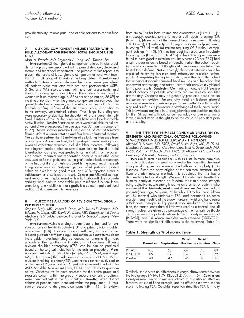
9 THE EFFECT OF HUMERAL CONDYLAR RESECTION ONSTRENGTH AND FUNCTIONAL OUTCOME FOLLOWINGSEMI-CONSTRAINED TOTAL ELBOW ARTHROPLASTYMichael D. McKee, MD, FRCS, David M.W. Pugh, MD, FRCS, M.Elizabeth Pederson, BSc, Caroline Jones, Emil H. Schemitsch, MD,FRCS, Robin R. Richards, MD, FRCS, St Michael’s Hospital andUniversity of Toronto, Toronto, Ontario, Canada
Purpose: In certain conditions, such as distal humeral nonunionor fracture, it is standard practice to excise the (nonunited) humeralcondyles during semi-constrained total elbow arthroplasty (TEA)insertion. Since the bony origins of the common extensors andflexor-pronator muscles are lost, it is postulated that this has adetrimental effect on strength. We sought to determine the effect ofhumeral condylar resection on forearm, wrist and hand strengthusing objective muscle strength testing on a series of patients whounderwent TEA. Methods, results, and discussion: We identified 32patients (mean age, 67 years; 22 females, 10 males; mean follow-up, 64 months) who underwent TEA, and performed objectivemuscle strength testing of the elbow, forearm, wrist and hand usinga Baltimore Therapeutic Equipment work simulator. To eliminatebias, the normal contralateral limb was used as a control, and allstrength values are given as a percentage of the normal side (Table1). There were 16 patients whose humeral condyles were intact(INTACT), and 16 whose condyles were resected (RESECTED).There were no significant differences in the following (Table 1).
Similarly, there were no differences in Mayo elbow score betweenthe two groups (INTACT 79, RESECTED 77, P � .67). Conclusion:Condylar resection has a minimal, clinically insignificant, effect onforearm, wrist and hand strength, and no effect on elbow outcomescore, following TEA. Condylar resection simplifies TEA for many
J Shoulder Elbow Surg ASES Abstracts 3Volume 12, Number 2
conditions, and our study supports this practice, as we could findno detrimental effects on distal muscle strength following condylarresection.
10 BIOMECHANICAL EVALUATION OF A NEW ELBOWULNAR COLLATERAL LIGAMENT RECONSTRUCTION USINGINTERFERENCE SCREW FIXATIONNeal S. ElAttrache, MD, Thay Q. Lee, PhD, Christopher S. Ahmad,MD, Kerlan-Jobe Orthopedic Clinic, Los Angeles, Calif
Introduction: Current techniques to reconstruct the ulnar collat-eral ligament (UCL) of the elbow are technically demanding. Thisstudy biomechanically evaluates a new technique of ulnar collat-eral ligament reconstruction designed to minimize muscle dissec-tion and risk of ulnar nerve injury while producing more reliableanatomic bone tunnels, ease of graft tensioning, and secure graftfixation. Methods: 10 matched cadaver elbow pairs were tested. Acontrol group was tested for failure strength and stiffness of theintact ligament. The reconstruction group was tested for kinematicdata, failure strength, and stiffness. Kinematic data was obtainedusing an testing apparatus that facilitates positioning of the elbowwith 5 degrees of freedom (constrained only to flexion angle) whileapplying a 3 Nm valgus load. Ultimate moment and stiffness weredetermined using a servohydraulic material testing machine toapply valgus load to failure with the elbow positioned at 70degrees of flexion. The UCL reconstruction was performed using atwo strand palmaris longus tendon graft. A single 5 mm tunnel wascreated at the isometric anatomic insertion sites for both the medialepicondyle and sublime tubercle. Graft fixation in the bone tunnelswas achieved with 5 � 15–mm soft-tissue interference screws.Results: Native ligaments failed by intrasubstance tears (8/10) andavulsions (2/10). Reconstructions failed by ulnar bone tunnel frac-ture (2/10), graft rupture (7/10), and graft and screw pullout(1/10). Native ligaments demonstrated significantly higher stiff-ness than reconstructed ligaments (P � .05). Ultimate moment fornative ligaments (34.0 Nm) was not significantly different thanreconstructed ligaments (30.6 Nm) (P � .05). Release of the UCLcaused valgus instability at all flexion angles with greatest instabil-ity at 60 degrees elbow flexion (average, 7.2 degrees). Recon-struction restored valgus stability to within one degree of the intactelbow for all flexion angles. Discussion and conclusion: Advan-tages of the anatomic interference fixation technique over previ-ously described techniques include less soft tissue dissection, lessrisk of ulnar nerve injury, and ease of graft insertion, tensioning,and fixation. The new technique described reconstructs the mostcentral fibers of the UCL to maximize graft isometry. Comprehen-sive biomechanical data presented confirms that the new techniquehas failure strength comparable to other reconstruction techniquesand reliably restores physiologic elbow kinematics over all flexionangles which is critical to the throwing athlete who stresses theelbow over a large range of motion.
11 ARTHROSCOPIC MANAGEMENT OF LARGE ANDMASSIVE ROTATOR CUFF TEARSChristopher K. Jones, MD, Felix Savoie, MD, Jackson, Miss
Introduction: Rotator cuff tears are a common cause of shoulderpain and dysfunction. Surgical outcomes for small to medium tears(�3 cm) have been shown to be good for open or arthroscopictechniques. However, the results for large and massive tears (�3-5cm) of the rotator cuff have been less successful. In recent years,with improved instrumentation and techniques, arthroscopic man-agement of these tears has improved and surgical outcomes areequal to or superior to open management. Purpose: The purpose ofthis study is to report our results with arthroscopic management oflarge and massive rotator cuff tears. Materials and methods: Sixtyconsecutive patients who had large or massive rotator cuff tendontears that were repaired arthroscopically were identified by retro-spective chart review. Inclusion criteria for the study population
included the presence of a large or massive rotator cuff tear thatwas repaired using an entirely arthroscopic technique, failure of aconservative treatment program and follow-up of at least 12months. Each patient also underwent arthroscopic subacromialdecompression and distal clavicle excision. Ten of these patientswere lost to follow-up before 12 months post-operatively. Theremaining 50 patients are the focus of this study. A modified UCLAshoulder rating scale was used to evaluate pre-operative andpost-operative shoulder pain, function, and range of motion,strength and patient satisfaction. Results: At average follow-up of32 months (range, 12-63), forty-four (88%) of the patients had agood or excellent outcome according to the modified UCLA shoul-der rating scale. The average post-operative UCLA score increasedan average of 17.1 points to 32.4. The pain scores improved 4.4points to 8.6. The function scores improved an average of fourpoints to 9.24, and forward flexion scores improved to 4.76 whilemeasured postoperative forward flexion increased an average of89 degrees to 170 degrees. Strength scores also improved anaverage of two points to 4.86. All of these improvements in scoreswere statistically significant (P � .000). Further, there was not adifference in the outcomes for patients with massive tears versuslarge tears. Six (12%) patients were considered failures by theshoulder rating scale, but 49 (98%) of the patients were satisfiedwith the result. Only one (2%) patient required reoperation. Thepatient who required a reoperation suffered a fall 2 months post-operatively in which he tore his rotator cuff. Following revisionarthroscopic repair, he had a good outcome with full functionalrecovery and pain only after heavy activities. Conclusion: Arthro-scopic management of large and massive tears of the rotator cuff isas successful as, and possibly more successful than, open manage-ment.
12 MOVING VALGUS STRESS TESTShawn W. O’Driscoll, PhD, MD, Richard L. Lawton, MD, PhD,Mayo Clinic, Rochester, Minn
Introduction: Diagnosing a painful partial tear of the medialcollateral ligament in overhead athletes is challenging, even forexperienced elbow surgeons. The purpose of this study was to testthe hypothesis a partial MCL can be diagnosed clinically by themoving valgus stress test. This test simulates the stresses, experi-enced by the elbow during throwing, that are responsible for MCLinjury and pain. Methods: A consecutive series of 15 patientsoperated on by a single surgeon to treat chronic MCL insufficiencyor to rule out this diagnosis and to treat other pathology of chronicvalgus overload were evaluated. The first two patients were re-viewed retrospectively, after which a prospective study was initi-ated on the second set of 13 patients. Inclusion criteria includedmedial elbow pain in an overhead athlete. Patients were dividedinto two groups: the test group had an MCL tear, which wasexplored surgically and confirmed (N � 6). The second group, thecontrol group (N � 9), had chronic valgus overload with posteriormedial impingement without a tear of the MCL. Results: The sensi-tivity of the moving valgus stress test was 90% and differencebetween the test and the control groups was highly significantstatistically (P � .001). With the elbow flexed fully, a moderatevalgus torque is applied to the elbow by the examiner. Whilemaintaining the valgus torque, the elbow is quickly extended. Apositive test is indicated by reproduction of the patient’s pain as theelbow passes between 120° and 90° of flexion, referred to as the“shear angle.” They were confirmed arthroscopically to have apositive arthroscopic valgus stress test, and stress radiographswere positive in each case. Patients in the control group all had anegative moving valgus stress test or a “false positive” test (ie, painwas only at the extension portion of the arch and not between 120°and 90°). They had negative arthroscopic valgus stress tests withless than 0.5 mm of opening and normal stress radiographs. Thesepatients were all confirmed to have a posteromedial lesion arthro-scopically and two patients also had dislocating ulnar nerves and
a snapping medial triceps. Discussion: The moving valgus stress testis a highly sensitive and specific physical examination maneuverthat, when performed and interpreted strictly, is virtually diagnosticfor a symptomatic tear of the MCL in an overhead athlete. Itreproduced that component of the throwing mechanics that isresponsible for both the tear in the first place and the pain associ-ated with the tear. As a constant valgus stress is applied to theelbow while the elbow moves from flexion to extension, internalshear stresses are generated between the fibers of the anteriorbundle of the MCL. Understanding the mechanism of how theseligament tears occur, and the physical diagnosis for them, willgreatly assist us in treatment decisions for our patients pursuingoverhead athletics.
13 BOTULINUM TOXIN (BOTOX) FOR THE PREVENTIONOF POST-TRAUMATIC ELBOW STIFFNESSCary B. Chapman, MD, Frank J. Raia, MD, H. Matthew Quitkin,MD, Themistocles S. Protopsaltis, MD, Melvin P. Rosenwasser, MD,The Trauma Training Center, New York Presbyterian Hospital–Columbia Campus, New York, NY
Purpose: Fracture or fracture/dislocation about the elbow jointoften leads to a stiff elbow with limited function. One of the majorcontributors to this stiffness is fibrosis and contracture of the elbowcapsule, which develops very quickly after surgery. It is our con-tention that voluntary and involuntary guarding and spasm of themuscles about the elbow, particularly the elbow flexors, aftersurgery limits the range of motion achievable during rehabilitation.The purpose of this pilot study was to evaluate the efficacy ofselective botulinum toxin (Botox) injections to the elbow flexormuscles as a means of preventing elbow stiffness. Botox creates areversible muscle weakness that lasts approximately 6 to 12 weeksby blocking presynaptic release of acetylcholine at the neuromus-cular junction. Since the early 1980s, Botox has been used safelyand effectively in the treatment of a wide range of disordersassociated with involuntary muscle spasm. Methods: Five womenand one man with an average age of 54 years (range, 27-91years) were enrolled in the study after suffering a traumatic fractureor fracture dislocation about the elbow. These included intra- andextra-articular distal humerus fractures and Monteggia fracture/dislocation. Patients underwent open reduction and internal fixa-tion of their fractures using standard techniques, following which50-100 units of Botox were injected into the brachialis and 100-200 units of Botox were injected into the biceps (divided betweenthe two heads). Patients began active-active assisted range ofmotion exercises between 7 and 10 days postoperatively with astatic progressive turnbuckle splint as an adjunct. In addition toROM measurements, Broberg-Morrey and DASH scores at 3 and12 months were used to gauge overall outcome. Results: Alloperatively treated fractures in this study healed. Biceps and bra-chialis function returned to normal by four months in all patients. Allpatients regained within 20 degrees of full elbow extension bythree months and had an arc of motion of at least 100 degrees. Atthree months, all patients were able to pronate at least 50 degrees;however, only 3 were able to achieve 50 degrees of supination.Broberg-Morrey scores at 3 months were excellent in 1 patient,good in 2 patients, and fair in 3 patients. At 1 year follow-up,Broberg-Morrey scores were excellent in 3 patients, good in 2patients, and fair in 1 patient. The average DASH score at 1 yearfollow-up was 14. Discussion and conclusion: The excellent elbowextension achieved by all patients in this study, regardless of age,supports the use of Botox as a post-operative adjunct to treatment offractures and dislocations about the elbow. Patients were able toregain elbow flexion to normal or near-normal levels in most casesand perform well on functional outcome analysis and in activities ofdaily living. Botulinum toxin injection prevented biceps and bra-chialis muscle spasm changing the paradigm of post-operativerecovery of motion, whereby extension is achieved earlier thanflexion. An ongoing randomized trial comparing elbow motion in
patients who received Botox following surgery with that in patientswho did not receive Botox following surgery will help to quantifythe effect.
14 CHRONICALLY RETRACTED RUPTURE OF THE DISTALBICEPS REPAIRED WITH SEMITENDINOSUS AUTOGRAFTRobert H. Bell, MD, William Wiley, MD, Thomas Dulaney, MD,Jeffrey S. Noble, MD, Akron, Ohio
Introduction: The chronic distal biceps tendon rupture presents aunique treatment dilemma. Treatment options include nonoperativebrachialis tenodesis, attempt at primary repair, reconstruction withautogenous flexor carpi radialis, palmaris longus and tensor fascialata with or without synthetic augmentation, semitendinosus andallograft. Indications for late reconstruction are continued pain,weakness, and fatigue for a patient that opted for nonoperativetreatment, delayed or misdiagnosis and failed primary repair. Thetechnique of using semitendinosus is a good salvage technique tomaster since even in the relatively acute setting the tendon can bescarred down and difficult to mobilize, retracted and foreshort-ened, or rarely a patient can have an unrecognized midsubstancetear where more tendon length is needed. Purpose: The purpose ofthis paper is to describe the surgical technique and present ourresults using a semitendinosus autograft in reconstructing the distalbiceps tendon in chronic ruptures. Methods: The study populationconsisted of 7 patients without surgery and 6 patients with au-tograft tendon reconstruction. The average time to surgery was 17weeks (4 to 42), the average time to follow-up for the nonoperativegroup was 30 months (5 to 109) and for the operative group was28 months (15 to 38). We utilized the two-incision technique in allcases. We evaluated functional outcome based on subjective out-come, physical exam and isokinetic testing of strength and endur-ance in flexion and supination comparing the injured to the non-injured extremity. Results: The autograft reconstruction allows therestoration of flexion and supination strength to within 5 percent ofnormal whereas the no surgery group lacked 20 percent of normal.Endurance in both groups was within the normal range. There wereno clinically detectable radial nerve injuries and no heterotopicossification. Conclusions: The use of autograft semitendinosus re-construction in chronic distal biceps tendon ruptures improvesflexion and supination strength when compared with conservativetreatment. It is a viable option that eliminates the need for syntheticaugmentation or allograft material.
15 RECALCITRANT LATERAL EPICONDYLITISWilliam H. Seitz, Jr, MD, Gary Sherman, MD, Louis Bley, MD,Cleveland, Ohio, Syracuse, NY, and Boston, Mass
Certain cases of lateral epicondylitis resist prolonged conserva-tive management and some even defy surgical intervention. Insome of these cases, histologic evaluation of the suspected site ofpathology fails to provide confirmatory pathologic evidence of thesuspected disease entity. In other cases where degenerative exten-sor tendinitis is confirmed, release or resection with or withoutrepair fails to completely alleviate symptomatology. This paperraises the question of the existence of an intraarticular source ofpain in patients who appear to have lateral epicondylitis but failstandard nonoperative and operative therapy. Twenty-three pa-tients with a diagnosis of lateral epicondylitis who failed conserva-tive management underwent localized extensor tendon release anddebridement, partial lateral epicondylectomy and fascial repairplus limited arthrotomy with resection of a thickened band ofsynovial tissue consistent with a radiocapitellar synovial “plica.”Histologic examination revealed focal inflammatory changes andslight hypertrophy within this synovial fold with erosive changes inthe radial head directly underlying this structure. Two additionalpatients had undergone a prior surgical procedure for lateralepicondylitis alone and failed to respond. A limited arthrotomy
J Shoulder Elbow Surg ASES Abstracts 5Volume 12, Number 2
alone was performed in those patients with confirmation of theintraarticular lesion which was then resected. Twenty-three of these25 patients have had complete relief of preoperative pain symp-toms. One has persistent diffuse pain and is seeking permanentpartial disability from workers’ compensation. Another workers’compensation patient has demonstrated complete relief of pain buthas a persistent sense of fatigability which has interfered with returnto work as a factory piece-worker. The remaining 23 patients havehad complete relief of symptoms and have returned to full dailyfunctions including work and athletics, with no recurrence at mini-mum two year follow-up. The presence of synovial bogginess, mildjoint effusion, a snapping sensation and tenderness at the radio-capitellar articulation with pronation and supination should raisethe suspicion of this clinical entity especially if local injection of thelateral epicondylar ridge alone with Xylocaine fails to completelyalleviate symptomatology.
16 THE VASCULAR ANATOMY OF THE GLENOHUMERALCAPSULE AND LIGAMENTS: AN ANATOMIC STUDYJohn L. Andary, MD, MBA, Steve A. Petersen, MD, Department ofOrthopaedic Surgery, Wayne State University
Introduction: While a detailed description of the vascular anat-omy of the shoulder capsule is lacking, surgical procedures (ie,capsular shifts or thermal modifications) may put this capsularblood supply at risk. The purpose of this study was to describe thevascular anatomy of the human glenohumeral capsule and liga-ments and its relevance to surgical treatment of the shoulder.Methods: In 24 fresh adult cadaveric shoulders the axillary arteryproximal to the thoracoacromial branch and the suprascapularartery were injected with India ink and treated with a modifiedSpalteholz technique. Results: The glenohumeral capsule demon-strated consistent arterial contributions from the anterior circumflex,posterior circumflex, circumflex scapular and suprascapular arter-ies. The arterial supply is centripetal in nature as the supplyingarteries enter the capsule superficially and penetrate to deeperlayers. The contributing vessels enter the capsule either laterally ormedially and arborize toward the middle of the capsule. Therotator cuff provides additional blood supply to the capsule throughperforating vessels. The dominant capsular vessels run horizontaland form intracapsular anastomosis via vertical branches. Theanterior and posterior bands of the IGHLC are vascularized byadjacent parallel vessels. In five of twelve capsules, a hypovascularzone was located near the humeral insertion of the anterior capsule2.5 to 3 centimeters medial to the intertubercular groove and justinferior to the rolled tendon of the subscapularis. In specimensdemonstrating a hypovascular zone near the supraspinatus inser-tion, there was an associated hypovascular zone of the underlyingcapsule. Discussion and conclusion: The glenohumeral capsule is awell-vascularized structure with predictable contributions but with avariable watershed area in the anterior capsule. Surgical ap-proaches to the shoulder that separate the rotator cuff from thecapsule may endanger the perforating blood supply from theoverlying rotator cuff. Approaches that open the rotator cuff andcapsule together as one unit and approaches that split the rotatorcuff muscle in line with the fibers are more likely to preserve thisperforating blood supply to the capsule. Horizontal capsular inci-sions are less likely to create vascular insult than vertical incisions.Surgical procedures including capsular incisions and thermalshrinkage that involve the hypovascular zone of the anterior cap-sule could experience a delay in healing. Capsular shrinkagetechniques that minimize capsular insult by spacing thermal expo-sure (ie, grid pattern techniques) and limit the depth of penetrationare more likely to leave capsular blood supply intact and allow forquicker healing of the capsule.
17 LIGAMENTOUS ANATOMY OF THE DISTAL CLAVICLEKevin J. Renfree, MD, Michael K. Riley, MD, Donna Wheeler, MD,Joseph G. Hentz, MD, Thomas W. Wright, MD, University ofFlorida, Gainesville, Fla
The acromioclavicular (AC) joint is a common site for pain,osteolysis, arthritis and instability. The purpose of this study is todescribe the insertional variations of the supporting ligaments of theacromioclavicular joint, especially with respect to patient gender.Forty-one clavicles with attached ligamentous structures (22 female,19 male) from 23 cadavers, were analyzed (19 matched pairs, 3unmatched). The average age was 79 years (range, 64-101). Thespecimens were carefully dissected under 3.5 loupe magnificationand the footprint of the coracoclavicular ligaments was measuredas a distance from the end of the clavicle using a caliper. Thephysician was blinded as to the sex of the specimen at the time ofthe dissection. The soft tissues were then removed with bleach andthe osseous footprints of the coracoclavicular ligaments weremapped and measured as to distance from the distal clavicle. Thesuperior AC ligament underwent a histological evaluation in threeadditional fresh specimens, 2 female and 1 male. Statistical anal-ysis of the measurements was performed using the two-sample ttest. The distance of insertion of the trapezoid ligament from thedistal end of the clavicle was not significantly different betweensexes, although that of the conoid ligament was significantlygreater in male specimens, as was the average anterior-posteriorwidth of the distal clavicle. The superior AC ligament blends withthe superior periosteum of the clavicle and acromion. Althoughthere exist significant gender-related differences in the insertionaldistances of the coracoclavicular ligaments, resection of less than11 mm should not violate the trapezoid ligament, and resection ofless than 24 mm the conoid ligament in either sex in 95% of thegeneral population. Resection of more than 7.5 mm of distalclavicle in a male and 5.2 mm in female, performed by anarthroscopic approach, may violate the superior AC ligament.
18 BIOMECHANICAL EVALUATION OF THE RELATIONBETWEEN REPAIR SITE SURFACE AREA AND STRENGTH OFTHE BONE-TENDON INTERFACERussell F. Warren, MD, Stephen Fealy, MD, Scott A. Rodeo, MD,John D. MacGillivray, MD, Hospital for Special Surgery, NewYork, NY
The purpose of this study is to test the hypothesis that thebiomechanical strength of a healed bone-tendon interface is re-lated to the cross-sectional area of tendon in contact with bone atrepair. 20 goats underwent rotator cuff repairs of the infraspinatustendon. The tendon was repaired using two anchors (Area A; N �20) on one shoulder and four anchors (Area B; N � 20) on thecontralateral shoulder. 10 goats were harvested at 4 wks (Group1) and 10 goats at 8 wks (Group 2) post-op. Ultimate failure wasreported for each shoulder. The mean load to failure in Group 1was 350.7 N, 619.4 N for Group 2 (P � .0002). In Group 1,Area A had mean load to failure of 317.3 N, specimens with AreaB had a mean load-to-failure of 375.5 N (P � .15). In Group 2,Area A had a mean load-to-failure of 635.8 N; Area B specimenshad a failure strength of 688.5 N (P � .45). Increasing the surfacearea of tendon in contact with bone at the repair site increasedload to failure of the repaired tendon at both 4 and 8 wks post-opby �10%. This was not a statistically significant increase in failurestrength in this model.
19 BIOMECHANICAL ANALYSIS OF PARTIAL ROTATORCUFF TEAR AND ITS SURGICAL REPAIRSteven D. Levin, MD, Gordon W. Nuber, MD, Jason L. Koh, MD,Mohsen Makhsous, PhD, Amanda F. Lin, PhD, Li-Qun Zhang, PhD,Chicago, Ill
Partial thickness rotator cuff tears (PTRCT) are seen more fre-quently than full thickness rotator cuff tears (FTRCT). The treatment
methods and results of FTRCT are well documented. Controversyexists, however, whether to treat partial thickness tears operativelyor non-operatively. The objective of this cadaveric study was toevaluate characteristic strain changes of the partially torn and thensurgically repaired supraspinatus tendon to improve our under-standing of the consequences of PTRCT and to help delineatepossible surgical indications. Four fresh-frozen shoulder specimenswere used to evaluate biomechanical changes of the supraspinatustendon caused by partial thickness tear and surgical repair. Thespecimens were obtained from male subjects (�60 years old) toobtain more truthful representation of intact supraspinatus tendons.The scapula was mounted rigidly onto a Teflon plate with theglenoid surface oriented vertically. The humerus was positioned at60° abduction (corresponding to 90° arm abduction) in the scap-ular plane. Ropes were sutured to individual muscles (anterior,middle, and posterior deltoid, supraspinatus, superior, middle, andinferior portions of the infraspinatus, superior, middle, and inferiorportions of the subscapularis, teres minor, and the long head ofbiceps) through fiberglass mesh. The muscles were loaded alongtheir line of action, proportional to their physiological cross-sec-tional areas. The supraspinatus was pulled along its line of actionthrough a rope and pulley system. The pulling force (ranging from20 to 80 N) and displacement were measured by a force sensorand a linear potentiometer, respectively. Strain at the anterior edgeof the supraspinatus tendon during the loading was measuredusing a differential variable reluctance transducer (MicroStrain,Burlington, Vt). Next, the supraspinatus tendon was cut partiallyfrom the bursal side near the insertion to simulate partial tear. Thetear was about 50% of tendon thickness and spanned the posterior3/4 of the tendon width. The same pulling evaluation describedabove was repeated to evaluate biomechanical changes causedby the partial tear. Afterwards, full-thickness tear was created andthe torn tendon was repaired using a Headed BioCorkscrew im-plant (Arthrex Inc). The same biomechanical test was then re-peated. Partial tear of the supraspinatus tendon increased the strainin the remaining intact portion of the tendon substantially. At 80 Nload, an intact supraspinatus tendon in a representative caseshowed a strain of about 0.6%. A partial tear of 50% of thethickness made the strain in the remaining tendon increase fivefoldto about 3%. Surgical reconstruction using Arthrex Headed Bio-Corkscrew over-corrected the excessive strain and made it lowerthan that in an intact tendon. This study gives biomechanicalevidence that PTRCT places considerably increased strain on theremaining intact portion of the tendon, and therefore may injure thetendon further over time. This serves to strengthen the argument torepair severe PTRCT surgically.
20 GLENOHUMERAL INSTABILITY IS ONLY PARTIALLYRESTORED AFTER REPAIR OF THE TYPE II SLAP LESIONAndreas Burkart, Richard E. Debski, Volker Musahl, Savio L.-Y.Woo, PhD, Patrick J. McMahon, Musculoskeletal Research Center,University of Pittsburgh, and Department of Orthopaedic SportsMedicine, Technical University of Munich. Investigation performedat the Musculoskeletal Research Center.
Superior labral anterior-to-posterior (SLAP) lesions can causedisabling shoulder pain partly from glenohumeral instability, yeteffects of repair remain uncertain. In 8 cadaver joints we used arobotic manipulator to measure joint translation in response toexternally applied loads of 50 newtons (N) anterior-directed, 50 Nsuperior-to-inferior (SI), 50 N anterior-directed combined with a 3Nm external rotation torque and lastly 50 N posterior-directedcombined with a 3 Nm internal rotation torque. A 44 N jointcompression load was maintained and two positions of glenohu-meral abduction were tested, 30° and 60°. We then created a typeII SLAP lesion by detaching the superior labrum and the bicepsanchor from the glenoid bone and testing was repeated. Repair ofthe type II SLAP by placing a tack through the labrum both anteriorand posterior to the biceps anchor was then performed and testing
again repeated. ANOVA was used to compare translation of theintact joint, that after the type II SLAP lesion was simulated and afterrepair. At 30° of abduction, anterior translation of the intact jointfrom anterior-directed loading was 18.5 � 8.5 mm and it in-creased significantly to 26.2 � 6.5 mm after simulation of the typeII SLAP lesion. Repair did not restore anterior translation (23.9 �8.6 mm) to that of the intact joint. At 60° of abduction, anteriortranslation of 16.6 � 9.6 mm in the intact joint was not significantlyincreased after simulation of the type II SLAP lesion 19.4 � 10.1(P � .0527). Anterior-directed loading also resulted in inferiortranslation. At 30° of abduction it was 3.8 � 4.0 mm in the intactjoint and the inferior translation increased to 8.5 � 5.4 mm afterthe type II SLAP lesion. After repair the inferior translation de-creased significantly to 6.7 � 5.3 mm. Although inferior transla-tions were less at 60 of abduction, results were similar to those at30 after repair. There were no significant increases in translationafter SI, anterior-directed combined external rotation torque orposterior-directed combined internal rotation torque loading. Gle-nohumeral translations increased in the anteroinferior directionafter simulation of the type II lesion while translations in the poste-rior and superior directions were unchanged. Repair of the type IISLAP lesion with an arthroscopic tack technique only partly re-stored translation to that of the intact joint. Improved techniques forrepair of the superior capsulolabral structures and the bicepsanchor are needed to restore stability after a type II SLAP lesion.
21 THE TWIST-LOCK CONCEPT OF TISSUE TRANSPORTAND TISSUE FIXATION WITHOUT KNOTS: OBSERVATIONSALONG THE HONG KONG SKYLINEStephen S. Burkhart, MD, Kiriacos A. Athanasiou, PhD, PE, SanAntonio, Tex
The senior author has devised a suture anchor system based onthe “twist-lock” concept. This concept was inspired by the knotlesslashings used to construct the hundred story high bamboo scaffold-ing frames that supported construction workers as they built thegreat Hong Kong skyscrapers. The twist-lock system enhancesinternal interference of the suture limbs by three mechanicallyverifiable friction-multiplier mechanisms: the twisting effect, thecable friction effect, and the wedge effect. After theoreticallyverifying the strength characteristics of the twist-lock system, theauthors chose to test its strength experimentally and to compare itsstrength to that of a standard screw-type suture anchor system thatis in common clinical use. Materials and methods: Unicellularpolyurethane, which has been demonstrated to accurately mimicthe mechanical properties of cancellous bone, was used for implan-tation of suture anchors for the purpose of comparing the load tofailure of the twist-lock system to the Corkscrew anchor system (N �10 for each system). Axial single pull loading was performed withan Instron testing machine. Results: The average load to failure forthe twist-lock group was 137.2 N, and the average for the Cork-screw group was 123.0 N, a difference of 14.2 N. This studyshowed that the twist-lock anchors failed at a load that was 12%higher than that of the Corkscrew group (P � .02). Conclusion: Thetwist-lock system achieves fixation of tendon to bone without theneed to tie knots, and with breaking strengths that are significantlyhigher than those of conventional suture anchors.
22 REMODELING OF THE GLENOHUMERAL JOINT AFTERSURGICAL RELEASE OF INTERNAL ROTATION CONTRAC-TURES SECONDARY TO BRACHIAL PLEXUS BIRTH PALSYMichael L. Pearl, MD, Darissa S. Kon, MD, Bradford W. Edgerton,MD, Ani B. Darakjian, Anne E. Kosco, Paul A. Kazimiroff, RaoulJ. Burchette
Introduction: Internal rotation contractures secondary to bra-chial plexus birth palsy frequently lead to developmental deformi-ties of the glenoid. The effectiveness of surgical release in prevent-ing deformity, or allowing the glenoid to remodel existing
J Shoulder Elbow Surg ASES Abstracts 7Volume 12, Number 2
deformity, has never been evaluated. Methods: Twenty-four chil-dren, aged 3 to 13 years, previously treated with surgical releaseof an internal rotation contracture were evaluated with MRI at twoor more years follow up. All children had received pre-operativearthrograms. The surgery performed was a subscapularis releaseas either an isolated procedure or along with a latissimus dorsitransfer. Results: At the time of the initial surgical release, 15children (63%) demonstrated arthrographic deformities of the gle-noid described as flat glenoids (3), biconcave glenoids (5) andpseudo-glenoids (7). At follow up, all but 4 of these childrenshowed extensive glenoid remodeling with normalization of poste-rior subluxation and improved conformity of the articular surfaces.One pseudoglenoid remained a pseudoglenoid, one pseudogle-noid became flat, one biconcave glenoid became a pseudoglenoidand one flat glenoid remained flat. The 9 children with concentricglenoids maintained them after contracture release. There was atrend towards younger and more involved children showing themost improvement but this was not statistically significant (P � .12and P � .06, respectively). Discussion and conclusion: The extent towhich a deformed glenoid can remodel is unknown. This studydocuments glenoid remodeling in most children after release oftheir internal rotation contracture. Timely surgical release to avoidglenoid deformity may be necessary even within the first year oflife. Internal rotation contractures secondary to birth palsy fre-quently lead to glenohumeral joint deformity. This study documentsremodeling after contracture release.
23 CORACOID IMPINGEMENT SYNDROME: A COMPARI-SON OF PRIMARY AND SECONDARY IMPINGEMENTCarl J. Basamania, MD, John F. Kragh, MD, Duke UniversityMedical Center, Durham, NC
Objective: To present a prospective study of primary and sec-ondary coracoid impingement syndrome, and compare treatmentsand outcomes. Methods: For six years, we followed patients withcoracoid impingement. Fifteen in the primary group ultimatelyrequired coracoplasty. Inclusion criteria for primary impingementincluded characteristic history and physical in addition to confir-matory plain radiographs, CT or MRI, diagnostic injection, EUAand arthroscopy. Ten patients had secondary impingement. Eighthad instability and 2 had previous coracoid fractures. Results:Follow-up for both groups averaged 2.8 years (range, 1.5 to 5.7years). We included fifteen shoulders (fourteen patients) in theprimary surgery group. All were athletes. Typically, pain wasreported in the anterior shoulder at a tender coracoid and wors-ened with push-ups and cross body adduction. Conservative treat-ment often failed. Hawkins’, Neer’s, and the modified abrasionsign were often positive. Injections at the coracoid tip confirmed thediagnosis. MRI suggested a narrow coracohumeral interval, espe-cially in internal rotation and adduction; the interval averaged 7.6mm (6-10 range). Arthroscopy ruled out other intra-articular pathol-ogy. All fifteen shoulders required coracoplasty. Ages averaged30.9 years (20-41). The duration of symptoms was 21.2 monthsprior to surgery (8-60). Improvements in visual analog pain scales(VAS) (5.6 average improvement, 3-9, P � .0001), Single Assess-ment Numerical Evaluation (SANE) (52.8, 22-82, P � .0001), andpatient satisfaction (100% excellent) showed good results. Tenpatients had secondary impingement. Three were misdiagnosed asprimary and had coracoplasties. Ages averaged 29 years (20-41).The duration of symptoms was 26.9 months (5-156). Improvementsin VAS (2.9 average improvement, 1-6, P � .08), SANE (22.0,0-40, P � .01), and satisfaction (50% satisfactory, 50% unsatis-factory) showed mixed results. Conclusion: Coracoid impingementis rare. Previous studies included only four case series, but only oneisolated coracoplasty for primary coracoid impingement was re-ported. Characteristic pain is anterior with a tender coracoid. Painis common during Neer’s, Hawkins’, and the modified abrasionsigns. MRI suggests the diagnosis, and pain relief with injectionsconfirms the diagnosis. Care needs to be taken to distinguish
primary from secondary coracoid impingement. If conservativetreatment fails, a coracoplasty can safely alleviate pain in carefullyselected patients.
24 ARTHROSCOPIC LOCALIZATION OF THE AXILLARYNERVE DURING CAPSULAR RELEASE OF THE SHOULDERLarry D. Field, MD, Felix H. Savoie, MD, Mississippi Sports Med-icine & Orthopaedic Center, Jackson, Miss
Introduction: Arthroscopic capsular release for resistant frozenshoulder is an accepted method of treatment. However, significantrisks to the axillary nerve exist during release and excision of theinferior capsule. This study reviews the experience of the author inidentification and exploration of the axillary nerve at the time ofinferior capsular release. Methods: The author has visually identi-fied the axillary nerve at the time of arthroscopic capsular releasein 27 patients in an effort to more safely release and excise aportion of the inferior capsule. Visualization and localization of thenerve was felt to be beneficial in an effort to reduce the chance ofinadvertent injury to the nerve during capsular release. The nervewas always identified in the area of the anteroinferior capsulethrough a cleavage plane established either at the time of initiationof the capsular release following gentle manipulation or by usingthe arthroscopic instruments to create a defect in the anteroinferiorcapsule. Following identification of the nerve, it can be tracedposteriorly and visualized arthroscopically throughout the remain-der of the inferior capsule release and excision. Blunt probes andbasket-type forceps can be used to gently retract the nerve ifnecessary. No axillary nerve injury occurred in this group ofpatients, and no additional portals or procedures were required tovisualize and/or expose the axillary nerve. Discussion: This seriesof patients represents a group who are undergoing ever increasingand complicated arthroscopic procedures both in the shoulder andelbow. As in open surgery, visualization and careful retraction ofnerves at the time of procedures is a time honored and valuablesurgical principle. Arthroscopic capsular release places motorizedand handheld cutting devices in immediate proximity to the axillarynerve as the nerve passes just inferior to this contracted thickenedcapsule. A thorough knowledge of the axillary nerve orientationand course is imperative in visualizing and arthroscopically retract-ing the nerve, but this technique appears valuable for some casesof arthroscopic capsular release.
25 PROSPECTIVE RANDOMIZED CLINICAL TRIAL COM-PARING THE EFFECTIVENESS OF IMMEDIATE ARTHRO-SCOPIC STABILIZATION VERSUS IMMOBILIZATION ANDREHABILITATION IN FIRST TRAUMATIC ANTERIOR DISLOCA-TIONS OF THE SHOULDER: EVALUATION OF LONG-TERMQUALITY OF LIFEAlexandra Kirkley, MD, R. Werstine, A. Rajtek, S. Griffin, FowlerKennedy Sport Medicine Clinic, London, Ontario, Canada
Introduction: It has been demonstrated in several studies thatarthroscopic stabilization after initial traumatic anterior dislocationcan significantly reduce the risk of redislocation. However, themore important question is “Does taking a wait and see approachadversely affect patients in the long run?” In 1991 we initiated aprospective randomized clinical trial comparing the effectivenessof immediate arthroscopic stabilization versus immobilization andrehabilitation in first traumatic anterior dislocations of the shoulder.The early results of this study have been published. At this time weare reporting on the long-term results (average, 6 years). Methods:40 subjects under the age of 30 years with a first traumatic anteriordislocation of the shoulder participated in the study. They wererandomized to receive either immediate anterior stabilization plusrehabilitation or immobilization �3 weeks followed by rehabilita-tion. For the long term follow-up, patients completed 3 quality of lifequestionnaires: the American Shoulder and Elbow Surgeon’s(ASES) Patient Assessment Form, the Disabilities of the Arm, Shoul-
der and Hand form, and the Western Ontario Shoulder InstabilityIndex (WOSI). Results: The average follow-up time from initialintervention was 75 months (range, 51-91); the average age atfollow-up in the surgical group was 28.1 years and in the tradi-tional group was 29.0 years. There was a significant difference inthe rate of redislocation between the groups (T � 75%, S � 15.4%,P � .03). Using an intention to treat analysis and with the numbersavailable, there was no statistical significant difference in shoulderfunction with the ASES (T � 94, S � 94.5, P � .92) and the DASH(T � 41, S � 39.5, P � .77). The mean difference between the twogroups with the WOSI index, the most responsive of the threeshoulder tools, estimates a small but clinically significant differencebetween the groups but with the numbers available for the study didnot reach statistical significance (T � 75.1%, S � 84.9%, P � .21).Conclusion: The surgical group demonstrated a lower rate of redis-location supporting the findings in the original study (32 monthfollow-up). Considering the long-term intention to treat analysis,immediate surgical treatment as the treatment for first, traumaticanterior dislocation of the shoulder continues to be appropriate forthis patient population.
26 PLLA TACK SYNOVITIS FOLLOWING ARTHROSCOPICSTABILIZATION OF THE SHOULDERDana Harms, MD, Daniel D. Buss, MD, Michael Q. Freehill, MD,Dogan Atlihan, MD, Shane M. Huber, ATC, Shoulder & SportsMedicine Specialists, PA, Minneapolis, Minn
Introduction: The use of bioabsorbable implants has increaseddramatically over the past few years. The purpose of this study is toreport on complications related to the use of Poly-L-Lactic acid(PLLA) implants during arthroscopic shoulder stabilization proce-dures. Materials and methods: Between 1997 and 1999 fifty-twopatients underwent arthroscopic stabilization utilizing a PLLA tack.Ten patients (19%) with an average age of 30 years (range, 15-48)developed a delayed onset of symptoms at an average time ofeight months post-operatively (range, 3-19 months). An average of2.2 PLLA implants were utilized in each patient. Presenting delayedsymptoms included pain in ten patients and progressive stiffness insix patients. Ten patients underwent MRI evaluation and arthro-scopic evaluation and debridement. Results: At arthroscopy ninepatients demonstrated gross implant debris. All patients demon-strated evidence of glenohumeral synovitis. Three patients hadsignificant full thickness chondral damage on the humeral head. Allpreexisting labral lesions were healed at the time of arthroscopy.Following arthroscopic debridement, loose body removal and sy-novectomy, seven patients reported no or minimal pain and fullreturn of motion at their latest post-operative visit. Two patientscontinued to have persistent pain with related stiffness, while onepatient reported discomfort with overhead throwing. Conclusion:This is the first study to demonstrate a high incidence of intra-articular synovitis and degeneration following arthroscopic stabili-zation utilizing PLLA implants. Patients presenting with delayedpain and progressive stiffness should be closely evaluated forsynovitis and chondral injury. Arthroscopic treatment providessignificant decrease in symptoms and increased range of motion.
27 ARTHROSCOPIC STABILIZATION OF RECURRENT POS-TERIOR SUBLUXATIONJeffrey S. Abrams, MD, Princeton Orthopaedic and RehabilitationAssociates, Princeton, NJ
Introduction: Posterior subluxation can become painful anddisabling to an athlete. Multiple surgical approaches to the softtissue restraints and the glenoid have been used to stabilize theshoulder. Arthroscopic evaluation can identify multiple lesions thatcan coexist, allowing for increased posterior translation. Multiplesites of repair give the best chance for stabilization. Materials andmethods: This is a retrospective review of 39 patients who under-went arthroscopic stabilization of recurrent posterior subluxation.
All patients had initial symptoms following a traumatic event. Mostpatients could demonstrate their instability during forward eleva-tion, or pain was reproduced during a posterior shift exam. Preop-erative load and shift exam demonstrated Grade III posteriortranslation in all cases. Patients were excluded with anterior insta-bility, but some demonstrated an inferior sulcus sign. Arthroscopicrepair included suture anchors posteriorly to repair torn labrum andredundant capsule. Anterior closure of the rotator capsular intervaland anterior labral repair was done, if detachment identified.Follow-up was a minimum of two years, ranging from 2.4 to 5.8years. History, exam and return to physical activities were as-sessed. Results: Arthroscopic findings included an enlarged pos-teroinferior pouch (39), posterior labral tear (20), anterior labraltear (11), SLAP lesion (3), rotator cuff tear (3), and loose bodies (2).Pain and instability were corrected in 92%. Twenty-one of twenty-five athletes returned to their sport. Strength testing was normal andsymmetric to the uninvolved shoulder at six months. Load-and-shiftposterior testing after two years demonstrated reduced but noteliminated translation. Conclusions: Arthroscopic evaluation of re-current posterior subluxation may identify multiple lesions in addi-tion to a posterior capsule-labral tear. A systematic approach tosoft tissue repair can improve patients’ functional level and returnthem to sports. Reinforcement of thin capsular tissue with anchorrepairs posteriorly and anteriorly may have improved results. Somepatients may develop capsular laxity during the remodeling phase,but not to the extent of their pretreatment state.
28 REVISION SURGERY FOR POSTERIOR SHOULDER IN-STABILITYC. Craig Satterlee, MD, Mike Walsh, MD, University of Missouri–Kansas City, Kansas City, Mo
Introduction: The surgical treatment of recurrent posterior shoul-der instability after failed previous surgical stabilization is not welldescribed in the literature. The purpose of this study was to retro-spectively review surgical treatment of 17 patients with recurrentposterior instability who failed previous operations for posteriorinstability. Methods: Our initial study group consisted of 9 men and8 women. Twelve had a traumatic event initiating their complaints.They were divided into two groups. Group I had multidirectionalinstability with the primary direction being posterior (10 patients).Group II had primary posterior instability (7 patients). A total of 37surgeries from 1992-1999 were performed elsewhere on the studygroup prior to surgery by the senior author (C.C.S.). These included7 anterior Bankart repairs, 6 posterior capsular shifts, 5 inferiorcapsular shifts, 5 arthroscopic surgeries, 2 posterior bone blocks, 2hardware removals, 2 anterior capsular shifts, 1 biceps tendontransfer, 1 rotator cuff repair, 1 posterior bone block revision, 3Bristow procedures, 1 thermal capsulorrhaphy, and 1 infraspinatusshortening. Average age was 25 years (18-40 years). The durationof complaints prior to operation was 3 months-20 years (average,35 months). Surgical indications included unacceptable pain andinstability. A combination of surgical interventions were employedincluding 13 posterior-inferior capsular shifts via posterior ap-proach, 2 inferior capsular shifts via anterior approach, 2 infraspi-natus repairs, 2 hardware removals, 1 adhesiolysis, 1 glenoid softtissue resurfacing fascioplasty, 1 subscapularis repair, 1 rotatorinterval repair, 1 Achilles tendon allograft, 1 posterior Bankartrepair, and 1 latissimus dorsi transfer. Intra-operative pathologywas documented. Of the 17 patients, 15 were examined postop-eratively and the follow-up was extended in two by telephoneinterview. Post-operative follow-up average 36 months. One pa-tient in each group was lost to follow-up at 3 and 9 months despiteexhaustive telephone and internet searches. Fifteen patients wereevaluated using the Simple Shoulder Test and an instability ratingsystem devised by Bigliani et al. Statistical significance for theBigliani test was determined by using t tests and confidence inter-vals. The SST was analyzed by McNemar tests and Wilcoxonsigned rank tests to determine significance. For SST evaluation, all
J Shoulder Elbow Surg ASES Abstracts 9Volume 12, Number 2
15 patients were grouped to achieve adequate sample size. Thegroups were analyzed separately for the Bigliani test. Pre-operativeand post-operative range of motion was also determined for activeand passive elevation, external and internal rotation. Results: At theindex surgery all patient demonstrated posterior instability (Biglianirating of 0). Redundant posterior capsule or capsular tears werefound in 15 shoulders. The Bigliani rating system showed multidi-rectional and posterior instability groups had average post-opera-tive ratings of 5.3 (fair) and 5.0 (fair) respectively. At 95% confi-dence intervals, post-operative improvement was demonstrated. Asexpected, no statistical post-operative difference between the twogroups was seen at confidence 95%. For the Simple Shoulder Test,statistical improvement (P � .05) was demonstrated for two cate-gories: the ability to place a coin on a shelf, and to do usual work.Borderline statistical significance (P � .07) was demonstrated forthe ability to tuck in shirt in back, place hand behind head, andwash to the back of the contralateral shoulder. Collectively, thesample group improved by an average 4 categories with statisticalsignificance P � .008. Pre- and post-operative ranges of motionwere not statistically different (P � .05) for either group, as ex-pected. One group I patient remained unstable. No post-operativeinstability was present in group II. Discussion and conclusion: In thisstudy revision for failed previous posterior instability repairs wassuccessful in most cases. The surgery was complex but usuallyincluded correction of capsular redundancy or tears. Statisticallydemonstrable improvements in Bigliani scores were found in bothgroups (P � .05). Significant improvements in overall SST werealso demonstrated (P � .05). Improvements for individual catego-ries were more difficult to prove secondary to small sample size. Asexpected no significant ROM improvements were demonstrated foreither group.
29 A LONG-TERM FOLLOW-UP OF POSTERIOR SHOUL-DER STABILIZATION FOR RECURRENT POSTERIOR GLENO-HUMERAL INSTABILITYRobin R. Richards, MD, Elaine Harniman, BScPT, Dorcas E. Beaton,PhD, St Michael’s Hospital, Toronto, Ontario, Canada
The purpose of this study was to review the long-term results ofthirty-one patients (33 shoulders) who had been treated with aposterior capsulorrhaphy combined with lateral advancement ofteres minor and infraspinatus insertions for recurrent posteriorglenohumeral instability. Patients were excluded if they had hadprevious shoulder stabilization surgery. There were 11 women and20 men and the average length of follow-up was 6.6 years (range,2.0 to 11.3 years). The subjective sensation of instability wasmeasured with a visual analogue scale. All patients completed theWestern Ontario Shoulder Instability Index, the Shoulder Pain andDisability Index, the Disabilities of the Arm, Shoulder, and Hand,and the Short Form-36. Before surgery the mean subjective insta-bility score (95% confidence interval) was 9.0 (8.6-9.4) while aftersurgery the mean score was 2.1 (1.3-2.9); the difference wassignificant (P � .0001). The mean post-operative scores for theWOSI, SPADI, DASH, SF-36 Physical and Mental ComponentScore were 31.8 (23.0-40.6), 22.3 (13.7-30.9), 17.2 (10.0-24.3), 46.9 (43.1-50.7), and 51.2 (47.5-55.0). The mean changescores for the WOSI, SPADI, and DASH were 42.8 (33.6-51.9),35.1 (25.8-44.4), and 26.7 (18.5-34.9), respectively, which wereall significant (P � .0001). One patient underwent further surgery(a de-rotational osteotomy) to stabilize his shoulder 2.5 years afterthe original posterior stabilization. Two patients required rotatorcuff decompression after posterior shoulder repair. Using a linearregression model, a significant (P � .0345) inverse relationshipwas found between age and change in WOSI score. The results ofposterior capsulorrhaphy combined with advancement of the teresminor and infraspinatus insertions are associated with a good levelof function and are maintained over time. Complications are rareand reoperation is usually not required. Summary: Thirty-one pa-tients (33 shoulders) were treated with a posterior capsulorrhaphy
combined with lateral advancement of teres minor and infraspina-tus insertions for recurrent posterior glenohumeral instability. Therewere 11 women and 20 men and the average length of follow-upwas 6.6 years (range, 2.0 to 11.3 years). Before surgery the meansubjective instability score (95% confidence interval) was 9.0(8.6-9.4) while after surgery the mean score was 2.1 (1.3-2.9) (P �.0001). The results of posterior capsulorrhaphy combined withadvancement of the teres minor and infraspinatus insertions areassociated with a good level of function and are maintained overtime. Complications are rare and reoperation is usually not re-quired.
30 SURGICAL MANAGEMENT OF COMPLICATIONS OFTHERMAL CAPSULORRHAPHYStephen C. Weber, MD, Sacramento, Calif
Shoulder instability has traditionally been treated surgicallyusing suturing techniques as an open procedure. More recently,thermal techniques with laser and radio frequency heat have beenused in an effort to allow these procedures arthroscopically withless technical difficulty than arthroscopic suture techniques. Nopeer-reviewed data is available regarding the outcome of theseprocedures, nor of complications. Presented here is a series of 17patients seen by a single surgeon with complications related tothermal capsulorrhaphy done elsewhere. Four patients presentedwith intractable stiffness; all were treated with arthroscopic lysis ofadhesions and manipulation. Three of four had residual stiffnessdespite recovery of motion at surgery. Seven patients had recurrentinstability requiring open stabilization; three of these had massiveareas of capsular necrosis requiring significant modification of thesurgical plan. Six of these seven have remained stable post openrepair, with one with marked pain and recurrent instability despiteopen capsular shift. One developed biceps subluxation due tonecrosis of the biceps anchor at the bicipital groove, treated withtenodesis. Four had intractable pain despite relatively normalappearance of the capsule and normal laxity under anesthesia,including one professional pitcher. Eight patients had EMGs aspart of their pre-revision workup; no axillary nerve injuries wereencountered. While thermal capsulorrhaphy is purported to be lessdifficult, it presents additional problems of capsular necrosis, nerveinjury, and refractory stiffness not normally encountered with suturecapsulorrhaphy. Preoperative recognition of these unique revisionproblems is important to the surgeon managing these challengingpatients.
31 LONG-TERM RESULTS AFTER SURGICAL TREATMENTOF ANTERIOR SHOULDER INSTABILITY: A COMPARISONBETWEEN BANKART AND BRISTOW-LATARJET PROCEDURESD. Mole, E. Villanueva, C. Gerber, P. Hardy, C. Levigne, G.Walch, Nancy, France
The aim of this retrospective multicentric study is to compare,with more than 10 years’ follow-up, the rate of recurrences, theincidence of residual pain or stiffness, and the frequency of osteo-arthritis after Bankart or Bristow-Latarjet procedures. Patients andmethod: Between 1982 and 1987, 134 patients were operated in5 centers for anterior recurrent post-traumatic dislocations or sub-luxations of the shoulder. Patients with hyperlaxity and patientsaged more than 40 years at the time of operation were excluded.All were reviewed for functional assessment and x-ray (130 pa-tients) evaluation of osteoarthritis (Samilson classification). 82 pa-tients (Group 1) had a Bankart procedure associated in 49 cases(60%) with an anterior capsular shift and in 46 cases (56%) with aPutti-Platt subscapularis plasty. Average follow-up is 16.5 years. 52patients (Group 2) had a Bristow-Latarjet procedure using a cora-coid transfer fixed with screws. Average follow-up is 12 years.Results: The rate of recurrence is 5% (4 cases) in Group 1 and 6%(3 cases) in Group 2. 67% of the patients in Group 1 and 69% inGroup 2 have a painfree shoulder. The average loss of external
rotation is 13° in Group 1 and 6° in group 2. Osteoarthritisoccurred in 41 among 80 cases (51%) after Bankart procedure(Samilson stage 1 in 32%, stage 2 in 8%, stage 3 in 11%). Theincidence of osteoarthritis in this group is higher (P � .004) whena capsular shift has been associated. Osteoarthritis occurred in 26cases (50%) after Bristow-Latarjet procedure (Samilson stage 1 in30%, stage 2 in 12%, stage 3 in 8%) mainly due to a lack of fusion(P � .01) or an overlapping position (P � .02) of the bone block.Conclusion: Long-term analysis of the results of open repair ofanterior instability shows a satisfying functional outcome with highincidence of x-ray osteoarthritis, without significant difference be-tween Bankart and Bristow-Latarjet procedures.
32 THE EFFECTS OF SHOULDER INSTABILITY ON UPPEREXTREMITY BLOOD FLOW IN PROFESSIONAL AND COLLE-GIATE BASEBALL PITCHERSSteven Bast, MD, C. Thomas Vangsness, Jr, MD, Fred Weaver,MD, Frank Jobe, MD, Department of Orthopaedic Surgery, Univer-sity of Southern California, Keck School of Medicine
Objectives: Complete occlusion and aneurysms of the subcla-vian and/or axillary arteries and veins in professional pitchershave been described in the literature. Musculoskeletal alterationssuch as instability can accentuate vascular compression, affectingblood flow in the upper extremity. These vascular insufficienciescan lead to a “dead arm” in the early stages, with catastrophicresults if clinically ignored. The purpose of this study is to correlatethe effects of shoulder instability on upper extremity blood flow,before and following pitching. Methods: Eighteen male pitchersunderwent a detailed shoulder evaluation via a fellowship-trainedshoulder specialist. This was then followed by a complete vascularexamination, including simultaneously non-invasive color velocityduplex imaging of bilateral extremities. Three vascular measure-ments were obtained: (1) prior to throwing, (2) following the lastpitch of 50 consecutive pitches, and (3) immediately following a20-minute ice-down session. Results: Seven of the eighteen pitchers(38%) examined had signs of instability. The average arterialvolume-flow for all pitchers at rest was 234 mL/min for the throwingarm and 256 mL/min for the non-dominant arm. This increased to397 mL/min and 482 mL/min after the last pitch. Following theice-down period, blood volume-flow decreased to 183 mL/min and243 mL/min, respectively. Following pitching, the average bloodflow to the throwing arm was 82% that of the non-dominant arm.Six of the seven pitchers with signs of instability had an averagearterial volume flow which was 46% that of the non-dominant armfollowing pitching (P � .05). The wrist-brachial index was alsosignificantly lower (0.79 to 0.86) in the players with instabilities.Conclusion: The violent act of repetitive overhand throwing gener-ates great stresses throughout the shoulder girdle leading to phys-iologic or pathologic changes in musculoskeletal and vascularstructures. An increase in humeral head subluxation appears toincrease the vascular compression during pitching motions, result-ing in significant decreases in arterial blood flow. Ultimately, thisinformation may be useful in limiting vascular insults, reducing armfatigue, optimizing pitching performance, and prolonging the ca-reers of throwing athletes.
33 ARTHROSCOPIC ROTATOR CUFF REPAIR: AN EVALUA-TION OF INTEGRITY, STRENGTH, AND FUNCTIONAL OUT-COMEAnthony A. Romeo, MD, Brian S. Cohen, MD, Scott Primack, DO,Susan Shott, PhD, Chicago, Ill
Introduction: The most common cause of shoulder pain anddysfunction in patients over the age of 40 is the failure of the rotatorcuff. The evaluation of patients following open repair of the rotatorcuff has been extensive, and includes results on integrity of therepair, strength of the cuff and patient outcome analysis. Arthro-
scopic repair of the rotator cuff is a new approach for the man-agement of rotator cuff tears. Early reports indicate comparableclinical results with open procedures with regards to functionaloutcome. However, there is skepticism with regards to the reliabilityof the cuff repair when repaired arthroscopically. The purpose ofthis study was to evaluate the integrity and strength of the rotatorcuff following arthroscopic repair, and examine the functionaloutcome of these patients. Methods: Fifty-nine arthroscopicallyrepaired rotator cuff tendons in 55 patients were evaluated forintegrity of the repair, strength of the rotator cuff and functionaloutcome. Ultrasonography was utilized to determine if the repairedtendon was intact, strength testing was analyzed for both shouldersusing the Isobex 3.0 (Cursor AG, Bern, Switzerland), and func-tional outcome was assessed with the SST, ASES and SF-36 scores.Results: At an average follow-up of 10.6 months, compared tohistorical follow-up studies of open rotator cuff repair, arthroscopicrepair has a similar success rate with regards to integrity of therepair for injuries involving the supraspinatus alone, and a similarre-tear rate with repairs involving more than the supraspinatustendon. Strength testing correlated with the cuff tendon beingintact, and patients demonstrated improvement in their strength forup to two years following surgery. Functional outcome improved forall patients, but there was a greater improvement in those patientsthat had an intact tendon. Conclusion: Arthroscopic repair ofrotator cuff tears provides results similar to open repairs in terms ofpatient satisfaction, pain relief, motion, strength, and tendon heal-ing to bone.
34 ARTHROSCOPIC VERSUS MINI-OPEN ROTATOR CUFFREPAIR: A COMPARISON OF CLINICAL OUTCOMEGerald R. Williams, Jr, MD, Andreas M. Sauerbrey, MD, MarcoPiancastelli, MD, UPHS-Presbyterian Medical Center, Penn Ortho-paedic Institute, Philadelphia, Pa
Introduction: The purpose of this study is to compare the out-come of rotator cuff repair using all arthroscopic or mini-openrepair techniques. Methods: We reviewed 54 patients who under-went either mini-open or arthroscopic rotator cuff repair. Therewere twenty-six patients in the mini-open group and twenty-eightpatients in the arthroscopic group. The follow-up period averaged33 months (range, 18-48 months) for the mini-open group and 19months (range, 13-26 months) for the arthroscopic group. Thepatients were compared with regard to age, activity level, associ-ated findings at surgery and tear size measured in square centime-ters. The outcome for the two groups was evaluated using themodified ASES analyzing scores for pain (30 points), satisfaction(10 points), and function (60 points). Statistical analysis was per-formed using Pearson correlations and the Student t test. Results:The two study populations were comparable with regard to age,activity level, and associated findings at the time of surgery. Thetear size averaged 2.0 cm2 for the arthroscopic group and 2.7 cm2
for the mini-open group. The total scores improved 37% for themini-open repair group and 44% for the arthroscopic group (P �.05). All patients showed significant improvement in their scores forpain, satisfaction and function at the time of follow-up. The pre:postoperative scores for the mini-open group were as follows: pain17/30:27/30, satisfaction 3/10:9/10, function 32/60:53/60,and total 52/100:89/100 (P � .05). For the arthroscopic groupthe scores were as follows: pain 12/30:26/30, satisfaction 2/10:9/10, function 28/60:51/60, and total 42/100:86/100 (P �.05). Although the improvement in scores within each group wassignificant, the difference in total scores comparing the two tech-niques was not statistically significant. Discussion/conclusion: Thisstudy confirms that the short term results for arthroscopic or mini-open rotator cuff repair are similar and supports the continued useof arthroscopic repair techniques.
J Shoulder Elbow Surg ASES Abstracts 11Volume 12, Number 2
35 FUNCTIONAL RESULTS OF L’EPISCOPO PROCEDUREFOR THE TREATMENT OF MASSIVE ROTATOR CUFF TEARS: APROSPECTIVE STUDYP. Habermeyer, S. Lichtenberg, P. Magosch, ATOS-Clinic, Heidel-berg, Germany
Aim of the study: In 1934 L’Episcopo reported of the combinedM. latissimus dorsi and M. teres major transfer for the treatment ofcongenital plexus injury. We initiated this procedure in patientswith a loss of external rotation and elevation in combined tears ofsupra- and infraspinatus, but intact subscapularis. The aim of thisprospective study is to evaluate the functional benefit of L’Episcopoprocedure in the treatment of massive rotator cuff tears as analternative to the classic latissimus dorsi transfer. Material andmethods: Since 1998 we performed the L’Episcopo procedure in19 patients (13 M, 6 F) with a mean age of 55.3 years (34-71) andcomplete postero-superior rotator cuff tears. The duration of symp-toms reached from 3 to 120 months (mean, 30 months). Thediagnosis was established in all patients clinically, by x-ray andMRI. The procedure was performed for patients with a positiveERO-lag sign, loss of active elevation, and pain. Subscapularisfunction had to be normal. In all patients the stage of tendonretraction was grade III according to Patte with muscle atrophygrade III according to Thomazeau in MRI with an intact subscap-ularis muscle. The tendon of latissimus dorsi and teres major musclewas detached from the humerus medially and reinserted laterally atthe proximal humerus using suture anchors. Postoperatively 17patients were followed up clinically, sonographically, and radio-logically. The results were graded according to the Constant score.Results: After a mean follow-up of 14.2 months (6-26) the non-weighted Constant score improved significantly (P � .007) from46.5 points (mean, 18-76) preoperatively to 65.5 points (mean,11-82) postoperatively. Strength increased form 0.9 kg to 2.9 kg,active ERO from 2.3°to 32.8° (P � .048), and elevation from127.2° to 159.3° (P � .01). Pain relief was achieved from 7.5points (maximum, 15 points) to 11.6 points. Conclusion: In ourhands the L’Episcopo procedure gives good functional results forexternal rotation and elevation in patients with postero-superiormassive rotator cuff tears.
36 PATTERNS OF VASCULAR AND ANATOMIC RE-SPONSE FOLLOWING ROTATOR CUFF REPAIRStephen J. O’Brien, MD, Frank A. Cordasco, MD, Stephen Fealy,MD, Ronald S. Adler, PhD, MD, Marck C. Drakos, BA, Anne M.Kelly, MD, Hospital for Special Surgery, New York, NY
Purpose: The “critical zone” of low vascular flow 1 cm medialto the supraspinatus insertion has been implicated as a cause ofrotator cuff tears. We sought to prospectively characterize thevascular and anatomic patterns in repaired rotator cuff tendonsusing power Doppler sonography (PDS) in a double-blinded fash-ion. Materials and methods: 50 patients (33 M, 17 F; mean age,57 years) undergoing rotator cuff repair (RCR) were enrolled in thisprospective study. There were 28 mini-open, 14 open, and 8arthroscopic RCR. 20 asymptomatic shoulders served as controls.Vascular assessment: Patients underwent PDS of affected shoulderat 6 wks (T#1), 3 mos (T#2), and 6 mos (T#3) post-op. Results wereblinded to patient, surgeon, and ultrasonographer. PDS analysisconsisted of examining several areas of RCR: peribursal, periten-dinous, musculotendinous, intratendinous, pericortical and alongthe cortical trough, including anchor site. A subjective scoringsystem (x/18) was created to assess blood flow in each region asabsent (0), sparse (1), moderate (2), or prominent (3). SpectralDoppler traces assessed the nature of vessels. Anatomic assess-ment: Gray scale features documented were discretely marginatedintrasubstance, partial thickness or full-thickness defects, focal thin-ning of repair, presence of bursal or joint fluid, and location ofanchors. Composition of defects was ascertained as soft tissue orfluid. Portions of the repair examined corresponded to those as-sessed for vascular response: peribursal, peritendinous, musculo-
tendinous, intratendinous, pericortical and along the corticaltrough, including the suture anchor site. For purposes of analysis,defects were categorized according to the Harryman classification.Results: Vascular: There is a significant and predictable decrease invascular scores following RCR over time. The average vascularscore was 11.6/T#1, 8.3/T#2, 7/T#3, and 2.4 for controls. Therewas a significant difference (P � .05) in vascular recruitment scoresbetween each time period. Vascular score was independent ofsocial or medical history. The most robust flow was at the periten-dinous region at each time point. The lowest vascular score wasfound at the anchor site/cancellous trough. Anatomic: 48% ofpatients were found to have a defect in RCR at some point post-op.Defects were seen in 50% of patients at T#1, 45% at T#2, and 43%at T#3. The majority of RC defects were classified as Type 1B, butseveral defects progressed from Grade 0 to 1A/1B. Averagedefect size at each time point was 1 � 1 cm2. These findings didnot correlate with functional assessment and outcome at 6 moswhen the group with RCR defects was compared to the groupwithout. There was no significant difference in vascular scorebetween defect and no defect groups. Average UCLA, L’Insalata,and ASES score at 6 mos was 28.6, 86.3, and 81.5, respectively.33% of asymptomatic controls were found to have a defect consis-tent with a rotator cuff tear that was 7.6 � 7.1 mm on average.Conclusion: This is the first study to prospectively evaluate vascularrecruitment patterns following RCR. There is an initial robust vascu-lar response following RCR that predictably drops off with time,which is not seen in asymptomatic shoulders. We are surprised tolearn that despite decortication, the cancellous trough is an area oflow flow relative to the repaired tendon. Knowledge of this vascularresponse, not previously documented, may have implications forthe biology of the repair. Nearly half of patients studied demon-strated areas of persistent rotator cuff defects following RCR thatdid not correlate with functional outcome and physical findings at6 mos.
37 REPAIR OF THE DIABETIC ROTATOR CUFFA.L. Chen, J.A. Shapiro, A.K. Ahn, J.D. Zuckerman, F. Cuomo,Hospital for Joint Diseases, New York, NY
Introduction: Open repair remains the gold standard for fullthickness tears of the rotator cuff. The presence of insulin-dependentdiabetes mellitus may confer additional theoretical risks, includinginfection, compromised tissue quality for repair, and a propensityfor problems with wound healing. A higher incidence of shoulderstiffness has been reported in insulin-dependent diabetic patients,with as high as a 35% incidence among all diabetic patients.Objective: The purpose of this investigation was to compare theresults of open repair of full-thickness rotator cuff tears in insulin-dependent diabetic patients to a matched control population.Methods: Between January 1995 and December 1998, 30 pa-tients with insulin-dependent diabetes mellitus (average age, 61;range, 34-77) underwent open repair of full-thickness rotator cuffdefects. Excluded were patients with a history of prior shoulderinjury or surgery, acromioclavicular pain, degenerative or inflam-matory arthrosis, neuropathic changes, or patients with fewer thantwenty-four months of postoperative follow-up. A matched controlgroup of 30 patients (average age, 59; range, 32-83) withouthistory of diabetes mellitus was used for comparison. All patientsunderwent open repair of full-thickness rotator cuff tears, withintraoperative measurement of tear size and subjective assessmentof tissue quality. A standardized passive range of motion exerciseregimen was begun immediately postoperatively, with active rangeof motion and resistive exercises instituted based on tear size.Range of motion was assessed preoperatively and postoperativelyat 6 weeks, 6 months, and final follow-up visit. Statistical analysiswas performed using a matched Student t test. Results: No signifi-cant differences were observed in the average passive preopera-tive range of motion of the diabetic (FE, 142°; ER, 46°; IR, L2) ascompared with the control (FE, 145.5°; ER, 48°; IR, L1) populations
(P � .30), or tear size observed intraoperatively (average, 3.1 cmvs 3.2 cm, respectively) (P � .9). Follow-up averaged 34 monthsfor each group. Significant differences in passive motion wereobserved postoperatively at 6 weeks (FE, 138° vs 153°, P � .006;ER, 35° vs 45°, P � .004; and IR, L4 vs L2, P � .009), 6 months(FE, 153° vs 164°, P � .006; ER, 44° vs 58°, P � .002; and IR, L2vs T11, P � .001), and final follow-up (FE, 155° vs 170°, P �.005; ER, 48° vs 63°, P � .001; and IR, T12 vs T9, P � .001).Complications in the diabetic group included two cases of deep
infection, one case of superficial infection, and one clinicallysignificant failure of repair requiring revision. In the control group,there were no complications and no clinically apparent failures ofrepair. Conclusion: Repair of full thickness tears of the rotator cuffmay be performed in the diabetic population with the expectationof improved range of motion and function, although less thanmatched controls. The surgeon should remain cognizant that ahigher rate of complications, infection in particular, may occur inthis population.
J Shoulder Elbow Surg ASES Abstracts 13Volume 12, Number 2