Amide Cis-Trans Isomerization in Aqueous Solutions of Methyl N-Formyl-D-glucosaminides and Methyl N-Acetyl-D-glucosaminides: Chemical Equilibria and Exchange Kinetics Xiaosong Hu, † Wenhui Zhang, † Ian Carmichael, ‡ and Anthony S. Serianni* ,† Department of Chemistry and Biochemistry and Radiation Laboratory, UniVersity of Notre Dame, Notre Dame, Indiana 46556 Received October 12, 2009; E-mail: [email protected]Abstract: Amide cis-trans isomerization (CTI) in methyl 2-deoxy-2-acylamido-D-glucopyranosides was investigated by 1 H and 13 C NMR spectroscopy. Singly 13 C-labeled methyl 2-deoxy-2-formamido-D- glucopyranoside (MeGlcNFm) anomers provided standard 1 H and 13 C chemical shifts and 1 H- 1 H and 13 C- 13 C spin-coupling constants for cis and trans amides that are detected readily in aqueous solution. Equipped with this information, doubly 13 C-labeled methyl 2-deoxy-2-acetamido-D-glucopyranoside (MeGlcNAc) anomers were investigated, leading to the detection and quantification of cis and trans amides in this biologically important aminosugar. In comparison to MeGlcNFm anomers, the percentage of cis amide in aqueous solutions of MeGlcNAc anomers is small (∼23% for MeGlcNFm versus ∼1.8% for MeGlcNAc at 42 °C) but nevertheless observable with assistance from 13 C-labeling. Temperature studies gave thermodynamic parameters ∆G°, ∆H°, and ∆S° for cis-trans interconversion in MeGlcNFm and MeGlcNAc anomers. Cis/trans equilibria depended on anomeric configuration, with solutions of R-anomers containing less cis amide than those of -anomers. Confirmation of the presence of cis amide in MeGlcNAc solutions derived from quantitative 13 C saturation transfer measurements of CTI rate constants as a function of solution temperature, yielding activation parameters E act , ∆G° q , ∆H° q , and ∆S° q for saccharide CTI. Rate constants for the conversion of trans to cis amide in MeGlcNFm and MeGlcNAc anomers ranged from 0.02 to 3.59 s -1 over 31-85 °C, compared to 0.24-80 s -1 for the conversion of cis to trans amide over the same temperature range. Energies of activation ranged from 16-19 and 19-20 kcal/mol for the cis f trans and trans f cis processes, respectively. Complementary DFT calculations on MeGlcNFm and MeGlcNAc model structures were conducted to evaluate the effects of an acyl side chain and anomeric structure, as well as C2-N2 bond rotation, on CTI energetics. These studies show that aqueous solutions of GlcNAc-containing structures contain measurable amounts of both cis and trans amides, which may influence their biological properties. Introduction Amide cis-trans isomerization (CTI) is an important ex- change process in biological systems. 1 In proteins, especially those that are proline-rich (e.g., collagen), the conformation of Xaa-Pro peptide bonds influences the pathway of protein folding and determines the final protein tertiary structure. 2 Prolyl cis-trans isomerases are well-known folding accessory proteins responsible for enzyme-catalyzed CTI of N Xaa-Pro C peptide bonds in ViVo. 3 Gly-Gly peptide bonds may also undergo spontaneous amide bond isomerization in ViVo due to the comparatively small energy difference between the cis and trans conformations caused by the absence of bulky R-groups. 4,5 Recently, secondary amide peptide bond cis-trans isomerases (APIases) have been reported to catalyze amide CTI in nonprolyl peptide bonds; an example of this class of enzyme is the Hsp60 chaperone, DnaK. 6 Amide CTI may also play a role in the chemo-mechanical cycle of motor proteins. 7 Amide CTI in glycobiology is less prominent than in proteobiology, largely because glycosidic linkages are involved in the assembly of oligo/polysaccharides rather than peptide (amide) bonds. However, some biologically important saccha- rides contain amide bonds as part of a side chain substituent, † Department of Chemistry and Biochemistry. ‡ Radiation Laboratory. (1) cis-trans Isomerization in Biochemistry; Dugave, C., Ed.; J Wiley & Sons: New York, 2006. (2) (a) Stein, R. L. AdV. Protein Chem. 1993, 44, 1–24. (b) Schmid, F. X.; Mayr, L. M.; Mu ¨cke, M.; Scho ¨nbrunner, E. R. AdV. Protein Chem. 1993, 44, 25–66. (c) Hur, S.; Bruice, T. C. J. Am. Chem. Soc. 2002, 124, 7303–7313. (d) Agarwal, P. K.; Geist, A.; Gorin, A. Biochemistry 2004, 43, 10605–10618. (e) Fangha ¨nel, J.; Fischer, G. Front. Biosci. 2004, 9, 3453–3478. (f) Lu, K. P.; Finn, G.; Lee, T. H.; Nicholson, L. K. Nat. Chem. Biol. 2007, 3, 619–629. (3) Voet, D.; Voet, J. G. Biochemistry, 3rd ed.; J. Wiley & Sons: New York, 2004; p 290. (4) Scherer, G.; Kramer, M. L.; Schutkowski, M.; Reimer, U.; Fischer, G. J. Am. Chem., Soc. 1998, 120, 5568–5574. (5) Schiene-Fischer, C.; Fischer, G. J. Am. Chem. Soc. 2001, 123, 6227– 6231. (6) Schiene-Fischer, C.; Habazettl, J.; Schmid, F. X.; Fischer, G. Nat. Struct. Biol. 2002, 9, 419–424. (7) Tchaicheeyan, O. FASEB J. 2004, 18, 783–789. Published on Web 03/12/2010 10.1021/ja9086787 2010 American Chemical Society J. AM. CHEM. SOC. 2010, 132, 4641–4652 9 4641
Transcript
Amide Cis-Trans Isomerization in Aqueous Solutions ofMethyl N-Formyl-D-glucosaminides and Methyl
N-Acetyl-D-glucosaminides: Chemical Equilibria andExchange Kinetics
Xiaosong Hu,† Wenhui Zhang,† Ian Carmichael,‡ and Anthony S. Serianni*,†
Department of Chemistry and Biochemistry and Radiation Laboratory, UniVersity of NotreDame, Notre Dame, Indiana 46556
Abstract: Amide cis-trans isomerization (CTI) in methyl 2-deoxy-2-acylamido-D-glucopyranosides wasinvestigated by 1H and 13C NMR spectroscopy. Singly 13C-labeled methyl 2-deoxy-2-formamido-D-glucopyranoside (MeGlcNFm) anomers provided standard 1H and 13C chemical shifts and 1H-1H and13C-13C spin-coupling constants for cis and trans amides that are detected readily in aqueous solution.Equipped with this information, doubly 13C-labeled methyl 2-deoxy-2-acetamido-D-glucopyranoside (MeGlcNAc)anomers were investigated, leading to the detection and quantification of cis and trans amides in thisbiologically important aminosugar. In comparison to MeGlcNFm anomers, the percentage of cis amide inaqueous solutions of MeGlcNAc anomers is small (∼23% for MeGlcNFm versus ∼1.8% for MeGlcNAc at42 °C) but nevertheless observable with assistance from 13C-labeling. Temperature studies gavethermodynamic parameters ∆G°, ∆H°, and ∆S° for cis-trans interconversion in MeGlcNFm and MeGlcNAcanomers. Cis/trans equilibria depended on anomeric configuration, with solutions of R-anomers containingless cis amide than those of �-anomers. Confirmation of the presence of cis amide in MeGlcNAc solutionsderived from quantitative 13C saturation transfer measurements of CTI rate constants as a function of solutiontemperature, yielding activation parameters Eact, ∆G°q, ∆H°q, and ∆S°q for saccharide CTI. Rate constantsfor the conversion of trans to cis amide in MeGlcNFm and MeGlcNAc anomers ranged from 0.02 to 3.59s-1 over 31-85 °C, compared to 0.24-80 s-1 for the conversion of cis to trans amide over the sametemperature range. Energies of activation ranged from 16-19 and 19-20 kcal/mol for the cisf trans andtransf cis processes, respectively. Complementary DFT calculations on MeGlcNFm and MeGlcNAc modelstructures were conducted to evaluate the effects of an acyl side chain and anomeric structure, as well asC2-N2 bond rotation, on CTI energetics. These studies show that aqueous solutions of GlcNAc-containingstructures contain measurable amounts of both cis and trans amides, which may influence their biologicalproperties.
Introduction
Amide cis-trans isomerization (CTI) is an important ex-change process in biological systems.1 In proteins, especiallythose that are proline-rich (e.g., collagen), the conformation ofXaa-Pro peptide bonds influences the pathway of proteinfolding and determines the final protein tertiary structure.2 Prolylcis-trans isomerases are well-known folding accessory proteinsresponsible for enzyme-catalyzed CTI of NXaa-ProC peptidebonds in ViVo.3 Gly-Gly peptide bonds may also undergospontaneous amide bond isomerization in ViVo due to the
comparatively small energy difference between the cis and transconformations caused by the absence of bulky R-groups.4,5
Recently, secondary amide peptide bond cis-trans isomerases(APIases) have been reported to catalyze amide CTI in nonprolylpeptide bonds; an example of this class of enzyme is the Hsp60chaperone, DnaK.6 Amide CTI may also play a role in thechemo-mechanical cycle of motor proteins.7
Amide CTI in glycobiology is less prominent than inproteobiology, largely because glycosidic linkages are involvedin the assembly of oligo/polysaccharides rather than peptide(amide) bonds. However, some biologically important saccha-rides contain amide bonds as part of a side chain substituent,
† Department of Chemistry and Biochemistry.‡ Radiation Laboratory.
(1) cis-trans Isomerization in Biochemistry; Dugave, C., Ed.; J Wiley &Sons: New York, 2006.
(2) (a) Stein, R. L. AdV. Protein Chem. 1993, 44, 1–24. (b) Schmid, F. X.;Mayr, L. M.; Mucke, M.; Schonbrunner, E. R. AdV. Protein Chem.1993, 44, 25–66. (c) Hur, S.; Bruice, T. C. J. Am. Chem. Soc. 2002,124, 7303–7313. (d) Agarwal, P. K.; Geist, A.; Gorin, A. Biochemistry2004, 43, 10605–10618. (e) Fanghanel, J.; Fischer, G. Front. Biosci.2004, 9, 3453–3478. (f) Lu, K. P.; Finn, G.; Lee, T. H.; Nicholson,L. K. Nat. Chem. Biol. 2007, 3, 619–629.
(3) Voet, D.; Voet, J. G. Biochemistry, 3rd ed.; J. Wiley & Sons: NewYork, 2004; p 290.
(4) Scherer, G.; Kramer, M. L.; Schutkowski, M.; Reimer, U.; Fischer,G. J. Am. Chem., Soc. 1998, 120, 5568–5574.
(5) Schiene-Fischer, C.; Fischer, G. J. Am. Chem. Soc. 2001, 123, 6227–6231.
(6) Schiene-Fischer, C.; Habazettl, J.; Schmid, F. X.; Fischer, G. Nat.Struct. Biol. 2002, 9, 419–424.
(7) Tchaicheeyan, O. FASEB J. 2004, 18, 783–789.
Published on Web 03/12/2010
10.1021/ja9086787 2010 American Chemical Society J. AM. CHEM. SOC. 2010, 132, 4641–4652 9 4641
notably, the N-acetyl side chains of N-acetyl-D-glucosamine(GlcNAc; NAG) and N-acetyl-neuraminic acid (Neu5Ac) (Scheme1). While it is commonly held that amide conformation inGlcNAc monomers is exclusively trans, it is known that, insome glycoprotein X-ray structures, NAG residues are observedwith the N-acetyl side chain in the cis conformation.8 Thisobservation led us to ponder whether the amide cis-transequilibrium can be detected and quantified in aqueous solutionsof methyl glycosides of GlcNAc and GlcNAc-related com-pounds (Scheme 2). In this report, we describe NMR approachesto detect and quantify cis and trans amides in aqueous solutionsof these glycosides. Using saturation-transfer 13C NMR methods,first-order rate constants and kinetic activation parameters havebeen measured for CTI in saccharides. We show that thecis-trans equilibrium and exchange kinetics depend on sac-charide anomeric configuration and side chain structure (N-acetylVs N-formyl) (Scheme 2), demonstrating the importance of localstructure on this side chain rearrangement.
Experimental Section
Methyl 2-Deoxy-2-[13C]formamido-r-D-glucopyranoside (1)(r-MeNFG) and Methyl 2-Deoxy-2-[13C]formamido-�-D-glu-copyranoside (2) (�-MeNFG). 2-Acetamido-2-deoxy-D-glucose(3.00 g, 13.6 mmol) was dissolved in 30 mL of absolute MeOH,and Dowex HCR-W2 (H+) resin was added with gentle stirring.The reaction mixture was refluxed for 1 h, cooled, and filtered toremove the resin, and the filtrate was concentrated to a syrup at 30°C in Vacuo to give a crude mixture of glycoside anomers (2.87 g,12.2 mmol). The syrup was dissolved in a minimal volume of H2O,and the solution applied to a column (3.0 cm × 50 cm) containingDowex 1 × 8 (200-400 mesh) (OH-) ion-exchange resin.9,10 The
column was eluted with distilled water at 2.7 mL/min, 20-mLfractions were collected, and the fractions were assayed foraminosugar using phenol-sulfuric acid.10,11 Methyl 2-acetamido-2-deoxy-R-D-glucopyranoside eluted first (fractions 17-19) (870mg, 3.70 mmol, 27.2%), followed by methyl 2-acetamido-2-deoxy-�-D-glucopyranoside (fractions 21-25) (1.15 g, 4.89 mmol, 36.0%).
Methyl 2-acetamido-2-deoxy-R-D-glucopyranoside (830 mg, 3.53mmol) was dissolved in 30 mL of H2O, and barium oxide12 wasadded batchwise to adjust the solution pH to ∼13. The reactionmixture was refluxed for 12 h. After cooling to room temperature,the precipitate was removed by filtration, and the filtrate was treatedwith dry ice to precipitate barium salts. After filtration to removethe salts, the filtrate was concentrated to a syrup at 30 °C in Vacuoto give methyl 2-amino-2-deoxy-R-D-glucopyranoside (677 mg, 3.53mmol, 100%).
Methyl 2-amino-2-deoxy-R-D-glucopyranoside (670 mg, 3.47mmol) was dissolved in a minimal volume of H2O, and EtOH (25mL) was added. Sodium [13C]formate (99 atom % 13C; CambridgeIsotope Laboratories, Inc.) (335 mg, 4.86 mmol, dissolved in 6 mLof H2O) and EEDQ10 (2-ethoxy-1-ethoxycarbonyl-1,2-dihydro-quinone) (857 mg, 3.47 mmol dissolved in 20 mL of EtOH) wereadded to the aminosugar solution with stirring. The reaction vesselwas sealed with a rubber septum and covered with aluminum foil,and the solution was stirred at 30 °C for 24 h. The reaction mixturewas then evaporated in Vacuo at 30 °C to a syrup to remove ethanoland water. Distilled water (20 mL) was added to dissolve theaminosugar syrup, and the solution was extracted with 6 × 15 mLof CH2Cl2. The resulting aqueous solution was deionized withbatchwise additions of excess Dowex 50 × 8 (20-50 mesh) (H+)and Dowex 1 × 8 (20-50 mesh) (OAc-) ion-exchange resins. Afterfiltration to remove the resins, the deionized filtrate was concentratedat 30 °C in Vacuo to remove acetic acid. The crude methyl 2-deoxy-2-[13C]formamido-R-D-glucopyranoside (1) was purified by chro-matography on a column (1.5 cm × 68 cm) containing Dowex 50
(8) Bode, W.; Wei, A. Z.; Huber, R.; Meyer, E.; Travis, J.; Neumann, S.EMBO J. 1986, 5, 2453–2458.
(9) Austin, P. W.; Hardy, F. E.; Buchanan, J. C.; Baddiley, J. J. Chem.Soc. 1963, 5350–5353.
(10) Zhu, Y.; Pan, Q.; Thibaudeau, C.; Zhao, S.; Carmichael, I.; Serianni,A. S. J. Org. Chem. 2006, 71, 466–479.
(11) Hodge, J. E.; Hofreiter, B. T. Methods Carbohydr. Chem. 1962, 1,380–394.
(12) Neumann, J.; Weingarten, S.; Thiem, J. Eur. J. Chem. 2007, 1130-1144. (These authors used Ba(OH)2 octahydrate in this prior work;BaO was used as a substitute in the current work.)
Scheme 1
Scheme 2
4642 J. AM. CHEM. SOC. 9 VOL. 132, NO. 13, 2010
A R T I C L E S Hu et al.
× 8 (200-400 mesh) ion-exchange resin in the Ca2+ form13 toafford 560 mg (2.52 mmol, 72.6%) of 1.
Methyl 2-deoxy-2-[13C]formamido-�-D-glucopyranoside (2) (�-MeNFG) was prepared by a procedure similar to that used for 1.A detailed description is available in Supporting Information.
Methyl 2-[1-13C]Acetamido-2-deoxy-r-D-[2-13C]glucopyrano-side (3) (r-MeNAG) and Methyl 2-[1-13C]Acetamido-2-deoxy-�-D-[2-13C]glucopyranoside (4) (�-MeNAG). 2-Amino-2-deoxy-D-[2-13C]glucose hydrochloride (400 mg, 1.85 mmol; OmicronBiochemicals, Inc.) was dissolved in a minimal volume of distilledH2O, and the solution pH was adjusted to 6.6 with the addition ofDowex 1 × 8 (200-400 mesh) (OH-) ion-exchange resin. Theresin was removed by filtration and washed with water, giving atotal filtrate volume of ∼6 mL to which was added 25 mL of EtOH.Sodium [1-13C]acetate (99 atom % 13C; Cambridge Isotope Labo-ratories, Inc.) (230 mg, 2.77 mmol, dissolved in a small amount ofdistilled H2O) and EEDQ10 (2-ethoxy-1-ethoxycarbonyl-1,2-dihy-droquinone) (684 mg, 2.77 mmol, dissolved in 10 mL of EtOH)were added to the aminosugar solution with stirring. The reactionvessel was sealed with a rubber septum and covered with aluminumfoil, and the reaction solution was stirred at 30 °C for 24 h. Thereaction solution was then concentrated in Vacuo at 30 °C to a stiffsyrup to remove ethanol and water. Distilled water (20 mL) wasadded to dissolve the syrup, and the solution was extracted with 6× 15 mL of CH2Cl2. The resulting aqueous solution was deionizedwith batchwise additions of excess Dowex 50 × 8 (20-50 mesh)(H+) and Dowex 1 × 8 (20-50 mesh) (OAc-) ion-exchange resins,and the deionized solution was concentrated at 30 °C in Vacuo toremove acetic acid. The crude yield of 2-[1-13C]acetamido-2-deoxy-D-[2-13C]glucopyranoses was 390 mg (1.75 mmol, 94.5%).
The 2-[1-13C]acetamido-2-deoxy-D-[2-13C]glucopyranoses (360mg, 1.61 mmol) were dissolved in 30 mL of absolute MeOH, andDowex HCR-W2 (H+) resin (∼0.5 g) was added with gentle stirring.The reaction mixture was refluxed for 1 h, cooled, and filtered toremove the resin, and the filtrate was concentrated to a syrup ofcrude glycosides (300 mg, 1.27 mmol). The syrup was dissolvedin a minimal volume of distilled H2O, and the solution was appliedto a column (3.0 cm × 50 cm) containing Dowex 1 × 8 (200-400mesh) (OH-) ion-exchange resin.9,10 The column was eluted withdistilled water at 2.7 mL/min, and 20-mL fractions were collected.Column fractions were assayed for aminosugar using a 1% (w/v)CeSO4/2.5% (w/v) (NH4)6Mo7O24/10% aq H2SO4 reagent14 (fractionsamples spotted on a silica gel TLC plate, followed by sprayingwith the reagent and charring on a hot plate). Methyl 2-[1-13C]acetamido-2-deoxy-R-D-[2-13C]glucopyranoside (3) eluted first(fractions 17-19) (98 mg, 0.414 mmol, 25.7%), followed by methyl2-[1-13C]acetamido-2-deoxy-�-D-[2-13C]glucopyranoside (4) (frac-tions 21-25) (138 mg, 0.582 mmol, 36.1%).
Methyl 2-[1-13C]Acetamido-2-deoxy-r-D-[1,2-13C2]glucopyrano-side (3′) (r-MeNAG) and Methyl 2-[1-13C]Acetamido-2-deoxy-�-D-[1,2-13C2]glucopyranoside (4′) (�-MeNAG). Compounds 3′and 4′ were prepared by a procedure similar to that used for 3 and4. A detailed description is available in Supporting Information.
NMR Spectroscopy. Detection and Quantification of Amidecis and trans Isomers of 1-4 in Aqueous Solution. 13C NMRsamples were prepared by dissolving ∼20 mg of 1-4 in 0.7 mL of2H2O to give ∼0.13 M solutions. The p2H values of the NMR solutionswere adjusted to 8.1 ( 0.1 with Dowex HCR-W2 (H+) ion-exchangeresin or dilute aqueous NaO2H to eliminate potential effects of different[2H+] on the measured equilibrium and rate constants.
The percentages of cis and trans forms in aqueous (2H2O)solutions of 1-4 at different temperatures were determined by13C{1H} NMR spectroscopy (150 MHz 13C). Data acquisition andprocessing conditions were as follows: 90° pulse width; 36764.7
Hz spectral window, 20 s (1/2) and 30 s (3/4) relaxation delays,1.8 s acquisition time. FIDs were treated with a 2-Hz exponentialline-broadening function and were zero-filled once to give finalspectral digital resolutions of ∼0.14 Hz/pt. Peak areas of the cisand trans carbonyl or C2 resonances were determined by computerintegration. Signals were carefully phased and starting and endingpoints selected to minimize integration errors. For each signal, theintegration was performed three times, and the average value wasused in the determination of solution percentages, from which trans/cis equilibrium constants, Ktrans/cis, were calculated.
1D 1H NMR spectra were obtained on a 600 MHz NMRspectrometer using a 90° pulse width, a spectral window of 9615.4Hz, a relaxation delay of 0.6 s, and a 3.4 s acquisition time. FIDswere zero-filled twice to give final spectral digital resolutions of∼0.04 Hz/pt. 2D 1H-1H gCOSY15 and 13C-1H gHSQC16 spectrawere performed using standard NMR software to confirm 1H and13C signal assignments.
Measurement of CTI Rate Constants by 13C Saturation-Transfer NMR Spectroscopy. First-order rate constants for CTIin 1-4 were measured as a function of temperature by 13Csaturation-transfer NMR (ST-NMR)17-20 using compounds 1-4selectively labeled with 13C at the carbonyl carbon of the N-acylside chain and/or the C2 ring carbon.
The Bloch equation, modified to account for chemical exchangeand describing the change in intensity (Mz) of the carbonyl carbon(or C2) resonance as a function of time, t, after the application ofa nonselective 90° pulse, is as follows:
Single-resonance saturation experiments were conducted at 150MHz (13C). The selected cis resonance was saturated for timesranging from 0.005 to 50 s before application of a 90° observepulse, and a relaxation delay of 20-30 s was used to allow forcomplete relaxation. In the saturation-transfer experiment, the cisresonance, denoted Mz
cis, is saturated, which causes Mzcis f 0 and
thus kcisftransMzcis(t) f 0. The resulting equation containing terms
for Mztrans, T1,trans, and ktransfcis can be integrated to the following
form:
where Mztrans(τ) is the intensity of the trans resonance at time τ
after onset of saturation of the cis resonance, and Mztrans(0) is the
intensity of the trans resonance in the absence of saturation.Furthermore,
(13) Angyal, S. J.; Bethell, G. S.; Beveridge, R. J. Carbohydr. Res. 1979,73, 9–18.
(14) Tropper, F. D.; Andersson, F. O.; Grand-Maitre, C.; Roy, R.Carbohydr. Res. 1992, 229, 149–154.
(15) (a) von Kienlin, M.; Moonen, C. T. W.; van der Toorn, A.; van Zijl,P. C. M. J. Magn. Reson. 1991, 93, 423–429. (b) Canet, D. Prog.NMR Spectrosc. 1997, 30, 101–135.
(16) (a) Vuister, G. W.; Boelens, R.; Kaptein, R.; Hurd, R. E.; John, B.;van Zijl, P. C. M. J. Am. Chem. Soc. 1991, 113, 9688–9690. (b) Kay,L. E.; Keifer, P.; Saarinen, T. J. Am. Chem. Soc. 1992, 114, 10663–10665.
(17) Forsen, S.; Hoffman, R. A. J. Chem. Phys. 1963, 39, 2892–2901.(18) Campbell, I. D.; Dobson, C. M.; Ratcliffe, R. G.; Williams, R. J. P.
J. Magn. Reson. 1978, 29, 397–417.(19) Serianni, A. S.; Pierce, J.; Huang, S.-G.; Barker, R. J. Am. Chem.
Soc. 1982, 104, 4037–4044.(20) Snyder, J. R.; Johnston, E. R.; Serianni, A. S. J. Am. Chem. Soc. 1989,
111, 2681–2687.
cis amide y\zkcisftrans
ktransfcis
trans amide
dMztrans
dt)
-[Mztrans(t) - Mz
trans(0)]
T1,trans- ktransfcisMz
trans(t) +
kcisftransMzcis(t) (1)
Mztrans(τ) ) Mz
trans(0)(τ1,trans
τtransexp( -τ
τ1,trans) +
τ1,trans
T1,trans) (2)
J. AM. CHEM. SOC. 9 VOL. 132, NO. 13, 2010 4643
Amide Cis-Trans Isomerization A R T I C L E S
where τtrans ) 1/ktransfcis and T1,trans is the spin-lattice relaxationtime of the trans resonance.
For 3 and 4 (MeNAG anomers), the small percentage of cis formin solution at equilibrium (<3%) allowed for rapid (i.e., within 0.005s) and selective saturation of the cis C1′ (carbonyl) carbonresonance. Consequently, ktransfcis was measured from a plot ofln(Mz
trans(τ) - Mztrans(∞)) Versus saturation time τ, where
Mztrans(∞) is the intensity of the trans C1′ carbon resonance an
“infinite” time (defined as 5(ktransfcis + 1/T1,trans)-1 after applyingthe saturating irradiation, giving a line with slope ) -1/τ1,trans. Therelaxation time was determined from the relationship
The values of Mztrans(0) and Mz
trans(∞) were determined using τvalues of 0.005 and 50 s, respectively. Therefore, both τ1,trans andT1,trans could be computed with precision, and eq 3 applied todetermine τtrans. From τtrans, the rate constant ktransfcis was calculated.From the Ktrans/cis equilibrium constant and ktransfcis at a giventemperature, kcisftrans was determined.
For 1 and 2 (MeNFG anomers), the relatively large percentageof cis form in solution (∼20%) prevented rapid and selectivesaturation of the cis signal within a short time period (i.e., 0.005s). Therefore a different procedure was followed to determinektransfcis values. The Mz
trans(∞)/Mztrans(0) ratio was determined by
conducting two saturation transfer experiments with identicalsaturation times (20 s), with saturation at the cis carbonyl carbonresonance frequency (on-resonance) in the first experiment and withsaturation at a frequency far from all other resonances in thespectrum (130 ppm; off-resonance) in the second. A relaxation delayof 20 s was used in both experiments. These two experimentsyielded the ratio, Mz
trans(∞)/Mztrans(0).. T1,trans was then determined
directly via an inversion-recovery experiment.21,22 With Mztrans(∞)/
Mztrans(0) and T1,trans available, τ1,trans was determined from eq 4,
and τtrans determined from eq 3. From τtrans, ktransfcis was calculated.Using the equilibrium constant measured at the same temperature,kcisftrans was calculated.
Values of Eact, ∆G°q, ∆H°q, and ∆S°q for cisftrans andtransfcis reactions of 1-4 were calculated as described inSupporting Information.
Calculations
Geometry optimizations were conducted with the use of Gauss-ian03,23 and density functional theory (DFT) was employed withthe B3LYP functional24 and the 6-31G* basis set25 (B3LYP/6-31G*). The effect of solvent water was evaluated using the self-consistent reaction field (SCRF),26 the integral equation formalism(polarizable continuum) model (IEFPCM),27 and the above-notedB3LYP/6-31G* computational method as implemented in Gauss-ian03. The united atom topological model (UAHF)28 was applied
in the calculation by using RADII)UAHF in the input file to ensureconvergence during energy minimization.
Model structures 5-8 were used to mimic compounds 1-4,respectively. Calculations were conducted on 5-8 in two limitingconformations about R, defined as the H2-C2-N2-H torsionangle; these conformations are denoted R-syn and R-anti. In bothR conformations, the � torsion angle, defined as H-N2-C1′-Oin 5/6 and C2-N2-C1′-C2′ in 7/8, was rotated in 15° incrementsthrough 360°, giving a total of 24 optimized structures each for5-8 (total of 96 optimized structures). Exocyclic torsion angles inboth R-syn and R-anti forms were constrained as follows: theC2-C1-O1-C7 and the C2-C3-O3-H torsion angles were fixedat 180°. In addition, the C3-C2-N2-C1′ torsion angle was fixedat 60° for R-syn and -120° for R-anti, the C1-C2-N2-C1′ torsionangle was fixed at -60° for R-syn and 120° for R-anti, and theH2-C2-N2-H torsion angle was fixed at 0° for R-syn and 180°for R-anti. All other geometric parameters were allowed to optimize.Energy data obtained for 5/6 and 7/8 were normalized within eachpair; for example, the lowest energy structure within the GlcNAcseries was 7 (solvated) in the R-anti conformation with aC2-N2-C1′-C2′ torsion angle of 180° (trans amide). The totalenergy of this structure was arbitrarily set at 0 kcal/mol, and theenergies of the remaining 95 structures within the series are reportedrelative to this reference structure. Data for 5/6 were treatedsimilarly.
Results and Discussion
General Experimental Strategy. The primary aim of thisinvestigation was to determine whether both cis and trans amideconformations of GlcNAc glycosides 3 and 4 are detectable inaqueous solution by NMR spectroscopy. It was expected thatvery minor amounts of the cis form would be present, makingits identification by NMR difficult (i.e., weak signals might arisefrom the presence of contaminants rather than from the cisisomer). To address these concerns, methyl glycosides ofN-formyl derivatives 1 and 2 were prepared, since prior workhad shown that cis and trans amides coexist in aqueous solutionin comparable amounts in related structures.29-32 Our underlyingassumption was that the effects of amide conformation on the13C chemical shifts of 3 and 4 would be similar to those observedin 1 and 2. In addition, 3 and 4 were prepared with 13C-labeling(21) Vold, R. L.; Waugh, J. S.; Klein, M. P.; Phelps, D. E. J. Chem. Phys.
1968, 48, 3831–3832.(22) Weiss, G. H.; Ferretti, J. A. Prog. NMR Spectrosc. 1988, 4, 317–335.(23) Frisch, M. J.; et al. Gaussian03, ReVision A.1; Gaussian, Inc.:
Pittsburgh, PA, 2003.(24) Becke, A. D. J. Chem. Phys. 1993, 98, 5648–5652.(25) Hehre, W. J.; Ditchfield, R.; Pople, J. A. J. Chem. Phys. 1972, 56,
2257–2261.(26) Cances, M. T.; Mennucci, B.; Tomasi, J. J. Chem. Phys. 1997, 107,
3032–3041.(27) Cammi, R.; Mennucci, B.; Tomasi, J. J. Phys. Chem. A 2000, 104,
5631–5637.(28) Barone, V.; Cossi, M.; Tomasi, J. J. Chem. Phys. 1997, 107, 3210–
3221.
(29) Katzenellenbogen, E.; Romanowska, E.; Kocharova, N. A.; Shashkov,A. S.; Knirel, Y. A.; Kochetkov, N. K. Carbohydr. Res. 1995, 273,187–195.
(30) Muldoon, J.; Shashkov, A. S.; Senchenkova, S. N.; Tomshich, S. V.;Komandrova, N. A.; Romanenko, L. A.; Knirel, Y. A.; Savage, A. V.Carbohydr. Res. 2001, 330, 231–239.
(31) Kocharova, N. A.; Maszewska, A.; Zatonsky, G. V.; Bystrova, O. V.;Ziolkowski, A.; Torzewska, A.; Shashkov, A. S.; Knirel, Y. A.;Rozalski, A. Carbohydr. Res. 2003, 338, 1425–1430.
(32) Breazeale, S. D.; Ribeiro, A. A.; McClerren, A. L.; Raetz, C. R. H.J. Biol. Chem. 2005, 280, 14154–14167.
1τ1,trans
) 1τtrans
+ 1T1,trans
(3)
Mztrans(∞)
Mztrans(0)
)τ1,trans
T1,trans(4)
4644 J. AM. CHEM. SOC. 9 VOL. 132, NO. 13, 2010
A R T I C L E S Hu et al.
at both C2 and C1′ (the side chain carbonyl carbon) to allowdetection of the geminal 2JC2,C1′ spin-coupling constant betweenthe labeled carbons. Observation of internally consistent 2JC2,C1′splitting patterns would thus provide further evidence for thecis isomer, because weak signals from contaminants wouldlikely show either no splitting or a splitting pattern differentfrom that expected. Upon detection of signals attributable to aputative cis isomer based on chemical shift and JCC arguments,13C-labeling at either the side chain carbonyl carbon or the C2ring carbon would then be exploited in quantitative 13Csaturation-transfer experiments to confirm the presence ofcisa trans chemical exchange and allow measurements of first-order rate constants as a function of temperature, from whichkinetics parameters could be determined. The magnitude of thelatter values would provide indirect evidence for cis a transequilibria in solution, since these parameters have been measuredpreviously in related systems.4,5,33
Synthesis of 13C-labeled GlcNAc methyl glycosides was ac-complished using methods described previously.10 2-Amino-2-deoxy-D-glucose was converted to its 2-deoxy-2-acetamido de-rivative using sodium acetate and EEDQ. The resulting N-acetyl-D-glucosamine was subsequently converted to an anomeric mixtureof methyl glycopyranosides, and the glycosides were separated bychromatography. D-[1-13C]Glucosamine, D-[1,2-13C2]glucosamine,and/or sodium [1-13C]acetate were used in the protocol to give twosets of 13C-isotopomers 3/4 and 3′/4′.
Synthesis of the methyl glycosides of NFG was less straight-forward. Attempts to glycosidate N-formyl-D-glucosamine led
to substantial amide hydrolysis (Scheme S1, Route A; seeSupporting Information). Consequently, an alternate route wasfollowed (Scheme S1, Route B; see Supporting Information)in which GlcNAc was first converted to its methyl glycopyra-nosides and the latter were hydrolyzed with aqueous BaO togive intermediate glycosides of the free amine. The latterhydrolysis occurred relatively smoothly and in reasonable yield.The free amine glycosides were then converted to the N-formylderivatives using sodium [13C]formate and EEDQ, and theglycopyranoside anomers were purified by chromatography.
Energies of the Cis and Trans Isomers of 5-8. DFT-Derivedenergies of 5-8 as a function of conformation about the R and� bonds are shown in Figure 1 and are summarized in TableS1 (see Supporting Information). In Vacuo and solvated energieswere calculated and compared (Figure 1). Inclusion of solventlowered absolute energies significantly as expected, but relativeenergies were largely unchanged. Activation barriers for theinterconversion of cis and trans isomers range from 18 to 29kcal/mol, and in most but not all cases, inclusion of solventenhanced the barriers. Activation barriers for cisf trans differslightly depending on the pathway of rotation, since the systemis asymmetric. In general, barriers are slightly higher for the-90° f 180° pathway than for the 90° f 180° pathway in5/6; for 7/8, no general pattern emerges from the data (TableS1). Experimental energies of activation (Eact) for cis f transof 65-82 kJ/mol (15.5-19.6 kcal/mol) have been reportedpreviously for the peptide bonds of oligopeptides.4
Within the solvated data set for MeNFG anomers, the most stablestructure was 5 (R-anti) in the trans amide conformation, followedclosely by 5 (R-anti) [+0.06 kcal/mol] (cis amide), 6 (R-anti)
(33) Wawra, S.; Fischer, G. In cis-trans Isomerization in Biochemistry;Dugave, C., Ed.; J. Wiley & Sons: New York, 2006; pp 167-193.
Figure 1. DFT-calculated total energies for 5 (A), 6 (B), 7 (C), and 8 (D) as a function of rotation of the N2-C1′ amide bond (0° ) pure cis; 180° ) puretrans). Solid symbols ) in Vacuo energies; open symbols ) solvated energies. Red symbols ) R-syn; green symbols ) R-anti.
J. AM. CHEM. SOC. 9 VOL. 132, NO. 13, 2010 4645
Amide Cis-Trans Isomerization A R T I C L E S
[+1.37 kcal/mol] (cis amide), and 6 (R-anti) [+1.70 kcal/mol](trans amide). For MeNAG anomers, the most stable structure was7 (R-anti) in the trans amide conformation, followed closely by 8(R-anti) (+1.98 kcal/mol (trans amide)). The latter findings areconsistent qualitatively with NMR data showing a greater propor-tion of N-acetyl-R-D-glucosamine than N-acetyl-�-D-glucosamine(68% R-pyranose; 32% �-pyranose) in aqueous solution and withthe commonly held assumption that R-anti conformations are morestable than R-syn conformations in GlcNAc anomers based onmagnitudes of 3JH2,NH values.34,35 In MeNFG models 5 and 6,R-anti structures appear favored in both anomers, but a morebalanced distribution of cis and trans amide conformations ispredicted in these structures. It should be noted, however, that theseenergy calculations will be influenced by subtle intramolecularH-bonding involving side chain atoms and adjacent OH groups atC1 and C3, which can skew energies in favor of forms that arenot significantly populated in solution, although the inclusion of apolarizable dielectric continuum in the calculation probably miti-gates this effect to some extent. In addition, the present calculationsexplore a limited set of exocyclic C-O bond torsions in the vicinityof the N-formyl/N-acetyl side chain, and thus only portions of thefull energy hypersurfaces were sampled. Therefore, although thecalculations yield reasonable cis-trans interconversion barriers,relative stabilities of pyranose anomers, and conformationalbehavior about R, they are not expected to yield quantitativeenergies of cis and trans isomers and thus accurate cis/transequilibria.
NMR Spectra of 1 and 2. 1D 1H NMR spectra of 1 and 2 showthe presence of cis and trans forms of each molecule in aqueoussolution (Figure 2; Table 1; for 1H-1H and 13C-1H spin-couplings,see Table S2 in Supporting Information). 1H nuclei most affectedby amide conformation are H1, H2, H3, OCH3, and the formylhydrogen, with the largest effect observed at H2 (∼0.5 ppm),followed by the formyl hydrogen (0.1-0.2 ppm) (Table 1). Thedirection of chemical shift changes observed upon the conversionof transf cis varies with proton site and anomeric configuration.The H2, H3, and formyl hydrogen signals shift upfield, whereasthe H1 and OCH3 signals shift downfield in 1; similar behavior isfound for 2, except in this case H1 shifts upfield. In contrast, thechemical shifts of H4-H6a/H6b in 1/2 are minimally affected byamide conformation (changes < 0.03 ppm). Presumably these
effects are caused by the shielding anisotropy of the exocycliccarbonyl group, with its influence on nearby 1H shifts determinedby differences in the orientation of the CdO bond relative tospecific ring protons. Ring protons remote from the side chain areless subject to this effect, and the small shift changes observedmay be due to subtle effects on ring molecular orbitals mediatedby amide conformation and/or small changes in pyranosyl ringconformation.
Virtually all 13C NMR signals in 1 and 2 are affected by amideconformation (Figure 3; Table 1; for 13C-13C spin-couplings,see Table 3). The largest differences upon conversion of transf cis are observed at C2 and the exocyclic formyl carbon (∼4.7and ∼3.2 ppm downfield shifts, respectively).
In summary, NMR data obtained on the NFG methylglycosides reveal chemical shift dependencies on side chainamide conformation that will be exploited in the followinganalyses of NMR data obtained on the NAG methyl glycosides3 and 4. The underlying assumption is that the direction of 1Hand/or 13C chemical shift changes observed in 1/2 and theirmagnitudes will be similar to those in 3/4.
The large difference in 13C chemical shifts of the formyl (C1′)carbon in the cis and trans forms and the presence of13C-labeling at C1′ facilitated measurements of Ktrans/cis in 1 and2 at different solution temperatures (Table 2). Ktrans/cis is affectedby anomeric configuration; at all temperatures studied, thepercentage of cis form in solution is greater in �-anomer 2 thanin R-anomer 1. For example, at 42°, Ktrans/cis is 4.22 ( 0.03 for1 and 2.84 ( 0.02 for 2, translating into ∼19% cis for 1 and∼26% cis for 2. van’t Hoff plots of the data in Table 2 (seeFigure S1 in Supporting Information) gave the followingstandard enthalpy changes, ∆Ho
cisftrans, for the conversion ofcis to trans forms: -1.1 ( 0.1 kcal/mol for 1; -0.8 ( 0.1 kcal/mol for 2. ∆Ho
cisftrans and standard entropy changes, ∆Socisftrans,
were determined from van’t Hoff plots and used to calculatestandard free energy changes, ∆Go
cisftrans: ∆Gocisftrans and
∆Socisftrans for 1, -0.9 ( 0.1 kcal/mol and -0.8 ( 0.3 cal/K/
mol; for 2, -0.7 ( 0.1 kcal/mol and -0.6 ( 0.2 cal/K/mol. In1 and 2, the conversion of cis f trans amide is enthalpicallyfavored but entropically disfavored, explaining the observedtemperature dependence of Ktrans/cis (higher temperatures reduceKtrans/cis and increase the percentage of cis form in solution).
13C NMR spectra of 1 and 2 yielded JCC values involvingthe side chain 13C-labeled formyl carbon (C1′) and the naturalabundance carbons of the pyranosyl ring; these spin-couplingsinclude 2JC2,C1′, 3JC1,C1′, and 3JC3,C1′ (Figure 3, Table 3). Whilethe latter two couplings are expected to depend strongly onC2-N2 bond conformation (Karplus dependency) and thus canvary appreciably in magnitude in different structures, thegeminal 2JC2,C1′ is likely to be less structure-sensitive and thusserves as a more reliable probe in the assignment of weak signalsarising from cis forms in 13C NMR spectra of 3 and 4. For thisreason, 3 and 4 were prepared each with two sites of 13Cenrichment to detect the CdO (C1′) and C2 signals selectively;
(34) (a) Horton, D.; Jewell, J. S.; Philips, K. D. J. Org. Chem. 1966, 31,4022–4025. (b) Neuberger, A.; Fletcher, A. P. Carbohydr. Res. 1971,17, 79–88. (c) Two arguments have been offered to explain thepredominant R-anomeric configuration of GlcNAc in aqueous solution.The first invokes a dipole-dipole interaction between the C1-O1 andthe amide CdO bonds that is minimized in the R-pyranose.34b Thesecond invokes hydrogen-bonding between the amide NAc side chainhydrogen and O1 as a means of preferentially stabilizing theR-pyranose (cis-fused five-membered ring).34a Both arguments pre-sume an approximate R-anti/�-trans conformation of the NAc sidechain in GlcNAc.
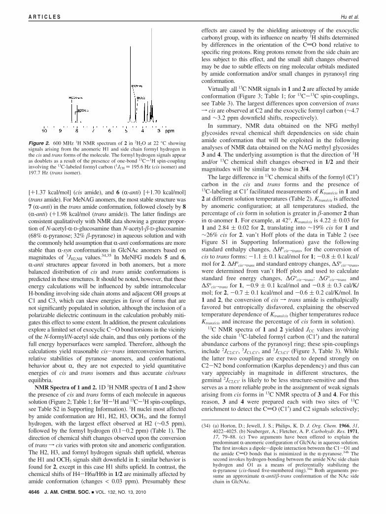
Figure 2. 600 MHz 1H NMR spectrum of 2 in 2H2O at 22 °C showingsignals arising from the anomeric H1 and side chain formyl hydrogen inthe cis and trans forms of the molecule. The formyl hydrogen signals appearas doublets as a result of the presence of one-bond 13C-1H spin-couplinginvolving the 13C-labeled formyl carbon (1JCH ) 195.6 Hz (cis isomer) and197.7 Hz (trans isomer).
4646 J. AM. CHEM. SOC. 9 VOL. 132, NO. 13, 2010
A R T I C L E S Hu et al.
both chemical shifts are sensitive to amide conformation asshown in 1/2 (Table 1), and the easily measured 2JC2,C1′ betweenboth 13C-labeled carbons could be used to validate C1′ and C2signal assignments in cis isomers.
NMR Spectra of 3 and 4. 1D 1H NMR spectra of 3 and 4 inaqueous (2H2O) solution contained signals from the dominanttrans amide form (Table 4). Efforts to detect and assign signalsarising from the cis amides were unsuccessful because of theirweak intensities in spectra containing significant signal overlap.
In contrast, 1D 13C{1H} NMR spectra of the doubly 13C-labeled 3 and 4 contained strong doublets attributed to the
labeled C2 and exocyclic CdO (C1′) carbons of the trans form(Figure 4). In each spectrum, identical splittings were observedin both signals due to the presence of 2JC2,C1′, thus confirming13C2-N2-13C1′ bond connectivity. On the basis of the 13Cchemical shift behaviors established for 1 and 2, a closeexamination was made of regions 4-6 ppm downfield of each
(35) Mobli, M.; Almond, A. Org. Biomol. Chem. 2007, 5, 2243–2251.(36) (a) Church, T.; Carmichael, I.; Serianni, A. S. Carbohydr. Res. 1996,
280, 177–186. (b) Serianni, A. S.; Bondo, P. B.; Zajicek, J. J. Magn.Reson. 1996, 112B, 69–74. (c) Zhao, S.; Bondo, G.; Zajicek, J.;Serianni, A. S. Carbohydr. Res. 1998, 309, 145–152.
Table 1. 1H and 13C Chemical Shift Assignmentsa for 1 and 2 in Their Cis and Trans Amide Conformations
a 1H chemical shifts in ppm relative to external dimethyl-2-silapentane-5-sulfonic acid (DSS); 13C chemical shifts in ppm relative to external DSS.Measured at 22 °C in 2H2O.
Figure 3. Partial 13C{1H} NMR spectra (150 MHz) of 2 in 2H2O at 22 °C. (A) Anomeric C1 signals in the cis and trans amides, showing splittings due to3JC1,C1′. (B, C) C2 signals in the cis and trans amides, showing splittings due to 2JC2,C1′. (D) C3 signals in the cis and trans amides showing splittings dueto 3JC3,C1′. (E) Intense signals from the labeled formyl (C1′) carbon in the cis and trans amides. See Table 3 for measured J-couplings.
J. AM. CHEM. SOC. 9 VOL. 132, NO. 13, 2010 4647
Amide Cis-Trans Isomerization A R T I C L E S
of these intense signals in an effort to detect signals arisingfrom cis forms. Weak signals were detected (Table 4; Figure4). Splittings of the CdO (C1′) and C2 signals attributed toputative cis forms were identical, suggesting that the signalsarose from carbons in the same molecule.
Further support for the cis and trans signal assignments in 3and 4 was obtained from 13C NMR spectra of the triply 13C-labeled isotopomers 3′ and 4′. In both anomers, the labeledcarbons were mutually coupled, giving rise to three-spin systemscontaining internally consistent J-couplings; these results areshown for 4′ in Figure S2 in Supporting Information.
Although the cis signals were weak, the use of 13C-labeledcompounds allowed determinations of the percentages of bothcis and trans amides of 3 and 4 at different solution temperaturesby integration of either the labeled C1′ (CdO) or C2 carbonsignals (Table 5; only data for C2 are shown). In 3, the cis amidevaried from 1.4% to 2.5% as temperature increased from 42.0to 84.6 °C (Table 5). In contrast, the cis amide varied from1.9% to 3.1% in 4 over a temperature range of 31.3-75.1 °C.At 52.7 °C, the percentage of cis form was greater in 4 (2.6%)than in 3 (1.6%), showing a dependence of Ktrans/cis on anomericconfiguration similar to that found for 1 and 2 (Table 2). Thesepercentages of cis amide, however, are roughly 10-fold lowerthan found for 1 and 2 at similar solution temperatures.
van’t Hoff plots of the data in Table 5 gave the following∆Ho
cisftrans values: for 3, -3.2 ( 0.4 kcal/mol; for 4, -2.6 (0.2 kcal/mol. These values are slightly more negative than thosefound for 1 and 2. ∆So
cisftrans Values were also determined fromvan’t Hoff plots, and ∆Go
cisftrans values were calculated from∆Ho
cisftrans and ∆Socisftrans; for 3, ∆Go
cisftrans )-2.7 ( 0.1 kcal/mol, ∆So
cisftrans ) -1.6 ( 1.2 cal/K/mol; for 4, ∆Gocisftrans )
like 1 and 2, the conversion of cisf trans amide is enthalpicallyfavored and entropically disfavored in 3 and 4.
Rate Constants for Amide Cis-Trans Isomerization in1-4. 13C Saturation-transfer (13C ST) experiments were conductedon 1-4 to test for the presence of chemical exchange and thusconfirm the assignment of cis and trans 13C NMR signals (Figure4), and to measure amide cis-trans isomerization (CTI) rateconstants and associated kinetic activation parameters.
Rate constants ktransfcis in 1 and 2 at different temperatures,obtained from steady-state 13C ST experiments (see NMRSpectroscopy section), are given in Table 6. At 64.4 °C, ktransfcis
for 1 was 0.15 ( 0.04 s-1; this value increased ∼5-fold to 0.77( 0.05 s-1 at 84.6 °C. In contrast, ktransfcis for 2 was 0.62 (0.05 s-1 at 64.4 °C, increasing to 3.59 ( 0.16 s-1 at 84.6 °C.The rate of conversion of trans f cis amide depends onanomeric configuration, with conversion more favored in 2.Energies of activation (Eact) for trans f cis were 19.6 ( 3.7kcal/mol for 1 and 19.6 ( 4.5 kcal/mol for 2.
Rate constants kcisftrans in 1 and 2 (Table 6) were calculatedfrom ktransfcis and Ktrans/cis values at each temperature. Energiesof activation (cis f trans) were 18.3 ( 3.9 kcal/mol for 1 and18.8 ( 4.5 kcal/mol for 2. Amide CTI appears more kineticallyfavored in 2 than in 1, on the basis of the larger ktransfcis andkcisftrans values observed in the former at any temperature.
CTI kinetics measurements were conducted on 3 and 4 usingtime-dependent 13C ST in which the C1′ (carbonyl) carbon ofthe cis form was saturated for varying times and the effect ofthis saturation monitored on the C1′ signal intensity of the trans
a In Hz ( 0.1 Hz at 22 °C in 2H2O. b Sign unknown. c Predicted signs based on analogous 2JC2,C4 in �-D-glucopyranosyl rings (ref 36). d Couplingsmeasured from 13C{1H} NMR spectra of 3′ and 4′.
Table 4. 1H and 13C Chemical Shift Assignmentsa for 3 and 4 in Their Cis and Trans Amide Conformations
a 1H chemical shifts in ppm relative to external dimethyl-2-silapentane-5-sulfonic acid (DSS); 13C chemical shifts in ppm relative to external DSS.Measured at 22 °C in 2H2O. b Measured from the 13C{1H} NMR spectra of 3′ and 4′.
Table 2. Ktrans/cis for 1 and 2 at Different Solution Temperatures
a Errors based on variations observed from multiple integration of 13CNMR signals in a given spectral data set.
4648 J. AM. CHEM. SOC. 9 VOL. 132, NO. 13, 2010
A R T I C L E S Hu et al.
form (Figure 5A). Data linearization at different solutiontemperatures (Figure 5B) gave ktransfcis (Table 7). For 3, ktransfcis
ranged from 0.02 to 0.9 s-1 over a temperature range of42.0-84.6 °C, whereas for 4, ktransfcis ranged from 0.05 to 2.6s-1 over a temperature range of 31.3-75.0 °C. As found for1/2, ktransfcis in 3/4 depends on anomeric (C1) configuration,with trans f cis conversion more favored in �-anomer 4 at alltemperatures investigated. Activation energies (Eact) (trans fcis) (Figure S3 in Supporting Information) were 19.2 ( 2.8 kcal/mol for 3 and 19.4 ( 3.0 kcal/mol for 4, values similar to thosefound for 1 and 2.
Rate constants, kcisftrans, in 3 and 4, calculated from ktransfcis
and Ktrans/cis values at each temperature, are also shown in Table7. Energies of activation (cis f trans) were 16.0 ( 3.2 kcal/mol for 3 and 16.7 ( 3.4 kcal/mol for 4, values statisticallysimilar to those observed for 1 and 2. Like 1/2, amide CTIappears more kinetically favored in �-anomer 4 than inR-anomer 3.
Additional activation parameters for the CTI of 1-4 aresummarized in Table 8. ∆G°q values range from 17 to 21 kcal/mol and ∆H°q from 15 to 19 kcal/mol, and the signs of bothparameters are positive. In contrast, ∆S°q values are uniformlynegative, ranging from -3 to -9 cal/mol/K. ∆S°q values appearmore negative for R-anomers 1 and 3 than for �-anomers 2 and4, but the large errors make this observation inconclusive.
Conclusions
N-Acetylation in biological systems is a common chemicalmodification that has important functional ramifications. Forexample, N-acetylation of L-lysine side chains of histones servesas a mechanism to reduce positive charge and thereby reduceelectrostatic attraction between histones and their negativelycharged nucleic acid binding partners.37 This reduced affinity playsan active role in modulating gene expression.38 In a similar vein,N-acetylation of 2-deoxy-2-aminosugars such as D-glucosamine 6Peliminates their charge properties and thus affects their biologicalbehaviors and functions. For example, D-galactosamine is toxic inanimals and thus is normally found in its N-acetylated form in ViVo(found as UDP-GalNAc produced via C4-epimerization of UDP-GlcNAc).39 In contrast, D-glucosamine 6P is synthesized in ViVofrom D-fructose 6P by the bifunctional enzyme, glutamine:fructose6P amidotransferase (GFA1), and can accumulate without toxiceffects. In ViVo conversion of D-glucosamine 6P to N-acetyl-D-glucosamine 6P is catalyzed by the CoA-dependent enzyme,D-glucosamine 6P N-acetyltransferase (GNA1).40
Figure 4. Partial 13C{1H} NMR spectra (150 MHz) of 4 in 2H2O at 22 °C. (A) Signals from the 13C-labeled carbonyl (C1′) carbon from cis (180.32 ppm)and trans (177.25 ppm) forms, with insets showing splittings due to 2JC2,C1′. (B, C) Signals from the 13C-labeled C2 carbon in the cis (62.58 ppm) and trans(58.00 ppm) forms, showing 2JC2,C1′ values internally consistent with values measured from the C1′ signals in A.
Table 5. Ktrans/cis for 3 and 4 at Different Solution Temperatures
a Values were determined from the integration of the C2 signals.Errors are based on variations observed from multiple integration of C2signals in a given spectral data set.
Table 6. Rate Constants (s-1), ktransfcis and kcisftrans, for 1 and 2 atDifferent Temperatures
1 (R) 2 (�)
T (°C) ktransfcis kcisftrans ktransfcis kcisftrans
Although N-acetylation is a ubiquitous saccharide modi-fication in biological systems, the chemical and biochemicalproperties conferred by this structural change are not fullyappreciated. It is well established that generic amide bondsassume both cis and trans conformations in solution, andconsiderable study has been directed toward understandingthe thermodynamics and kinetics of amide CTI in simplesystems. For example, free energy differences between cisand trans formamides and acetamides have been reported torange from 0.6 to 9.3 kcal/mol, and energy barriers to amidebond rotation in simple amides range from 18 to 30 kcal/mol depending on the nature of the substituents appended tothe carbonyl carbon and amide nitrogen.41
To date, little attention has been paid to amide cis-transequilibria and kinetics in N-acylated saccharides, specificallyin GlcNAc, GalNAc, and N-acetyl-neuraminic acid (Neu5Ac).It is normally assumed that, in solution, the trans amidepredominates in these and related structures, and the potentialpresence of cis isomers and their role in biology have beenoverlooked. The latter is especially noteworthy in light of thedistribution of cis and trans GlcNAc residues in crystalstructures of glycoproteins,42 where a broad and nearly continu-ous span of rotamers about the N-CO bond is observed despitethe relatively high energies required to rotate the partial C-Ndouble-bond into nonplanar conformations (i.e., geometries otherthan the idealized cis and trans forms) (Figure 6A). This energylikely derives from crystal packing forces, highlighting the highdegree of structural deformation accessible in crystal structuresof conformationally flexible molecules or fragments thereofunder certain packing conditions. Additional analysis of the samedata set with respect to the more easily rotatable C2-N2 bond(R) shows a preference for R-anti (i.e., H2 anti to NH) (Figure6B), a result consistent with prior interpretations of 3JH2,NH spin-couplings in GlcNAc and related structures measured insolution.35 Correlation between C2-N2 (R) and amide (�) bondconformation (Figure 6C) shows a clustering of data points near180° for both torsions, as expected, but also a discerniblehorizontal clustering of points attributable to the cis amide overa wide range of R conformations. The distribution of torsionsis more uniform for R than for �, presumably as a result of thepartial double bond character of the latter that destabilizestorsions between 10° to 60° and -10° to -60°.
Prior studies have shown that aqueous solutions of N-formylated aminosugars contain comparable amounts of cis andtrans amides.29–32 The small N-formyl hydrogen apparentlystabilizes the cis form (relative to methyl in the N-acetylderivative) by minimizing steric interactions with the proximal
(37) Legube, G.; Trouche, D. EMBO Rep. 2003, 4, 944–847.(38) Verdone, L.; Agricola, E.; Caserta, M.; Di Mauro, E. Briefings Funct.
Genomics Proteomics 2006, 5, 209–221.(39) McMillan, J. M.; McMillan, D. C. Toxicology 2006, 222, 175–184.
(40) Hurtado-Guerrero, R.; Raimi, O. G.; Min, J.; Zeng, H.; Vallius, L.;Shepherd, S.; Ibrahim, A. F. M.; Wu, H.; Plotnikov, A. N.; van Aalten,D. M. F. Biochem. J. 2008, 415, 217–223.
(41) Isaacs, N. Physical Organic Chemistry, 2nd ed.; Longman Scientificand Technical: Harlow, England, 1995; p 350.
(42) This survey was conducted on 1311 pdb files (from the RCSB PDBdatabase) containing a total of 7994 GlcNAc residues. Within the lattergroup, 917 residues contained a H2-C2-N2-H torsion angle between60° and-60°, producing an ∼11% R-syn population; the remaining∼89% is R-trans. More pertinent to the present work, 463 residuescontained a C2-N2-C1′-C2′ torsion angle between 60° and-60°,giving an ∼6% �-cis population.
Figure 5. (A) Plots of C1′trans signal intensity in 3 as a function of saturation time of the C1′cis signal. Data were collected at different temperatures: black) 42.0 °C; blue ) 52.7 °C; green ) 64.4 °C; red ) 75.1 °C; orange ) 84.6 °C. (B) Linearization of the data shown in A following eq 1, from which ktransfcis
values were determined.
Table 7. Rate Constants (s-1), ktransfcis and kcisftrans, for 3 and 4 atDifferent Temperatures
3 (R) 4 (�)
T (°C) ktransfcis kcisftrans ktransfcis kcisftrans
C2-H2 bond.43 In the present work, NMR characterization ofthe N-formyl glycosides was a prerequisite to the more chal-lenging detection and quantification of cis amide in solutionsof GlcNAc glycosides. The success of the latter work hingedon the assumption that the effects of amide conformation on1H and 13C chemical shifts for nuclei in the vicinity of the amidebond were similar in the N-formyl and N-acetyl derivatives, thusallowing signal assignments in the latter despite the lowabundance of cis amide. While this assumption proved valid,
additional support for cis and trans signal assignments in 3/4was obtained from studies of internally consistent 13C-13C spin-couplings in 13C-labeled GlcNAc isotopomers and by thedemonstration and quantification of cis-trans chemical ex-change by 13C saturation transfer.
The equilibrium between cis and trans amides in the N-formyland N-acetyl derivatives of methyl D-glucosaminide dependson anomeric configuration. The cis amide is less stable inR-anomers of NFG and NAG methyl glycosides than in thecorresponding �-anomers. Given the proximity of the N-acylside chain to the anomeric center in these structures, thisdependency might be expected. Inspection of molecular models,however, does not provide a structural rationale for the observeddependency if preferred conformation about R in the cis amidesis anti (as observed in the trans amides based on 3JH2,NH
values35). On the other hand, if R is syn in cis amides, or partlyso, in solution, then the acyl side chain and the axial C1-O1bond would be in close proximity in R-anomers, therebyproducing a destabilizing steric interaction.45 This problem andthe effects of acyl chain structure and length on the anomericdependency of Ktrans/cis will require further investigation.
The relatively small rate constants for saccharide cis-transisomerization call attention to the potential implications of thisprocess in biological receptor binding and enzyme catalysis. Inhypothetical cases in which the cis amide of GlcNAc is theproductive form for binding, the overall rate of binding couldbe determined by the rate of conversion of the trans to cis form.Whether such a scenario occurs in ViVo and whether biologicalsystems have evolved enzymes to facilitate CTI in saccharidesin order to circumvent this problem demand further scrutiny.
Given the importance of anomeric configuration on amidecis-trans equilibria in 2-deoxy-2-formamido and 2-deoxy-2-acetamido sugars, it is natural to ponder the effects of otherstructural perturbations on the cis-trans equilibria and CTIkinetics in saccharides. For example, in structures like methyl�-chitobioside 9 (and the chitin polymer derived from it) andhyaluronic acid 10, the multiple N-acetyl groups may displaydifferent cis-trans equilibria and exchange kinetics due to theirdifferent chemical environments and/or states of solvation.Having established the fundamental NMR characteristics of cisand trans amides in simple GlcNAc glycosides, and the utilityof selective 13C labeling to enable such studies, it is now feasibleto investigate N-acetyl side chain behavior in more complex
(43) Eliel and Wilen have suggested that the preference for trans amideconformations in solution is due to a combination of steric (attractivein trans, repulsive in cis) and charge interaction factors. See ref 44for further discussion.
(44) Eliel, E. L.; Wilen, S. H. Stereochemistry of Organic Compounds;Wiley: New York: 1994; pp 620-621.
(45) Preliminary data on the behavior of R in the cis and trans forms of 2was obtained by measuring 3JH2,NH values in DMSO-d6 solvent (FigureS4 in Supporting Information). For 2, nearly identical couplingmagnitudes were observed in both amide conformations (9.6 Hz in�-cis; 9.5 Hz in �-trans), suggesting similar bond conformations aboutbond R. On the other hand, the effect of amide conformation on R isprobably smaller for N-formyl than for N-acetyl substituents. Atpresent, similar J-coupling measurements have not been attempted inMeGlcNAc anomers because of the considerably less intense cissignals.
Figure 6. (A) Distribution of C2-N2-C1′-C2′ dihedral angles observedin a survey of crystal structures of GlcNAc residues in glycoproteins takenfrom the PDB.42 The trans amide predominates (180°), but a continuousdistribution of angles is observed over the full 360° rotation, including thatfor the cis amide (0°). (B) Distribution of R dihedral angles in the sameensemble of X-ray structures as in A. (C) Correlation between conformationsof the R and � bonds in the same group of GlcNAc structures sampled inA and B.
J. AM. CHEM. SOC. 9 VOL. 132, NO. 13, 2010 4651
Amide Cis-Trans Isomerization A R T I C L E S
structures. Likewise, studies of N-acetyl side chain behavior inNeu5Ac, where the side chain resides in a different chemicalenvironment than exists in GlcNAc, may reveal new structuralproperties that contribute to its biological properties. It isnoteworthy that Neu5Ac is commonly found as a terminal (andthus exposed) residue in the oligosaccharide chains of N-linkedglycoproteins, thus increasing the likelihood that, in someinstances, recognition between Neu5Ac residues and biorecep-tors may depend on side chain amide conformation.
Acknowledgment. This work was supported by a grant (to A.S.)from the National Institutes of Health (GM059239). The NotreDame Radiation Laboratory is supported by the Office of BasicEnergy Sciences of the United States Department of Energy. Thisis Document No. NDRL-4815 from the Notre Dame RadiationLaboratory. The authors thank Omicron Biochemicals, Inc. for gifts
of 2-amino-2-deoxy-D-[2-13C]glucose hydrochloride and 2-amino-2-deoxy-D-[1,2-13C2]glucose hydrochloride.
Supporting Information Available: Routes for the synthesisof NFG glycosides; DFT-calculated total energies of 5-8 (inVacuo and solvated); 1H-1H spin-coupling constants in 1 and2; van’t Hoff plots for 1 and 2; partial 13C{1H} NMR spectraof 4′; Arrhenius plot of ktransfcis in 4; partial 1H NMR spectrumof 2 in DMSO-d6; description of the synthesis of 2; descriptionof the synthesis of 3′ and 4′; description of calculations of kineticactivation parameters Eact, ∆G°q, ∆H°q, and ∆S°q; full ref 23.This material is available free of charge via the Internet at http://pubs.acs.org.