J. Clin. Endocrinol. Metab. 2010 95: 2529-2535, doi: 10.1210/jc.2010-0180
Fausto Bogazzi, Luigi Bartalena and Enio Martino
Approach to the Patient with Amiodarone-Induced Thyrotoxicosis
Society please go to: http://jcem.endojournals.org//subscriptions/ or any of the other journals published by The EndocrineJournal of Clinical Endocrinology & MetabolismTo subscribe to
Approach to the Patient with Amiodarone-Induced Thyrotoxicosis
Fausto Bogazzi, Luigi Bartalena, and Enio Martino
Department of Endocrinology (F.B., E.M.), University of Pisa, 56124 Pisa, Italy; andDepartment of Clinical Medicine (L.B.), University of Insubria, 21100 Varese, Italy
Amiodarone, a benzofuranic iodine-rich antiarrhythmic drug, causes thyroid dys-function in 15–20% of cases. Although amiodarone-induced hypothyroidismposes no particular problem, amiodarone-induced thyrotoxicosis (AIT) is a diag-nostic and therapeutic challenge. There are two main forms of AIT: type 1, a formof iodine-induced hyperthyroidism, and type 2, a drug-induced destructive thy-roiditis. However, mixed/indefinite forms exist that may be caused by both patho-genic mechanisms. Type 1 AIT usually occurs in abnormal thyroid glands, whereastype 2 AIT develops in apparently normal thyroid glands (or small goiters). Diag-nosis of thyrotoxicosis is easy, based on the finding of increased free thyroidhormone concentrations and suppressed TSH levels. Thyroid radioactive iodine(RAI) uptake values are usually very low/suppressed in type 2 AIT, most commonlylow or low-normal, but sometimes normal or increased in type 1 AIT despite theiodine load. Color flow Doppler sonography shows absent hypervascularity intype 2 and increased vascularity in type 1 AIT. Mixed/indefinite forms may havefeatures of both AIT types. Thionamides represent the first-line treatment fortype 1 AIT, but the iodine-replete gland is not very responsive; potassium per-chlorate, by inhibiting thyroid iodine uptake, may increase the response to thio-namides. Type 2 AIT is best treated by oral glucocorticoids. The response verymuch depends on the thyroid volume and the severity of thyrotoxicosis. Mixed/indefinite forms may require a combination of thionamides, potassium perchlor-ate, and steroids. RAI is usually not feasible in AIT due to low RAI uptake values.Thyroidectomy represents a valid option in cases resistant to medical therapy.(J Clin Endocrinol Metab 95: 2529–2535, 2010)
The Case
A 66-yr-old man was referred to our Department because ofthyrotoxicosis. He had a 2-yr history of paroxysmal atrialfibrillationtreatedbyelectroconversion. Inthe last6months,sinus rhythm had been maintained by oral amiodarone (200mg/d) given in association with antiplatelet therapy. Therewas no information as to his thyroid function and morphol-ogy before the initiation of amiodarone therapy, but he hadno history of previous thyroid diseases. In the last 4 wk, hecomplained of nervousness, palpitations, weight loss (3 kg)
Abbreviations: AIT, Amiodarone-induced thyrotoxicosis; CFDS, color flow Doppler sonog-raphy; FT4, free T4; MIBI, [99mTc]2-methoxy-isobutyl-isonitrile; RAI, radioactive iodine;RAIU, RAI uptake.
Accreditation and Credit Designation StatementsTheEndocrineSociety isaccreditedbytheAccreditationCoun-cil for Continuing Medical Education to provide continuingmedical education for physicians. The Endocrine Society hasachieved Accreditation with Commendation.The Endocrine Society designates this educational activityfor a maximum of 1 AMA PRA Category 1 Credit(s)TM.Physicians should only claim credit commensurate with theextent of their participation in the activity.Learning ObjectivesUpon completion of this educational activity, participantsshould be able to• Differentiate the two main forms of AIT• Choose the more appropriate therapy for either form of
AIT• Predict the response to glucocorticoids in patients with
type 2 AIT• Select patients who might take advantage from
thyroidectomyTarget AudienceThis continuing medical education activity should be of sub-stantial interest to endocrinologists.Disclosure PolicyAuthors, editors, and Endocrine Society staff involved inplanning this CME activity are required to disclose to learn-ers any relevant financial relationship(s) that have occurredwithin the last 12 months with any commercial interest(s)whose products or services are discussed in the CME con-tent. The Endocrine Society has reviewed all disclosures andresolved or managed all identified conflicts of interest, asapplicable.The following individuals reported relevant financialrelationships:Leonard Wartofsky, M.D., has received speaker honorar-ium from Genzyme Corp.The following individual reported NO relevant financialrelationships:Enio Martino, M.D., reported no relevant financialrelationships.Fausto Bogazzi, M.D., reported no relevant financialrelationships.Luigi Bartalena, M.D., reported no relevant financialrelationships.Endocrine Society staff associated with the development ofcontent for this activity reported no relevant financialrelationships.Acknowledgement of Commercial SupportThis activity is not supported by grants, other funds, or in-kind contributions from commercial supporters.Privacy and Confidentiality StatementThe Endocrine Society will record learner’s personal infor-mation as provided on CME evaluations to allow for issu-ance and tracking of CME certificates. No individual per-formance data or any other personal information collectedfrom evaluations will be shared with third parties.Method of ParticipationThis Journal CME activity is available in print and online asfull text HTML and as a PDF that can be viewed and/orprinted using Adobe Acrobat Reader. To receive CMEcredit, participants should review the learning objectivesand disclosure information; read the article and reflect on itscontent; then go to http://jcem.endojournals.org and findthe article, click on CME for Readers, and follow the in-structions to access and complete the post-activity test ques-tions and evaluation. The estimated time to complete thisactivity, including review of material, is 1 hour. If you havequestions about this CME activity, please direct them [email protected] release date: June 2010Activity expiration date: June 2011
S P E C I A L F E A T U R E
A p p r o a c h t o t h e P a t i e n t
J Clin Endocrinol Metab, June 2010, 95(6):2529–2535 jcem.endojournals.org 2529
not associated with changes in appetite, insomnia, and amodest increase in bowel movements.
On physical examination, this patient, whose familyhistory was negative for thyroid disorders, had tachy-cardic atrial fibrillation (120 beats/min). Blood pressurewas 145/55 mm Hg. The thyroid gland was neither in-creased in volume nor tender, no nodules were palpable,and no bruit was appreciated over the gland. There wereno symptoms or signs of Graves’ ophthalmopathy.
Serum biochemical tests were as follows: free T4
(FT4), 65 pg/ml (84 pmol/liter; normal values, 7.5–15.5pg/ml); free T3 (FT3), 9.8 pg/ml (15.2 pmol/liter; normalvalues, 3.5–5.7 pg/ml); and TSH, less than 0.01 mU/liter(normal values, 0.4 –3.5 mU/liter). Antithyroglobulin,antithyroperoxidase, and anti-TSH receptor antibodieswere undetectable. Erythrocyte sedimentation rate, C-reactive protein, and cell blood count were normal. Uri-nary iodine excretion was markedly increased (9100�g/24 h; normal values, 100 –300 �g/24 h). No iodine-containing contrast agents had been recently adminis-tered to this patient.
Thyroid ultrasonography evidenced a slightly hypo-echogenic gland with an estimated volume of 18 ml andwith no nodules; color flow Doppler sonography (CFDS)showed a pattern 0 (absent hypervascularity) and peaksystolic velocity measured at the level of inferior thyroidartery was normal (3 cm/sec, normal range 2–7 cm/sec);thyroid radioactive iodine (RAI) uptake (RAIU) was 0.7%after 3 h and 0.9% at 24 h.
Background
The use of amiodarone, a benzofuranic iodine-rich (37%of its weight) antiarrhythmic drug, is complicated in 15–20% of cases by the occurrence of thyroid dysfunction,either thyrotoxicosis [amiodarone-induced thyrotoxicosis
(AIT)] or hypothyroidism (1). The relative proportion ofAIT and hypothyroidism partly depends upon environ-mental iodine intake, because AIT is relatively more fre-quent in iodine-deficient areas and hypothyroidism iniodine-sufficient areas (2–5). AIT, at variance withspontaneous hyperthyroidism, is more common in menthan in women (1).
Two main mechanisms can lead to AIT (Table 1): io-dine-induced hyperthyroidism (type 1 AIT), a form of JodBasedow, or destructive thyroiditis (type 2 AIT), caused byamiodarone itself and its high iodine content (6). How-ever, the two mechanisms may concur to AIT in the samepatient (mixed or indefinite AIT) (Table 1). Recent datashowed that type 2 AIT is by far the most frequent form(7). This is probably due to the improved pretherapy eval-uation of candidates to amiodarone treatment and theavoidance of this drug (if feasible) in patients with preex-isting thyroid abnormalities. In fact, type 1 AIT usuallyoccurs in patients with preexisting nodular goiter or latentGraves’ disease, whereas type 2 AIT generally develops inpatients without clinical, biochemical, and morphologicalevidence of thyroid disease (1). A small, sometimes mod-erately painful, goiter may, however, be present also intype 2 AIT (8).
AIT may develop early during amiodarone treatment oreven several months after drug withdrawal (1). This isbecause amiodarone and its metabolites (mainly deseth-ylamiodarone) have a long half-life (up to 100 d) and arestored invarious tissues, particularly in the fat, fromwhichthey are released very slowly (9, 10). The onset of AIT isoften sudden and explosive.
Diagnostic Evaluation
Our patient had relatively few and mild symptoms of thy-rotoxicosis associated with the evidence of iodine load
TABLE 1. Clinical and pathogenic features of type 1 and type 2 AIT
Type 1 Type 2 Ref.Underlying thyroid disease Yes NoThyroid ultrasound Diffuse or nodular goiter Normal (hypoechoic) gland (small goiter) 38CFDS Increased vascularity Absent hypervascularity 13Thyroidal RAIU Low/normal/increased Low/absent 39, 40MIBI Thyroid retention Absent uptake 17Thyroid antibody Sometimes present Usually absent 41–43Pathogenesis Iodine-induced hyperthyroidism Destructive thyroiditis 44, 45Spontaneous remission No Possible 47Preferred medical therapy Thionamides (plus KClO4) Glucocorticoids 6, 31Subsequent hypothyroidism Unlikely Possible 46Subsequent therapy for the
underlying thyroid diseaseLikely No
Mixed/indefinite forms of AIT are still hypothetical and not yet formally described; their clinical findings are believed to be a mix of amiodarone-induced destructive thyroiditis and iodine-induced hyperthyroidism.
2530 Bogazzi et al. Amiodarone-Induced Thyrotoxicosis J Clin Endocrinol Metab, June 2010, 95(6):2529–2535
(attributable to chronic amiodarone administration) ap-parently in the absence of thyroid abnormalities; his mainclinical picture was the worsening of cardiac arrhyth-mia (recurrence of atrial fibrillation) heralded by pal-pitations. AIT may, however, be heavily symptomatic,especially in younger patients, whose clinical manifes-tations are indistinguishable from those of spontaneoushyperthyroidism (11, 12).
Identification of the different subtypes of AIT is crucialbecause this affects the therapeutic approach (Table 1).Thyroid ultrasonography is useful because it shows thepresence or absence of a diffuse or a nodular goiter. CFDSof the thyroid is a very important diagnostic tool (13, 14).Type 2 AIT is in most cases characterized by absent hy-pervascularity (pattern 0), whereas type 1 cases usuallyshow an increased vascularity (patterns 1–3) and bloodflow velocity (13–15).
Thyroidal 131I uptake (RAIU) is another useful diag-nostic tool (1). RAIU is usually very low (�3%) in type 2AIT and low-normal, normal, or even increased (despitethe iodine load) in type 1 AIT (8, 15). One study failed todistinguish the two main forms of AIT on the basis ofRAIU values (16). The likely explanation is that the pres-ence of diffuse or nodular goiter associated with low thy-roidal RAIU does not exclude a destructive or mixed formof AIT.
Recently, thyroid [99mTc]2-methoxy-isobutyl-isoni-trile (MIBI) scintigraphy has been suggested as a usefuldiagnostic tool in a study of 20 consecutive patients withAIT (17). In this study, MIBI diffuse retention, which isindicative of a hyperfunctioning tissue, was present in alltype 1 AIT patients, whereas no significant uptake, sug-gestive of a destructive process, was found in type 2 AIT;the four patients with mixed/indefinite AIT had either afaint persistent MIBI uptake or a quick washout of thetracer (17). The real usefulness of this expensive procedurein the identification of more complex and more difficult totreat mixed/indefinite forms of AIT needs to be confirmedby larger studies (18).
Other serum biochemical markers, such as IL-6 (usu-ally elevated in type 2 AIT) (1) or C-reactive protein (19),seem to have a marginal diagnostic role in differentiatingthe different forms of AIT because of their poor specificity(8). Search for thyroid-directed autoantibodies (particu-larly TSH receptor autoantibody) is relevant only in AITpatients whose underlying and preexisting thyroid disor-der is Graves’ disease (1).
Thus, although diagnosis of AIT is not difficult per se,identification of the pathogenic mechanisms of thyrotox-icosis may be difficult. This is reflected by the results ofquestionnaire-based surveys in Europe, North America,and Latin America (3–5), with a substantial proportion of
respondents unable to distinguish with certainty type 1and type 2 AIT. However, in general, CDFS and thyroidRAIU determination appear to be the most useful diag-nostic techniques in the initial assessment of the patient(Table 1).
Therapeutic Approach
AIT is a dangerous and critical situation for the patientwith underlying cardiac abnormalities. Indeed, AIT isbound to an increased mortality, especially in older pa-tients with impaired left ventricular function (20). Thus,restoration and stable maintenance of euthyroidismshould be achieved as quickly as possible. On the otherhand, the diagnostic uncertainties underscored in the men-tioned surveys (3–5) often have an impact on the thera-peutic approach; if the physician is unable to define thepathogenic mechanism of AIT, the optimal treatment maynot be selected or all possible pharmacological weaponsmay need to be used at the same time. This, in turn, mayimply a higher risk of side effects and complications oftherapy.
Type 1 AIT, a form of true hyperthyroidism triggeredby the iodine load, is best treated by antithyroid drugs (1).However, an iodine-replete thyroid gland is less respon-sive to the inhibitory action of thionamides. Thus, higherdrug dosages (40–60 mg/d methimazole or equivalentdoses of propylthiouracil) and longer periods of therapyare required before euthyroidism is restored. This is ob-viously not ideal in patients with cardiac problems. Toincrease the sensitivity of the thyroid gland and the re-sponse of thyrotoxicosis to thionamides, potassium per-chlorate, which decreases thyroid iodine uptake, is addedfor 2–6 wk (21). To minimize the adverse effects of thedrug (particularly on the kidney and blood marrow), dosesof 1 g/d or lower of potassium perchlorate should be used(1). The use of the combined thionamide-potassium per-chlorate treatment seems to be more popular in Europethan in North America, where most thyroidologists em-ploy thionamides alone as first-line treatment for type 1AIT (5).
Type 2 AIT, a form of drug-induced destructive thy-roiditis caused by amiodarone itself, is best treated by ste-roids (5, 6). Type 2 AIT may be self-limiting, and contin-uation of amiodarone has been suggested by some authorsnot to influence the effectiveness of steroids (22). Initialprednisone dose is about 0.5–0.7 mg/kg body weight perday, and the treatment is usually continued for 3 months(1). Although the response to treatment often is dramatic,and 50% of patients are cured within 4 wk (23), a delayedresponse is sometimes observed. However, using a math-ematical model, euthyroidism should be reached after 40 d
J Clin Endocrinol Metab, June 2010, 95(6):2529–2535 jcem.endojournals.org 2531
of treatment, unless goiter is large and initial thyrotoxi-cosis is particularly severe (23). Thionamides are not ef-fective in type 2 AIT. A recent retrospective cohort studyshowed that after about 6 wk of therapy, more than 85%of patients treated with thionamides were still thyrotoxiccompared with 24% of prednisone-treated patients (24).
The most difficult challenge is represented by mixed/indefinite forms of AIT. In these cases, both pathogenicmechanisms (increased thyroid hormone synthesis andthyroid hormone discharge due to glandular damage) arelikely operating. Thus, the best treatment is represented bya combination of thionamides (with or without potassiumperchlorate) and oral glucocorticoids (1). However,mixed/indefinite forms of AIT, although proposed as aseparate entity, have not been fully characterized so far.The fact that features of hyperthyroidism and destructivethyroiditis may concomitantly be present suggests that de-structive phenomena may superimpose to a hyperfunc-tioning gland. However, how these findings will affectthyroid tests, allowing a clear-cut identification of the un-derlying process, still is unsettled. If a patient initiallytreated with thionamides alone (because of a diagnosis oftype 1 AIT) does not respond within 4 wk, most thyroid-ologists usually add potassium perchlorate and/or oralglucocorticoids (3–5). Amiodarone-induced destructivethyroiditis may occur in patients with goiter, making dif-ferentiation of type 1 and mixed/indefinite forms very dif-ficult. In these cases, a relevant proportion of thyroidolo-gists (more frequently in North America than in Europe)treat these particular patients with a combination of an-tithyroid drugs and steroids from the beginning (5). Manythyroidologists think that glucocorticoids may be not easytohandle inpatientswithcardiacdiseases;accordingly, ithasbeensuggestedtostartmedical therapyof type2AITpatientswith thionamides for at least a month and to associate ste-roids if response is poor or absent (25). In our opinion, thissomehow expectant strategy may be harmful for a patientwith (often serious) cardiac abnormalities, whose thyrotox-icosis should be promptly corrected. In addition, no evidencesupports such a therapeutic approach.
The use of lithium has been proposed for AIT (26), butthe evidence is too limited to support its effectiveness. Io-panoic acid has been initially proposed as a medical ther-apy for AIT patients (27), but this drug is less effective thanglucocorticoids (28). The main advantage of iopanoic acidadministration is in the preparation of AIT patients forthyroidectomy, because it rapidly lowers serum T3 con-centrations (29). Plasmapheresis is generally not used be-cause of its transient effects, its costs, and because notwidely available (1).
RAI therapy is in principle not feasible in AIT patientsbecause of the low RAIU values either due to the iodine
load or the destructive process (1). An open study sug-gested, however, that RAI may have some value also inthese cases (30). Thyroid RAIU values might to some ex-tent be increased by the administration of recombinanthuman TSH in type 1 AIT, thus allowing RAI therapy (31).This approach may be risky, because recombinant hu-man TSH administration in these patients may be fol-lowed by a sustained increase in serum thyroid hormoneconcentrations (32), which is obviously harmful for pa-tients with underlying cardiac abnormalities.
Total thyroidectomy is not the first-line treatment forAIT,alsoinviewofthepotentialsurgicalriskinthesepatientswith underlying cardiac disorders. However, this approachmay be necessary in patients who are resistant to other treat-ments (29, 33, 34). To obtain a better (although transient)control of thyrotoxicosis before surgery, a short course ofiopanoic acid (in association with antithyroid drugs) can beused (29);1g iopanoicacidperdayusuallynormalizes serumT3 concentrations in 1–3 wk, whereas T4 remains un-changed. The surgical risk is probably further reduced by acombination of minimally invasive thyroid surgery and localanesthesia (35). We believe that continuing iopanoic acid for7–10 d after surgery, particularly in patients with very highserum thyroid hormone concentrations before surgery, maybe beneficial to avoid serum T3 surge after iopanoic acidwithdrawal. Iopanoic acid has now become unavailable inthe market, and scientific authorities and medical societiesshould consider the possibility to strongly recommend itsproduction for severe cases of AIT.
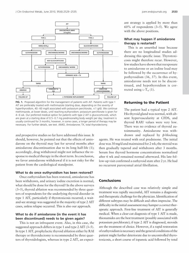
A therapeutic algorithm of AIT is offered in Fig. 1.
Several issues remain as to the optimal approach to thepatient with AIT.
Should amiodarone be withdrawn once adiagnosis of AIT is made?
The decision of whether amiodarone therapy can bediscontinued requires a strict interaction between cardi-ologists and endocrinologists. In a recent survey amongEuropean and North American thyroidologists, amioda-rone withdrawal was considered necessary by 90% of Eu-ropeans and 79% of North Americans in the case of type1 AIT; this proportion decreased to 80 and 66%, respec-tively, in the case of type 2 AIT (5). This question is un-settled, because mild cases of type 2 AIT may remit with-out treatment and/or amiodarone withdrawal (8). For thetime being, we favor amiodarone withdrawal (if feasiblefrom the cardiological point of view), but no controlled
2532 Bogazzi et al. Amiodarone-Induced Thyrotoxicosis J Clin Endocrinol Metab, June 2010, 95(6):2529–2535
and prospective studies so far have addressed this issue. Itshould, however, be pointed out that the effects of amio-darone on the thyroid may last for several months afteramiodarone discontinuation due to its long half-life (1);accordingly, drug withdrawal might not influence the re-sponse to medical therapy in the short term. In conclusion,we favor amiodarone withdrawal if it is not risky for thepatient from the cardiological standpoint.
What to do once euthyroidism has been restored?Once euthyroidism has been restored, amiodarone has
been withdrawn, and urinary iodine excretion is normal,what should be done for the thyroid? In the above surveys(3–5), thyroid ablation was recommended by three quar-ters of respondents for the underlying thyroid disorder intype 1 AIT, particularly if thyrotoxicosis recurred; a wait-and-see strategy was suggested in the majority of type 2 AITcases, unless relapse occurred. This is also our approach.
What to do if amiodarone (in the event it hasbeen discontinued) needs to be given again?
This is not an infrequent event. Also, in this case, thesuggested approach differs in type 1 and type 2 AIT (3–5).In type 1 AIT, prophylactic thyroid ablation either by RAItherapy or thyroidectomy is recommended by three quar-ters of thyroidologists, whereas in type 2 AIT, an expect-
ant strategy is applied by more than60% of respondents (3–5). We agreewith the above positions.
What may happen if amiodaronetherapy is restarted?
This is an unsettled issue becausethere are no longitudinal studies ad-dressing this specific issue. Thyrotoxi-cosis might therefore recur. However,few studies have shown that reexposureto amiodarone or an iodine load maybe followed by the occurrence of hy-pothyroidism (36, 37). In this event,amiodarone needs not to be discon-tinued, and hypothyroidism is cor-rected using L-T4 (1).
Returning to the Patient
The patient had a typical type 2 AIT.His thyroid gland was normal, with ab-sent hypervascularity at CFDS, andthyroid RAIU values were very low.There was no evidence of thyroid au-toimmunity. Amiodarone was with-drawn and replaced by �-blocking
agents. He was treated with oral prednisone. The initialdose was 30 mg/d and maintained for 2 wk; the steroid wasthen gradually tapered and withdrawn after 3 months.Serum free thyroid hormone concentrations normalizedafter 6 wk and remained normal afterward. His late fol-low-up visit confirmed a euthyroid state after 2 yr. He hadno recurrent paroxysmal atrial fibrillation.
Conclusions
Although the described case was relatively simple andtreatment was rapidly successful, AIT remains a diagnosticand therapeutic challenge for the physician. Identification ofdifferent subtypes may be difficult and often imprecise. Thedifficulty in the initial assessment may hamper a correct ther-apeutic approach. First-line treatment of AIT is generallymedical. When a clear-cut diagnosis of type 1 AIT is made,thionamides are the best treatment (possibly associated withpotassium perchlorate); if type 2 AIT is diagnosed, steroidsare the treatment of choice. However, if a rapid restorationofeuthyroidismisnecessaryandthegeneralconditionsof thepatient might further deteriorate due to uncontrolled thyro-toxicosis, a short course of iopanoic acid followed by total
FIG. 1. Proposed algorithm for the management of patients with AIT. Patients with type 1AIT are preferably treated with methimazole (starting dose, depending on the severity ofhyperthyroidism, 40–60 mg/d associated with potassium perchlorate, �1 g/d). We continuemethimazole, at lower doses, until reaching euthyroidism; potassium perchlorate is given for4–6 wk. Our preferred medical option for patients with type 2 AIT is glucocorticoids, whichare given at a starting dose of 0.5–0.7 mg prednisone/kg body weight per day; treatment isusually continued for 3 months; however, in some cases, a longer period of therapy may benecessary. For further details, see text. AMIO, Amiodarone; TX, total thyroidectomy.
J Clin Endocrinol Metab, June 2010, 95(6):2529–2535 jcem.endojournals.org 2533
thyroidectomy is a valid therapeutic option. RAI has a mar-ginal role in the management of AIT.
Acknowledgments
Address all correspondence and requests for reprints to: Prof.Enio Martino, Department of Endocrinology, University of Pisa,Ospedale Cisanello, Via Paradisa, 2, 56124 Pisa, Italy. E-mail:[email protected].
This work was supported by grants from the University ofPisa to Enio Martino and Fausto Bogazzi and from the Universityof Insubria to Luigi Bartalena.
Disclosure Summary: The authors have nothing to declare.
References
1. Martino E, Bartalena L, Bogazzi F, Braverman LE 2001 The effectsof amiodarone on the thyroid. Endocr Rev 22:240–254
2. Martino E, Safran M, Aghini-Lombardi F, Rajatanavin R, LenziardiM, Fay M, Pacchiarotti A, Aronin N, Macchia E, Haffajee C 1984Environmental iodine intake and thyroid dysfunction duringchronic amiodarone therapy. Ann Intern Med 101:28–34
3. Bartalena L, Wiersinga WM, Tanda ML, Bogazzi F, Piantanida E,Lai A, Martino E 2004 Diagnosis and management of amiodarone-induced thyrotoxicosis in Europe: results of an international surveyamong members of the European Thyroid Association. Clin Endo-crinol (Oxf) 61:494–502
4. Diehl LA, Romaldini JH, Graf H, Bartalena L, Martino E, AlbinoCC, Wiersinga WM 2006 Management of amiodarone-induced thy-rotoxicosis in Latin America: an electronic survey. Clin Endocrinol(Oxf) 65:433–438
5. Tanda ML, Piantanida E, Lai A, Liparulo G, Sassi L, Bogazzi F,Wiersinga WM, Braverman LE, Martino E, Bartalena L 2008 Di-agnosis and management of amiodarone-induced thyrotoxicosis:similarities and differences between North American and Europeanthyroidologists. Clin Endocrinol (Oxf) 69:812–818
6. Bartalena L, Brogioni S, Grasso L, Bogazzi F, Burelli A, Martino E1996 Treatment of amiodarone-induced thyrotoxicosis, a difficultchallenge: results of a prospective study. J Clin Endocrinol Metab81:2930–2933
7. Bogazzi F, Bartalena L, Dell’Unto E, Tomisti L, Rossi G, Pepe P,Tanda ML, Grasso L, Macchia E, Aghini-Lombardi F, Pinchera A,Martino E 2007 Proportion of type 1 and type 2 amiodarone-in-duced thyrotoxicosis has changed over a 27-year period in Italy. ClinEndocrinol (Oxf) 67:533–537
8. Eskes SA, Wiersinga WM 2009 Amiodarone and thyroid. Best PractRes Clin Endocrinol Metab 23:735–751
9. Han TS, Williams GR, Vanderpump MP 2009 Benzofuran deriva-tives and the thyroid. Clin Endocrinol (Oxf) 70:2–13
10. Kurt IH, Yigit T, Karademir BM 2008 Atrial fibrillation due to lateamiodarone-induced thyrotoxicosis. Clin Drug Invest 28:527–531
11. Basaria S, Cooper DS 2005 Amiodarone and the thyroid. Am J Med118:706–714
12. Cohen-Lehman J, Dahl P, Danzi S, Klein I 2010 Effects of amioda-rone therapy on thyroid function. Nat Rev Endocrinol 6:34–41
13. Bogazzi F, Bartalena L, Brogioni S, Mazzeo S, Vitti P, Burelli A,Bartolozzi C, Martino E 1997 Color flow Doppler sonography rap-idly differentiates type I and type II amiodarone-induced thyrotox-icosis. Thyroid 7:541–545
14. Eaton SE, Euinton HA, Newman CM, Weetman AP, Bennet WM2002 Clinical experience of amiodarone-induced thyrotoxicosis
over a 3-year period: role of colour-flow Doppler sonography. ClinEndocrinol (Oxf) 56:33–38
15. Bogazzi F, Martino E, Dell’Unto E, Brogioni S, Cosci C, Aghini-Lombardi F, Ceccarelli C, Pinchera A, Bartalena L, Braverman LE2003 Thyroid color flow Doppler sonography and radioiodine up-take in 55 consecutive patients with amiodarone-induced thyrotox-icosis. J Endocrinol Invest 26:635–640
16. Erdogan MF, Gulec S, Tutar E, Ba°kal N, Erdogan G 2003 A step-wise approach to the treatment of amiodarone-induced thyrotoxi-cosis. Thyroid 13:205–209
17. Piga M, Cocco MC, Serra A, Boi F, Loy M, Mariotti S 2008 Theusefulness of 99mTc-sestaMIBI thyroid scan in the differential di-agnosis and management of amiodarone-induced thyrotoxicosis.Eur J Endocrinol 159:423–429
18. Tanda ML, Bogazzi F, Martino E, Bartalena L 2008 Amiodarone-induced thyrotoxicosis: something new to refine the initial diagno-sis? Eur J Endocrinol 159:359–361
19. Pearce EN, Bogazzi F, Martino E, Brogioni S, Pardini E, PellegriniG, Parkes AB, Lazarus JH, Pinchera A, Braverman LE 2003 Theprevalence of elevated serum C-reactive protein levels in inflamma-tory and noninflammatory thyroid disease. Thyroid 13:643–648
20. O’Sullivan AJ, Lewis M, Diamond T 2006 Amiodarone-inducedthyrotoxicosis: left ventricular dysfunction is associated with in-creased mortality. Eur J Endocrinol 154:533–536
21. Martino E, Aghini-Lombardi F, Mariotti S, Lenziardi M, BaschieriL, Braverman LE, Pinchera A 1986 Treatment of amiodarone as-sociated thyrotoxicosis by simultaneous administration of potas-sium perchlorate and methimazole. J Endocrinol Invest 9:201–207
23. Bogazzi F, Bartalena L, Tomisti L, Rossi G, Tanda ML, Dell’UntoE, Aghini-Lombardi F, Martino E 2007 Glucocorticoid response inamiodarone-induced thyrotoxicosis resulting from destructive thy-roiditis is predicted by thyroid volume and serum free thyroid hor-mone concentrations. J Clin Endocrinol Metab 92:556–562
24. Bogazzi F, Tomisti L, Rossi G, Dell’Unto E, Pepe P, Bartalena L,Martino E 2009 Glucocorticoids are preferable to thionamides asfirst-line treatment for amiodarone-induced thyrotoxicosis due todestructive thyroiditis: a matched retrospective cohort study. J ClinEndocrinol Metab 94:3757–3762
25. Vanderpump MP 2009 Thyroid gland: use of glucocorticoids in ami-odarone-induced thyrotoxicosis. Nat Rev Endocrinol 5:650–651
26. Dickstein G, Shechner C, Adawi F, Kaplan J, Baron E, Ish-ShalomS 1997 Lithium treatment in amiodarone-induced thyrotoxicosis.Am J Med 102:454–458
27. Chopra IJ, Baber K 2001 Use of oral cholecystographic agents in thetreatment of amiodarone-induced hyperthyroidism. J Clin Endocri-nol Metab 86:4707–4710
28. Bogazzi F, Bartalena L, Cosci C, Brogioni S, Dell’Unto E, Grasso L,Aghini-Lombardi F, Rossi G, Pinchera A, Braverman LE, MartinoE 2003 Treatment of type II amiodarone-induced thyrotoxicosis byeither iopanoic acid or glucocorticoids: a prospective, randomizedstudy. J Clin Endocrinol Metab 88:1999–2002
29. Bogazzi F, Miccoli P, Berti P, Cosci C, Brogioni S, Aghini-LombardiF, Materazzi G, Bartalena L, Pinchera A, Braverman LE, Martino E2002 Preparation with iopanoic acid rapidly controls thyrotoxicosisin patients with amiodarone-induced thyrotoxicosis before thyroid-ectomy. Surgery 132:1114–1117; discussion 1118
30. Czarnywojtek A, Czepczynski R, Ruchala M, Wasko R,Zgorzalewicz-Stachowiak M, Szczepanek E, Zamyslowska H,Bartkowiak Z, Florek E, Sowinski J 2009 Radioiodine therapy inpatients with amiodarone-induced thyrotoxicosis (AIT). Neuroen-docrinol Lett 30:209–214
31. Albino CC, Paz-Filho G, Graf H 2009 Recombinant human TSH asan adjuvant to radioiodine for the treatment of type 1 amiodarone-induced thyrotoxicosis (AIT). Clin Endocrinol (Oxf) 70:810–811
32. Bogazzi F, Tomisti L, Ceccarelli C, Martino E 2010 Recombinanthuman TSH as an adjuvant to radioiodine for the treatment of type
2534 Bogazzi et al. Amiodarone-Induced Thyrotoxicosis J Clin Endocrinol Metab, June 2010, 95(6):2529–2535
1 amiodarone-induced thyrotoxicosis: a cautionary note. Clin En-docrinol (Oxf) 72:133–134
33. Houghton SG, Farley DR, Brennan MD, van Heerden JA, ThompsonGB, Grant CS 2004 Surgical management of amiodarone-associatedthyrotoxicosis: Mayo Clinic experience. World J Surg 28:1083–1087
34. Gough J, Gough IR 2006 Total thyroidectomy for amiodarone-associated thyrotoxicosis in patients with severe cardiac disease.World J Surg 30:1957–1961
35. Berti P, Materazzi G, Bogazzi F, Ambrosini CE, Martino E, MiccoliP 2007 Combination of minimally invasive thyroid surgery and localanesthesia associated to iopanoic acid for patients with amiodarone-induced thyrotoxicosis and severe cardiac disorders: a pilot study.Langenbecks’ Arch Surg 392:709–713
36. Ryan LE, Braverman LE, Cooper DS, Ladenson PW, Kloos RT 2004Can amiodarone be restarted after amiodarone-induced thyrotox-icosis? Thyroid 14:149–153
37. Roti E, Minelli R, Gardini E, Bianconi L, Braverman LE 1993 Thy-rotoxicosis followed by hypothyroidism in patients treated withamiodarone. A possible consequence of a destructive process in thethyroid. Arch Intern Med 153:886–892
38. Roti E, Bianconi L, De Chiara F, Minelli R, Tosi C, Gardini E, SalviM, Braverman LE 1994 Thyroid ultrasonography in patients witha previous episode of amiodarone induced thyrotoxicosis. J Endo-crinol Invest 17:259–262
39. Martino E, Aghini-Lombardi F, Lippi F, Baschieri L, Safran M,Braverman LE, Pinchera A 1985 Twenty-four hour radioactive io-dine uptake in 35 patients with amiodarone associated thyrotoxi-cosis. J Nucl Med 26:1402–1407
40. Wiersinga WM, Touber JL, Trip MD, van Royen EA 1986 Unin-hibited thyroidal uptake of radioiodine despite iodine excess in ami-odarone-induced hypothyroidism. J Clin Endocrinol Metab63:485–491
41. Foresti V, Parisio E, Scolari N, Carini L, Lovagnini-Scher CA 1985 Ami-odarone and antithyroid antibodies. Ann Intern Med 103:157–158
42. Monteiro E, Galvao-teles A, Santos ML, Mourao L, Correia MJ,Lopo Tuna J, Ribeiro C 1986 Antithyroid antibodies as an earlymarker for thyroid disease induced by amiodarone. Br Med J (ClinRes Ed) 292:227–228
43. Martino E, Macchia E, Aghini-Lombardi F, Antonelli A, LenziardiM, Concetti R, Fenzi GF, Baschieri L, Pinchera A 1986 Is humoralthyroid autoimmunity relevant in amiodarone iodine-induced thy-rotoxicosis (AIIT)? Clin Endocrinol (Oxf) 24 :627–633
44. Rajatanavin R, Safran M, Stoller WA, Mordes JP, Braverman LE1984 Five patients with iodine-induced hyperthyroidism. Am J Med77:378–384
45. Wimpfheimer C, Staubli M, Schadelin J, Studer H 1982 Prednisonein amiodarone-induced thyrotoxicosis. Br Med J (Clin Res Ed) 284:1835–1836
46. Bogazzi F, Dell’Unto E, Tanda ML, Tomisti L, Cosci C, Aghini-Lombardi F, Sardella C, Pinchera A, Bartalena L, Martino E 2006Long-term outcome of thyroid function after amiodarone-inducedthyrotoxicosis, as compared to subacute thyroiditis. J EndocrinolInvest 29: 694–699
47. Wiersinga WM 1997 Amiodarone and the thyroid. In Weetman AP,Grossman A, eds. Pharmacotherapeutics of the thyroid gland. Hei-delberg: Springer; 227–287
Join The Endocrine Society and network with endocrine thought leaders from around the world.
www.endo-society.org/join
J Clin Endocrinol Metab, June 2010, 95(6):2529–2535 jcem.endojournals.org 2535


![VITAMIN E AMELIORATES THE TOXIC EFFECT OF AMIODARONE ON THYROID GLAND ... · peripheral neuropathy and thyroid dysfunction[4,5,6,7]. Recently, bilateral vestibulopathy was described](https://static.documents.pub/doc/80x56/5e77c33b15630f2b0e6f2b60/vitamin-e-ameliorates-the-toxic-effect-of-amiodarone-on-thyroid-gland-peripheral.jpg)

