The charts and radiographs of 12 of 1 ,520 patients who developed amnesia after transfemoral cerebral angiography performed with local anesthesia and minimal sedation were retrospectively reviewed . In three younger patients , exacerbation of organic or idiopathic temporal lobe epilepsy was thought to be the cause of the amnesia, while the amnesia of the nine patients over 40 years old had the characteristics of transient global amnesia . Eight of the patients had had similar episodes before angiography , and three others had other evidence of vertebrobasilar disease. Emboli from atheromatous debris or catheter clot due either to catheter manipulation problems, an inexperienced operator, or the use of a large catheter were considered likely causes in 11 cases, while in three patients contrast emboli or toxicity may have been the etiologic factor.
Amnesia after angiography is se ldom mentioned in reviews of the complications of ce rebral angiography performed by transfemoral catheterizati on [1]. Wishart [2] briefly mentioned six cases in 231 ve rtebral angiograms. Wales and Nov [3] described two cases of transient g loba l amnes ia complicating cerebral ang iography . Memory d isturbance was described in nine patients after ve rtebral angiography by de Tribo let et al. [4] , while two cases have been record ed during coronary angiography with the catheter positioned in the aorta [5]. Our experience is that amnes ia is more common than the literature suggests; th is paper presents 12 cases seen in our department over the past 4 years.
MClterials and Methods
Between April 1977 and April 1981 , 1,520 patients had cerebral angiog raphy performed by transfemoral catheteri zation under local anesthesia w ith premed ication using either 10 mg Valium or 30 mg Serax given orally about 1 hr before the angiog ram. The posterior c ircu lation was stud ied in 1,321 of th e patients. Forty percent o f the pati ents were examined for suspected carotid stenosis, 11 % fo r possible vertebrobasi lar ischemia, 30% for possible intracranial b leed ing, 10% for suspec ted brain tumors, and 9% for misce ll aneous reasons.
One neuroradiolog ist used a red Kifa 6 French catheter for 750 angiograms. The other neurorad iolog ist obtained 3 13 angiograms w ith a 7 .3 French Cook Head Hunter catheter on cases presenting wi th transient ischemic attacks and vertebral basi lar di sease , and used a Man i 5 French ca th eter for th e o th er 457 angiog rams. Fort y-e ight ang iog rams were obtained by a resident under supervision, and the two experienced neurorad iolog ists obtained the oth er 1 ,472.
Conray-260 was used in all cases; the catheters were frequently flushed with heparini zed saline (4 ,000 U/ L). Subc lavian or innominate injec tions were made in 519 patients;
selec ti ve vertebral injections were made in 802. For each subc lavian or innominate artery series, 10 ml o f contrast materia l was used per injec tion , while 2 '12-6 ml o f contrast materia l was injec ted each time a vertebral artery was selec ti ve ly stu died . The to tal amount of contrast materia l injec ted into the vertebrobasil ar c ircu lation by th e neurorad iolog ist using the Mani and Cook ca th eters was 20 ml , and the o ther neurorad iologist never injected more than 3 5 ml. If both vertebral arteries needed to be ca theteri zed, both invest igators
980 PEXMAN AND COATES AJNR:4, Jul. / Aug. 1983
studied one before and the other after the anterior c irculation had been examined so that there was a '/2-1 hr interval between the two series of vertebrobasilar inject ions.
The smallest artery cath eterized in each examination of the posterior c irculat ion is recorded in table 1.
Results
The central nervous system (CNS) complicati ons are li sted in table 2 and are div ided into four types as described
TABLE 1: Relation of Catheter Size and Artery Injected to Amnesic Complications
Artery Injected: Catheter Type and French Size
Subclavian: Head Hunter 7.3 Kifa 6 Mani 5
Vertebral: Head Hunter 7.3 Kifa 6 Mani 5
Tolals
284 225
10
13 420 369
No. Patients
With Amnesic Complica tions (Ages)
5(57,57,59,64,72) o o
o 5(22,36,43,43,62) 1 (65)
Note.-A 12th patient (aged 24) developed amnesia aft er a right carot id angiogram was obtained .
TABLE 2: Central Nervous System Complications in 1,520 Patients after Femorocerebral Angiography
Type No. (%) Notes
Transient minor 14 (0. 9) Five < 5 min; all < 20 min Transient major 17 (1.1) 12 amnesias < 24 hr; three
strokes < 10 days; one corti-ca l blindness < 24 hr; one hallucination < 48 hr
Permanent 3 (0.2) Two strokes; one internuc lear ophthalmople9 ia
Deaths 0
Total 34 (2.2)
by Mani et al. [1]. A transient minor complication lasted less than 10 days and did not significantly affect the hea lth of the patient, while a transient major complicat ion was of the same duration, but the activ ity and hea lth of the patient were affected . A permanent complication persisted for more than 10 days . Technical problems during angiography were recorded on our radiologic reports . The data concern ing amnes ia and other complications were recorded at the time of the angiogram and from information supplied during our daily neuroscience case conferences. Details of the course of amnesia were derived retrospectively from the patients ' charts.
Twelve patients developed sudden amnesia for recent events and could not remember where they were , why they were there, or what had happened to them (table 3). Frequently, they asked a question that they had asked just a few minutes before and to which they had been g iven an answer. Some felt vaguely that something was wrong and others felt shivery.
In three younger patients (cases 1-3), the amnesic episodes were thought to be due to a temporal lobe epilepsy because the patients were more ag itated , restl ess, or belligerent, and because they had previously had a similar attack labe led as temporal lobe epilepsy. These patients, two men and one woman aged 22-36 years, had no evidence of cerebrovascular disease. The temporal lobe epi lepsy was idiopathi c in one patient, the result of a viral encephaliti s in another, and produced by a thalamic g lioma in the third . The amnesia started during or at the end of the angiograms and lasted 3 , 6 , and 24 hr, respectively. The posterior c irculation was studied angiographically in two patients, but in the patient with the thalamic glioma on ly a right carotid angiogram was obtained . In this patient the right carotid artery supplied the right posterior cerebral artery.
In nine older patients , six men and three women 43-72 years old (mean , 58 years), transient g lobal amnesia developed after angiography. This was recognized during or at the end of angiography in eight patients, while in one patient (case 12), the amnesia was recognized only 8 hr after
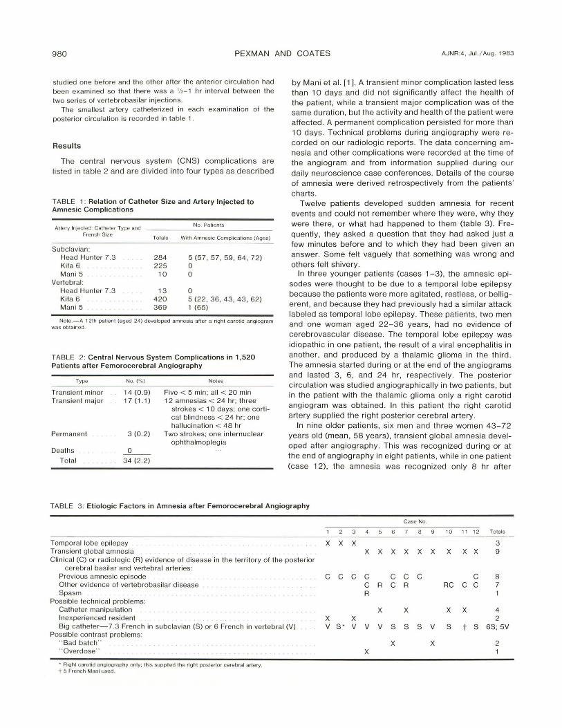
TABLE 3: Etiologic Factors in Amnesia after Femorocerebral Angiography
Case No.
2 3 5 6 8 9 10 11 12 TOlals
Temporal lobe epilepsy X X X 3 Transient global amnesia X X X X X X X X X 9 Clinica l (C) or rad iologic (R) evidence of disease in th e territory of the posterior
cerebral basilar and vertebral arteries: Previous amnesic episode C C C C C C C C 8 Other evidence of vertebrobasilar disease C R C R RC C C 7 Spasm R 1
Possible technical problems: Catheter manipulation X X X X 4 Inexperienced resident X X 2 Big catheter-7 .3 French in subc lavian (S) or 6 French in vertebral (V) V S ' V V V S 8 8 V 8 t 8 68; 5V
Possible contrast problems: " Bad batch" . . . . . . . . . . . . . . X X 2 " Overdose " . . . . . . . . . . . . . . . . . . . X
. Right carotid angiog raphy only: this supplied the ri ght posterior cerebral artery. t 5 French Mani used .
AJNR:4. Jul. / Aug . 1983 ANGIOGRAPHIC AMNESIA 981
angiography. The nine patients had both their anterior and posterior c irculations stud ied. Cases 4-7 and 10-12 had cl inical and/or radiologic evidence of cerebrovascular d isease. Five of the nine cases (cases 4 , 6-8 , and 12) also had had a previous episode of transient g lobal amnesia. The precise duration of that amnesia was difficult to evaluate, but five patients had recovered within 4 hr and all had recovered by 24 hr.
Seven patients (cases 1, 2, 6, 7, and 9-1 1), questioned at various times during their hospital stay, could not recall the angiogram . Four patients (cases 3, 5, 8, and 12) were not asked if they cou ld remember the angiogram. Case 4 was amnesic for 3 hr after angiography, but later c laimed to remember the angiogram . One of the older patients, case 7, developed transient signs indicative of vertebrobasi lar ischemia during his amnesia. No new signs occurred in any other patient. Computed tomography (CT) was performed on all patients before angiography and was normal in all except case 2, the patient with the thalamic glioma.
Clinical vertebrobasilar d isease and a history of transient global amnesia was prominent in those patients who developed transient g lobal amnesia after angiography. Seven (4%) of 167 patients presenting with vertebrobasilar disease developed postangiographic transient g loba l amnesia. Excluding all patients who may conceivably have had temporal lobe ep ilepsy, only one (0.1 %) in 1,200 of those patients without vertebrobasilar disease developed postangiographic transient g lobal amnesia. A patient who developed postangiographic transient g lobal amnesia was at least 10 (and not more than 30) times more li kely to have a history of transient global amnesia than one who did not develop this complication.
In case 4 , a patient with spasm of the basilar artery in whom it was important clinically to prove that a basilar tip aneurysm was present , four injections of 6 ml of contrast material were made into the left vertebral artery, and it was estimated in this case that 30 ml of the total 50 ml of con trast agent used for the investigation entered the posterior circulation.
Technical problems with catheter manipulation were recorded in four patients (cases 5, 7 , 10, and 11). All the amnesic complications occurred in the hands of experienced neuroradiologists with the exception of two of the younger patients (cases 1 and 3), who were catheterized by a resident under supervision.
In five patients the amnesic episodes followed subclavian injections using a 7.3 French Head Hunter catheter . There were no episodes of amnesia when subclavian angiography was carr ied out using 6 French Kifa or 5 French Mani catheters. In five patients the amnesic episodes followed selective vertebral injections using a 6 French Kifa catheter. One patient developed amnesia after a selective vertebral injection with a 5 French Mani catheter. In the patient in whom the posterior cerebral artery originated from the carotid artery , the amnesic episode followed carotid angiography using a 7.3 French Head Hunter cathete r. When the re lation of catheter size and the smallest artery injected in age-matched groups is studied (table 1), a soft size 6 catheter is safer than a stiff size 7.3 catheter in a subclavian
artery and a size 5 catheter is safer than a size 6 catheter in a vertebral artery.
Cases 6 and 9 developed amnesia with the same batch of contrast material (table 3), and at that time six of nine consecutive patients developed complicat ions. Five of these had neurologic comp licat ions, and one had an allergic complication. Three of the patients had the presenting neurologic symptoms reproduced. The probability that two of five consecutive patients would develop amnesia after angiography when the approximate incidence of amnesia is 1 % (12 in 1,520) is 9.7 X 10- 4
, and the probability of five of nine patients developing neurologic comp licat ions when our neurologic complication rate is 2 .2% (table 2) is 6 x 10- 7
using binomial probabili ty . Three patients had a history of temporal lobe epilepsy
and five patients had a history of transient g loba l amnesia. Seven patients had either c lin ical or radiologic evidence of vertebrobasi lar disease (table 3). Overall , therefore , 11 of 12 patients had ev idence of disease in the territories of the posterior cerebral arteries; case 9 was the on ly exception . These 11 patients were regarded as having " susceptible brains. " Situations likely to produce atheromatous debri s or catheter c lot emboli occurred in 11 cases (table 3). In three patients contrast emboli or toxicity may have contributed to the development of the amnesia, and in case 9 it was most strong ly suspected. This 60-year-old man presenting with amaurosis fug ax had no radiologic or c linical evidence of vertebrobasilar disease , and his technically uneventfu l angiogram was obtained by an experienced operator using a 6 French Kifa catheter in the vertebral artery. Thi s patient was one of the six of nine consecutive patients who developed complications during ang iography using the same batch of contrast material.
Discussion
Transient global amnesia is a syndrome described by Fisher and Adams [6] in 1964 in which there is a sudden loss of memory for the present and recent past in individuals in or past middle age, and the patients cannot retain new facts although the remote memory is intact . Charac teristicall y, the patient keeps asking the same question. There is no loss of consc iousness, no physical signs, and the pati ent recovers in several hours but has amnesia for the period of the attack. Single occurrences are the rul e but multiple attacks have been reported [7 -9]. The authors speculated that the cause of the transient global amnesia might be a localized ep ileptic discharge in the temporal lobe, a theory subsequently favored by few [10 , 11], or an ep isode of ischemia involving the limbic system of the medial temporal lobe. The generally accepted theory is that it is due to transient vascular insufficiency of the arteries supplying the medial temporal lobe [12]. Our series has two distinct groups of patients-a younger group of three labeled as temporal lobe epi lepsy and an o lder group of nine classified as transient global amnesia .
Of our 1 ,520 patients, 12 (0 .8% or one in 125) developed postangiographic amnesia, yet thi s complicati on is rarely recorded by others [1 -5]. The com plication rate for patients
982 PEXM AN AND COATES AJNR:4 , Ju l. / Aug . 1983
with cerebrovascular disease and subarachnoid hemorrhage in the 2,316 cases of Mani and Eisenberg [1 3] was 1.7%. They did not say whether am nesia occurred . Eisenberg et al. [1 4] reported a complication rate of 1.3% in 301 cases of cerebrovascular d isease, but they d id not attempt either ve rtebral or subclavian catheteri zation. They reported no am nesic com plicati ons. Although our CNS complicati on rate was 2 .2%, we had a high number of minor transient CNS complications (0.9%) compared with Mani et al. [1] , possibly due to our light sedati on. Because our overa ll comp licati on rates are similar to those of other investigators, we speculate that others may not have recog nized amnesia due to heavier sedation. We would have been unaware of these com pl icati ons if the pati ents had been premedicated with neuroleptanalgesia [15] or Demerol or had the investi gati on been performed under general anesthes ia. The type of sedation used by Mani et al. [1 ] and by Eisenberg et al. [14] is not recorded. Palmer et al. [1 6] , who injected the innominate and subclavian arteri es, may not have noticed amnesic complicat ions because of the neuroleptanalgesia. Silent radiolog ic emboli from cerebrofemoral angiography were described by Cronqvi st [17].
Our statist ics for vertebral and subclavian angiography suggest that a smaller , softer catheter will produce the fewest complicati ons. Eisenberg et al. [14] stated that a 5 French catheter has a surface area only half that of a 7 French catheter, which would correspondingly decrease the potential for emboli , and that the stiffer catheters, whose torque facilitates catheterizati on, may also damage the intima by flailing of the catheter tip . Wishart [2] , who recorded six cases of amnesia in 447 cases, used an 8 French Hinck Head Hunter catheter [1 8]. De Tribolet et al. [4 ], who described Korsakoff syndrome in nine of 832 vertebral angiograms, used a 6.5 French BD red catheter, and we used a 7 .3 French Head Hunter in six of our cases. Lin [19] recommended only using bigger Head Hunters for difficult cases, preferring, as do Mani et al. [ 1] and Eisenberg et al. [1 4 ], the soft 5 French catheter.
Wishart [2 ] also stated that most of his cases were done by residents, and Mani et al. [1] found in 5, 000 cases that res idents prod uced 4 .5 times as many complicati ons as more experienced operators. Although Olivecrona [20], who revi ewed a similar number of cases, d isag rees, comparisons suggest that residents in Californ ia and Stockholm have
sim ilar complication rates. Two of our cases occurred in the hands of a resident .
In Europe, fi ve common brands of contrast medi um have been found to contain intrinsic parti c les [21]. In rubbertipped via ls there are between 6 1 and 369 partic les measuring 5- 10 fLm, 45 to 183 measuring 10 - 30 fL m , and three to f ive measuring greater than 30 fLm in va ri ous brands in 1 ml of contrast materi al. These small part ic les could embolize into the territori es of both posterior cerebral arteri es, the reg ions thought by Horel [22] to be responsible fo r amnesia, and cou ld account for our cases of amnesia in which verteb robasilar disease or temporal lobe epilepsy was present in 11 of our 12 patients. Two cases described by Wales and Nov [3] occurred wh ile using the same batch of contrast medium (Con ray-60), and it was theorized that the compli-
cati ons were due to contrast emboli . We had two similar cases associated wi th one batch of contrast medium .
One of our cases of amnesia occurred when about 30 ml of contrast materi al was injected into a ve rtebra l artery to diag nose a bas ilar aneurysm in the presence of some spasm. Th is amnesia may be due to contrast tox ic ity in a brain made susceptible by atherosc lerosis and arteri al spasm. Tox ic effects of contrast medium can occur wi th very high doses of modern agents [23].
In 14 patients with transient global amnesia reported by Matthew and Meyer [7] , 11 had c linical and 12 had rad iolog ic evide nce of vertebrobas ilar vascular disease; and in our nine cases of transient global amnesia, seve n had c linical and three had rad iolog ic evid ence of vertebrobasilar disease. Five of our nine cases of transient global amnesia had a previous amnesic episode, and all of our cases di ag nosed as temporal lobe epilepsy had had a similar epi sode. All these factors suggest that there are areas in the hippocampi made susceptible by disease processes that can be tri ggered either by an operator or contrast- induced factor. Overd oses with diazepam may produce transient global amnesia [24] , but in our cases 10 mg oral Val ium or 30 mg Serax was chosen to produce mild tranquil ization with minimal drowsiness.
REFERENCES
1. Man i RL, Ei senberg RL, McDonald EJ, Pollock JA, Man i JR . Complicat ions of catheter cerebral arteriog raphy: analysis of 5000 procedures. 1. Criteria and inc idence. AJR 1978; 131 :861-865
2. Wishart DL. Complications of vertebral angiography as compared to non-vertebral cerebral angiog raphy in 447 stud ies. AJR 1971 ;113:527-537
3. Wales LR, Nov AA. Transient global amnesia: complication of cerebral ang iography. AJNR 1981 ;2 : 275 - 277
4. de Tribolet N, Assai G, Oberson R. Syndrome de Korsakoff et cecite corticale transi toires apres ang iog raphie vertebrale. Schweiz Med Wochenschr 1975; 105: 1 506-1 509
5. Shuttleworth EC, Wise GR. Transient global amnesia due to arterial embolism. Arch Neuro/1973; 29: 340-342
6. Fisher CM, Adams RD. Transient global amnesia. Acta Neurol Scand 1964 ;40[SuppI 9 ];7 - 83
7. Matthew NT, Meyer JS. Pathogenesis and natural history of transient global amnesia. Stroke 1974;5: 303-311
9. Poser CM, Sieg ier OK. Temporary amnesia as a manifestat ion of cerebral vascular insuffic iency. Trans Am Neurol Assoc 1960;85:22 1-223
10. Green HH , Bennett DR. Transient global amnesia with previously unreported EEG abnormality. Electroencephalogr Clin Neurophysio /1974 ;36: 409- 413
11 . Lou HOC. Repeated episodes of transient global amnesia. Acta Neurol Scand 1968;44: 612- 6 18
12. Shuping JR, Rollinson RD, Toole JF. Transient global amnesia. Ann Neuro/1980 ;7: 28 1-285
13. Mani RL , Eisenberg RL. Complicat ions of catheter cerebral arteriog raphy: analys is of 5000 proced ures. II. Relation of complication rates to clinical and arteriographic diagnosis. AJR 1978;131 :867- 869
14. Eisenberg RL, Bank WO, Hedgcock MW. Neurolog ic compli-
AJNR:4, Jul. / Aug. 1983 ANGIOGRAPHIC AMNESIA 983
cations of angiography for cerebrovascular d isease. Neuro logy (N Y) 1980;30 : 895-8 97
15 . Hoy RJ , Sorby WA. " Minineuro lept. " A note on neuroleptanalgesia in radiologic procedures. Austra las Radiol 1968; 12:268 - 269
16. Palmer FJ, Bri scoe PJ , Sorby WA, Barry BP, Williams RM . Transfemoral selec tive angiography in the investigation of cerebral ischaemic di sease. Review of 400 consecutive studies. 1. Technique and evaluation of method . Australas Radiol 1977;2 1 :31-38
17 . Cronqvist S, Efsing HO, Palacios E. Emboli complications in cerebral angiography with the catheter technique. Acta Radiol [Diagn] (Stockh) 1970; 10 : 9 7 -1 07
18. Hinck VC, Judkins MP , Paxton HO . Simplified selecti ve femo-
rocerebral angiography. Radiology 1967;89: 1 048-1 052 19. Lin JP. Techniques of cerebral angiog raphy. Radiol Clin North
Am 1974;12:223-240 20 . Olivecrona H. Complications of cerebral angiog raphy. Neuro
radiology 1977; 14: 1 75-1 8 1 2 1 . Windin g O. Intrinsic parti c les in angiog raphic con trast med ia.
Radiology 1980; 134: 317 -320 22. Horel JA. Neuroanatomy of amnesia-a criti que of hippocam
pal memory hypothesis. Brain 1978; 101 : 403-445 23. Murphy OJ . Cerebrovascular permeability after meg lumine io
thalamate admini strati on. Neurology (NY) 1973;23 : 926-936 24. Gilbert JJ , Benson OF. Transient global amnesia. Report of two
cases with definit e etiolog ies. J Nerv Ment Dis 1972; 154 : 461 -464