10/28/11 OSN Supersite 1/5 www.osnsupersite.com/print.aspx?rid=30182 Thomas John Amniotic membrane transplant key to pterygium surgical method Technique may provide a more prolonged antifibrotic effect and a decreased recurrence rate, but further study is needed. By Thomas John, MD; John A. Hovanesian, MD, FACS; Andrew Behesnilian, BS Introduction There is a continued search among ophthalmic surgeons for the optimal surgical technique for pterygium surgery. This is partly because pterygium surgery is associated with potential postoperative complications, including recurrence of pterygium. My guests in this corneal dissection column, John A. Hovanesian, MD, FACS, and Andrew Behesnilian, BS, describe their surgical technique of using amniotic membrane transplantation as a “biologic implant” beneath the normal conjunctiva surrounding the region where the pterygium is excised and replaced with a conjunctival autograft. They said the recurrence rate of 5% after pterygium surgery combined with conjunctival autograft is similar to the eyes receiving amniotic membrane transplantation in the region where the pterygium is excised. The guests are of the opinion that placing the amniotic membrane graft in the surgically created subconjunctival space may provide a more prolonged antifibrotic effect, as compared with placing it on the ocular surface, and may contribute to a decreased recurrence rate of the pterygium. This technique must be further examined in larger studies to address all of the ocular effects of such a procedure. The preferred anesthesia for this technique is with a peribulbar block of bupivacaine and lidocaine 1% with epinephrine. Although subconjunctival infiltration of anesthetic agents is also effective for the procedure, this may result in more postoperative patient discomfort. Story continues below! ADVERTISEMENT
Transcript
10/28/11 OSN Supersite
1/5www.osnsupersite.com/print.aspx?rid=30182
ThomasJohn
Amniotic membrane transplant key to pterygium surgical methodTechnique may provide a more prolonged antifibrotic effect and a decreased recurrence rate, but further
study is needed.By Thomas John, MD; John A. Hovanesian, MD, FACS; Andrew Behesnilian, BS
Introduction
There is a continued search among ophthalmic surgeons for the optimal surgical technique for pterygium surgery. This is partly becausepterygium surgery is associated with potential postoperative complications, including recurrence of pterygium.
My guests in this corneal dissection column, John A. Hovanesian, MD, FACS, and Andrew Behesnilian, BS, describetheir surgical technique of using amniotic membrane transplantation as a “biologic implant” beneath the normal
conjunctiva surrounding the region where the pterygium is excised and replaced with a conjunctival autograft. Theysaid the recurrence rate of 5% after pterygium surgery combined with conjunctival autograft is similar to the eyesreceiving amniotic membrane transplantation in the region where the pterygium is excised. The guests are of the
opinion that placing the amniotic membrane graft in the surgically created subconjunctival space may provide a moreprolonged antifibrotic effect, as compared with placing it on the ocular surface, and may contribute to a decreased
recurrence rate of the pterygium. This technique must be further examined in larger studies to address all of the oculareffects of such a procedure.
The preferred anesthesia for this technique is with a peribulbar block of bupivacaineand lidocaine 1% with epinephrine. Although subconjunctival infiltration of anesthetic agents is also
effective for the procedure, this may result in more postoperative patient discomfort.
Story continues below!
ADVERTISEMENT
10/28/11 OSN Supersite
2/5www.osnsupersite.com/print.aspx?rid=30182
Step-by-step surgery
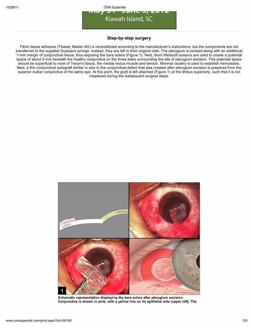
Fibrin tissue adhesive (Tisseel, Baxter AG) is reconstituted according to the manufacturer’s instructions, but the components are nottransferred to the supplied Duploject syringe. Instead, they are left in their original vials. The pterygium is excised along with an additional1-mm margin of conjunctival tissue, thus exposing the bare sclera (Figure 1). Next, blunt Westcott scissors are used to create a potentialspace of about 5 mm beneath the healthy conjunctiva on the three sides surrounding the site of pterygium excision. This potential spaceshould be superficial to most of Tenon’s fascia, the medial rectus muscle and tendon. Minimal cautery is used to establish hemostasis.
Next, a thin conjunctival autograft similar in size to the conjunctival defect that was created after pterygium excision is prepared from thesuperior bulbar conjunctiva of the same eye. At this point, the graft is left attached (Figure 1) at the limbus superiorly, such that it is not
misplaced during the subsequent surgical steps.
Schematic representation displaying the bare sclera after pterygium excision.Conjunctiva is shown in pink, with a yellow line on its epithelial side (upper left); Thepterygium has been excised and the autograft prepared and reflected onto the cornea at
10/28/11 OSN Supersite
3/5www.osnsupersite.com/print.aspx?rid=30182
pterygium has been excised and the autograft prepared and reflected onto the cornea atthe superior limbus (upper right); Freeze-dried human amniotic membrane is cut into aC-shaped graft (lower left); The amniotic membrane is placed in the subconjunctivalregion surrounding the site of pterygium excision. The location of the amnioticmembrane graft is shown in red, surrounding the area where the conjunctival autograftwill be placed (lower right).
Images: Hovanesian JA
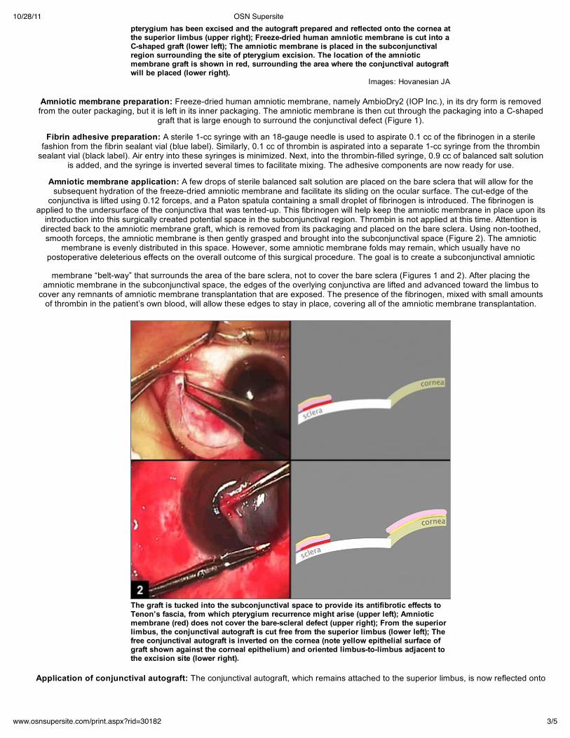
Amniotic membrane preparation: Freeze-dried human amniotic membrane, namely AmbioDry2 (IOP Inc.), in its dry form is removedfrom the outer packaging, but it is left in its inner packaging. The amniotic membrane is then cut through the packaging into a C-shaped
graft that is large enough to surround the conjunctival defect (Figure 1).
Fibrin adhesive preparation: A sterile 1-cc syringe with an 18-gauge needle is used to aspirate 0.1 cc of the fibrinogen in a sterilefashion from the fibrin sealant vial (blue label). Similarly, 0.1 cc of thrombin is aspirated into a separate 1-cc syringe from the thrombin
sealant vial (black label). Air entry into these syringes is minimized. Next, into the thrombin-filled syringe, 0.9 cc of balanced salt solutionis added, and the syringe is inverted several times to facilitate mixing. The adhesive components are now ready for use.
Amniotic membrane application: A few drops of sterile balanced salt solution are placed on the bare sclera that will allow for thesubsequent hydration of the freeze-dried amniotic membrane and facilitate its sliding on the ocular surface. The cut-edge of the
conjunctiva is lifted using 0.12 forceps, and a Paton spatula containing a small droplet of fibrinogen is introduced. The fibrinogen isapplied to the undersurface of the conjunctiva that was tented-up. This fibrinogen will help keep the amniotic membrane in place upon its
introduction into this surgically created potential space in the subconjunctival region. Thrombin is not applied at this time. Attention isdirected back to the amniotic membrane graft, which is removed from its packaging and placed on the bare sclera. Using non-toothed,
smooth forceps, the amniotic membrane is then gently grasped and brought into the subconjunctival space (Figure 2). The amnioticmembrane is evenly distributed in this space. However, some amniotic membrane folds may remain, which usually have no
postoperative deleterious effects on the overall outcome of this surgical procedure. The goal is to create a subconjunctival amniotic
membrane “belt-way” that surrounds the area of the bare sclera, not to cover the bare sclera (Figures 1 and 2). After placing theamniotic membrane in the subconjunctival space, the edges of the overlying conjunctiva are lifted and advanced toward the limbus to
cover any remnants of amniotic membrane transplantation that are exposed. The presence of the fibrinogen, mixed with small amountsof thrombin in the patient’s own blood, will allow these edges to stay in place, covering all of the amniotic membrane transplantation.
The graft is tucked into the subconjunctival space to provide its antifibrotic effects toTenon’s fascia, from which pterygium recurrence might arise (upper left); Amnioticmembrane (red) does not cover the bare-scleral defect (upper right); From the superiorlimbus, the conjunctival autograft is cut free from the superior limbus (lower left); Thefree conjunctival autograft is inverted on the cornea (note yellow epithelial surface ofgraft shown against the corneal epithelium) and oriented limbus-to-limbus adjacent tothe excision site (lower right).
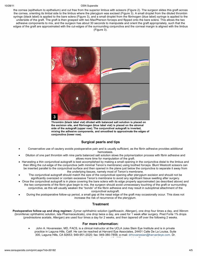
Application of conjunctival autograft: The conjunctival autograft, which remains attached to the superior limbus, is now reflected ontothe cornea (epithelium to epithelium) and cut free from the superior limbus with scissors (Figure 2). The surgeon slides this graft across
10/28/11 OSN Supersite
4/5www.osnsupersite.com/print.aspx?rid=30182
the cornea (epithelium to epithelium) and cut free from the superior limbus with scissors (Figure 2). The surgeon slides this graft acrossthe cornea, orienting its limbal side to the limbus where the pterygium was excised (Figure 3). A small droplet from the diluted thrombinsyringe (black label) is applied to the bare sclera (Figure 3), and a small droplet from the fibrinogen (blue label) syringe is applied to the
underside of the graft. The graft is then grasped with two MacPherson forceps and flipped onto the bare sclera. This allows the twoadhesive components to mix, and the surgeon has about 30 seconds to manipulate and orient the graft appropriately, such that the
edges of the graft are approximated with the cut-edges of the surrounding conjunctiva and the corneal margin is aligned with the limbus(Figure 3).
Thrombin (black label vial) diluted with balanced salt solution is placed onthe excision site, and fibrinogen (blue label vial) is placed on the stromalside of the autograft (upper row); The conjunctival autograft is inverted,mixing the adhesive components, and smoothed to approximate the edges ofconjunctiva (lower row).
Surgical pearls and tips
Conservative use of cautery avoids postoperative pain and is usually sufficient, as the fibrin adhesive provides additionalhemostasis.
Dilution of one part thrombin with nine parts balanced salt solution slows the polymerization process with fibrin adhesive andallows more time for manipulation of the graft.
Harvesting a thin conjunctival autograft is best accomplished by making a small opening in the conjunctiva distal to the limbus andthen lifting the cut-edge of the conjunctiva (with minimal Tenon’s membrane) using toothed forceps. Blunt Westcott scissors canbe inserted parallel to the conjunctival surface and then opened in the plane just below the conjunctiva to separate it away from
the underlying tissues, namely most of Tenon’s membrane.The conjunctival autograft should match the size of the conjunctival opening after pterygium excision and should not be
significantly oversized or contain excessive Tenon’s membrane to avoid any significant tissue swelling after surgery.Once the conjunctival autograft is in place covering the bare sclera with its edge properly approximated (as described above) and
the two components of the fibrin glue begin to mix, the surgeon should avoid unnecessary touching of the graft or surroundingconjunctiva, as this will usually weaken the “bonds” of the fibrin adhesive and may result in suboptimal attachment of the
conjunctival autograft.During the postoperative follow-up period, a small gap at the nasal edge of the graft may occasionally occur. This does not
increase the risk of recurrence of the pterygium.
Treatment
Postoperative follow-up and drug regimen: Zymar ophthalmic solution (gatifloxacin, Allergan), one drop four times a day, and Xibrom(bromfenac ophthalmic solution, Ista Pharmaceuticals), one drop twice a day, are used for 1 week after surgery. Pred Forte 1% drops
(prednisolone acetate, Allergan) are used four times a day for 2 weeks, and then tapered off over the following 2 weeks.
For more information:John A. Hovanesian, MD, FACS, is a clinical instructor at the UCLA Jules Stein Eye Institute and is in private
practice in Laguna Hills, Calif. He can be reached at Harvard Eye Associates, 24401 Calle De La Louisa, Suite300, Laguna Hills, CA 92653; 949-951-2020; fax: 949-380-7856; e-mail: [email protected]. Dr.
Hovanesian is a consultant to Allergan, Baxter AG, IOP Inc. and Ista Pharmaceuticals.
10/28/11 OSN Supersite
5/5www.osnsupersite.com/print.aspx?rid=30182
Hovanesian is a consultant to Allergan, Baxter AG, IOP Inc. and Ista Pharmaceuticals.Thomas John, MD, is a clinical associate professor at Loyola University at Chicago and is in private practice in
Tinley Park and Oak Lawn, Ill. He can be reached at 708-429-2223; fax: 708-429-2226; e-mail:[email protected]. Dr. John has no direct financial interest in the products discussed in this article, nor is he a
paid consultant for any companies mentioned.
References:
Chui J, Di Girolamo N, Wakefield D, Coroneo MT. The pathogenesis of pterygium: Current concepts and theirtherapeutic implications. Ocul Surf. 2008;6:24-43.
De la Hoz F, Montero JA, et al. Efficacy of mitomycin C associated with direct conjunctival closure and slidingconjunctival graft for pterygium surgery. Br J Ophthalmol. 2008;92:175-178.
Hovanesian JA, Behesnilian A. Results of pterygium excision using amniotic membrane beneath the healthyconjunctiva surrounding a conjunctival autograft. Presented at: Annual Meeting of the American Society of
Cataract and Refractive Surgery; 2008; Chicago.Kenyon KR, Wagoner MD, Hettinger ME. Conjunctival autograft transplantation for advanced and recurrent
pterygium. Ophthalmology 1985;92:1461-1470.Ma DH, See LC, Liau SB, Tsai RJ. Amniotic membrane graft for primary pterygium: Comparison with conjunctival
autograft and topical mitomycin C treatment. Br J Ophthalmol. 2000;84:973-978.Memarzadeh F, Fahd AK, Shamie N, Chuck RS. Comparison of de-epithelialized amniotic membrane
transplantation and conjunctival autograft after primary pterygium excision. Eye. 2008;22:107-112.