47
AMOEBAE M.PRASAD NAIDU MSc, (Medical) Ph.D. (Medical)
| Date post: | 30-Dec-2015 |
| Category: |
Documents |
| Upload: | mprasadnaidu |
| View: | 21 times |
| Download: | 0 times |

AMOEBAE
M.PRASAD NAIDUMSc, (Medical) Ph.D. (Medical)

• An amoeba (also ameba, amœba or amoeboid) is a type of cell or organism which has the ability to alter its shape, primarily by extending and retracting pseudopods. Amoebae do not form a single taxonomic group, but are found in every major lineage of eukaryotic organisms (domain Eukaryota).

• Amoeoboid cells occur not only among the protozoa, but also fungi, algae and animals.• Among microbiologists, the terms
"amoeboid" and "amoebae" are often used interchangeably for any organism that exhibits amoeboid movement.

• The best known amoeboid protists are the "giant amoebae" Chaos carolinense and Amoeba proteus, both of which are widely cultivated and studied in classrooms and laboratories. Other well known species include the so-called "brain-eating amoeba" Naegleria fowleri, the intestinal parasiteEntamoeba histolytica, which causes amoebic dysentery, and the multicellular "social amoeba" Dictyostelium discoideum.

Naegleria fowleri :
Primary Amoebic Meningo
Encephalitis
(PAM)
Fowler & Carter (1965 ).
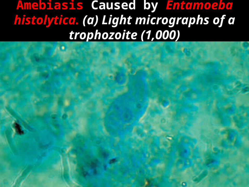
Amebiasis Caused by Entamoeba histolytica. (a) Light micrographs of a trophozoite (1,000)

A cyst (1,000).

• TROPHOZOITE :
Vegetative & Feeding stage .
CSF &Tissue .
10 -20 µm , Karyosome with halo .
Actively motile (Lobopodia )
Binary fission .
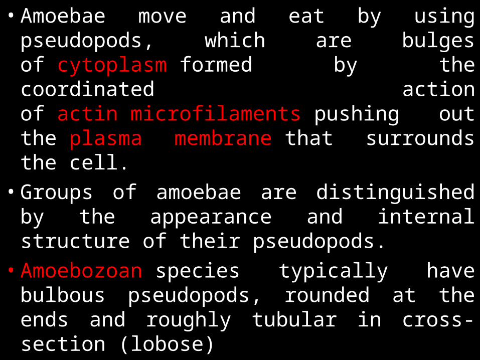
• Amoebae move and eat by using pseudopods, which are bulges of cytoplasm formed by the coordinated action of actin microfilaments pushing out the plasma membrane that surrounds the cell.
• Groups of amoebae are distinguished by the appearance and internal structure of their pseudopods.
• Amoebozoan species typically have bulbous pseudopods, rounded at the ends and roughly tubular in cross-section (lobose)

• Cercozoan amoeboids, such as Euglypha and Gromia, have slender, thread-like (filose) pseudopods. Foraminiferan emit fine, branching pseudopods that merge with one another to form net-like (reticulose) structures. Some groups, such as the Radiolaria and the amoeboids loosely called Heliozoa, have stiff, needle-like, radiating actinopods supported from within by bundles of microtubules

SHAPE, MOVEMENT
AND NUTRITION
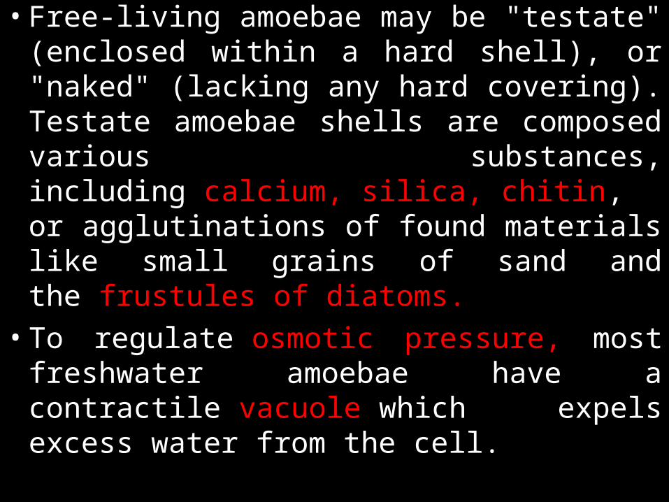
• Free-living amoebae may be "testate" (enclosed within a hard shell), or "naked" (lacking any hard covering). Testate amoebae shells are composed various substances, including calcium, silica, chitin, or agglutinations of found materials like small grains of sand and the frustules of diatoms.
• To regulate osmotic pressure, most freshwater amoebae have a contractile vacuole which expels excess water from the cell.

• This organelle is necessary because freshwater has a lower concentration of solutes (such as salt) than the amoeba's own internal fluids (cytosol). Because the surrounding water is hypotonic with respect to the contents of the cell, water is transferred across the amoeba's cell membrane by osmosis. Without a contractile vacuole, the cell would fill with excess water and, eventually, burst. Marine amoebae do not usually possess a contractile vacuole, because the concentration of solutes within the cell are in balance with the tonicity of the surrounding water.
• The food sources of amoebae vary. Some amoebae are predatory and live by consuming bacteria and other protists.

• Some are detritivores and eat dead organic material. Amoebae typically ingest their food by phagocytosis, extending pseudopods to encircle and engulf live prey or particles of scavenged material.
• Amoeboid cells do not have a mouth or cytostome, and there is no fixed place on the cell at which phagocytosis normally occurs. Some amoebae also feed by pinocytosis, imbibing dissolved nutrients through vesicles formed within the cell membrane.

AMOEBAE IN MULTICELLULAR
ORGANISMS: ANIMALS AND SLIME MOLDS
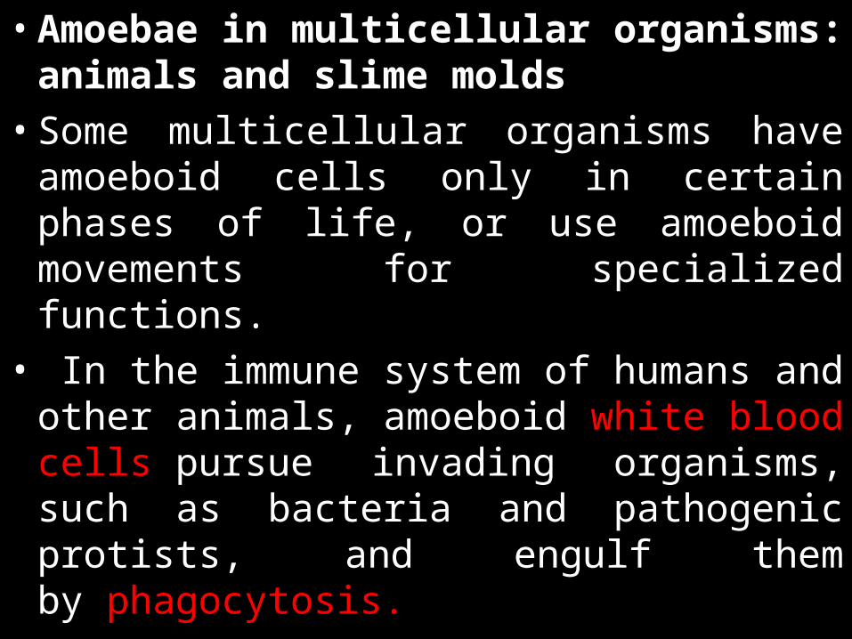
• Amoebae in multicellular organisms: animals and slime molds
• Some multicellular organisms have amoeboid cells only in certain phases of life, or use amoeboid movements for specialized functions.
• In the immune system of humans and other animals, amoeboid white blood cells pursue invading organisms, such as bacteria and pathogenic protists, and engulf them by phagocytosis.
• Amoeboid stages also occur in the multicellular fungus-like protists, the so-called slime molds. • Both the plasmodial slime molds,
currently classified in the class Myxogastria, and the cellular slime molds of the groups Acrasida and Dictyosteliida, live as amoebae during their feeding stage.

• The cells of the former form a giant multinucleate amoeboid organism,while the cells of the latter live separately until food runs out, at which time the amoebae aggregate to form a multicellular migrating slug which functions as a single organism

LIFE CYCLE :
ONE HOST : Man .
Asexual Generation cycle .
MOI : Swimming in Contaminated water .
Infective form : Trophozoites .
Cysts (Inhalation ) –Rare .
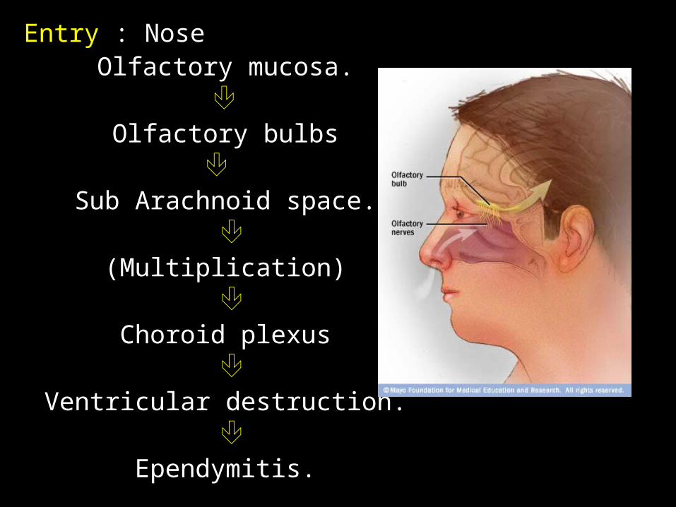
Entry : Nose Olfactory mucosa.
Olfactory bulbs
Sub Arachnoid space.
(Multiplication)
Choroid plexus
Ventricular destruction.
Ependymitis.


• Trophozoites Cysts (unfavorable conditions ) .
• PATHOGENESIS :
IP : 2 -15 days .(5 days )
Neurotropic .
Brain tissue destruction .
Acute Hemorrhagic Necrotising meningo
encephalitis .
Cysts are absent in humans .

CLINICAL FEATURES :
Children & Young adults .
PAM .
Rapid onset & Fulminant .

Sudden severe Persistent Bifrontal /Bitemporal
Headache ,Nausea , Projectile vomiting .
Ageusia (loss of taste function).
Parosmia (olfactory dysfunction).
Generalized seizures
Photophobia
Coma
Death
Poor Prognosis .(95 % death )
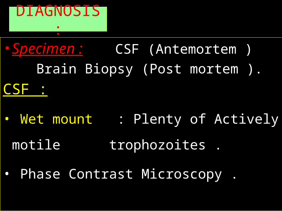
DIAGNOSIS :
• Specimen : CSF (Antemortem ) Brain Biopsy (Post
mortem ).
CSF :
• Wet mount : Plenty of Actively motile
trophozoites .
• Phase Contrast Microscopy .
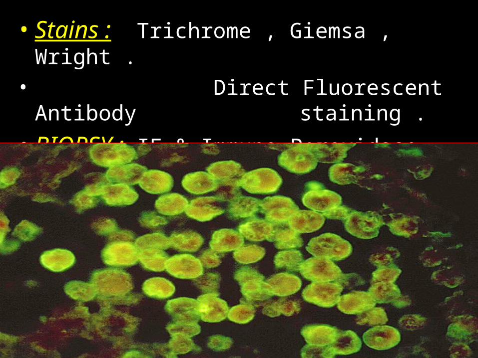
• Stains : Trichrome , Giemsa , Wright .• Direct Fluorescent Antibody
staining .• BIOPSY : IF & Immuno Peroxidase method .

• Serodiagnosis : Not Useful .
• Molecular methods : DNA probes & PCR .
• CSF : Features of Pyogenic
Meningitis .

• Treatment : Amphotericin B & Rifampicin,Miconazole .
• Prevention :
Acanthamoeba• Opportunistic pathogens .
1. A.castellani .
2. A.astronyxis .
3. A.polyphaga .
4. A.culbertsoni .

• HABITAT : Dust , Soil , Sand , Rivers ,
Ponds , Tap water .
• MORPHOLOGY :
2 stages .
1. Trophozoite .
2. Cyst .
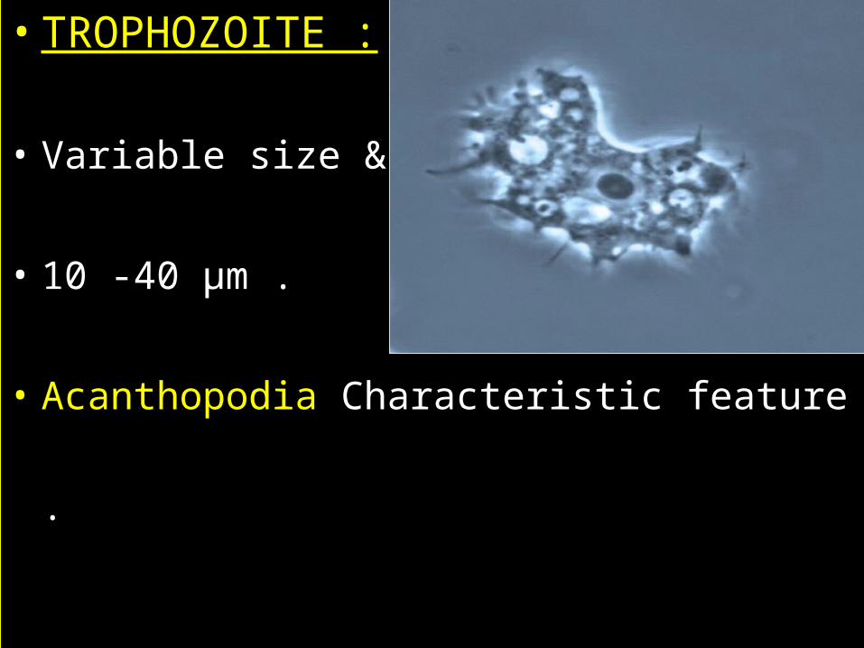
• TROPHOZOITE :
• Variable size & shape .
• 10 -40 µm .
• Acanthopodia Characteristic feature .

• CYST :
Polygonal /Spherical /Star shaped .
15 -20 µm.
Double layered cyst wall .

• LIFE CYCLE :
• MOI : Invasion of Broken skin .
• Inhalation of Cysts & Trophozoites .
• ROUTE : Lungs to Blood stream
(Multiplication )
• CNS .
• Sites of Localisation :Basal ganglia ,
• Posterior fossa ,
Cerebellum .


• PATHOGENESIS :
• Lesions of Brain , Eye , Lungs & Skin .
• Brain: Patchy , Sub Acute lesions of
granulomatous encephalitis .
• Skin: Nodular & Ulcerative lesion. Abscess .
• Cornea : Epithelial inflammation, Hypopyon .


• CLINICAL MANIFESTATIONS :
Granulomatous Amoebic Encephalitis (GAM)
Acanthamoeba Keratitis .
Cutaneous Lesions .

GAE:
• Rare .
Risk factors :
• AIDS ,
• Immunosupression,Organ transplantation
• Malnourished .
• Clinical features : Low grade fever,stiff neck , Altered Mental status . Seizures , Cranial palsies , Hemiparesis , Ataxia , Photophobia Coma , Multi organ Failure , Death .

• Acanthamoeba Keratitis :
Contact lens wearers .(trauma )
Chronic , Progressive , Ulceration .
Annular Infiltration & Congested Cornea .
Perforation .
Blindness .

• CUTANEOUS LESIONS :
• Face & Extremities .
• Nodules , papules & ulcers .
• Poor Prognosis .
• GAE Fatal .
• DIAGNOSIS :
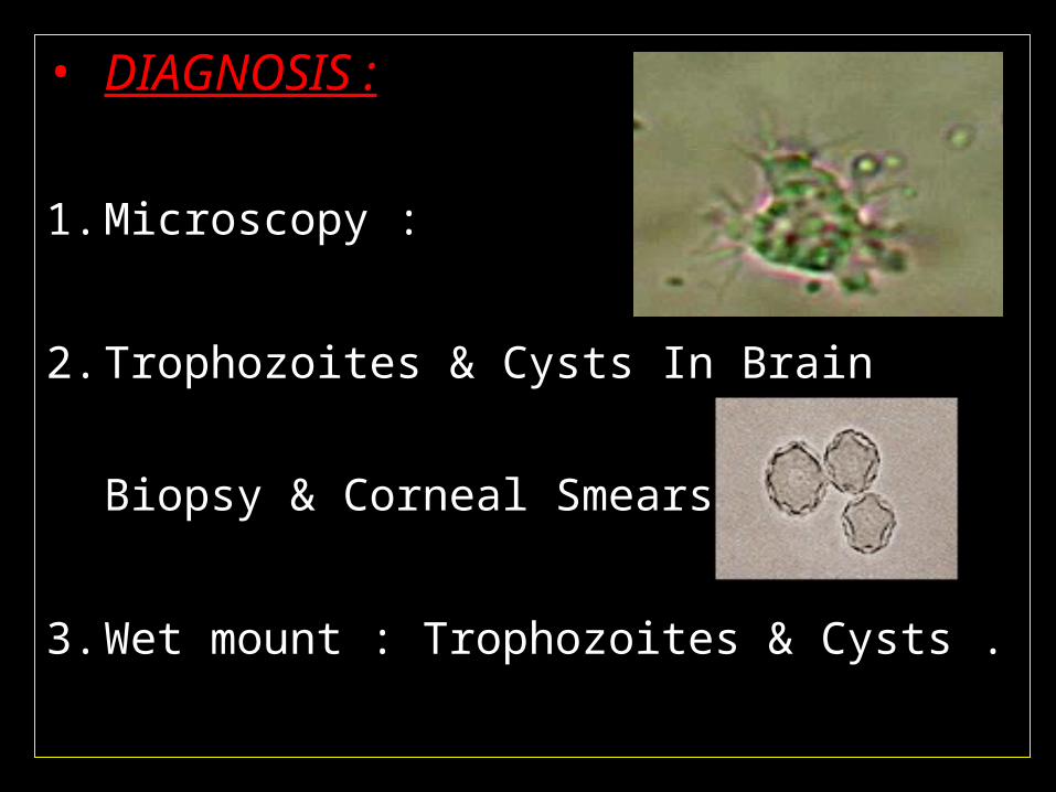
1. Microscopy :
2. Trophozoites & Cysts In Brain Biopsy & Corneal
Smears .
3. Wet mount : Trophozoites & Cysts .
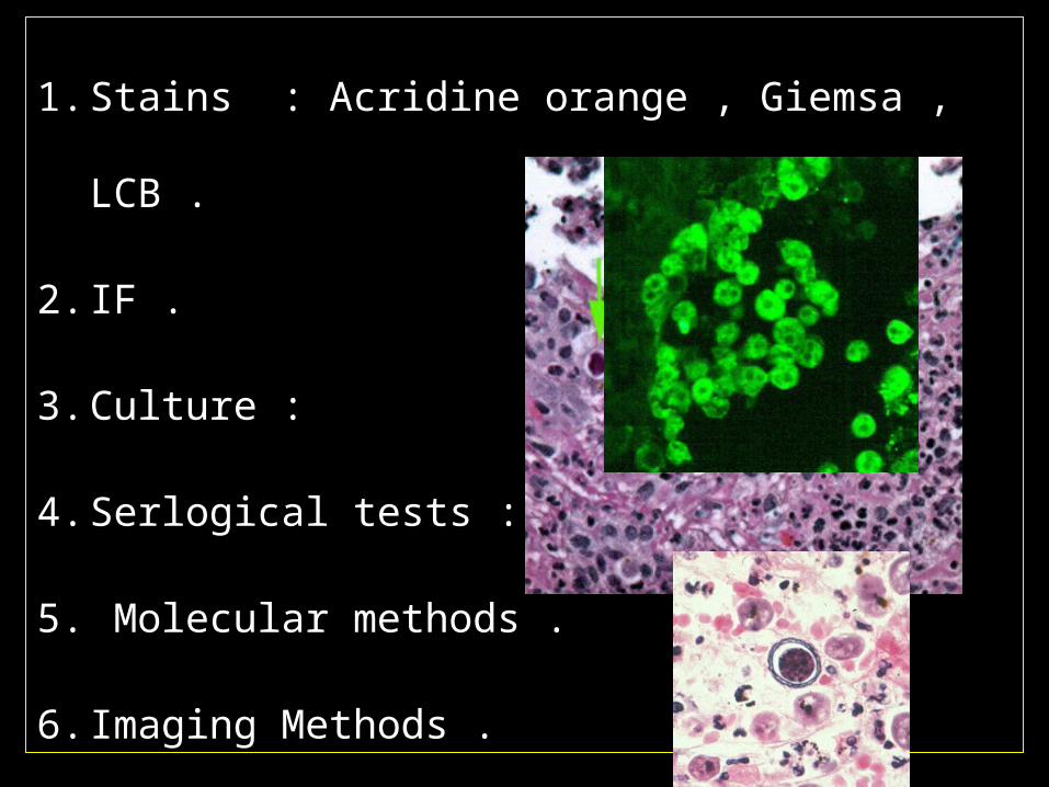
1. Stains : Acridine orange , Giemsa , LCB .
2. IF .
3. Culture :
4. Serlogical tests : Not useful .
5. Molecular methods .
6. Imaging Methods .
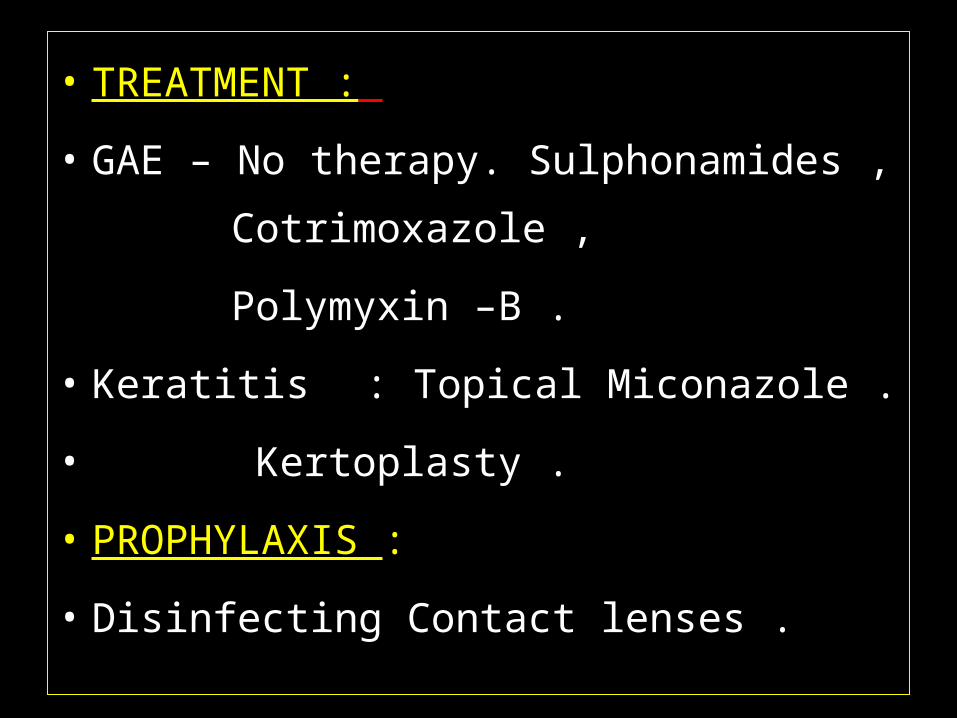
• TREATMENT :
• GAE – No therapy. Sulphonamides ,
Cotrimoxazole ,
Polymyxin –B .
• Keratitis : Topical Miconazole .
• Kertoplasty .
• PROPHYLAXIS :
• Disinfecting Contact lenses .
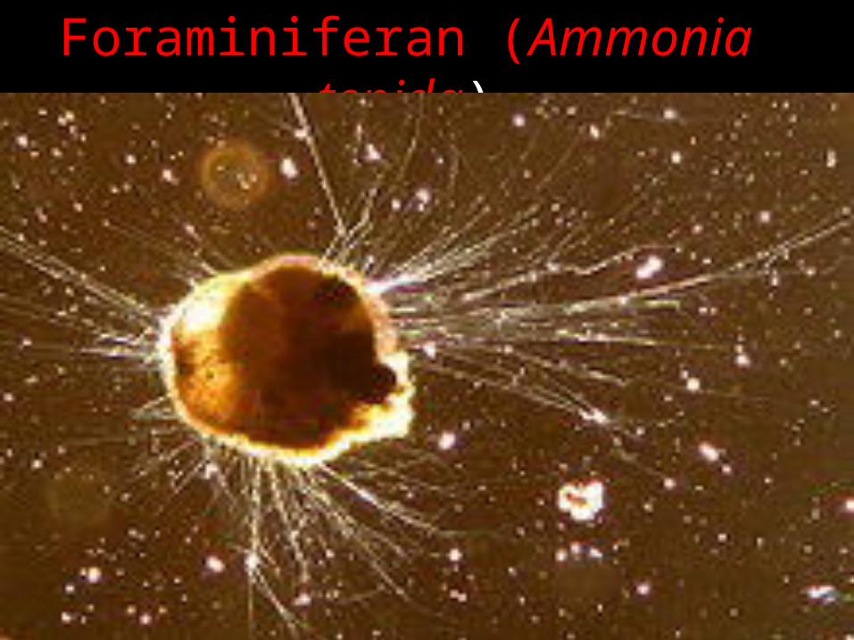
Foraminiferan (Ammonia tepida)

Shell of the testate amoeba Difflugia acuminata, made up of mineral particles

THANK YOU


