Plant-pathogen interactions are complex and dynamic and in-volve diverse recognition and signal transduction networks. At the heart of these interactions, massive gene expression chang-es govern the outcome. The mechanisms initiating and regulat-ing gene expression are of particular interest in understanding plant-pathogen interactions. The manipulation of host chromatin is a powerful strategy to alter gene expression, but the mech-anistic understanding of plant chromatin changes during plant- pathogen interactions, particularly how pathogens regulate host chromatin changes, remains largely obscure. The full relevance of this mechanism in plant-pathogen interactions is still emerg-ing (Alvarez et al., 2010; Berr et al., 2012; Dowen et al., 2012; Yu et al., 2013; Ding and Wang, 2015; Rambani et al., 2015; Yang et al., 2015; Zhu et al., 2016). Pathogens deliver a repertoire of effectors into plant cells that counteract defense responses or alter host cells to modulate cellular processes to support pathogen survival. Cyst nema-todes are plant-parasitic animals that reprogram plant root cells by secreting effectors to create a large, highly metabolically
active nutrient sink known as the syncytium, from which they feed (Hewezi and Baum, 2013; Mitchum et al., 2013; Hewezi et al., 2015). Obviously, effectors are of particular interest when exploring pathogen-triggered gene expression changes in the host. Here, we present the function of the 32E03 effector of the sugar beet cyst nematode Heterodera schachtii, which also in-fects the model plant Arabidopsis thaliana. H. schachtii effector 32E03 is a homolog of the uncharacterized soybean cyst nema-tode (Heterodera glycines) 32E03 effector (GenBank accession number AF500036) (Gao et al., 2003). Our analyses unveil that the 32E03 effector interacts with the Arabidopsis FK506 bind-ing protein FKBP53 and the plant-specific tuin-type histone deacetylase (HDAC) HDT1 in the plant nucleolus. FKBP53 is an immunophilin-type peptidyl propyl cis-trans isomerase and a histone chaperone (Li and Luan, 2010). Tuin-type HDACs play roles in plant growth and responses to environmental stimuli (Colville et al., 2011; Luo et al., 2012; Yano et al., 2013; Zhao et al., 2015; Han et al., 2016). Guided by this discovery, we show that the 32E03 protein acts as a potent inhibitor of plant his-tone deacetylase activities. Because we had identified HDT1 and FKBP53 as 32E03 interaction partners, we functionally characterized 32E03 deploying the reported HDT1/FKBP53 effects on rDNA regulation as an example of how HDAC inhi-bition by a pathogen effector can alter host gene expression. In these studies, we determined that the 32E03 effector medi-ates a dose-dependent epigenetic control of plant rRNA gene expression, which regulates rRNA gene dosage and influences cyst nematode parasitism.
An Effector from the Cyst Nematode Heterodera schachtii Derepresses Host rRNA Genes by Altering Histone Acetylation[OPEN]
Paramasivan Vijayapalani,a Tarek Hewezi,b Frederic Pontvianne,c,d and Thomas J. Bauma,1
aDepartment of Plant Pathology and Microbiology, Iowa State University, Ames, Iowa 50011 bDepartment of Plant Sciences, University of Tennessee, Knoxville, TN 37996cUniversité de Perpignan Via Domitia, Laboratoire Génome et Développement des Plantes, UMR5096, F-66860 Perpignan, FrancedUniversité de Perpignan Via Domitia, Laboratoire Génome et Développement des Plantes, UMR5096, F-66860, Perpignan, France
Cyst nematodes are plant-pathogenic animals that secrete effector proteins into plant root cells to alter host gene expres-sion and reprogram these cells to form specialized feeding sites, known as syncytia. The molecular mechanisms of these effectors are mostly unknown. We determined that the sugar beet cyst nematode (Heterodera schachtii) 32E03 effector protein strongly inhibits the activities of Arabidopsis thaliana histone deacetylases including the HDT1 enzyme, which has a known function in the regulation of rRNA gene expression through chromatin modifications. We determined that plants ex-pressing the 32E03 coding sequence exhibited increased acetylation of histone H3 along the rDNA chromatin. At low 32E03 expression levels, these chromatin changes triggered the derepression of a subset of rRNA genes, which were conducive to H. schachtii parasitism. By contrast, high levels of 32E03 caused profound bidirectional transcription along the rDNA, which triggered rDNA-specific small RNA production leading to RNA-directed DNA methylation and silencing of rDNA, which inhib-ited nematode development. Our data show that the 32E03 effector alters plant rRNA gene expression by modulating rDNA chromatin in a dose-dependent manner. Thus, the 32E03 effector epigenetically regulates plant gene expression to promote cyst nematode parasitism.
1Address correspondence to [email protected]. The author responsible for distribution of materials integral to the findings presented in this article in accordance with the policy described in the Instructions for Authors (www.plantcell.org) is: Thomas J. Baum ([email protected]).[OPEN]Articles can be viewed without a subscription.www.plantcell.org/cgi/doi/10.1105/tpc.18.00570
Effector 32E03 Is Important for H. schachtii Pathogenicity
We determined by in situ hybridization that 32E03 mRNA ac-cumulates in the dorsal esophageal gland cell of H. schachtii (Figure 1A), which is a hallmark characteristic of many nem-atode effectors. Furthermore, we confirmed the presence of 32E03 mRNA in preparasitic and parasitic developmental stag-es of H. schachtii by RT-qPCR analyses (Figure 1B). In order to determine the biological relevance of 32E03 in cyst nematode- Arabidopsis interactions, we tested the pathogenicity of H. schachtii nematodes in which 32E03 gene expression was strongly reduced by RNA interference (RNAi). After confirming the downregulation of 32E03 mRNA in the RNAi nematodes by RT-qPCR analyses (Figure 1C), RNAi and control nematodes (incubated in YFP double-stranded RNA [dsRNA] or only buffer) were used separately to inoculate wild-type Arabidopsis plants. RNAi nematodes produced fewer adult female nematodes com-pared with control nematodes (Figure 1D), revealing reduced pathogenicity. The infection assay data thus confirmed that 32E03 is a crucial effector in cyst nematode parasitism. In addition to depriving infective nematodes of this effector function by RNAi, we also expressed the 32E03 coding sequence without the secretory signal peptide sequence (Figure 2A) under control of the 35S promoter in Arabidopsis (32E03 line) to as-sess effector function. It can be expected and has been shown repeatedly that in planta expression of an effector will profoundly alter plant morphology and will either increase or decrease plant susceptibility (Hewezi et al., 2008, 2010, 2015). While screening for nonsegregating homozygous 32E03-expressing transgenic lines in the T3 generation, we determined that a portion of these lines showed strong morphological phenotypes (small leaves, short roots, and an overall stunted growth), while other lines showed no noticeable phenotype and resembled the wild-type
Arabidopsis plants (Figure 2B). This observation suggested a dose effect of the 32E03 transgene in planta. When these two types of transgenic lines were assayed for 32E03 mRNA and protein expression, we found high 32E03 mRNA and protein ex-pression in the transgenic lines that displayed distinct morpho-logical phenotypes, whereas the transgenic lines without visi-ble phenotype changes showed relatively lower expression of 32E03 (Figures 2C and 2D). We chose at least three homozygous lines each from these two groups for further study and designated transgenic Arabidopsis lines showing high or low expression of 32E03 as 32E03-H or 32E03-L, respectively. We assessed sus-ceptibility to H. schachtii of the two types of transgenic lines. Interestingly, we observed a severe reduction in the susceptibil-ity of 32E03-H lines, while 32E03-L lines were more susceptible when compared with wild-type Arabidopsis plants (Figure 2E). These results imply that relatively low 32E03 expression levels are conducive to parasitism. By contrast, high 32E03 expression levels are detrimental to the plant and the nematode. Further-more, these data show that 32E03 has a powerful function in planta and that the mode of action of this effector influences the plant-nematode interaction. In order to discern that the lower susceptibility of the 32E03-H line is not just due to the smaller root size of these lines but due to an actual change in plant-nematode interactions, we measured the size of syncytia developed at later stages in the requisite Ara-bidopsis lines. We found a significant reduction in average size of syncytia found in the tested 32E03-H line (56,116 µm2) when compared with those found in the tested 32E03-L line (145,145 µm2) and the wild-type plants (138,308 µm2). While root size likely plays a role in the reduced number of females developing on the 32E03-H line (we determined that fewer nematodes penetrated into the 32E03-H line roots than into wild-type plant roots; Figure 2F), there also are significant syncytial changes taking place as a function of high 32E03 levels that lead to smaller syncytia and likely to lower numbers of developing females.
Figure 1. H. schachtii Effector 32E03 Has Important Pathogenicity Function.
(A) 32E03 mRNA is abundantly expressed in the dorsal esophageal gland (DG) of H. schachtii. In situ hybridization of digoxigenin-labeled 32E03 anti-sense- or sense-cDNA probes to 32E03 transcripts expressed in the DG of third-stage (J3) nematodes. S, stylet. Bar = 10 µm.(B) 32E03 mRNA is detectable throughout the life cycle of H. schachtii. Total RNA was extracted from eggs, second-stage (J2), third-stage (J3), fourth-stage (J4), and adult female nematodes. cDNA was synthesized, and abundance of 32E03 mRNA was quantified by qPCR in each life stage in three technical replicates. β-ACTIN mRNA abundance was used to normalize 32E03 expression. The fold values indicate values relative to that of eggs ± se.(C) and (D) RNAi of 32E03 expression in H. schachtii inhibits pathogenicity. (C) Downregulation of 32E03 expression in RNAi H. schachtii. Pools of newly hatched H. schachtii J2 nematodes were soaked in 32E03 dsRNA, YFP dsRNA, or only buffer. Total RNA of nematode pools was extracted, cDNA was synthesized and abundance of 32E03 was quantified by qPCR. β-ACTIN mRNA abundance was used to normalize 32E03 expression. Expression values are shown as fold changes relative to nematodes soaked in buffer. The experiment was repeated three times, each with three technical replicates. Similar results were obtained from three independent experiments and only data from one representative experiment are shown. Shown data are means ± se. 5′ or 3′ indicates 5′ or 3′ region of the 32E03 mRNA, respec-tively. Mean values significantly different from that of nematodes soaked in buffer were determined by unadjusted paired t test and are indicated by an asterisk (P < 0.1%).(D) Downregulation of 32E03 expression in H. schachtii inhibits pathogenicity. Arabidopsis wild-type plants were inoculated with RNAi nematodes or nematodes soaked in buffer, and 4 weeks after inoculation, the number of adult females per plant was determined. Data are the average number of adult females ±se (n = 30). The experiment was repeated at least three times. Similar results were obtained from three independent experiments. Data from one representative experiment are shown. Mean values significantly different from that of the nematode soaked in buffer were determined by unadjusted paired t test (P < 0.05) using the SAS statistical software package and are indicated by an asterisk.
2798 The Plant Cell
Figure 2. Expression of 32E03 in Arabidopsis Alters Morphology and Susceptibility to H. schachtii.
(A) Amino acid sequence of 32E03 effector of H. schachtii. N terminus of 32E03 contains a secretory signal peptide (in bold). Bipartite nuclear local-ization signal predicted by PSORT algorithm is underlined.(B) Morphology of transgenic Arabidopsis plants expressing 32E03. Arabidopsis wild-type plants were transformed with a construct containing the 32E03 coding sequence without the secretory signal peptide under control of the 35S promoter. In the T3 generation, two types of homozygous lines (32E03-H and 32E03-L) varying in morphology were identified. Root length is the average measurement of 20 plant roots ± se.(C) Quantification of 32E03 mRNA in transgenic Arabidopsis lines. Total RNA of Arabidopsis 32E03-H and 32E03-L lines was extracted and the levels of 32E03 mRNA were quantified by qPCR. ACTIN2 was amplified as reference. Data are the mean ± se. The experiment consisted of three independent biological replicates, each encompassing three technical replicates.(D) Quantification of 32E03 protein in transgenic Arabidopsis lines. Total protein of Arabidopsis 32E03-H and 32E03-L lines was resolved in Novex 4-16% Tris-glycine SDS-PAGE, electroblotted onto a PVDF membrane, probed with anti-32E03 antibodies, and detected using LumiSensor Chemilu-minescent HRP Substrate. Rubisco was detected as loading control.(E) Expression of 32E03 in Arabidopsis plant affects susceptibility to H. schachtii. Five independent Arabidopsis 32E03-H and 32E03-L lines each were inoculated with H. schachtii J2 nematodes, and 4 weeks after inoculation, the number of adult females per plant were counted. H. schachtii- inoculated Arabidopsis wild-type plant was used as control. Each experiment was repeated three times. Similar results were obtained in three independent
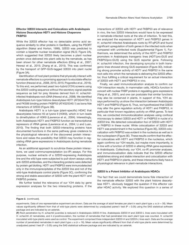
Effector 32E03 Interacts and Colocalizes with Arabidopsis Histone Deacetylase HDT1 and Histone Chaperone FKBP53
While the 32E03 effector has no detectable amino acid se-quence similarity to other proteins in GenBank, using the PSORT algorithm (Nakai and Horton, 1999), 32E03 was predicted to contain a bipartite nuclear localization signal (NLS; Figure 2A), which suggested that 32E03 likely becomes a plant nuclear protein once delivered into plant cells by the nematode, as has been shown for other nematode effectors (Elling et al., 2007; Hewezi et al., 2015; Zhang et al., 2015). This was confirmed by the transport of GFP-GUS-tagged 32E03 into plant nuclei (Figure 3A). Identification of host plant proteins that physically interact with nematode effectors is a promising approach to elucidate effector function (Hewezi et al., 2008, 2010, 2015; Pogorelko et al., 2016). To this end, we performed yeast two-hybrid (Y2H) screens using the 32E03 coding sequence without the secretory signal peptide sequence as bait for prey libraries derived from H. schachtii- infected Arabidopsis root cDNA (Hewezi et al., 2008). We identified Arabidopsis tuin-type histone deacetylase HDT1 (AT3G44750.1) and FK506 binding protein FKBP53 (AT4G25340.1) as bona fide interactors of 32E03 (Figure 3B). Arabidopsis HDT1 is a tuin-type (plant-specific) HDAC that deacetylates histone H3 at lysine 9 (H3K9), which in turn leads to dimethylation of H3K9 (Lawrence et al., 2004). Interestingly, both Arabidopsis HDT1 and FKBP53 function as transcriptional repressors of rRNA genes (Lawrence et al., 2004; Li and Luan, 2010). The finding that both 32E03-interacting proteins have documented functions in the same pathway gives credence to the physiological relevance of the discovered protein interac-tions and raises the possibility that 32E03 may function in reg-ulating rRNA gene expressions in Arabidopsis during nematode infection. As an additional approach to scrutinize these protein interac-tions, we used coimmunoprecipitation (co-IP) assays. For this purpose, nuclear extracts of a 32E03-expressing Arabidopsis line and the wild type were subjected to pull-down assays using anti-32E03 antibodies, and the interacting proteins were detected by protein gel blotting. HDT1 and FKBP53 were immunodetected only in the immunoprecipitates of the 32E03 line and not of the wild-type Arabidopsis control plants (Figure 3C), confirming the strong and stable association of 32E03 with the plant HDT1 and FKBP53 proteins. We further tested the relevance of our Y2H data by gene expression analyses for the two interacting proteins. If the
interactions of 32E03 with HDT1 and FKBP53 are of relevance in vivo, the two 32E03 interactors would have to be expressed in nematode-infected roots at the site of infection. To test this, we analyzed the expression of HDT1 and FKBP53 genes in H. schachtii-infected Arabidopsis roots by RT-qPCR and found significant upregulation of both genes in the infected roots when compared with uninfected roots (Supplemental Figure 1). Fur-thermore, we determined the activity of the HDT1 and FKBP53 promoters in Arabidopsis transgenic lines (HDT1pro:GUS and FKBP53pro:GUS) using the GUS reporter gene. Following H. schachtii infection, the developing syncytia in both trans-genic lines showed strong GUS expression (Figure 3D), indicat-ing strong promoter activity of HDT1 and FKBP53 in the same root cells into which the nematode is delivering the 32E03 effec-tor, thus fulfilling a critical requirement for an actual interaction of 32E03 with HDT1 and FKBP53 in vivo. Finally, we used immunolocalization analysis to confirm our Y2H interaction results. In mammalian cells, HDACs function in concert with nuclear FKBP proteins in regulating gene expressions (Yang et al., 2001), and one can postulate that similar functions may also be conserved in plants. In support of this, Y2H as-says performed by us show the interaction between Arabidopsis HDT1 and FKBP53 (Figure 4). Thus, we hypothesized that 32E03 may alter the gene regulation activity of HDT1 and FKBP53, in particular their known regulatory activity on rRNA genes. To test this, we conducted immunolocalization analyses using confocal microscopy to detect 32E03 and HDT1 or FKBP53 in nuclei of a 32E03 line. We detected colocalization foci of 32E03 with HDT1 or FKBP53 in these nuclei. While colocalization of 32E03 and HDT1 was predominant in the nucleolus (Figure 3E), 32E03 colo-calization with FKBP53 was evident in the nucleolus as well as in the nucleoplasm (Figure 3E). These results confirm that the effec-tor colocalizes with HDT1 and FKBP53 in the nucleolus, which again confirms our Y2H results, but maybe more importantly, is in line with a function of 32E03 in altering rRNA gene expression in Arabidopsis. Collectively, our Y2H, co-IP, promoter analyses, and immunolocalization data indicate that the 32E03 effector establishes strong and stable interaction and colocalization with HDT1 and FKBP53 in planta, and these interactions likely have a physiological relevance in plant-nematode interactions.
32E03 Is a Potent Inhibitor of Arabidopsis HDACs
The fact that we could demonstrate bona fide interaction of the nematode effector 32E03 with the plant histone deacety-lase HDT1, obviously begged the question if this effector can alter HDAC activity. We explored this question in a series of
experiments. Data of one representative experiment are shown. Data are the average of adult females per plant in each plant type ± se (n = 30). Mean values significantly different from that of wild-type plants were determined by unadjusted paired t test (P < 0.05) using the SAS statistical software package and are indicated by an asterisk.(F) Root penetration by H. schachtii juveniles is reduced in Arabidopsis 32E03-H line. Arabidopsis 32E03-H and 32E03-L lines were inoculated with H. schachtii J2 nematodes, and 4 d postinoculation, the number of nematodes that had penetrated into each plant type was counted. H. schachtii inoculated wild-type plants were used as control. The experiment comprised three independent 32E03-H and 32E03-L lines each. Data are the average number of penetrated nematodes in each plant type ± se (n = 16). Mean values significantly different from that of wild-type plants were determined by unadjusted paired t test (P < 0.05) using the SAS statistical software package and are indicated by an asterisk.
Figure 3. 32E03 Expressed in Arabidopsis Interacts and Colocalizes with HDT1 and FKBP53 Proteins.
(A) 32E03 accumulates in the plant nucleus. A plasmid containing the 32E03 coding sequence without the secretory signal peptide fused to the GFP-GUS gene was delivered into onion epidermal cells using biolistic bombardment, and the bombarded cells were analyzed by epifluorescence microscopy. Bar = 100 µm.(B) 32E03 interacts with Arabidopsis HDT1 and FKBP53 in yeast. Yeast cells cotransformed with the 32E03 bait plasmid and the HDT1 or FKBP53 prey plasmid were grown on a low stringency double dropout (DDO) medium and a high stringency quadruple dropout (QDO) medium in the presence of X-α Gal to confirm protein interaction. Empty prey vector or prey vector containing human Lamin C served as controls.(C) 32E03 synthesized in Arabidopsis forms a complex with endogenous HDT1 and FKBP53. Nuclear extract of a 32E03-expressing Arabidopsis line was immunoprecipitated with anti-32E03 antibodies, and the immunoprecipitates (IP) were analyzed by protein gel blot using anti-32E03, anti-HDT1, or anti-FKBP53 antibodies. HDT1, FKBP53, and ACTIN2 in input nuclear extract was detected as loading control.(D) H. schachtii infection upregulates Arabidopsis HDT1 and FKBP53 promoter activities. Arabidopsis transgenic plants harboring the GUS gene under the control of the HDT1 (HDT1pro:GUS) or FKBP53 (FKBP53pro:GUS) promoter were inoculated with H. schachtii, and the infected roots were analyzed for GUS expression by histochemical assays. dpi, days postinoculation. N, nematode; S, syncytium; P, lateral root primordium. Bar = 10 µm.(E) 32E03 colocalizes with endogenous Arabidopsis HDT1 and FKBP53. Nuclei of 32E03-expressing Arabidopsis line were immunostained with anti-32E03 antibodies in combination with anti-HDT1 or anti-FKBP53 antibodies, probed with secondary antibodies conjugated to Alexa Fluor 488 or Alexa Fluor 594 and counterstained with DAPI. About 200 nuclei in each preparation were analyzed by confocal microscopy. no, nucleolus; np, nucleoplasm. Bar = 5 mm.
experiments. First, we measured total HDAC activities in nu-clear extracts from 7-d-old whole wild-type and 32E03 expres-sion seedlings. In the extracts from 32E03-H and 32E03-L lines, HDAC activity was significantly reduced when compared with wild-type plants (Figure 5A), and the reduction in enzyme activ-ity was more pronounced in the 32E03-H line, suggesting that 32E03 is the cause of inhibition of total HDAC activity. To con-firm this, we measured HDAC activity in wild-type plant nuclear extract as a function of added purified recombinant 32E03 pro-tein. In the presence of 32E03, HDAC activity was significantly inhibited when compared with enzyme activity in the absence of 32E03 (Figure 5B). The level of HDAC inhibitory action of 32E03 in the wild-type plant nuclear extract was comparable
to that of the potent HDAC inhibitor trichostatin, which was added to a set of wild-type plant nuclear extract (Figure 5B). Our results convincingly show that 32E03 inhibits HDAC ac-tivity in planta to a degree comparable to that of the HDAC inhibitor trichostatin. We then determined if our HDAC activity assay in fact mea-sures HDT1 activity by comparing HDAC activity between nu-clear extracts of an HDT1 overexpression line (HDT1 expression was driven by the 35S promoter; Supplemental Figure 2) and wild-type plants. In the HDT1 overexpression line, HDAC activity was significantly increased relative to the wild type (Figure 5B), which documented that HDT1 activity was indeed measured as a part of total HDAC activity in our assays. Interestingly, we determined in subsequent experiments that the HDAC activity measured in the nuclear extracts of wild type plants and the 32E03 expression line is largely due to HDAC enzymes other than HDT1 because HDAC activity in the extracts of a HDT1 knockdown mutant (hdt1) was not different from that of wild-type plants (Figure 5B). In other words, while we showed upreg-ulation of the HDT1 promoter in the syncytium, HDT1 expression in whole plants appears relatively low. In order to determine if 32E03 also inhibits HDT1, we needed to employ an indirect approach because we were not aware of a specific HDT1 activity assay in planta. For this purpose, we measured HDAC activity in nuclear extracts of the HDT1 over-expression line as a function of added purified recombinant 32E03 protein at two concentrations. Both 32E03 preparations inhibited the elevated HDAC activity in the nuclear extracts of the HDT1 overexpression line and the higher 32E03 concentra-tion had an almost complete HDAC inhibitory effect comparable to that of trichostatin (Figure 5B). These data showed that the
Figure 4. Arabidopsis HDT1 and FKBP53 interact.
Yeast cells cotransformed with the HDT1 bait plasmid and the FKBP53 prey plasmid were grown on a low stringency double dropout (DDO) me-dium and a high stringency quadruple dropout (QDO) medium in the presence of X-α Gal to confirm protein interaction. Empty prey vector or prey vector containing human Lamin C served as controls.
Figure 5. 32E03 Inhibits HDAC Activities.
(A) Expression of 32E03 in Arabidopsis inhibits HDAC activities. HDAC activities of the 32E03-H and 32E03-L lines were compared with that of the wild-type plants.(B) Recombinant 32E03 inhibits HDAC activities. HDAC activities in the wild-type, HDT1, and hdt1 plants were measured in the presence or absence of recombinant 32E03 protein (r32E03; 500 nM, r32E03a;1500 nM) or trichostatin (TSA; 500 nM).In (A) and (B), plants of the tested genotypes were grown in a randomized block design. For each biological replicate, plants were sampled randomly to prepare pools for each line. Nuclei of Arabidopsis pools were isolated and nuclear extracts were prepared for HDAC assays. The experiment comprised three biological replicates, each with three technical replicates. Data are the mean values ± se. Statistically significant changes in HDAC activity were determined by unadjusted paired t test and are indicated by an asterisk (P ≤ 0.1).
32E03 effector is a powerful and promiscuous inhibitor of HDAC activities including that of HDT1. Because of this wide inhibition of HDACs by 32E03, we per-formed additional targeted Y2H assays in order to explore which other HDAC enzymes might interact with 32E03. Given the large size of the HDAC gene family, we only assayed the tuin-type HDACs HDT2 (AT5G22650.1), HDT3 (AT5G03740.1), and HDT4 (AT2G27840.1) as the closest HDT1 relatives. In addition, we included the RPD3-type HDAC HDA6 (AT5G63110.1) because, similar to HDT1, it has known functions in rRNA gene regulation (Earley et al., 2010). Interestingly, none of these proteins inter-acted with 32E03 in the YTH assays (Figure 6). While strong Y2H interaction is a promising indicator that the proteins in question truly interact, the absence of protein interaction in Y2H assays does not preclude possible protein interactions in vivo. Because a more detailed analysis of HDAC interactions with 32E03 is be-yond the scope of this paper, we did not further explore which specific HDACs are inhibited by 32E03 at this point. However, we took this analysis one step further by conducting genetic analyses of the hdt1 and hda6 mutants. Even though we could not show 32E03 interaction with HDA6, we included the hda6 mutant because of the documented role of HDA6 in rRNA gene regulation (Earley et al., 2010). As one could expect from the broad HDAC inhibitory function of 32E03, the hdt1 and hda6 mutant lines showed no morphological or nematode suscepti-bility phenotypes when compared with the wild type (Figure 7), suggesting robust functional redundancy among HDACs in Ara-bidopsis. We also assayed if mRNA expression of HDT1 or HDA6 is altered in the 32E03-H and 32E03-L lines and determined that the steady state mRNA abundance of these genes is not altered by expression of the effector (Figure 8). Our data convincingly show broad HDAC activity inhibition by 32E03. Furthermore, we showed 32E03 interaction with and in-hibition of HDT1, a HDAC that has been shown to regulate rRNA gene expression through chromatin modifications. These con-clusions directed our attention to the regulation of rRNA genes as a function of 32E03.
Expression of 32E03 Mediates rDNA Chromatin Modifications and Alters 45S Pre-rRNA Abundance
As mentioned above, HDT1 has been shown to deacetylate H3K9 along rDNA chromatin, which subsequently leads to dimeth-ylation of H3K9 and repression of rDNA expression (Lawrence et al., 2004; Pontes et al., 2007; Li and Luan, 2010). Our finding of inhibition of HDAC activities by 32E03 in Arabidopsis plants naturally begged the question if the presence of this effector in plant cells would modulate the acetylation and methylation status of H3K9 along the rDNA chromatin and would alter rRNA gene expression. In A. thaliana rRNA genes are tandemly arrayed head-to-tail at chromosomal loci known as nucleolus organizer regions (NORs), and the Arabidopsis genome has two such NORs. Each rRNA gene is separated from adjacent genes by an intergenic spacer (IGS). RNA polymerase I (Pol I) transcribes 45S pre-rRNA primary transcripts, which are pro-cessed into catalytic rRNAs (18S, 5.8S, and 25S) by sequential cleavage of the external and internal transcribed spacers (ETS and ITS) in the nucleolus. To further delineate the function of
32E03, levels of H3K9Ac and H3K9me2 along the rDNA chro-matin stretches as shown in Figure 9A were compared between 32E03-L and -H lines and wild type Arabidopsis plants by chro-matin immunoprecipitation (ChIP)-qPCR assays. Confirming the HDAC inhibitory function of 32E03, we found elevated levels of H3K9Ac throughout the coding and noncoding regions of rDNA in the 32E03-H and 32E03-L lines as compared with wild-type Arabidopsis plants (Figure 9B), while H3K9me2 levels were sub-stantially reduced in the same locations (Figure 9B). In both the 32E03-H and 32E03-L lines, the H3K9 modifications assayed were unaltered at ACTIN2 and AtSN1 retrotransposon loci when compared with wild type Arabidopsis plants (Figure 9B). In other words, these ChIP-qPCR data indicate that the 32E03 effector modulates histone modifications along the rDNA chromatin in Arabidopsis plants. The above discoveries lead us to hypothesize that 32E03- mediated H3K9 hyperacetylation along the rDNA chromatin would open rDNA chromatin, thereby allowing an increased transcrip-tion of rRNA genes. To test this, we quantified 45S pre-rRNA transcripts in 32E03-L and -H lines by RT-qPCR. While we in-deed confirmed the expected high abundance of 45S pre-rRNA
Figure 6. 32E03 Does Not Interact with Other Tuin-Type HDACs or HDA6 of Arabidopsis in the Y2H System.
Yeast cells cotransformed with the 32E03 bait plasmid and the HDT2, HDT3, HDT4, or HDA6 prey plasmid were grown on a low stringency double dropout (DDO) medium and a high stringency quadruple dropout (QDO) medium in the presence of X-α Gal to confirm protein interaction. Empty prey vector or prey vector containing human Lamin C served as controls.
transcripts in the 32E03-L line, we surprisingly observed a sig-nificant reduction in 45S pre-rRNA transcripts in the 32E03-H line (Figure 9C). This result raised the distinct possibility that the difference in 45S pre-rRNA abundance in 32E03-L and 32E03-H may be the cause for the earlier described variation in their morphology and susceptibility phenotypes. If this were true, then increased 45S-pre-rRNA abundance would be beneficial to nematode infection, while a severe reduction in pre-rRNA abun-dance would be detrimental. To validate this conclusion, we compared the levels of 45S pre-rRNA transcripts in Arabidopsis
root segments containing H. schachtii-induced syncytia and neighboring root segments without syncytia. We found a sig-nificant increase in 45S pre-rRNA abundance (7.4-fold) in root segments containing syncytia when compared with the root segments without syncytia (Figure 9D). These data demonstrate that H. schachtii infection indeed upregulates the rRNA gene expressions in or around the syncytial feeding cells.
High Levels of 32E03 in Arabidopsis Trigger RNA-Directed DNA Methylation of rDNA
It remained unclear why the 32E03-H line in spite of increased acetylation of H3K9 along the rDNA chromatin exhibited a strong repression of the rRNA genes. We hypothesized that the repression of rRNA genes in the 32E03-H line is a plant response to out-of-control transcription events triggered by high concentrations of 32E03. To test this hypothesis, we used ChIP-qPCR analyses to evaluate if the 32E03-mediated open structure of rDNA chromatin in the 32E03-H line is accompa-nied by increased RNA polymerase II (Pol II) occupancy. Sig-nificantly elevated Pol II-mediated transcription along the rDNA would be expected for serendipitous transcription triggered by an opened chromatin state rather than the normal core-promot-er-triggered Pol I-mediated transcription of rRNA genes. We documented an ∼7- to 23-fold increased Pol II occupancy in the rDNA coding and non-coding regions in the 32E03-H line as compared with wild-type Arabidopsis plants (Figure 10A). Im-portantly, occupancy of Pol II at IGS regions in the 32E03-H line was increased to 19-fold when compared with wild type plants. In contrast and as expected, Pol II occupancy was not elevated in the 32E03-L line when compared with wild-type Arabidopsis plants (Supplemental Figure 3A). Pol II ChIP signals at ACTIN2 and AtSN1 transposons did not vary between the two 32E03 transgenic lines and wild-type plants (Figure 10A; Supplemental Figure 3A), indicating the likely enhanced transcription activity of Pol II along the rDNA chromatin in the 32E03-H line. We then
Figure 7. Susceptibility to H. schachtii Is Not Altered in Arabidopsis hdt1 and hda6 Lines.
Three independent lines of Arabidopsis hdt1 and hda6 each were inoculated with H. schachtii J2 nematodes, and 4 weeks after inoculation, the number of adult females per plant were counted. H. schachtii-inoculated Arabidopsis wild type plants were used as control. The experiment was repeated three times. Similar results were obtained in three independent experiments. Data of one representative experiment are shown. Data are the average of adult females per plant in each plant type ± se (n = 30). Mean values significantly different from that of wild-type plants were determined by unadjusted paired t tests (P < 0.05) using the SAS statistical software package.
Figure 8. Expression of HDT1 and HDA6 Is Unaltered in Arabidopsis 32E03-H and 32E03-L Lines.
Root total RNA of Arabidopsis wild-type plants and the 32E03-H and 32E03-L lines was extracted, cDNA was synthesized and HDT1 and HDA6 expression was quantified by qPCR. Wild-type plants were used as control. ACTIN2 was amplified as reference. Tested genotypes were grown in randomized block designs. For each biological replicate, plants were sampled randomly to prepare pools for each genotype. The ex-periment consisted of three biological replicates, each encompassing three technical replicates. Data are the mean ± se. Statistically significant difference in the mean values was analyzed by unadjusted paired t test (P = 0.05).
Figure 9. Expression of the 32E03 Coding Sequence in Arabidopsis Mediates rDNA Chromatin Modifications and Alters 45S pre-rRNA Abundance.
(A) Diagram showing Arabidopsis rDNA regions. The indicated regions were amplified in qPCR assays shown in Figures 9B and 10A and Supplemental Figure 3A. 25S and 18S, coding region; +1, transcription start site.(B) 32E03 expression in Arabidopsis causes histone H3 modifications along the rDNA. Chromatin of 32E03-H and 32E03-L lines was immunoprecipi-tated with anti-H3K9Ac or anti-H3K9me2 antibodies and subjected to qPCR to quantify the rDNA regions indicated in (A). Wild-type plants were used as control. ACTIN2 and SN1 were amplified as reference. Pro, promoter.(C) Abundance of 45S pre-rRNA in Arabidopsis 32E03-H and 32E03-L lines. Total RNA of roots of Arabidopsis wild-type plants and 32E03-H and 32E03-L lines was extracted. Wild-type plants were used as control. 45S pre-rRNA in the 32E03 expression lines was determined relative to wild-type plants.(D) Abundance of 45S pre-rRNA in Arabidopsis wild-type root segments enriched in H. schachtii-induced syncytia. Wild-type plants were inoculated with H. schachtii J2s. Root segments enriched in H. schachtii-induced syncytia (root+syncytium) and adjacent root segments without syncytia (root-syncytia; control) were dissected at 10 d postinoculation.For (B) to (D), plants of the tested genotypes/treatments were grown in randomized block designs. For each biological replicate, plants were sampled randomly to prepare pools for each genotype/treatment. Each experiment comprised three technical replicates. Similar results were obtained from three independent experiments. Data from one representative experiment each are shown. Data are the means ± se.For (C) and (D), root cDNA was synthesized and 45S pre-rRNA was quantified by qPCR. Arabidopsis ACTIN8 was amplified as reference.
Figure 10. High Levels of 32E03 in Arabidopsis Trigger RNA-Directed DNA Methylation of rDNA.
(A) Increased RNA Pol II occupancy along the rDNA in the Arabidopsis 32E03-H line. Chromatin of wild-type plants and the 32E03-H line was im-munoprecipitated with anti-RNA Pol II antibodies, and rDNA regions shown in Figure 9A were qPCR amplified. Wild-type plants served as control. Arabidopsis ACTIN2 and SN1 served as reference. The experiment was repeated three times, each with three technical replicates. Similar results were obtained from three independent experiments. Data from one representative experiment are shown. Data are the mean ± se.(B) Diagram showing Arabidopsis rDNA regions. The indicated regions were amplified in (C) and (D).(C) Enhanced bidirectional transcription along the rDNA IGS in 32E03-H line. cDNA of wild-type plants and the 32E03-H line was used to amplify the IGS regions indicated in (B) by RT-PCR and analyzed in 1% agarose gel electrophoresis. Wild-type plants served as control. Band intensity of sense and antisense strand amplicons of each plant type was quantified using the ImageJ software and the ratio is indicated in parenthesis. Arabidopsis ACTIN2 was amplified as reference. +/−RT, with or without reverse transcriptase.(D) Enhanced rDNA IGS-specific small RNA biogenesis in the Arabidopsis 32E03-H line. Small RNA of wild-type plants and the 32E03-H line was resolved in a 15% TBE-urea gel, electroblotted, hybridized with siRNA probes as indicated in (B), and detected using chemiluminescence reagent. Wild-type plants were used as control. Small nuclear RNA U6 (snRNA), loading control.In (C) and (D), the experiment was repeated at least two times. Similar results were obtained from the two independent experiments. Data from one
2806 The Plant Cell
assessed IGS-derived transcript levels in 32E03-H plants by random-primed and strand-specific RT-PCR assays in selected IGS regions (Figure 10B). As could be expected from the elevated Pol II occupancy along the IGS in the 32E03-H line, we documented enhanced sense as well as anti-sense IGS transcripts in the 32E03-H line when compared with wild-type plants (Figure 10C), which is indicative of profound bidirectional transcription along the IGS regions in the 32E03-H line. In con-trast, bidirectional transcription along the IGS was not elevated in the Arabidopsis 32E03-L line (Supplemental Figure 3B). These findings are consistent with an enhanced derepression of cryp-tic Pol II transcription units along the rDNA in the 32E03-H line, which is likely the result of a 32E03-mediated opened state of the rDNA chromatin. Discovering the enhanced bidirectional tran-scription along IGS regions in the 32E03-H line prompted us to postulate that bidirectional transcription would result in the pro-duction of dsRNA, which could trigger biogenesis of small RNAs (sRNAs) in the 32E03-H line. Therefore, we analyzed the accumu-lation of IGS-derived sRNAs in the 32E03-H line by RNA gel blot analysis. Using probes corresponding to the IGS regions (Figure 10B), we detected an increase in accumulation of 21- and 24- nucleotide sRNAs in the tested 32E03-H line relative to wild-type Arabidopsis plants (Figure 10D). The presence of these sRNAs in the 32E03-H line pointed toward the possibility that RNA-directed de novo DNA methylation (RdDM) could be responsible for the observed repression of rRNA genes in the 32E03-H line. A similar phenotype has been described in an Arabidopsis hda6 knockout mutant, in which cryptic RNA Pol II transcriptional activity was ac-companied by an over accumulation of small RNAs that directed de novo DNA methylation and gene silencing (Earley et al., 2010). In Arabidopsis, stable gene silencing is mediated by DNA methylation (Zilberman et al., 2007; Lister et al., 2008; Becker et al., 2011; Schmitz et al., 2011). While cytosine methylation in CG and CHG contexts is maintained by methyltransferase MET1 and plant-specific CMT3 methyltransferase, respectively (Lindroth et al., 2001; Kankel et al., 2003), maintenance of asym-metric CHH methylation relies on the RdDM pathway (Matzke, 2016). To test our hypothesis that silencing of rRNA genes in the 32E03-H line is the regulatory mechanism postulated above, we compared the cytosine methylation levels at the core rDNA promoter region (Figure 10E) of the 32E03-H line with that of wild-type Arabidopsis plants. Bisulphite sequence analyses revealed an approximately threefold hypermethylation of the rDNA promoter in the CHH context in the 32E03-H line when compared with wild-type Arabidopsis plants (Figures 10F and
10G), which indicates that high levels of 32E03 in Arabidopsis plants triggered the RdDM pathway. Interestingly, we discovered that methylation in the CG and CHG contexts also were elevated, which suggests that high 32E03 levels triggered additional reg-ulatory mechanism that resulted in the hypermethylation of the rDNA promoter region. These major quantitative changes in cytosine methylation are consistent with the observed reduc-tion in 45S pre-rRNA abundance in the 32E03-H line. Therefore, presence of 32E03 at high levels as found in the 32E03-H line led to silencing of rRNA genes, which significantly interfered with cyst nematode parasitism. In a wider sense, the 32E03 effector-triggered hypermethylation of rDNA renders plant cells unable to sustain normal syncytium function and therefore causes decreased parasitism. In contrast to the hypermethylation of the rDNA promoter found in the Arabidopsis 32E03-H line, cyto-sine methylation of the rDNA promoter did not vary between the 32E03-L line and wild-type plants (Supplemental Figure 3C).
Low Levels of 32E03 in Arabidopsis Derepress a Subset of VAR1 rRNA Genes
Our findings that the 32E03-L line showed an increase in 45S pre-rRNA abundance and an overall phenotype conducive to H. schachtii parasitism suggested that 32E03 is a positive regula-tor (i.e., a derepressor) of rRNA genes and, thus, of H. schachtii parasitism. We set out to obtain molecular data f to test this hypothesis. The specific nature of rDNA variants in Arabidop-sis provided an opportunity to further dissect the mechanism of 32E03 function in planta. In A. thaliana ecotype Col-0, there are at least four rRNA gene variants (VAR1-4), based on sequence variation within the repetitive region in the 3′ ETS (Pontvianne et al., 2010). These four rRNA variants are expressed in newly germinated seeds, but by 10-14 days after germination and throughout the remaining vegetative development, the majority of VAR1, accounting for ∼50% of the total rRNA gene pool, is selectively silenced by an epigenetic mechanism (Pontvianne et al., 2012, 2013). The rRNA gene dosage is controlled accord-ing to the cellular demand for ribosomes and protein synthesis. The silenced rRNA gene subtypes were mapped to the NOR on chromosome 2, while the active rRNA gene subtypes are mapped to the NOR on chromosome 4 (Chandrasekhara et al., 2016). Therefore, it is a tempting hypothesis that the 32E03 effector function leading to an increase in 45S pre-rRNA tran-scription in the 32E03-L line is due to a derepression of rRNA genes that are normally silenced in growing plants.
representative experiment each are shown.(E) Diagram highlighting the Arabidopsis rDNA promoter analyzed by bisulphite sequencing (BS).(F) and (G) Arabidopsis 32E03-H line rDNA promoter is hypermethylated. (F) Analysis of cytosine methylation. Genomic DNA of wild-type plants and the32E03-H line was digested with BamHI and subjected to sodium bisul-phite conversion. The rDNA promoter region indicated in (E) was amplified by PCR, cloned into pGEM-T Easy vector, and analyzed by the CyMATE algorithm. Wild-type plants were used as control. Approximately 25 promoter clones per genotype were analyzed.(G) Percentage of cytosine methylation in wild-type plants and the 32E03-H line in the three cytosine contexts. Total numbers of CG, CHG, or CHH present in the rDNA promoter region are shown in parenthesis.In (A), (C), (D), and (F), plants of the tested genotypes were grown in a randomized block design. For each experiment, plants were sampled randomly to prepare pools for each genotype.
To test this hypothesis, we took advantage of the single nu-cleotide polymorphisms (SNPs) naturally existing within the ETS and ITS of Arabidopsis VAR1, VAR2, and VAR3 rRNA variants, which create unique restriction endonuclease recognition sites (Chandrasekhara et al., 2016). We adapted cleaved amplified polymorphic sequence (CAPS) assays to analyze expression of rRNA subtypes VAR1 (6645), VAR2 (4302), and VAR3 (7122) in the 32E03-L line and wild type Arabidopsis plants. For this, root cDNA was PCR-amplified, digested with VAR1-6645, VAR2-4302, or VAR3-7122 SNP-specific restriction enzyme and ana-lyzed by agarose gel electrophoresis. Among the rRNA subtypes analyzed, VAR1-6645C was detected only in the 32E03-L line and not in wild type Arabidopsis plants (Figure 11A), which in-dicated derepression of the VAR1-6645C rRNA subtype as a function of the 32E03 effector. To determine if this derepression also can be found in the H. schachtii-induced syncytium, rRNA subtypes were analyzed in wild-type Arabidopsis root segments containing H. schachtii- induced syncytia and in neighboring root segments without syncytia. Interestingly, VAR1-6645C was detected only in root segments containing syncytia and not in segments without syn-cytia (Figure 11B). Thus, these CAPS data confirmed that the derepression of rRNA subtype VAR1-6645C occurs in Arabidop-sis root cells into which the nematode had delivered the 32E03 effector during the infection process. Though derepression of a single rRNA subtype by the 32E03 effector is documented here, the possibility of derepression of multiple rRNA subtypes by 32E03 cannot be ruled out. We further elaborated on this phenomenon by comparing the proportion of VAR1 in rRNA pools of the Arabidopsis 32E03-L line and wild-type plants. For this purpose, we determined the ratio of VAR1 to 45S pre-rRNA (VAR1:45S) by RT-qPCR analyses. In the Arabidopsis 32E03-L line, we found a remarkable increase in the VAR1:45S ratio rela-tive to wild-type plants at both time points analyzed (Figure 11C). In addition, analysis of the VAR1:45S ratio in wild-type Arabi-dopsis root segments containing H. schachtii-induced syncytia revealed an increase in the VAR1:45S rRNA to 3.5-fold when compared with root segments without syncytia (Figure 11D). Collectively, the CAPS and VAR1:45S ratio data further confirm the function of 32E03 effector in the derepression of rRNA genes in host plant cells. In summary, our data document that 32E03 is a potent cyst nematode effector that the parasite deploys to inhibit the func-tion of Arabidopsis HDACs (including HDT1) to mediate rDNA
Figure 11. A Subset of VAR1 rRNA Variant Is Derepressed and VAR1:45S pre-rRNA Ratio Is Altered in the Arabidopsis 32E03-L Line.
(A) Expression of subtypes of rRNA variants in roots of Arabidopsis 32E03-L line analyzed by SNP analysis. Wild-type roots were used as control.(B) Expression of subtypes of rRNA variants in Arabidopsis wild-type root segments enriched in H. schachtii-induced syncytia analyzed by SNP analysis. Wild-type plants were inoculated with H. schachtii J2s and root segments enriched in H. schachtii-induced syncytia (root+ syncytium) and adjacent root segments without syncytia (root-syncy-tium; control) were dissected at 10 d postinoculation. In (A) and (B), whole root or root segment cDNA was synthesized, sub-types of rRNA variants were amplified by PCR, gel-eluted, digested with SphI, AluI, or MspI to detect VAR1-6645, VAR2-4302, or VAR3-7122 subtype, respectively. DNA fragments were visualized by 2.5% agarose gel electrophoresis. The experiment comprised at least two biological replicates. Similar results were obtained in the two independent experi-ments. Data of one representative experiment are shown.(C) Quantification of VAR1 rRNA and 45S pre-rRNA in Arabidopsis 32E03-L line (14 and 18 d old) by qPCR. Wild-type plants were used as control.(D) Quantification of rRNA VAR1 and 45S pre-rRNA in wild-type Ara-bidopsis root segments enriched in H. schachtii-induced syncytia by
qPCR. Wild-type plants were inoculated with H. schachtii J2s and root segments enriched in H. schachtii-induced syncytia (root+syncytium) and adjacent root segments without syncytia (root-syncytium; control) were dissected at 10 d postinoculation. In (C) and (D), whole roots or root segments cDNA was synthesized, and VAR1 and 45S pre-RNA were quantified by qPCR. ACTIN8 was ampli-fied as reference. The experiments comprised three biological replicates, each consisting of three technical replicates. Similar results were ob-tained in the three independent experiments. Data of one representative experiment are shown.For (A) to (D), plants of the tested genotypes/treatments were grown in randomized block designs. For each biological replicate, plants were sampled randomly to prepare pools for each genotype/treatment.
2808 The Plant Cell
chromatin modifications with the outcome of a derepression of rRNA genes. This regulation of plant genes by the 32E03 ef-fector not only provides key insights into plant-parasite interac-tions, but also reveals the apparent requirement of fine-tuning of rRNA gene dosage in the nematode induced syncytium. In ad-dition, there likely are additional, so far unknown consequences of 32E03-mediated inhibition of HDACs. Certain HDACs have been documented to play roles in modulating defense gene expressions and the manifestation of plant resistance (Zhou et al., 2005; Kim et al., 2008; Choi et al., 2012; Ding et al., 2012). Furthermore, tuin-type HDACs have been shown to act as nega-tive regulators of elicitor-induced plant cell death (Bourque et al., 2011; Dahan et al., 2011). Interestingly, the HC toxin produced by the plant-pathogenic fungus Cochliobolus carbonum (Brosch et al., 1995; Ransom and Walton, 1997; Sindhu et al., 2008) and the Depudecin toxin of the fungus Alternaria brassicicola (Wight et al., 2009) inhibit plant HDACs to suppress defense responses and to enable the necrotrophic life style of these fungi within their hosts. Here, we report a very different, and so far, unique molecu-lar mechanism of how a parasite deploys an effector to modulate a plant-specific HDAC (and likely a histone chaperone although not further studied in this report) to fine-tune host rRNA dosage to sustain the demands and rigors of nematode parasitism. Taken one step further, it is highly interesting, yet not surprising, that plants have evolved a unique mechanism that is triggered by effector-mediated chromatin modulation, and it remains to be seen if such mechanisms are also triggered by other phytopathogens. In this cyst nematode pathosystem, it is evident that the nema-tode parasite is “walking a tight rope” by having to increase rRNA abundance without triggering the host plant’s gene silencing through DNA hypermethylation. Although a variety of epigenetic mechanisms in plants are associated with pathogen interactions, in particular bacterial and fungal pathogen infections (Ding and Wang, 2015; Zhu et al., 2016), direct evidence for how patho-gen effectors may manipulate epigenetic regulation in the host remains very limited. The TrAP protein of two plant Geminiviruses inhibits H3K9 methylation in Arabidopsis to counter host defense (Castillo-González et al., 2015). Recently, it has been shown that an effector of the oomycete pathogen Phytophthora sojae acts as a modulator interfering with the function of the plant histone acetyltransferase GCN5 complex and suppresses defense genes at an epigenetic level (Kong et al., 2017). The RomA effector of the human bacterial pathogen Legionella pneumophila acts as a histone methyltransferase to directly methylate host histones, which represses immune gene expression (Rolando et al., 2013). Finally, an effector of the animal parasite Toxoplasma manipu-lates the function of a host histone deacetylase complex, which is linked to blocking of immune gene expression (Olias et al., 2016). The 32E03 effector function documented here reveals a powerful mechanism for how a parasite alters plant chromatin structure to achieve gene expression changes required for infection success.
METHODS
Plant Material
Arabidopsis thaliana plants were grown under sterile conditions on Murashige and Skoog (MS) medium containing vitamins (Plant Media)
and 2% sucrose at 26°C or in soil at 23°C in a growth chamber under long-day (16-h-light/8-h-dark photoperiod with fluorescent bulbs gen-erating soft white light). For stable plant expression, the 32E03 coding sequence was PCR-amplified from Heterodera schachtii cDNA, while the HDT1 coding sequence was amplified from Arabidopsis cDNA. Amplified products were individually cloned into the binary vector pBI121. Arabi-dopsis (ecotype Col-0 for 32E03 or C24 for HDT1) was transformed by the floral-dip method (Clough and Bent, 1998). Transformants were screened on MS medium containing 50 mg/L kanamycin, and homozygous lines were identified in the T3 generation. Arabidopsis hdt1 (CS348580) and hda6 (Murfett et al., 2001) mutant seeds were obtained from the ABRC.
Nematode Infection Assay
Ten-day-old Arabidopsis seedlings grown on modified Knop’s medium (Sijmons et al., 1991) at 24°C under 16 h light/8 h dark were inoculated with J2 H. schachtii nematodes (Baum et al., 2000). Four weeks post inoculation, adult females in each plant were counted, and the data were analyzed by a modified t test using the Statistical Software Package (SAS; P < 0.05). Root segments containing H. schachtii-induced syncytia and adjacent root segments without syncytia were dissected under a light microscope as described (Hermsmeier et al., 2000).
Nematode Penetration Assay
Penetration of H. schachtii into roots of Arabidopsis seedlings was de-termined 4 d postinoculation (Hewezi et al., 2008). The number of pen-etrating nematodes in each root system was counted under bright-field illumination using a Zeiss Axiovert 100 microscope. Each plant line was replicated 16 times, and three independent experiments were conducted. Average numbers of penetrating nematodes were calculated, and statis-tically significant differences were determined in a modified t test using the SAS statistical software package (P < 0.05).
Syncytial Measurements
Size of syncytia was measured 21 d postinoculation of Arabidopsis with H. schachtii (Hewezi et al., 2008). For each line, 20 single-female syncytia were randomly selected, size was measured, and average size for each line was determined. Statistically significant differences were determined in a modified t test using the statistical software package SAS (P < 0.05).
RNA Extraction and cDNA Synthesis
H. schachtii eggs, preparasitic J2 juveniles from a hatch chamber, para-sitic J2, J3, J4, and adult females from nematode-infected Brassica oler-acea were collected and frozen. Total RNA was extracted from nematode and plant tissues using the Versagene RNA tissue kit (Gentra Systems) or RNeasy Plant Mini Kit (Qiagen). After treating the RNA with RNase-free DNase I (Invitrogen), cDNA was synthesized using the qScript cDNA SuperMix (Quanta Biosciences).
In Situ Hybridization
Parasitic H. schachtii J3 nematodes were isolated from infected Ara-bidopsis plants as described (Gao et al., 2001), and 32E03 mRNA was detected by in situ hybridization (de Boer et al., 1998) with a gene-specific digoxigenin (DIG)-labeled (Boehringer) antisense- or sense- cDNA probe synthesized by asymmetric PCR (de Boer et al., 1998). Hybridization signals were detected using anti-DIG antibodies conjugated to alkaline phosphatase (ALP) (diluted 1:100) and 5-bromo-4-chloro-3-indolyl-phosphate with nitro blue tetrazolium as substrate in a Zeiss Axiovert 100 inverted compound light microscope.
32E03 expression in preparasitic H. schachtii J2s was downregulated by the dsRNA soaking method (Sukno et al., 2007). Two nonoverlap-ping coding regions (5′: 1–200 bp and 3′: 286–486 bp) of the 32E03 coding sequence without the secretory signal peptide sequence were PCR-amplified from H. schachtii cDNA. A YFP gene sequence (1–195 bp) was amplified from p35S-SPYNE (provided by Jörg Kudla, University of Münster). The PCR products were used as templates to synthesize dsRNA transcripts in vitro using the MEGAscript RNAi kit (Ambion). Freshly hatched nematodes were soaked in M9 buffer (43 mM Na2HPO4, 22 mM KH2P04, 2 mM NaCl, and 4.6 mM NH4Cl) containing dsRNA (3.5 mg/mL), 50 mM octopamine (Sigma-Aldrich), 1 mM spermidine (Sigma-Aldrich), and 0.05% gelatin in a moisture chamber at 28°C for 24 h.
Y2H Screening and Protein Interaction Assays
The 32E03 coding sequence without the secretory signal peptide was PCR-amplified from H. schachtii cDNA with an artificial start codon and fused in-frame to the GAL4 DNA binding domain in plasmid pGB-KT7 (Clontech). The resultant bait construct was designated as pTH22. cDNA of H. schachtii-infected Arabidopsis roots was cloned into plasmid pGADT7 (Clontech) to construct prey libraries (Hewezi et al., 2008). Yeast AH109 strain harboring the prey library and Y187 strain harboring the bait construct were mated and screened on a double dropout medium (SD/-Leu/-Trp; DDO) and subsequently on a high stringency quadruple dropout medium (SD/-Leu/-Trp/-Ade/-His; QDO) containing X-α-Gal (5-bromo-4-chloro-3-indolyl α-d-galactopyranoside) using the BD Match-maker Library Screening kit (Clontech). From yeast cells that displayed a positive protein interaction, prey plasmids were rescued in Esche-richia coli and sequenced. For protein interaction assays, Arabidopsis HDT1 was cloned into pGBKT7, while Arabidopsis HDT2, HDT3, HDT4, HDA6, and FKBP53 were cloned into pGADT7. A prey vector harboring the human Lamin C gene (Clontech) served as control. DNA and protein sequences were analyzed with the BLAST alogrithms (http://blast.ncbi.nlm.nih/Blast.cgi).
HDT1 and FKBP53 Promoter Assay
Promoter constructs of HDT1 and FKBP53 were generated by ligating 1006 and 970 bp DNA fragments upstream of Arabidopsis HDT1 or FKBP53 coding regions, respectively, into the pBI101 binary vector to drive expression of a GUS reporter gene. Wild-type Arabidopsis plants were transformed with either of the binary constructs. Stable homozygous transgenic lines were infected with H. schachtii and GUS expression was analyzed by histochemical staining (Jefferson et al., 1987) in a Zeiss SV-11 microscope. Images were captured using a Zeiss AxioCam MRc5 digital camera and processed using Zeiss Axiovision software (version 4.8).
Protein Synthesis in Planta
For subcellular localization analyses, the PCR-amplified 32E03 coding sequence without the secretory signal peptide coding sequence was cloned between the 35S promoter and the GFP-GUS fusion reporter gene in a modified pRJG23 vector (Grebenok et al., 1997). The construct was delivered into onion epidermal cells by particle bombardment, and the bombarded samples were incubated at 25°C in the dark for 16 h. Flu-orescence signals were analyzed with a Zeiss Axiovert 100 microscope.
Protein Synthesis in E. coli and Purification
The 32E03 coding sequence without the secretory signal peptide was PCR-amplified from H. schachtii cDNA with a start codon, a 6X histidine tag at the 3′ end and a stop codon and cloned into plasmid pET28a
(Novagen). E. coli strain C41 (DE3) (Lucigen) was transformed with this construct. Transformants were grown at 37°C in Luria Bertoni medium (supplemented with 100 µg/mL ampicillin) to A
600 0.5 and induced with 0.6 mM isopropyl-d-thiogalactopyranoside for 3 h. Cells were harvest-ed, resuspended in PBS (0.05 M phosphate, pH 7.4, 0.25 M NaCl, 2 mM phenylmethanesulfonyl fluoride, and protease inhibitor cocktail tablets [Roche]), sonicated, and centrifuged at 10,000g. The lysate was applied onto cobalt resin (Pierce Biotechnology) and washed with PBS containing increasing concentrations of imidazole (35, 50, or 60 mM). Resin-bound 32E03 recombinant protein was eluted with 500 mM imid-azole, dialyzed in PBS, and purity of the protein was verified in a Novex 8-16% Tris-glycine SDS-PAGE (Life Technologies). Polyclonal antibodies against recombinant 32E03 were generated in mouse at the Iowa State University Hybridoma Facility.
Co-IP and Immunodetection
For co-IP assays, nuclei were isolated from Arabidopsis plants and lysed as described (Wierzbicki et al., 2008). Nuclear lysate was immunoprecip-itated with mouse anti-32E03 antibodies overnight at 4°C. For immuno-detection of proteins in Arabidopsis plants, total protein was extracted in extraction buffer (10 mM Tris-HCl, pH 7.4, 300 mM NaCl, 5 mM EDTA, 1 mM PMSF, and 1 mM DTT). The immunoprecipitate or total protein was separated in Novex 4-16% Tris-glycine SDS-PAGE (Life Technolo-gies) and electroblotted onto a PVDF membrane (Bio-Rad). The blot was probed with anti-32E03 antibodies (dilution 1:2000), rabbit anti-HDT1 polyclonal antibodies (provided by Craig Pikaard, Indiana University) (di-lution 1:1000) rabbit anti-FKBP53 polyclonal antibodies (developed to oligopeptide representing FKBP53 amino acids 350–363 by Genscript) (dilution 1:1000), or mouse anti-ACTIN monoclonal antibodies (ABclonal) (1:1000). The total protein blot was developed with goat anti-mouse anti-bodies conjugated to horseradish peroxidase (HRP) (Genscript) (dilution 1:10,000) and detected using the LumiSensor Chemiluminescent HRP Substrate kit (GenScript). The immunoprecipitate sample blot was de-veloped using anti-mouse or anti-rabbit antibodies conjugated to HRP and detected using the SuperSignal Western Femto Maximum Sensitivity Substrate (Thermo Fisher Scientific).
Immunostaining
Nuclei of Arabidopsis plants were isolated and immunostained as de-scribed (Durut et al., 2014). A combination of anti-32E03 antibodies and anti-HDT1 antibodies or anti-FKBP53 antibodies at a dilution of 1:100 in PBS was applied onto a slide precoated with nuclei and incubated over-night at 4°C. The nuclei were labeled with anti-rabbit-Alexa Fluor 488 and anti-mouse-Alexa Fluor 594 antibodies (Abcam) at a dilution of 1:1000, counterstained with DAPI (4′,6-diamidino-2-phenylindole), mounted using the Vectashield medium (Vector Laboratories), and analyzed in a Leica SP5 X inverted confocal microscope. The images were processed using the Leica Application Suite 2.3.0. All images are projections of optical sections.
Histone Deacetylase Assay
Nuclear extract of Arabidopsis plants (7 d old) was prepared using the Epiquick Nuclear Extraction Kit I (Epigentek), and total histone deacetyl-ase activity in nuclear extracts was measured in the presence or absence of recombinant 32E03 (500 or 1500 nM) or trichostatin (500 nM) using the Epigenase HDAC Activity/Inhibition Direct Assay Kit (Epigentek). Protein concentration in the nuclear extract was determined using the Coomassie Protein Assay Reagent (Thermo Fisher Scientific).
ChIP-qPCR
Nuclei of Arabidopsis seedlings were isolated, and chromatin was immu-noprecipitated using anti-H3AceK9 antibodies (Thermo Fisher Scientific),
anti-H3me2K9 antibodies (Abcam), or anti-RNA polymerase II antibodies (Santa Cruz Biotechnology) as described (Wierzbicki et al., 2008). Abun-dances of rDNA regions, SN1, and ACTIN2 in ChIP samples relative to input were determined by qPCR.
Random and Strand-Specific RT-PCR and qPCR
For random RT-PCR and strand-specific RT-PCR, total RNA of Arabidop-sis roots was treated with DNase I, and using random or strand-specific primers and the RevertAid First-Strand cDNA synthesis kit (Thermo Fisher Scientific), first-strand cDNA was synthesized. The first-strand cDNA was PCR-amplified using the amplicon-specific primers and analyzed by agarose gel (1.5%) electrophoresis followed by SYBR Safe staining. Images of the strand-specific RT-PCR products were analyzed using the ImageJ software (https://imagej.nih.gov/ij/). For qPCR, 10-fold diluted cDNA or genomic DNA, 10 pmol primer, and iQ SYBR Green Supermix (Bio-Rad) were used for amplification in an iCycler IQ system (Bio-Rad Laboratories). Data were analyzed using the comparative CT method (Livak and Schmittgen, 2001). Gene expression in Arabidopsis plants and nematodes were normalized to ACTIN gene expression. qPCR con-ditions were as follows: 95°C for 3 min, followed by 40 cycles of each of 10 s at 95°C, 30 s at 60°C. A dissociation curve was produced at the end of the cycling phase to ensure that a single PCR product was produced with no primer dimers.
rRNA Variant SNP Analysis
rRNA variants in 12-d-old Arabidopsis plants were analyzed as described (Chandrasekhara et al., 2016). cDNA of Arabidopsis roots or root segments enriched in H. schachtii syncytia was used to amplify rRNA variants by PCR. The products were gel eluted, digested with SphI (VAR1-6645), AluI (VAR2-4302), or MspI (VAR3-7122), and resolved in 2.5% agarose gels followed by SYBR Safe staining.
Small RNA Gel Blot Hybridization
Small RNAs of Arabidopsis seedlings were isolated using the Nucleospin miRNA kit (Machery Nagel), resolved in a 15% TBE-urea gel (Life Tech-nologies) and blotted onto a nylon membrane (GenScreen Plus). Oligo-nucleotide probes corresponding to regions indicated in Figure 10B were synthesized using the mirVana probe construction kit (Ambion), purified with the Performa DTR Gel Filtration Cartridge (EdgeBio), hybridized to small RNAs on the blots at 42°C overnight, and recognized using anti-DIG-ALP antibodies (Roche) at RT for 45 min. The blot was processed using the DIG Wash and Block Buffer Set reagents (Roche) and hybrid-ization signal was detected using the CDP-Star Chemiluminescence Reagent (Perkin-Elmer).
DNA Methylation Analysis
Genomic DNA of Arabidopsis seedlings was extracted using the DNA Easy Plant Mini kit (Qiagen), and 500 ng of DNA was digested with BamHI prior to bisulphite conversion using the Epitect Bisulphite kit (Qiagen). The rDNA promoter sequence was PCR-amplified, cloned in the pGEM-T Easy vector (Promega), and the clones were analyzed using the CyMATE method (Hetzl et al., 2007).
Sequences of all the primers used in this study are listed in Supple-mental Table 1.
Accession Numbers
TAIR accession numbers of Arabidopsis genes are as follows: AT3G44750 (HDT1), AT5G22650 (HDT2), AT5G03740 (HDT3), AT2G27840 (HDT4), AT5G63110 (HDA6), AT4G25340 (FKBP53), and AT1G49240 (ACTIN 8).
The GenBank accession number of H. schachtii β-ACTIN is AY443352 and that of Heterodera glycines 32E03 is AF500036.
Supplemental Data
Supplemental Figure 1. Expression of HDT1 and FKBP53 in H. schachtii infected Arabidopsis wild-type plants.
Supplemental Figure 2. Expression of HDT1 in Arabidopsis HDT1 and hdt1 lines.
Supplemental Figure 3. Cytosine methylation of rDNA promoters does not vary between Arabidopsis 32E03-L line and wild-type plants.
Supplemental Table 1. Sequence of primers.
ACKNOWLEDGMENTS
This work was supported by Hatch Act and State of Iowa funds and by grants to T.J.B. from the Iowa Soybean Association, the North Cen-tral Soybean Research Project, and the USDA NIFA-AFRI (Grant 2015-67013-23511). F.P. was supported by the French Laboratory of Excellence project TULIP (ANR-10-LABX-41 and ANR-11-IDEX-0002-02). We thank Craig S. Pikaard (Indiana University) for providing polyclonal antibod-ies to Arabidopsis HDT1. We thank Jörg Kudla (University of Münster) for sharing the p35S-SPYNE vector. We thank Tom Maier (Iowa State University) for technical assistance.
AUTHOR CONTRIBUTIONS
P.V. conceived, designed, and performed the Y2H interaction, colocal-ization, co-IP, RNAi experiments, and all the experiments related to func-tional characterization of 32E03. T.H. isolated the effector, designed and conducted localization, Y2H screening and GUS assays, and generated yeast prey libraries and transgenic lines. F.P. performed cytosine meth-ylation data analyses and participated in designing the colocalization and rRNA experiments. T.J.B. supervised and guided the project. P.V. and T.J.B. wrote the manuscript with input from all authors. All authors reviewed and commented on the manuscript.
Received July 30, 2018; revised September 25, 2018; accepted October 14, 2018; published October 17, 2018.
REFERENCES
Alvarez, M.E., Nota, F., and Cambiagno, D.A. (2010). Epigenetic con-trol of plant immunity. Mol. Plant Pathol. 11: 563–576.
Baum, T.J., Wubben, M.J., Hardyy, K.A., Su, H., and Rodermel, S.R. (2000). A screen for Arabidopsis thaliana mutants with altered sus-ceptibility to Heterodera schachtii. J. Nematol. 32: 166–173.
Becker, C., Hagmann, J., Müller, J., Koenig, D., Stegle, O., Borgwardt, K., and Weigel, D. (2011). Spontaneous epigenetic variation in the Arabidopsis thaliana methylome. Nature 480: 245–249.
Berr, A., Ménard, R., Heitz, T., and Shen, W.H. (2012). Chromatin mod-ification and remodelling: a regulatory landscape for the control of Arabidopsis defence responses upon pathogen attack. Cell. Micro-biol. 14: 829–839.
Bourque, S., Dutartre, A., Hammoudi, V., Blanc, S., Dahan, J., Jeandroz, S., Pichereaux, C., Rossignol, M., and Wendehenne, D. (2011). Type-2 histone deacetylases as new regulators of elicitor- induced cell death in plants. New Phytol. 192: 127–139.
Brosch, G., Ransom, R., Lechner, T., Walton, J.D., and Loidl, P. (1995). Inhibition of maize histone deacetylases by HC toxin, the host-selective toxin of Cochliobolus carbonum. Plant Cell 7: 1941–1950.
Chandrasekhara, C., Mohannath, G., Blevins, T., Pontvianne, F., and Pikaard, C.S. (2016). Chromosome-specific NOR inactivation explains selective rRNA gene silencing and dosage control in Arabidopsis. Genes Dev. 30: 177–190.
Choi, S.M., Song, H.R., Han, S.K., Han, M., Kim, C.Y., Park, J., Lee, Y.H., Jeon, J.S., Noh, Y.S., and Noh, B. (2012). HDA19 is required for the repression of salicylic acid biosynthesis and salicylic acid- mediated defense responses in Arabidopsis. Plant J. 71: 135–146.
Clough, S.J., and Bent, A.F. (1998). Floral dip: a simplified method for Agrobacterium-mediated transformation of Arabidopsis thaliana. Plant J. 16: 735–743.
Colville, A., Alhattab, R., Hu, M., Labbé, H., Xing, T., and Miki, B. (2011). Role of HD2 genes in seed germination and early seedling growth in Arabidopsis. Plant Cell Rep. 30: 1969–1979.
Dahan, J., Hammoudi, V., Wendehenne, D., and Bourque, S. (2011). Type 2 histone deacetylases play a major role in the control of elicitor- induced cell death in tobacco. Plant Signal. Behav. 6: 1865–1867.
de Boer, J.M., Yan, Y., Smant, G., Davis, E.L., and Baum, T.J. (1998). In-situ hybridization to messenger RNA in Heterodera glycines. J. Nematol. 30: 309–312.
Ding, B., and Wang, G.L. (2015). Chromatin versus pathogens: the func-tion of epigenetics in plant immunity. Front. Plant Sci. 6: 675.
Ding, B., Bellizzi, Mdel.R., Ning, Y., Meyers, B.C., and Wang, G.L. (2012). HDT701, a histone H4 deacetylase, negatively regulates plant innate immunity by modulating histone H4 acetylation of defense- related genes in rice. Plant Cell 24: 3783–3794.
Dowen, R.H., Pelizzola, M., Schmitz, R.J., Lister, R., Dowen, J.M., Nery, J.R., Dixon, J.E., and Ecker, J.R. (2012). Widespread dynamic DNA methylation in response to biotic stress. Proc. Natl. Acad. Sci. USA 109: E2183–E2191.
Durut, N., et al. (2014). A duplicated NUCLEOLIN gene with antagonis-tic activity is required for chromatin organization of silent 45S rDNA in Arabidopsis. Plant Cell 26: 1330–1344.
Earley, K.W., Pontvianne, F., Wierzbicki, A.T., Blevins, T., Tucker, S., Costa-Nunes, P., Pontes, O., and Pikaard, C.S. (2010). Mechanisms of HDA6-mediated rRNA gene silencing: suppression of intergenic Pol II transcription and differential effects on maintenance versus siRNA- directed cytosine methylation. Genes Dev. 24: 1119–1132.
Elling, A.A., Davis, E.L., Hussey, R.S., and Baum, T.J. (2007). Active uptake of cyst nematode parasitism proteins into the plant cell nucle-us. Int. J. Parasitol. 37: 1269–1279.
Gao, B., Allen, R., Maier, T., Davis, E.L., Baum, T.J., and Hussey, R.S. (2001). Molecular characterisation and expression of two venom allergen-like protein genes in Heterodera glycines. Int. J. Parasitol. 31: 1617–1625.
Gao, B., Allen, R., Maier, T., Davis, E.L., Baum, T.J., and Hussey, R.S. (2003). The parasitome of the phytonematode Heterodera glycines. Mol. Plant Microbe Interact. 16: 720–726.
Grebenok, R.J., Pierson, E., Lambert, G.M., Gong, F.C., Afonso, C.L., Haldeman-Cahill, R., Carrington, J.C., and Galbraith, D.W. (1997). Green-fluorescent protein fusions for efficient characterization of nuclear targeting. Plant J. 11: 573–586.
Han, Z., Yu, H., Zhao, Z., Hunter, D., Luo, X., Duan, J., and Tian, L. (2016). AtHD2D gene plays a role in plant growth, development, and response to abiotic stresses in Arabidopsis thaliana. Front. Plant Sci. 7: 310.
Hermsmeier, D., Hart, J.K., Byzova, M., Rodermel, S.R., and Baum, T.J. (2000). Changes in mRNA abundance within Heterodera schachtii- infected roots of Arabidopsis thaliana. Mol. Plant Microbe Interact. 13: 309–315.
Hetzl, J., Foerster, A.M., Raidl, G., and Mittelsten Scheid, O. (2007). CyMATE: a new tool for methylation analysis of plant genomic DNA after bisulphite sequencing. Plant J. 51: 526–536.
Hewezi, T., and Baum, T.J. (2013). Manipulation of plant cells by cyst and root-knot nematode effectors. Mol. Plant Microbe Interact. 26: 9–16.
Hewezi, T., Howe, P., Maier, T.R., Hussey, R.S., Mitchum, M.G., Davis, E.L., and Baum, T.J. (2008). Cellulose binding protein from the para-sitic nematode Heterodera schachtii interacts with Arabidopsis pectin methylesterase: cooperative cell wall modification during parasitism. Plant Cell 20: 3080–3093.
Hewezi, T., Howe, P.J., Maier, T.R., Hussey, R.S., Mitchum, M.G., Davis, E.L., and Baum, T.J. (2010). Arabidopsis spermidine synthase is targeted by an effector protein of the cyst nematode Heterodera schachtii. Plant Physiol. 152: 968–984.
Hewezi, T., Juvale, P.S., Piya, S., Maier, T.R., Rambani, A., Rice, J.H., Mitchum, M.G., Davis, E.L., Hussey, R.S., and Baum, T.J. (2015). The cyst nematode effector protein 10A07 targets and recruits host posttranslational machinery to mediate its nuclear trafficking and to promote parasitism in Arabidopsis. Plant Cell 27: 891–907.
Jefferson, R.A., Kavanagh, T.A., and Bevan, M.W. (1987). GUS fusions: beta-glucuronidase as a sensitive and versatile gene fusion marker in higher plants. EMBO J. 6: 3901–3907.
Kim, K.C., Lai, Z., Fan, B., and Chen, Z. (2008). Arabidopsis WRKY38 and WRKY62 transcription factors interact with histone deacetylase 19 in basal defense. Plant Cell 20: 2357–2371.
Kong, L., et al. (2017). A Phytophthora effector manipulates host histone acetylation and reprograms defense gene expression to promote in-fection. Curr. Biol. 27: 981–991.
Lawrence, R.J., Earley, K., Pontes, O., Silva, M., Chen, Z.J., Neves, N., Viegas, W., and Pikaard, C.S. (2004). A concerted DNA methylation/ histone methylation switch regulates rRNA gene dosage control and nucleolar dominance. Mol. Cell 13: 599–609.
Li, H., and Luan, S. (2010). AtFKBP53 is a histone chaperone required for repression of ribosomal RNA gene expression in Arabidopsis. Cell Res. 20: 357–366.
Lindroth, A.M., Cao, X., Jackson, J.P., Zilberman, D., McCallum, C.M., Henikoff, S., and Jacobsen, S.E. (2001). Requirement of CHROMOMETHYLASE3 for maintenance of CpXpG methylation. Sci-ence 292: 2077–2080.
Lister, R., O’Malley, R.C., Tonti-Filippini, J., Gregory, B.D., Berry, C.C., Millar, A.H., and Ecker, J.R. (2008). Highly integrated single-base resolution maps of the epigenome in Arabidopsis. Cell 133: 523–536.
Livak, K.J., and Schmittgen, T.D. (2001). Analysis of relative gene expression data using real-time quantitative PCR and the 2(-Delta Delta C(T)) Method. Methods 25: 402–408.
Luo, M., Wang, Y.Y., Liu, X., Yang, S., Lu, Q., Cui, Y., and Wu, K. (2012). HD2C interacts with HDA6 and is involved in ABA and salt stress response in Arabidopsis. J. Exp. Bot. 63: 3297–3306.
Matzke, N.J. (2016). The evolution of antievolution policies after Kitzmiller versus Dover. Science 351: 28–30.
Mitchum, M.G., Hussey, R.S., Baum, T.J., Wang, X., Elling, A.A., Wubben, M., and Davis, E.L. (2013). Nematode effector proteins: an emerging paradigm of parasitism. New Phytol. 199: 879–894.
Murfett, J., Wang, X.J., Hagen, G., and Guilfoyle, T.J. (2001). Identifi-cation of Arabidopsis histone deacetylase HDA6 mutants that affect transgene expression. Plant Cell 13: 1047–1061.
Nakai, K., and Horton, P. (1999). PSORT: a program for detecting sort-ing signals in proteins and predicting their subcellular localization. Trends Biochem. Sci. 24: 34–36.
2812 The Plant Cell
Olias, P., Etheridge, R.D., Zhang, Y., Holtzman, M.J., and Sibley, L.D. (2016). Toxoplasma effector recruits the Mi-2/NuRD complex to re-press STAT1 transcription and block IFN-γ-dependent gene expres-sion. Cell Host Microbe 20: 72–82.
Pogorelko, G., Juvale, P.S., Rutter, W.B., Hewezi, T., Hussey, R., Davis, E.L., Mitchum, M.G., and Baum, T.J. (2016). A cyst nematode effector binds to diverse plant proteins, increases nematode susceptibility and affects root morphology. Mol. Plant Pathol. 17: 832–844.
Pontes, O., Lawrence, R.J., Silva, M., Preuss, S., Costa-Nunes, P., Earley, K., Neves, N., Viegas, W., and Pikaard, C.S. (2007). Postem-bryonic establishment of megabase-scale gene silencing in nucleolar dominance. PLoS One 2: e1157.
Pontvianne, F., et al. (2010). Nucleolin is required for DNA methylation state and the expression of rRNA gene variants in Arabidopsis thali-ana. PLoS Genet. 6: e1001225.
Pontvianne, F., Blevins, T., Chandrasekhara, C., Feng, W., Stroud, H., Jacobsen, S.E., Michaels, S.D., and Pikaard, C.S. (2012). Histone methyltransferases regulating rRNA gene dose and dosage control in Arabidopsis. Genes Dev. 26: 945–957.
Pontvianne, F., Blevins, T., Chandrasekhara, C., Mozgová, I., Hassel, C., Pontes, O.M.F., Tucker, S., Mokros, P., Muchová, V., Fajkus, J., and Pikaard, C.S. (2013). Subnuclear partitioning of rRNA genes between the nucleolus and nucleoplasm reflects alternative epiallelic states. Genes Dev. 27: 1545–1550.
Rambani, A., Rice, J.H., Liu, J., Lane, T., Ranjan, P., Mazarei, M., Pantalone, V., Stewart, C.N., Jr., Staton, M., and Hewezi, T. (2015). The methylome of soybean roots during the compatible interaction with the soybean cyst nematode. Plant Physiol. 168: 1364–1377.
Ransom, R.F., and Walton, J.D. (1997). Histone hyperacetylation in maize in response to treatment with HC-toxin or infection by the filamentous fungus Cochliobolus carbonum. Plant Physiol. 115: 1021–1027.
Rolando, M., Sanulli, S., Rusniok, C., Gomez-Valero, L., Bertholet, C., Sahr, T., Margueron, R., and Buchrieser, C. (2013). Legionella pneu-mophila effector RomA uniquely modifies host chromatin to repress gene expression and promote intracellular bacterial replication. Cell Host Microbe 13: 395–405.
Schmitz, R.J., Schultz, M.D., Lewsey, M.G., O’Malley, R.C., Urich, M.A., Libiger, O., Schork, N.J., and Ecker, J.R. (2011). Transgener-ational epigenetic instability is a source of novel methylation variants. Science 334: 369–373.
Sijmons, P.C., Grundler, F.M.W., Vonmende, N., Burrows, P.R., and Wyss, U. (1991). Arabidopsis thaliana as a new model host for plant-parasitic nematodes. Plant J. 1: 245–254.
Sindhu, A., Chintamanani, S., Brandt, A.S., Zanis, M., Scofield, S.R., and Johal, G.S. (2008). A guardian of grasses: specific origin and
conservation of a unique disease-resistance gene in the grass lin-eage. Proc. Natl. Acad. Sci. USA 105: 1762–1767.
Sukno, S.A., McCuiston, J., Wong, M.Y., Wang, X., Thon, M.R., Hussey, R., Baum, T., and Davis, E. (2007). Quantitative detection of double-stranded RNA-mediated gene silencing of parasitism genes in Heterodera glycines. J. Nematol. 39: 145–152.
Wierzbicki, A.T., Haag, J.R., and Pikaard, C.S. (2008). Noncoding tran-scription by RNA polymerase Pol IVb/Pol V mediates transcriptional silencing of overlapping and adjacent genes. Cell 135: 635–648.
Wight, W.D., Kim, K.H., Lawrence, C.B., and Walton, J.D. (2009). Bio-synthesis and role in virulence of the histone deacetylase inhibitor depudecin from Alternaria brassicicola. Mol. Plant Microbe Interact. 22: 1258–1267.
Yang, L., Li, B.S., Zheng, X.Y., Li, J.G., Yang, M., Dong, X.N., He, G.M., An, C.C., and Deng, X.W. (2015). Salicylic acid biosynthesis is en-hanced and contributes to increased biotrophic pathogen resistance in Arabidopsis hybrids. Nat. Commun. 6: 8145.
Yang, W.M., Yao, Y.L., and Seto, E. (2001). The FK506-binding protein 25 functionally associates with histone deacetylases and with tran-scription factor YY1. EMBO J. 20: 4814–4825.
Yano, R., Takebayashi, Y., Nambara, E., Kamiya, Y., and Seo, M. (2013). Combining association mapping and transcriptomics identify HD2B histone deacetylase as a genetic factor associated with seed dormancy in Arabidopsis thaliana. Plant J. 74: 815–828.
Yu, A., Lepère, G., Jay, F., Wang, J., Bapaume, L., Wang, Y., Abraham, A.L., Penterman, J., Fischer, R.L., Voinnet, O., and Navarro, L. (2013). Dynamics and biological relevance of DNA demethylation in Arabidopsis antibacterial defense. Proc. Natl. Acad. Sci. USA 110: 2389–2394.
Zhang, L., Davies, L.J., and Elling, A.A. (2015). A Meloidogyne incog-nita effector is imported into the nucleus and exhibits transcriptional activation activity in planta. Mol. Plant Pathol. 16: 48–60.
Zhao, J., Zhang, J., Zhang, W., Wu, K., Zheng, F., Tian, L., Liu, X., and Duan, J. (2015). Expression and functional analysis of the plant-specific histone deacetylase HDT701 in rice. Front. Plant Sci. 5: 764.
Zhou, C., Zhang, L., Duan, J., Miki, B., and Wu, K. (2005). HISTONE DEACETYLASE19 is involved in jasmonic acid and ethylene signaling of pathogen response in Arabidopsis. Plant Cell 17: 1196–1204.
Zhu, Q.H., Shan, W.X., Ayliffe, M.A., and Wang, M.B. (2016). Epigen-etic Mechanisms: An Emerging Player in Plant-Microbe Interactions. Mol. Plant Microbe Interact. 29: 187–196.
Zilberman, D., Gehring, M., Tran, R.K., Ballinger, T., and Henikoff, S. (2007). Genome-wide analysis of Arabidopsis thaliana DNA methylation uncovers an interdependence between methylation and transcription. Nat. Genet. 39: 61–69.
DOI 10.1105/tpc.18.00570; originally published online October 17, 2018; 2018;30;2795-2812Plant Cell
Paramasivan Vijayapalani, Tarek Hewezi, Frederic Pontvianne and Thomas J. BaumAltering Histone Acetylation
Derepresses Host rRNA Genes byHeterodera schachtiiAn Effector from the Cyst Nematode
This information is current as of May 24, 2020
Supplemental Data /content/suppl/2018/10/17/tpc.18.00570.DC1.html