An endogenous dAMP ligand in Bacillus subtilis class Ib RNR promotes assembly of a noncanonical dimer for regulation by dATP Mackenzie J. Parker a , Ailiena O. Maggiolo b,1 , William C. Thomas c,1 , Albert Kim a , Steve P. Meisburger c , Nozomi Ando c,2 , Amie K. Boal b,d,2 , and JoAnne Stubbe a,e,2 a Department of Chemistry, Massachusetts Institute of Technology, Cambridge, MA 02139; b Department of Biochemistry and Molecular Biology, The Pennsylvania State University, University Park, PA 16802; c Department of Chemistry, Princeton University, Princeton, NJ 08544; d Department of Chemistry, The Pennsylvania State University, University Park, PA 16802; and e Department of Biology, Massachusetts Institute of Technology, Cambridge, MA 02139 Edited by Amy C. Rosenzweig, Northwestern University, Evanston, IL, and approved April 10, 2018 (received for review January 8, 2018) The high fidelity of DNA replication and repair is attributable, in part, to the allosteric regulation of ribonucleotide reductases (RNRs) that maintains proper deoxynucleotide pool sizes and ratios in vivo. In class Ia RNRs, ATP (stimulatory) and dATP (inhibitory) regulate activity by binding to the ATP-cone domain at the N terminus of the large α subunit and altering the enzyme’s quaternary structure. Class Ib RNRs, in contrast, have a partial cone domain and have generally been found to be insensitive to dATP inhibition. An ex- ception is the Bacillus subtilis Ib RNR, which we recently reported to be inhibited by physiological concentrations of dATP. Here, we demonstrate that the α subunit of this RNR contains tightly bound deoxyadenosine 5′-monophosphate (dAMP) in its N-terminal domain and that dATP inhibition of CDP reduction is enhanced by its presence. X-ray crystallography reveals a previously unobserved (noncanonical) α 2 dimer with its entire interface composed of the partial N-terminal cone domains, each binding a dAMP molecule. Using small-angle X-ray scattering (SAXS), we show that this noncanonical α 2 dimer is the predominant form of the dAMP-bound α in solution and further show that addition of dATP leads to the formation of larger oligo- mers. Based on this information, we propose a model to describe the mechanism by which the noncanonical α 2 inhibits the activity of the B. subtilis Ib RNR in a dATP- and dAMP-dependent manner. nucleotide metabolism | allostery | ribonucleotide reductase | ATP-cone | dAMP C lass I ribonucleotide reductases (RNRs) reduce ribonucleo- side diphosphates (NDPs) to supply de novo the 2ʹ-deoxyri- bonucleotides (dNTPs) required for DNA replication and repair in all eukaryotes and most bacteria. These enzymes are com- posed of two subunits termed “α,” which houses the catalytic site where the reduction occurs (C-site), and “β,” which houses a dimetallo-tyrosyl radical (Y•) cofactor responsible for transiently generating a catalytically essential cysteine thiyl radical in the C-site of α [C382 in the modeled canonical dimer (1, 2) in Fig. 1A]. During turnover, the subunits of class I RNRs form an α 2 β 2 complex, allowing the oxidation of the C-site Cys by the dimetallo- Y • cofactor in β to occur via a long-range (>35-Å) radical transfer (RT) pathway involving redox-active tyrosines in both subunits (3). The dimetallo-Y• cofactor composition serves as the basis for the subclassification of class I RNRs, with most falling into either class Ia (α = NrdA, β = NrdB), which use a diferric-Y•, or class Ib (α = NrdE, β = NrdF), which use a dimanganic-Y • (4). RNRs contribute to the high fidelity of DNA replication and repair by being subjected to many levels of regulation (5) to control the intracellular dNTP pool sizes and ratios. All RNRs employ a sophisticated system of allosteric regulation that allows a single enzyme to reduce four different substrates (CDP, UDP, ADP, and GDP). In addition to having a conserved C-site, all class I α subunits have specificity sites (S-sites) located at the interface of a canonical α 2 dimer (Fig. 1A). Each protomer contributes two helices and flexible loops to which dNTP and ATP effectors bind. These structures couple the C- and S-sites and control substrate preference (6). The substrate/effector (S/E) pairs are universally conserved in all RNRs (5): ATP and dATP stimulate the reduction of CDP and UDP, whereas dGTP and TTP stimulate the reduction of ADP and GDP, respectively. A regulatory mechanism which is distinct between class Ia and Ib RNRs is allostery controlling the enzyme’s overall activity. NrdAs have a second effector binding site (A-site) to which ATP and dATP competitively bind and alter the enzyme’s quaternary structure between active and inactive states, respectively (7–9). The A-site is located within the evolutionarily mobile ATP-cone domain fused to α’s N terminus (10). ATP-cone domains are thought to have evolved independently on different NrdAs via horizontal gene transfer, resulting in the ability of Ia RNRs to form at least three distinct inhibited complexes (α 4 , α 6 , and α 4 β 4 ) with subunit interfaces that are entirely or partially composed of this domain (11). In each of these structures, RNR activity is inhibited by disrupting the RT pathway between the α and β Significance Negative feedback regulation of ribonucleotide reductase (RNR) activity by dATP is important for maintaining balanced in- tracellular 2ʹ-deoxynucleoside triphosphate (dNTP) pools es- sential for the high fidelity of DNA replication and repair. To date, this type of allostery has been nearly universally associ- ated with dATP binding to the N-terminal ATP-cone domain of the class Ia RNR large subunit (canonical α 2 ), resulting in an altered quaternary structure that is unable to productively bind the second subunit (β 2 ). Here, we report our studies on activity inhibition by dATP of the Bacillus subtilis class Ib RNR, which lacks a traditional ATP-cone domain. This unprecedented allo- stery involves deoxyadenosine 5′-monophosphate (dAMP) binding to a newly identified site in a partial N-terminal cone domain, forming an unprecedented noncanonical α 2 . Author contributions: M.J.P., A.O.M., W.C.T., N.A., A.K.B., and J.S. designed research; M.J.P., A.O.M., W.C.T., A.K., S.P.M., N.A., and A.K.B. performed research; S.P.M. contributed new reagents/analytic tools; M.J.P., A.O.M., W.C.T., A.K., S.P.M., N.A., A.K.B., and J.S. analyzed data; and M.J.P., A.O.M., W.C.T., N.A., A.K.B., and J.S. wrote the paper. The authors declare no conflict of interest. This article is a PNAS Direct Submission. This open access article is distributed under Creative Commons Attribution-NonCommercial- NoDerivatives License 4.0 (CC BY-NC-ND). Data deposition: The atomic coordinates and structure factors have been deposited in the Protein Data Bank (PDB), www.wwpdb.org (PDB ID codes 6CGL, 6CGM, and 6CGN). 1 A.O.M. and W.C.T. contributed equally to this work. 2 To whom correspondence may be addressed. Email: [email protected], [email protected], or [email protected]. This article contains supporting information online at www.pnas.org/lookup/suppl/doi:10. 1073/pnas.1800356115/-/DCSupplemental. Published online April 30, 2018. E4594–E4603 | PNAS | vol. 115 | no. 20 www.pnas.org/cgi/doi/10.1073/pnas.1800356115 Downloaded by guest on November 7, 2021
Transcript
An endogenous dAMP ligand in Bacillus subtilis class IbRNR promotes assembly of a noncanonical dimerfor regulation by dATPMackenzie J. Parkera, Ailiena O. Maggiolob,1, William C. Thomasc,1, Albert Kima, Steve P. Meisburgerc, Nozomi Andoc,2,Amie K. Boalb,d,2, and JoAnne Stubbea,e,2
aDepartment of Chemistry, Massachusetts Institute of Technology, Cambridge, MA 02139; bDepartment of Biochemistry and Molecular Biology, ThePennsylvania State University, University Park, PA 16802; cDepartment of Chemistry, Princeton University, Princeton, NJ 08544; dDepartment of Chemistry,The Pennsylvania State University, University Park, PA 16802; and eDepartment of Biology, Massachusetts Institute of Technology, Cambridge, MA 02139
Edited by Amy C. Rosenzweig, Northwestern University, Evanston, IL, and approved April 10, 2018 (received for review January 8, 2018)
The high fidelity of DNA replication and repair is attributable, inpart, to the allosteric regulation of ribonucleotide reductases (RNRs)that maintains proper deoxynucleotide pool sizes and ratios in vivo.In class Ia RNRs, ATP (stimulatory) and dATP (inhibitory) regulateactivity by binding to the ATP-cone domain at the N terminus of thelarge α subunit and altering the enzyme’s quaternary structure.Class Ib RNRs, in contrast, have a partial cone domain and havegenerally been found to be insensitive to dATP inhibition. An ex-ception is the Bacillus subtilis Ib RNR, which we recently reportedto be inhibited by physiological concentrations of dATP. Here, wedemonstrate that the α subunit of this RNR contains tightly bounddeoxyadenosine 5′-monophosphate (dAMP) in its N-terminal domainand that dATP inhibition of CDP reduction is enhanced by its presence.X-ray crystallography reveals a previously unobserved (noncanonical)α2 dimer with its entire interface composed of the partial N-terminalcone domains, each binding a dAMP molecule. Using small-angleX-ray scattering (SAXS), we show that this noncanonical α2 dimer isthe predominant form of the dAMP-bound α in solution and furthershow that addition of dATP leads to the formation of larger oligo-mers. Based on this information, we propose a model to describe themechanism by which the noncanonical α2 inhibits the activity of theB. subtilis Ib RNR in a dATP- and dAMP-dependent manner.
Class I ribonucleotide reductases (RNRs) reduce ribonucleo-side diphosphates (NDPs) to supply de novo the 2ʹ-deoxyri-
bonucleotides (dNTPs) required for DNA replication and repairin all eukaryotes and most bacteria. These enzymes are com-posed of two subunits termed “α,” which houses the catalytic sitewhere the reduction occurs (C-site), and “β,” which houses adimetallo-tyrosyl radical (Y•) cofactor responsible for transientlygenerating a catalytically essential cysteine thiyl radical in theC-site of α [C382 in the modeled canonical dimer (1, 2) in Fig.1A]. During turnover, the subunits of class I RNRs form an α2β2complex, allowing the oxidation of the C-site Cys by the dimetallo-Y• cofactor in β to occur via a long-range (>35-Å) radical transfer(RT) pathway involving redox-active tyrosines in both subunits (3).The dimetallo-Y• cofactor composition serves as the basis for thesubclassification of class I RNRs, with most falling into either classIa (α = NrdA, β = NrdB), which use a diferric-Y•, or class Ib (α =NrdE, β = NrdF), which use a dimanganic-Y• (4).RNRs contribute to the high fidelity of DNA replication and
repair by being subjected to many levels of regulation (5) tocontrol the intracellular dNTP pool sizes and ratios. All RNRsemploy a sophisticated system of allosteric regulation that allowsa single enzyme to reduce four different substrates (CDP, UDP,ADP, and GDP). In addition to having a conserved C-site, allclass I α subunits have specificity sites (S-sites) located at theinterface of a canonical α2 dimer (Fig. 1A). Each protomercontributes two helices and flexible loops to which dNTP and
ATP effectors bind. These structures couple the C- and S-sitesand control substrate preference (6). The substrate/effector (S/E)pairs are universally conserved in all RNRs (5): ATP and dATPstimulate the reduction of CDP and UDP, whereas dGTP and TTPstimulate the reduction of ADP and GDP, respectively.A regulatory mechanism which is distinct between class Ia and
Ib RNRs is allostery controlling the enzyme’s overall activity.NrdAs have a second effector binding site (A-site) to which ATPand dATP competitively bind and alter the enzyme’s quaternarystructure between active and inactive states, respectively (7–9).The A-site is located within the evolutionarily mobile ATP-conedomain fused to α’s N terminus (10). ATP-cone domains arethought to have evolved independently on different NrdAs viahorizontal gene transfer, resulting in the ability of Ia RNRs toform at least three distinct inhibited complexes (α4, α6, and α4β4)with subunit interfaces that are entirely or partially composed ofthis domain (11). In each of these structures, RNR activity isinhibited by disrupting the RT pathway between the α and β
Significance
Negative feedback regulation of ribonucleotide reductase (RNR)activity by dATP is important for maintaining balanced in-tracellular 2ʹ-deoxynucleoside triphosphate (dNTP) pools es-sential for the high fidelity of DNA replication and repair. Todate, this type of allostery has been nearly universally associ-ated with dATP binding to the N-terminal ATP-cone domain ofthe class Ia RNR large subunit (canonical α2), resulting in analtered quaternary structure that is unable to productively bindthe second subunit (β2). Here, we report our studies on activityinhibition by dATP of the Bacillus subtilis class Ib RNR, whichlacks a traditional ATP-cone domain. This unprecedented allo-stery involves deoxyadenosine 5′-monophosphate (dAMP)binding to a newly identified site in a partial N-terminal conedomain, forming an unprecedented noncanonical α2.
Author contributions: M.J.P., A.O.M., W.C.T., N.A., A.K.B., and J.S. designed research; M.J.P.,A.O.M., W.C.T., A.K., S.P.M., N.A., and A.K.B. performed research; S.P.M. contributed newreagents/analytic tools; M.J.P., A.O.M., W.C.T., A.K., S.P.M., N.A., A.K.B., and J.S. analyzeddata; and M.J.P., A.O.M., W.C.T., N.A., A.K.B., and J.S. wrote the paper.
The authors declare no conflict of interest.
This article is a PNAS Direct Submission.
This open access article is distributed under Creative Commons Attribution-NonCommercial-NoDerivatives License 4.0 (CC BY-NC-ND).
Data deposition: The atomic coordinates and structure factors have been deposited in theProtein Data Bank (PDB), www.wwpdb.org (PDB ID codes 6CGL, 6CGM, and 6CGN).1A.O.M. and W.C.T. contributed equally to this work.2To whom correspondence may be addressed. Email: [email protected],[email protected], or [email protected].
This article contains supporting information online at www.pnas.org/lookup/suppl/doi:10.1073/pnas.1800356115/-/DCSupplemental.
subunits and by prohibiting the subunits from forming an activeα2β2 complex. In contrast, NrdEs lack a complete ATP-conedomain, and, until recently, the activity of previously studiedclass Ib RNRs has been reported to be insensitive to inhibitionby dATP (12–19). These results have led to the belief that ATP-cones are modular and uniquely responsible for overall activityallosteric regulation in RNRs (5).Contrary to this belief, we recently reported that physiologi-
cally relevant dATP concentrations (5–10 μM) are able to inhibitthe activity of the Bacillus subtilis Ib RNR (20). This result is anexample of RNR inhibition occurring independently of an ATP-cone and suggests that nature has evolved other methods fornegative feedback regulation of elevated and/or imbalanceddNTP pools. Here, we report the results of further biochemicaland biophysical studies undertaken to understand the allostericregulation of the B. subtilis RNR and to gain insight into thestructural basis for dATP inhibition. Surprisingly, we discoveredthat the inhibitory potency of dATP is dependent on an equiv-alent of deoxyadenosine 5′-monophosphate (dAMP) tightlybound to recombinantly expressed NrdE (dAMP/α). Further-more, isolation of endogenous NrdE as the NrdE·NrdF (NrdEF)complex from B. subtilis and analysis by high-resolution massspectrometry also reveals bound dAMP, providing support for itsphysiological importance. Structural characterization of NrdE by
analytical ultracentrifugation (AUC), X-ray crystallography, andanion exchange (AEX) chromatography-coupled small-angleX-ray scattering (SAXS) reveals a previously uncharacterizedNrdE dimer (Fig. 1B). The interface of this noncanonical α2 dimeris formed by a partial N-terminal cone domain, which houses onedAMP-binding site per α. Using AUC and SAXS, we further showthat the addition of dATP to dAMP-bound NrdE leads to theformation of higher-order oligomers in a manner that is distinctfrom but reminiscent of dATP inhibition in class Ia RNRs. Basedon these results, a model for the basis of overall activity allostericregulation of the B. subtilis Ib RNR is proposed.
ResultsSubstrate Specificity Allosteric Regulation of the Mn(III)2–Y•–DependentEnzyme Is Similar to Other Class I RNRs. Given the surprising suscep-tibility of the B. subtilis Ib RNR to dATP inhibition (20), we ex-amined its allosteric regulation in more detail. The steady-statekinetics using as-isolated NrdE (aiNrdE) with Mn(III)2–Y•–loadedNrdF were measured spectrophotometrically with endogenous re-ductants. The results, summarized in Fig. 2 and SI Appendix, TableS1, show that its regulation of substrate specificity conforms to thegeneral scheme observed previously with Ia and Fe(III)2–Y•–loadedIb RNRs (5). ATP and low dATP (<5 μM) stimulate pyrimidinereduction, TTP stimulates GDP reduction, dGTP stimulates ADPreduction, and dCTP has no effect on activity. The previouslyreported inhibitory effect of dATP at ≥10 μM on B. subtilis RNR isalso observed with UDP reduction (SI Appendix, Fig. S1A). Thekinetics follows Michaelis–Menten behavior with pyrimidine sub-strates but not with purine substrates, ADP (SI Appendix, Fig. S1B),or GDP (SI Appendix, Fig. S1C). These data fit best to the Hillequation and revealed negative (nH = 0.7 ± 0.1) and positive (nH =1.8 ± 0.3) cooperative binding for ADP and GDP, respectively. Theformer result may explain the unusually large apparent affinity(Kapp
m ) for ADP relative to other class I RNRs (SI Appendix, TableS1). The apparent affinity of B. subtilis RNR for its other substrates(SI Appendix, Table S1) and effectors (SI Appendix, Table S2)showed characteristics similar to those of other class I enzymes:GDP is the tightest-binding substrate, UDP the weakest, and the S-site exhibits an ∼200-fold greater affinity for dNTPs relative to ATP.Finally, kcat values (1–2·s
−1) are on the low side but are comparableto those measured with the Escherichia coli Ia RNR in the absenceof effectors (2·s−1) vs. in the presence of effectors (5–10·s−1) (21).The kcat·Km
−1 values, on the other hand, are within the range previously
Fig. 1. Comparison of the modeled canonical and noncanonical dimericforms of B. subtilis NrdE. (A) A model of the canonical dimer was generated viasecondary-structure matching structural superposition (1) of the dAMP-boundB. subtilis NrdE monomer structure against the Salmonella typhimurium NrdEα2 dimer (PDB ID code 1PEM) (2). Disordered S-site residues are shown asdashed lines. (B) Crystal structure of the noncanonical dimer of dAMP-boundB. subtilis NrdE at pH 4. In both panels, the dimer interface is indicated by asolid black line. The N-terminal partial cone (green), the connector region(pink), and the two helices at the S-site in the canonical NrdE dimer (dark gray)are colored by domain. These colors are used throughout this report.
Fig. 2. Allosteric regulation of B. subtilis RNR substrate specificity using Mn(III)2–Y• NrdF and aiNrdE (described subsequently). Activities are expressed asa percentage relative to the maximum activity observed for a given substrate.Maximum activities for S (E) in α2β2 are ADP (dGTP) = 420 ± 20 nmol·min−1·mg−1;GDP (TTP) = 245 ± 10 nmol·min−1·mg−1; CDP (ATP) = 385 ± 20 nmol·min−1·mg−1;and UDP (ATP) = 435 ± 12 nmol·min−1·mg−1. Assays (500 μL) were conducted at37 °C and contained a 1:1 mixture of 0.5-μM His6-tagged subunits, the endog-enous reducing system (40-μM TrxA, 0.4-μM TrxB, and 0.2-mM NADPH), and Sand E nucleotides at the concentrations indicated in the figure.
observed for other class I RNRs (103–105·M−1·s−1). The observation ofpatterns of allosteric regulation (Fig. 2 and SI Appendix, TableS1) similar to class Ia RNRs and Ib RNRs with diferric-Y•s isconsistent with the formation of a canonical α2 structure forNrdE (Fig. 1A) and the assembly of an active α2β2 complex duringturnover. The kinetic parameters also suggest that the presumedconformational change(s) gating RT with the Mn(III)2–Y•
cluster in NrdFs might be slower than and distinct from that withthe Fe(III)2–Y• cluster in NrdBs, resulting in lower turnover numbers.
dATP and TTP Have Contrasting Effects on NrdE’s Quaternary Structure.We next utilized sedimentation velocity analytical ultracentrifu-gation (SV-AUC) to compare the effects of dATP and TTP on thequaternary structure of NrdE. Previously, we showed that, in theabsence of nucleotides, aiNrdE at physiological concentrations(1-μM α) sediments with a sedimentation coefficient (s20.w) valueof 5.6 S, consistent with that predicted for a monomer (5.4 S) (20).Similar results (5.5 ± 0.1 S) are observed here using 1.1 ± 0.1-μMsamples (SI Appendix, Fig. S2, top curves). Addition of dATPleads to the appearance of a second peak (SI Appendix, Fig. S2A).At 25-μM dATP, for example, ∼30% of aiNrdE appears to sedi-ment as a monomer (5.6 S), while the remainder of the samplesediments with an apparent s20.w of 7.5 S. The position of thissecond peak is dependent on dATP concentration and shifts from∼6.5 S at 1-μM dATP to 9.3 S at 100-μM dATP (SI Appendix, Fig.S2A). Interestingly, the peak position does not asymptoticallyapproach the expected s20,w for a canonical NrdE dimer (8.0 S)and instead continues to shift to higher s20,w with increasing dATP.This result, along with the simultaneous shift of the monomerpeak to higher s20,w values, is indicative of a higher-order speciesthat is exchanging on a faster time-scale than sedimentation.Additionally, SV-AUC data at 100-μM dATP showed the pres-ence of even larger species that completed sedimentation withinthe first 10–15 scans (details are given in SI Appendix). In contrast,the specificity effector TTP had little effect on the quaternarystructure of NrdE at concentrations up to 10-fold its Kapp
m (0.12–12 μM) (SI Appendix, Fig. S2B). Thus, while our steady-state kineticsstudies indicate that B. subtilis RNR conforms to the canonicalscheme of specificity regulation, these AUC results suggest anunanticipated oligomerization-dependent mechanism for dATPinhibition.
NrdE Contains Tightly Bound dAMP. Our preliminary biophysicalstudies of His6-aiNrdE with AUC and AEX chromatographyrevealed minor quaternary structural heterogeneity independent ofprotein concentration. Given these results and our previous ex-perience with the purine biosynthetic enzyme formylglycineamideribonucleotide synthetase, which requires tightly bound, non-catalytic Mg2+–ADP to assemble the active form (22), we exam-ined the possibility of a bound nucleotide as a potential source ofheterogeneity in aiNrdE. Two denaturation methods were pur-sued: aiNrdE was either dialyzed against water and then heatdenatured, or it was buffer exchanged to remove glycerol andDTT and treated with 1% HClO4. With the acid protocol thesupernatant was neutralized (KOH). The precipitated protein inthe former case and the KClO4 salt in the latter case were thenremoved by centrifugation, and the supernatants were analyzed byUV-visible (UV-vis) spectroscopy. Spectra with a maximum ab-sorbance (λmax) = 259–260 nm, minimum absorbance (λmin) =227–229 nm, and an A280:A260 ratio = 0.16–0.19 were observed,consistent with the presence of (deoxy)adenosine analog(s) (23).To establish the phosphorylation state of the compound(s), thesupernatant from heat-denatured aiNrdE was analyzed by poly-ethyleneimine (PEI)-cellulose TLC with several developmentconditions and compared with mono-, di-, and triphosphorylatedadenosine (deoxyadenosine) nucleotide standards (24, 25). Un-expectedly, with His6-tagged protein we observed a mixture ofmono-, di-, and triphosphate-containing species (dAXP, X = M,
D, T) with Rf values similar to the authentic standards, whereaswith tagless NrdE, we only observed a monophosphate (26).Molecular identification was performed by 1H-NMR spectros-
copy. Compounds isolated by different workups from His6-taggedNrdE were chromatographed on a DEAE A25 anion exchangecolumn with a triethylammonium bicarbonate (TEAB) gradientfrom 50–600 mM. For the compounds recovered with the heatdenaturation procedure, the majority of the loaded material elutedat 200-mM TEAB, suggesting that the monophosphate was themajor species associated with aiNrdE. Subsequent to TEAB re-moval, the compound was analyzed by 1H-NMR, which revealed asingle species with a spectrum almost identical to dAMP (Fig. 3);the small differences in chemical shifts are due to small differencesin the pH between standards and sample, salts associated with thesample preparation, and concentration differences. UV and TLCanalyses also revealed variable amounts of nucleoside di- and tri-phosphates eluting from the column at 400- and 600-mM TEAB.NMR analysis of these species revealed chemical shifts in the baseand anomeric proton spectral regions consistent with purine-likenucleotides (26). However, the amount of dAMP was always>50%. With the HClO4 protocol on tagless-NrdE (see SI Appendixfor details), only dAMP (∼0.7 equiv per monomer) was recovered.Importantly, control experiments revealed that nonspecific break-down of dATP during NrdE denaturation by heat or acid treatmentwas not occurring. Thus, the results establish that dAMP is themajor nucleotide associated with recombinant aiNrdE.
dAMP Copurifies with Endogenous RNR. The tight association ofdAMP with recombinant NrdE was an unexpected result. Wetherefore examined whether dAMP is also associated with NrdEisolated from B. subtilis. Molecular identification and quantita-tion were performed with high-resolution LC/MS as endogenousRNR levels are low in all organisms, including B. subtilis. Tofurther facilitate measurement, RNR was isolated from theB. subtilis strain 1B-UP that we previously engineered to increase
Fig. 3. Comparison of 1H-NMR spectra of the deoxynucleoside mono-phosphate recovered from aiNrdE (black trace) with authentic dAMP (redtrace), dADP (blue trace), and dATP (green trace) standards. The panels showregions of the spectra containing peaks that correspond to protons associ-ated with the adenine base (A), the anomeric carbon (C1′) (B), the 2′ carbon(C), and carbons C3′, C4′, and C5′ (D). Note that H3′ for dATP is masked bythe H2O/DOH solvent peak.
E4596 | www.pnas.org/cgi/doi/10.1073/pnas.1800356115 Parker et al.
its levels ∼17-fold relative to wild type (27). The RNR as isolatedfrom two different preparations was composed of an ∼1:1 mix-ture of NrdE and Mn-loaded NrdF, and the purity, as judged bySDS/PAGE, was ∼80%. In each case, NrdEF was precipitatedwith HClO4 and separated from the supernatant by centrifuga-tion. UV analysis of the neutralized, desalted supernatantrevealed an absorption feature with a λmax of 260 nm, corre-sponding to ∼0.4 dAXP/NrdEF (see SI Appendix for details).High-resolution electrospray ionization (ESI)-MS of this mate-rial yielded a major [M + H]− ion with a mass of 331.0609 and aMS/MS fragmentation pattern identical to that of a dAMPstandard. dAMP thus copurifies with NrdEF. Consistent with theUV analysis, the amount of dAMP recovered from NrdEF wasdetermined to be 0.3/α by generating a standard curve usingknown amounts of dAMP that was subjected to the same treat-ment as the nucleotide isolated from NrdEF. Precise quantita-tion is not possible, as endogenously isolated NrdEF is not pureand the subunit ratio is determined by Western analysis. How-ever, despite these limitations, the data strongly supportdAMP copurification with NrdEF and hence its physiologicalimportance.
Recombinant aiNrdE Can Be Separated into Nucleotide-Free (Apo) anddAMP-Bound (Holo) Protein by AEX Chromatography. An averageloading of 0.7 ± 0.1 dAXP/α was measured in seven differentpreparations of recombinant aiNrdE using the heat-denaturationmethod. In an effort to investigate the effects of dAMP on RNRactivity and structure, we developed a method capable of sepa-rating apo- and holo-NrdE by FPLC using MonoQ AEX chro-matography and elution with a 50- to 500-mM linear NaClgradient. The initial separation produced a complex chromato-gram (SI Appendix, Fig. S3A) with multiple unresolved species,perhaps reflecting the heterogeneity of the bound nucleotidesand/or the quaternary structure of NrdE. Quantitation of dAXP,however, revealed that the protein eluting at 250- to 270-mMNaCl contained little to no nucleotide and that eluting at 330- to350-mM NaCl had close to one equiv. By pooling the respectivefractions and rechromatographing them, we obtained nearlyhomogenous preparations of apo-NrdE (SI Appendix, Fig. S3B,0.02 ± 0.02 dAXP/α, average of five preparations) and holo-NrdE(SI Appendix, Fig. S3C, 1.1 ± 0.2 dAMP/α, average of eightpreparations). Subsequent PEI-cellulose TLC analysis revealedthat only a monophosphate was associated with holo-NrdE. Anynucleoside di- and triphosphates that could be associated withaiNrdE are thus lost during this purification procedure.
Sensitivity of Apo- and Holo-NrdE to dATP Inhibition. Spectrophoto-metric assays using apo- and holo-NrdE revealed that the presenceof dAMP has a pronounced effect on the inhibition of RNR ac-tivity by dATP (Fig. 4). The stimulatory effect of dATP on CDPreduction from 0.25 to 1 μM is very similar for both forms ofNrdE, consistent with S-site binding. Higher dATP concentrationsresult in potent inhibition of holo-NrdE, with only ∼15% activityremaining at 25 μM. In contrast, apo-NrdE activity continues toincrease until dATP concentrations reach 5–10 μM. Milder in-hibition sets in above 10-μM dATP, with 60% activity stillremaining at 100 μM. Furthermore, the activity of apo-NrdE re-mains significantly higher (∼7.5-fold on average) relative to holo-protein, even up to 0.5-mM dATP. The data thus reveal thatdAMP enhances RNR’s susceptibility to dATP inhibition.
dAMP Binds to an N-Terminal Effector Site to Facilitate Assembly of aNoncanonical α2 Dimer. To identify the location of the dAMP-binding site, holo-NrdE was characterized by X-ray crystallog-raphy. Initial crystallization conditions [0.1 M citric acid (pH 4.0)and 1.6 M ammonium sulfate] yielded a 3.20-Å resolutionstructure (SI Appendix, Table S3) solved by molecular re-placement using the coordinates of Salmonella typhimurium
NrdE [Protein Data Bank (PDB) ID code 1PEM] as the initialsearch model (2). Subsequent refinements revealed a surface-accessible pocket at the N terminus of the protein containingextra electron density that could be reasonably modeled as dAMP(Fig. 5 A and D). The asymmetric unit (ASU) of the crystalstructure contains two NrdE monomers that interact by a longcarboxylate-rich loop (D396–E402) distal to the dAMP-bindingsite (SI Appendix, Fig. S4A). Analysis of this quaternary inter-face indicates that it is likely a crystallographic artifact. Inspectionof symmetry-related monomers in the crystal lattice reveals asecond interface near the N terminus involving the dAMP-bindingsites of each protomer. The interaction constitutes a symmetricdimer interface that buries ∼620 Å2 of protein surface area (Fig.1B) (28) and includes two consecutive Phe residues (F47 and F48)(Fig. 5 C and D) that buttress the dAMP site in the adjacentmonomer. The new dimer surface is fully distinct from the ca-nonical α2 dimer interface (Fig. 1A) observed in class Ia RNRs(29) and in the other class Ib NrdE structure (2).An additional structure was obtained at pH 7 in a different
space group, and it exhibits a distinct packing arrangement withone NrdE monomer in the ASU (SI Appendix, Table S3). Theaforementioned carboxylate-rich loop and specificity site loopfrom two different symmetry-related molecules now approachthe N-terminal dAMP pocket of NrdE (SI Appendix, Fig. S4 Band C). Consequently, the new diffraction datasets lack electrondensity for a nucleotide in this region, despite initial dAMP oc-cupancy of the protein sample at 90–100%, and the side chains inthe vicinity of the original site (F37, N42, and R117) are eitherdisordered or in alternative conformations (SI Appendix, Fig.S4C). A soak of dAMP (10 mM at pH 7.0) into the crystalsresulted in reincorporation of the nucleotide into the N-terminalbinding site (SI Appendix, Fig. S4D) with a significant im-provement in resolution (2.26 Å) relative to the originalpH 4 structure. The interactions with dAMP can now be modeledin more detail (Fig. 5 B and E), but the quaternary structurerearrangement into a noncanonical dimer is prevented by thecrystal lattice.
Fig. 4. Tightly bound dAMP associated with NrdE enhances inhibition ofthe B. subtilis Ib RNR by dATP. Spectrophotometric assays (500 μL) wereconducted at 37 °C using 1-mM CDP, the indicated concentrations of dATP,the endogenous reducing system (40-μM TrxA, 0.4-μM TrxB, 0.2-mMNADPH), a 1:1 ratio of Mn(III)2–Y• NrdF (0.9 Y•/β2) and either apo-NrdE (blacksquares, 0.05 dAMP/α) or holo-NrdE (red squares, 1.2 dAMP/α). Each datapoint is the average of two replicates, and in most cases the error bars (±1SD) are smaller than the marker.
Although the pH 4 crystallization condition is seemingly lessrepresentative of the protein structure under physiological con-ditions inside the cell, in this case we believe that the low-pHcondition is beneficial in minimizing the impact of disruptivelattice contacts, particularly those involving the carboxylate-richloop (SI Appendix, Fig. S4 A and B). While the function of thisloop is unknown, it is interesting to note that B. subtilis NrdFharbors a similar surface loop containing a stretch of four con-secutive carboxylate side chains (E278–D281) of unknownfunction, and the analogous region complicates crystallographicanalysis of that subunit as well (30). In the NrdE structures atboth pH conditions, the surface-exposed location of the dAMP-binding site was unexpected given that the ligand copurifies withrecombinant NrdE and endogenous NrdEF through multiplechromatography steps. However, the open nature of the bindingsite could be a consequence of either the dimeric quaternarystructure or a crystallographic artifact. In the soak experiment,nucleotide binding induces ordering in neighboring loops con-tributed by adjacent monomers (SI Appendix, Fig. S4 C and D),suggesting that dAMP incorporation triggers local protein struc-tural changes that might promote further burial of the nucleotidein solution.
Comparison of the N-Terminal Nucleotide-Binding Sites in Class Ia andIb RNRs. The domain architecture of a prototypical RNR α sub-unit from E. coli (NrdA) (Fig. 6 A and C) was originally de-scribed as a 480-residue α/β barrel housing the active site witha 220-residue α-helical N-terminal domain (29). The first ∼100residues of this domain (residues 5–95) (Fig. 6C) constitute anATP-cone motif that is characterized by a four-helix bundle witha conserved nucleotide-binding sequence in an N-terminal
β-strand cap (10). The remainder of the N-terminal domain(residues 96–225) (Fig. 6C) forms a globular structure thatconnects the ATP-cone to the catalytic domain. Spatially, thisconnector domain is positioned between the ATP-cone (Fig. 6A,green) and the specificity site (Fig. 6A, dark gray). An explicitrole for the connector domain (Fig. 6A, magenta) in RNR ca-talysis has not been defined, but its position between the ATP-cone (Fig. 6A, green) and the specificity site (Fig. 6A, dark gray)suggests it could facilitate communication between the twoestablished regulatory nucleotide-binding sites in RNRs.The sole X-ray crystal structure of a class Ib RNR catalytic
subunit reported before this study, from S. typhimurium NrdE,revealed a domain architecture similar to that of E. coliNrdA (Fig.6C and SI Appendix, Fig. S5 A and B), except the S. typhimuriumNrdE structure was initially described as completely devoid of theATP-cone domain (2). A comparison of the NrdE structures fromS. typhimurium and B. subtilis with those of E. coli NrdA (29),which contains a single ATP-cone domain at the N terminus,suggests that class Ib RNRs instead retain a shorter version of thismotif (residues 3–49) (Fig. 6C). The truncated N-terminal domainin the NrdE structures preserves two of the four helices thatcomprise the ATP-cone in NrdAs (Fig. 6B and SI Appendix, Fig. S5A and B). In the B. subtilis NrdE structure, the second helix of thetruncated cone, along with a region of the connector domain, isclearly implicated in binding and positioning of dAMP at the baseof the half-cone (Fig. 6B and SI Appendix, Fig. S5).A side-by-side comparison of the two NrdE proteins crystal-
lographically characterized to date (Bs and St) shows that theN-terminal nucleotide-binding site is structurally conserved, andmany of the residues that line the pocket are either strictly(F37, R90, F91) or moderately (H34, N42, R117) conserved
Fig. 5. (A and B) The N-terminal dAMP-binding site in the noncanonical holo-NrdE dimer at pH 4 (A) and the dAMP-soaked NrdE monomer at pH 7 (B). A 2Fo-Fcelectron density map (gray mesh) is contoured at 1.0σ in A and 2.0σ in B, and Fo-Fc omit maps (green mesh) are contoured at 3.0σ. Selected amino acid side chainsare shown as sticks and are colored by atom type. (C) A zoomed-in view of the symmetric N-terminal dimer interface shows a stacked pair of consecutive Pheresidues (F47 and F48) that link the dAMP-binding sites in each monomer. (D and E) Ligand-interaction diagrams for the holo-NrdE pH 4 structure (D) anddAMP-soaked pH 7 structure (E). Interactions with symmetry-related monomers are shown in light gray in D and in light gray and dark gray in E.
E4598 | www.pnas.org/cgi/doi/10.1073/pnas.1800356115 Parker et al.
(SI Appendix, Fig. S5 A and B, Insets and SI Appendix, Fig. S6A).The similarity is surprising, given the reported lack of dATP in-hibition in the S. typhimurium enzyme (15). A comparable pocketlined with slightly different functional groups in S. typhimuriumNrdE raises the possibility of an as-yet undiscovered capacity foroverall activity modulation in other NrdE systems (SI Appendix, Fig.S5 A and B), perhaps involving priming ligands other than dAMP.Interestingly, a subset of class Ib RNRs homologous to B. subtilisNrdE (including those of mycobacterial pathogens) exhibits highsequence identity in the residues that interact directly with dAMP(SI Appendix, Fig. S6), suggesting that the dAMP-dependentoverall activity regulation observed in B. subtilis NrdE is likelyshared with this group.
Mutagenesis of the dAMP-Binding Site. In the dAMP-bound B.subtilis NrdE structures, the adenine of the nucleotide is foundinserted into a large hydrophobic pocket lined by V33 and F37 inhelix 2 of the truncated ATP-cone (Fig. 5 A and B, green) andF91 and the methylene arm of R90 in the connector domain (Fig.5A, magenta and Fig. 5B, blue). The adenine base is recognized ina base-specific fashion via H-bonding interactions between theN1 nitrogen and the N6 amino group to the backbone amide and
carbonyl groups of F91 (Fig. 5 D and E) in the connector domain,enabled by a helix-strand transition at 87-KKFRFP-92. The base-specific F91 backbone interaction is an example of a reverseadenine-binding interaction (31), unusual in RNRs (6, 9, 32), al-though recent reports of multiple ATP molecules bound to a singleATP-cone in Pseudomonas aeruginosa class Ia RNR suggests di-verse binding modes are possible (9). The motif is common to di-verse classes of adenine nucleotide–binding proteins (31), implyinghigh specificity of this site for effectors containing an adenine base.In contrast to the typical nucleotide-binding motifs found in RNRs,the dAMP site in B. subtilis NrdE exhibits more limited interactionwith the sugar–phosphate moiety of the ligand and lacks a co-ordinating divalent cation. N42 and H34 in the truncated ATP-cone are within H-bonding distance of the furanose oxygen andone of the phosphate oxygens of dAMP, respectively (Fig. 5 D andE). N42 is additionally a possible H-bond donor/acceptor to the3ʹ-OH of the dAMP ribose, and E53 in the noncanonical dimerpartner also resides within 4 Å of this functional group.To further validate the crystallographically observed dAMP-
binding site (Fig. 5), H34 and F37 were mutated to Gln and Ile,respectively. The NrdE variants were purified in a manner sim-ilar to the wild-type enzyme and assayed for activity and dAMPincorporation. H34Q NrdE had activity higher than the wild-typeprotein (850–900 vs. 600 nmol·min−1·mg−1 α2), and HClO4 de-naturation of the protein in the AEX fractions yielded littledAMP, as monitored by A260. Additionally, H34Q NrdE dis-played a dATP-inhibition profile (SI Appendix, Fig. S7) similar toapo-NrdE (Fig. 4, black) although inhibition was observed athigh dATP (0.5 mM). Thus, the H34Q mutation partially dis-rupts dAMP binding and renders the enzyme largely insensitiveto dATP inhibition while maintaining high catalytic activity. Inthe case of F37I NrdE, all AEX fractions exhibited low specificactivity (80–90 nmol·min−1·mg−1 α2) and no detectable A260 peakupon protein precipitation. The preliminary results are consis-tent with the crystallographic observations, which suggest thatF37 is most important for dAMP binding because it forms part ofthe adenine-binding pocket. Residue H34 may be less criticalbecause it is one of two residues within H-bonding distance ofthe dAMP phosphate, and the substituted Q may retain some ofthis functionality (Fig. 5 B and E).Given the observed inhibition of holo-NrdE activity by dATP
(Fig. 4, red), it is interesting to consider whether the dAMP sitein the N terminus of the protein could accommodate deoxy-adenosine 5′-diphosphate (dADP) or dATP via displacement ofthe endogenous dAMP ligand. The dAMP-soaked structure atpH 7 provides some insight into this question because it containsunequivocal electron density for a free phosphate positionednext to the terminal phosphate of the dAMP ligand (Fig. 5B).Although the origin of the phosphate is unknown, the structureprovides a view of how this site might accommodate a deoxy-adenosine di- or triphosphate. Residue K88, located near theadenine-binding motif in the helix–strand transition of the con-nector domain (Fig. 5B), is within H-bonding distance of the freephosphate but is too far away from the terminal dAMP phos-phate to bind directly to the ligand. Interaction between theterminal phosphate of a di- or triphosphorylated nucleotide via aLys or Arg side chain is a common pattern in other nucleotide–effector binding sites in RNR structures (6, 9, 32) and otherATP-binding proteins, including P-loop nucleotide switches (33).In the latter group of proteins, the charged residue functions inreadout of the phosphorylation state of the nucleotide to com-municate the status to other regions of the protein via long-rangeconformational changes. In B. subtilis NrdE, K88 could com-municate N-terminal domain dAMP/dADP/dATP interactionsto the specificity site or noncanonical dimer partner to modulateactivity and/or oligomeric state. Analysis of electrostatic surfacepotential in the N-terminal and connector domains of B. subtilisNrdE reveals an extended positively charged region that includes
E. coli NrdA
B. subtilis NrdE
N
C
C439
β2 interface indATP-inhibitedα4β4 complex
ATP
connectordomain
ATP-cone
N
C
C382
dAMP
truncatedATP-cone
E. coli NrdA
B. subtilis NrdE
736 290 278 249 234 225 95 5C439NC
689C
238 222 194 179 170 49C382 3N
α/β barrel catalytic domain canonicaldimer
interface
connectordomain
ATP-cone
A
B
C
non-canonicaldAMP-inducedα2 dimer interface
connectordomain
canonical α2
dimer interface
canonical α2
dimer interface
Fig. 6. (A and B) A comparison of the structure of the N-terminal nucleotide-binding domains in E. coli NrdA (PDB ID code 3R1R) (A) and B. subtilis NrdE(B). (C) B. subtilis NrdE retains a shorter version of the ATP-cone motif foundin E. coli NrdA and many other class I RNRs.
the observed dAMP pocket and further supports the use of thispocket to bind nucleoside di- and triphosphates (SI Appendix,Fig. S8). The analysis indicates a second adjacent cavity thatcould bind another nucleotide simultaneously, as has been ob-served recently in one of the two cone domains of the class IaRNR from P. aeruginosa (9).
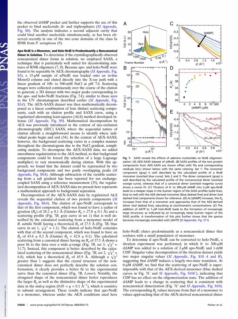
Apo-NrdE Is a Monomer, and Holo-NrdE Is Predominantly a NoncanonicalDimer in Solution. To determine if the crystallographically observednoncanonical dimer forms in solution, we employed SAXS, atechnique that is particularly well suited for deconvoluting mix-tures of RNR oligomers (7, 8). Because apo- and holo-NrdE werefound to be separable by AEX chromatography (SI Appendix, Fig.S3), a 15-μM sample of aiNrdE was loaded onto an in-lineMonoQ column and eluted directly into the X-ray path with alinear gradient of 100- to 500-mM NaCl at pH 7.6. Scatteringimages were collected continuously over the course of the elutionto generate a 3D dataset with two major peaks corresponding tothe apo- and holo-NrdE fractions (Fig. 7A), similar to those seenin the UV chromatogram described earlier (SI Appendix, Fig.S3A). The AEX–SAXS dataset was then mathematically decom-posed as a linear combination of four distinct scattering compo-nents, each with an elution profile and SAXS curve, using aregularized alternating least-squares (ALS) method developed in-house (SI Appendix, Fig. S9). Mathematical decomposition byALS was previously introduced in the context of size-exclusionchromatography (SEC) SAXS, where the sequential nature ofelution affords a straightforward means to identify where indi-vidual peaks begin and end (34). In the context of AEX–SAXS,however, the background scattering varies in a complex mannerthroughout the chromatogram due to the NaCl gradient, compli-cating analysis. To decompose the AEX–SAXS data, we addedsmoothness regularization to the ALS method, so that backgroundcomponents could be forced (by selection of a large Lagrangemultiplier) to vary monotonically during elution. With this ap-proach, we found that the dataset could be represented by twobackground components and two partly overlapping peaks (SIAppendix, Fig. S9A). Although subtraction of the variable scatter-ing from a salt gradient has been reported for ion exchange-coupled SAXS using a reference measurement (35), the regular-ized decomposition of AEX–SAXS data we present here representsa mathematical approach to background separation.Decomposition of the AEX–SAXS data by regularized ALS
reveals the sequential elution of two protein components (SIAppendix, Fig. S9A). The elution of apo-NrdE corresponds tothat of the first component, which was found to have a radius ofgyration (Rg) of 28.2 ± 0.1 Å (Guinier Rg = 27.8 ± 0.1 Å) and ascattering profile (Fig. 7B, gray curve in set 1) that is well de-scribed by the calculated scattering from a monomer model ofB. subtilis NrdE having a theoretical Rg of 27.5 Å (Fig. 7B, bluecurve in set 1, √χ2 = 1.1). The elution of holo-NrdE coincideswith that of the second component, which was found to have anRg of 43.6 ± 0.2 Å (Guinier Rg = 42.8 ± 0.1). The calculatedscattering from a canonical dimer having an Rg of 37.5 Å shows apoor fit to the data over a wide q-range (Fig. 7B, set 3, √χ2 =11.7). Instead, this component is better described by the calcu-lated scattering of the noncanonical dimer (Fig. 7B, set 2, √χ2 =6.8), which has a theoretical Rg of 45.9 Å. Although a √χ2greater than 1 suggests that the crystal structure of the non-canonical dimer does not perfectly describe the solution con-formation, it clearly provides a better fit to the experimentalcurve than the canonical dimer (Fig. 7B, Lower). Notably, theelongated shape of the noncanonical dimer better reproducesthe larger Rg as well as the distinctive shape of the experimentaldata in the mid-q region (0.05 < q < 0.1 Å−1), which is sensitiveto subunit arrangement. These results indicate that apo-NrdEis a monomer, whereas under the AEX conditions used here
holo-NrdE elutes predominantly as a noncanonical dimer thatcoelutes with a small population of monomer.To determine if apo-NrdE can be converted to holo-NrdE, a
titration experiment was performed, in which 0- to 300-μMdAMP was added to a solution of 2-μM apo-NrdE and 1-mMCDP. Singular value decomposition of the titration dataset yieldstwo major singular values (SI Appendix, Fig. S10 A and B),suggesting that dAMP induces a largely two-state transition. At0-μM dAMP, we find that the scattering of apo-NrdE is super-imposable with that of the AEX-derived monomer (blue dashedcurves in Fig. 7C and SI Appendix, Fig. S10C), indicating thatCDP has no effect on the oligomerization state. The addition ofdAMP leads to a change in scattering that is consistent withnoncanonical dimerization (Fig. 7C and SI Appendix, Fig. S10).The corresponding Rg values increase from that of a monomer tovalues approaching that of the AEX-derived noncanonical dimer
Frame Number
I(q)
(a.
u.)
5001000
1500q (Å )-10
0.050.1
0
10
20
30
A
[dAMP] (µM)0 2 4 6 8 10 300
Rg
(Å)
30
34
38
42
//
//
26
C
D
[dAMP]
10-2 10-1
I(q)
(a.
u.)
10-1
100
101
102
103
104
E
B
Apo
Holo
-200
20
q (Å-1)
q2 (Å-2) x10-3
res.
(1)
(2)
(3)
0 0.4 0.8 1.2 1.6 2
ln[I(
q)/I(
0)]
-1
-0.8
-0.6
-0.4
-0.2
0
q2 (Å-2) x10-30 0.2 0.4 0.6 0.8 1
ln[I(
q)/I(
0)]
-2
-1.6
-1.2
-0.8
-0.4
0
0 µM dATP
20 µM dATP
50 µM dATP
Fig. 7. SAXS reveals the effects of adenine nucleotides on NrdE oligomeri-zation. (A) AEX–SAXS dataset of aiNrdE. (B) SAXS profiles of the two proteincomponents from AEX–SAXS are shown offset with fits and correspondingresiduals (res.) shown below with the same coloring. Set 1: The monomercomponent (gray) is well described by the calculated profile of a NrdEmonomer (overlaid blue curve). Sets 2 and 3: The dimer component (gray) iswell described by the calculated profile of the noncanonical dimer (overlaidorange curve), whereas that of a canonical dimer (overlaid magenta curve)shows a worse fit. (C) Titration of 0- to 300-μM dAMP into 2-μM apo-NrdEleads to a steeper slope in the Guinier region of the SAXS profiles (solid lines,blue to red) with the AEX-derived monomer (blue dashed line) and dimer (reddashed line) components shown for reference. (D) As [dAMP] increases, the Rgincreases from that of a monomer and approaches that of the AEX-deriveddimer (red dashed line), saturating at stoichiometric concentrations. (E) Theaddition of dATP to 1-μM holo-NrdE leads to the formation of increasinglylarge structures, as indicated by an increasingly steep Guinier region of theSAXS profile. A transformation of this plot further shows that the speciesbecomes increasingly extended with [dATP] (SI Appendix, Fig. S11).
E4600 | www.pnas.org/cgi/doi/10.1073/pnas.1800356115 Parker et al.
(Fig. 7D), saturating at stoichiometric concentrations of dAMP.At 300-μM dAMP, the scattering becomes superimposable tothat of the AEX-derived noncanonical dimer (SI Appendix, Fig.S10C, red dashed curve). This titration thus provides evidencethat, while apo-NrdE is isolated as a dAMP-free monomer, it hasa strong affinity for dAMP, such that stoichiometric concentra-tions are sufficient to restore a holo-like structure.
dATP Induces a Higher-Order Oligomerization of Holo-NrdE. Havingestablished the structure of holo-NrdE by crystallography andSAXS, we next revisited the effect of dATP on quaternarystructure. To mirror the AUC experiments performed withaiNrdE, the scattering of 1-μM holo-NrdE was measured in thepresence of 0-, 20-, and 50-μM dATP and 1-mM CDP. At thislow protein concentration, the scattering profiles are noisier.However, addition of dATP leads to a clear change in the low-qregion of the Guinier plots (Fig. 7E), corresponding to a steepincrease in Rg. At 0-μM dATP, Guinier analysis yields an Rg of49.5 ± 2.1 Å, consistent with a sample that is predominantlynoncanonical dimer. At 20-μM dATP, the Rg increases to 76.5 ±6.7 Å, and the forward scattering intensity, which is an indicatorof mass, increases by a factor of 2.5. Further increasing the dATPconcentration to 50-μM leads to an even greater Rg of 107.5 ±13.3 Å and 5.7-fold increase in forward scattering relative to0-μM dATP. Thus, we find that the size and mass of NrdE, asreported by Rg and the forward scattering intensity, do not sat-urate within the physiologically relevant range of dATP con-centrations examined here. Instead, both our AUC data andSAXS data suggest that dATP inhibition in B. subtilis RNR in-volves the formation of an increasingly larger species. Addi-tionally, the SAXS data provide evidence that this species iselongated. When replotted as a cross-sectional Guinier plot [i.e.,ln(I × q) vs. q2], the addition of dATP leads to increased linearityin the low-q region, which is indicative of an extended shape (SIAppendix, Fig. S11) (8). Such a species would be possible if dATPbinds the specificity sites to promote the formation of a canon-ical interface between two adjacent noncanonical NrdE dimers.
DiscussionBefore this work, negative-feedback allosteric regulation bydATP was considered a hallmark of class Ia RNR regulation inthe maintenance of balanced dNTP pools inside the cell. In thesecases, inhibition occurs via an ATP-cone domain that interactsreadily with other α or β chains to form ring-shaped inhibitedstructures (7–9). Recently, we found the B. subtilis class Ib RNRto be inhibited by dATP (20), in contrast to the previouslystudied unregulated members of this subclass (12–19). Sequencealignments and structural analyses indicated that all class IbRNRs lack complete ATP-cones (36). Thus, dATP inhibition ofthe B. subtilis enzyme was a surprising result that implied amechanism of overall activity regulation divergent from that ofboth Ia and other Ib RNRs. The work presented herein suggeststhat a tightly bound small-molecule modulator is a criticalcomponent of allosteric feedback regulation in B. subtilis RNR.Interaction between this ligand and the truncated cone in theN-terminal domain promotes the formation of inhibited structuresdistinct from those seen in previously studied class Ia systems.The discovery of tightly bound dAMP associated with NrdE in
protein obtained recombinantly from E. coli and endogenouslyfrom B. subtilis suggests that this nucleotide plays an importantrole in regulation and/or stability of the catalytic subunit. Al-though little is known about the metabolic origin(s) of dAMP inB. subtilis, the presence of ymaB in the Bs nrd operon is pro-vocative (26, 37). While the function of YmaB has yet to be fullyexplored, it is homologous to members of the Nudix hydrolasesuperfamily reported to cleave dATP to dAMP and pyrophos-phate (38–40). Thus, we speculate that YmaB might play aunique role in producing the dAMP necessary for the formation
of holo-NrdE. Indeed, preliminary studies of recombinant YmaBhave shown that the enzyme possesses dNTP pyrophosphohy-drolase activity (26), suggesting that at the very least it mayparticipate in controlling dATP concentrations and allostericregulation of RNR. Further study is required to better charac-terize this protein and establish YmaB’s function inside the cell.We note that, of the Ib RNRs identified from other bacteriathat could potentially exhibit similar allosteric regulation bydAMP and dATP (SI Appendix, Fig. S6A), only those belongingto the genus Bacillus have a homolog of YmaB encoded in theRNR operon.Although dAMP is the primary nucleotide recovered from
NrdE, other unidentified nucleoside di- and triphosphates werealso detected. An outstanding issue that is related to this result isthe mechanism(s) by which dATP inhibits the B. subtilis RNR.One possibility is that dATP displaces dAMP from its bindingsite to exert its inhibitory effects on enzymatic activity. Indeed,the observation of a phosphate in our dAMP-soaked NrdEcrystal structure suggests that the dAMP-binding site may be ableto accommodate dATP. A second possibility is that there is anadditional binding site in the noncanonical dimer created bydAMP for dATP. Analysis of electrostatic surface potential inthe N-terminal and connector domains of NrdE reveals an ex-tended positively charged region that includes both the observeddAMP pocket and an adjacent cavity (SI Appendix, Fig. S8).Therefore, dAMP-induced formation of the noncanonical NrdEdimer observed in the pH 4 crystal structure may create a thirdeffector site at the interface to which dATP can bind to furtherstabilize the noncanonical dimer. Such versatility has precedentin the ATP-cones of class Ia RNRs, which can form differenttypes of quaternary contacts (7–9), bind nucleotides with dif-ferent phosphorylation states (41, 42), and interact with multiplenucleotides at once (9). Flexibility in the overall activity allostericsites of RNRs, whether it be the ATP-cone or the N-terminallytruncated cone identified here, may give the cell the ability tofinely tune enzymatic activity in response to a broader range ofnucleotide (deoxynucleotide) cues that indicate imbalances inthe in vivo dNTP pools.The recovery of dAMP associated with B. subtilis NrdEF in-
dicates that the holo-form of NrdE occurs in vivo. The SAXSanalyses at pH 7.6 show that in vitro dAMP induces a shift inequilibrium from monomer to a noncanonical dimer. However,given that full S-sites do not form within the noncanonical NrdEdimer (Fig. 1B), at least at low [dATP], we believe that thisstructure cannot be the active form in NDP reduction. Based onour current understanding of substrate specificity allostery inother class I RNRs, the canonical dimer interface is required tofully form the S-site (Fig. 1A). This configuration minimizes thedistance between the S- and C-sites so that the correct pairing ofsubstrate and effector nucleotides occurs (43–46). The fact thatB. subtilis RNR conforms to the universal scheme of substratespecificity allostery (Fig. 2) indicates that NrdE is able to form acanonical dimer (5, 6) in addition to the noncanonical form wehave described here. We propose that the dAMP-bound non-canonical dimer is an inhibited form of the protein and thatcanonical dimerization is favored when a monomer of NrdEbinds to NrdF (β2), a second α, and effector to form the activeα2β2 complex. The existence of an α2β2 structure is supported byour previous SV-AUC studies (20).A notable feature of the canonical and noncanonical dimers is
that the uncomplexed dimer interfaces are solvent-exposed andtherefore are capable of interacting with other NrdE species. IfdATP is able to stabilize both interfaces, we can envision higher-order association of NrdE into oligomeric structures (Fig. 8).Unlike the dATP-inhibited oligomers found in RNRs with ATP-cones (7–9), the N-terminal region of B. subtilis NrdE is likelynot sufficiently long or flexible to twist the oligomer to the degreenecessary to terminate in a relatively compact ring structure,
leading to the increasingly large oligomers observed experimen-tally. As in other class I RNRs, the formation of higher-orderoligomeric structures after subunit dissociation of the α2β2 com-plexes following turnover could attenuate RNR activity. The exactmolecular basis for dATP inhibition in B. subtilis class Ib RNRrequires further study. An initial analysis of the structure of twodocked models of α2β2 complexes linked via the noncanonical αsubunit dimer interface, for example, leads to a steric clash be-tween the β2 components, potentially a route to inactivation viaquaternary structure disruption (SI Appendix, Fig. S12).Based on these observations, we propose the model shown in
Fig. 8 to describe the overall activity allosteric regulation of theB. subtilis Ib RNR. As in enzymes with ATP-cone domains, wepropose that high concentrations of dATP inhibit the enzyme bydisruption of the RT pathway between the subunits via seques-tration of NrdE into inhibited states, here represented by boththe noncanonical dimer and higher-order oligomers. A possibleroute to the assembly of larger structures is indicated by thedashed line in Fig. 8. In this proposal, high [dATP] promotesaddition of the NrdE monomer to the noncanonical dimer, po-tentially via a canonical dimer interface or a structurally similarequivalent. The relative locations of these binding sites in B.subtilis NrdE on opposite sides of the protein would result inextended structures consistent with SAXS analysis of the dATP-bound form. In this model, the key to controlling the amount ofactive RNR is the quaternary structural equilibrium of NrdEmonomer and noncanonical dimer (Fig. 8 and SI Appendix, Fig.S2). The dATP ligand signals elevated or imbalanced dNTPpools in vivo and reduces RNR activity by shifting the equilib-rium toward higher-order oligomers composed of noncanonicaldimers. The absence of dATP promotes dissociation into mono-meric NrdE, capable of binding NrdF dimers to form an active α2β2,thus leading to enzyme turnover. On-going studies in our groups arefocused on investigating this model and other alternatives, includingone in which NrdF (β) is a component of the inhibited state. The
latter option has precedent in the dATP-inhibited α4β4 complex ofthe E. coli class Ia RNR (7, 32).Finally, sequence comparison of B. subtilis NrdE with other
class Ib RNRs reveals that only a limited subset may exhibit in-hibition by dATP and/or dAMP. These include the phylogeneti-cally close relatives of B. subtilis, Mycobacterium tuberculosis, andseveral other soil bacteria that are known to be opportunistichuman pathogens (SI Appendix, Fig. S6A). Curiously, M. tuber-culosis NrdE, which has all the dAMP-interacting residues con-served, and the Corynebacterium NrdEs, which have all but anH34 equivalent, have previously been reported to be insensitive todATP inhibition (13, 19). The insensitivity ofM. tuberculosis NrdEmay be a result of its low activity measured using NrdF loadedwith a diferric-Y• cofactor and DTT as the reductant (19). Theresults with Corynebacterium ammoniagenes RNR require furtherstudies (13, 47). Given what we now know about the assembly ofthe physiologically relevant Mn(III)2–Y• cofactor and the impor-tance of the endogenous reductant, it will be interesting to reex-amine the effects of dATP and dAMP on the activity of theseenzymes. Likewise, further study of other closely related enzymesis warranted, as this distinctive form of overall activity allosterymay present opportunities for the development of newantibiotics.The work reported here suggests that negative-feedback reg-
ulation of RNR activity to ensure the high fidelity of DNAreplication and repair may well be more nuanced than previouslythought. Traditionally, ATP-cone domains were considered tobe the universal activity regulator of class Ia RNRs, operating asa discrete, indivisible unit that can be added or subtracted toimbue or remove activity regulation (9, 36). Our studies ofB. subtilis Ib RNR illustrate that regulatory function can also beimparted to an ATP-cone by truncation. Additionally, the dis-covery of the unique role of dAMP in B. subtilis NrdE regulationsuggests that RNRs may be connected in unexpected ways tonucleotide metabolism in the cell. Future examination of theallosteric regulation of other RNRs lacking ATP-cones may re-veal other forms of overall activity allostery mediated by neweffector molecules.
Materials and MethodsHis6-tagged NrdE (720 nmol·min−1·mg−1·α2), NrdF, NrdI, B. subtilis TrxA (200nmol·min−1·mg−1), and B. subtilis TrxB (18 μmol·min−1·mg−1) were prepared asdescribed previously (20, 27, 48). Tagless variants of NrdE wild type (NrdE-wt,710 nmol·min−1·mg−1 α2), NrdE-H34Q, NrdE-F37I, NrdF, and NrdI were producedusing SUMO gene fusion technology (49, 50) as described in SI Appendix. NrdFwas reconstitutedwithMn(III)2–Y• and the holo-proteins (1,300 nmol·min−1·mg−1 β2)purified on a MonoQ column as described previously (20). Apo- and holo-NrdEwere also separated by AEX chromatography on a MonoQ column as de-scribed in SI Appendix. Unless noted otherwise, NrdE and NrdF concentrationsare reported relative to dimer. BsNrdEF was overexpressed in the IB-UP strainand was purified to ∼80% purity as previously described (27). Detailed de-scriptions of materials and methods, including cloning, purification, spectro-photometric assays, SV-AUC, nucleotide isolation, identification, andquantification, X-ray crystallography, and SAXS are given in SI Appendix.
ACKNOWLEDGMENTS. We thank the Massachusetts Institute of Technol-ogy’s Biophysical Instrumentation Facility for the Study of Complex Macro-molecular Systems, supported by National Science Foundation (NSF) GrantNSF-0070319, and in particular Deborah Pheasant, for AUC instrument accessand assistance in experimental setup and execution; Drs. Richard Gillilan andJesse Hopkins for assistance with beamline setup at the Cornell High EnergySynchrotron Source (CHESS); and the Advanced Photon Source (APS), a US De-partment of Energy (DOE) Office of Science User Facility operated for the DOEOffice of Science by Argonne National Laboratory under Contract DE-AC02-06CH11357, for use of its resources. The General Medical Sciences and NationalCancer Institute Collaborative Access Team at the APS was funded in whole or inpart with Federal funds, National Cancer Institute Grant ACB-12002 and NationalInstitute of General Medical Sciences (NIGMS) Grant AGM-12006. The Eiger 16Mdetector was funded by NIH Office of Research Infrastructure Programs, High-End Instrumentation Grant 1S10OD012289-01A1. Use of the Life SciencesCollaborative Access Team Sector 21 was supported by the Michigan Eco-nomic Development Corporation and Michigan Technology Tri-Corridor
Fig. 8. A model describing the overall activity allosteric regulation of theB. subtilis class Ib RNR. NrdE, when loaded with dAMP (orange hexagons),exists in an equilibrium between monomer and the noncanonical dimer. Themonomer pool is capable of forming a canonical dimer and becoming active forNDP reduction when it binds to Mn(III)2–Y• NrdF with a second monomer, and inthe presence of the specificity effector (yellow circles) it potentiates α2β2 complexformation. At high dATP concentrations, noncanonical dimers and possiblymonomers (shown as a dashed line with a red box and a question mark) canassociate into inhibited, extended oligomeric structures involving the canonicaldimer interface, shown in dark gray. The N-terminal dATP-binding site of theoligomer is represented by an open circle suggesting two possible cases: in one,dATP displaces dAMP, and in a second, both dAMP and dATP bind.
E4602 | www.pnas.org/cgi/doi/10.1073/pnas.1800356115 Parker et al.
Grant 085P1000817. We also acknowledge the Berkeley Center for Struc-tural Biology at the Advanced Light Source (ALS), supported in part bythe NIH, NIGMS, and the Howard Hughes Medical Institute. The ALS is aDOE Office of Science User Facility under Contract DE-AC02-05CH11231.SAXS data were collected at the CHESS, which is supported by NSF Grant
DMR-1332208, and at the MacCHESS facility, which is supported by NIHNIGMS Grant GM-103485. This work was supported by NIH GrantsGM117757 (to S.P.M.), GM119707 (to A.K.B.), GM100008 (to N.A.), GM124847(to N.A.), and GM081393 (to J.S.) and by start-up funds from PrincetonUniversity (N.A.).
1. Krissinel E, Henrick K (2004) Secondary-structure matching (SSM), a new tool for fastprotein structure alignment in three dimensions. Acta Crystallogr D Biol Crystallogr60:2256–2268.
2. Uppsten M, et al. (2003) Structure of the large subunit of class Ib ribonucleotide re-ductase from Salmonella typhimurium and its complexes with allosteric effectors.J Mol Biol 330:87–97.
3. Minnihan EC, Nocera DG, Stubbe J (2013) Reversible, long-range radical transfer inE. coli class Ia ribonucleotide reductase. Acc Chem Res 46:2524–2535.
4. Cotruvo JA, Stubbe J (2011) Class I ribonucleotide reductases: Metallocofactor as-sembly and repair in vitro and in vivo. Annu Rev Biochem 80:733–767.
5. Hofer A, Crona M, Logan DT, Sjöberg BM (2012) DNA building blocks: Keeping controlof manufacture. Crit Rev Biochem Mol Biol 47:50–63.
6. Eriksson M, et al. (1997) Binding of allosteric effectors to ribonucleotide reductaseprotein R1: Reduction of active-site cysteines promotes substrate binding. Structure 5:1077–1092.
7. Ando N, et al. (2011) Structural interconversions modulate activity of Escherichia coliribonucleotide reductase. Proc Natl Acad Sci USA 108:21046–21051.
8. Ando N, et al. (2016) Allosteric inhibition of human ribonucleotide reductase by dATPentails the stabilization of a hexamer. Biochemistry 55:373–381.
9. Johansson R, et al. (2016) Structural mechanism of allosteric activity regulation in aribonucleotide reductase with double ATP cones. Structure 24:906–917.
10. Aravind L, Wolf YI, Koonin EV (2000) The ATP-cone: An evolutionarily mobile, ATP-binding regulatory domain. J Mol Microbiol Biotechnol 2:191–194.
11. Meisburger SP, Thomas WC, Watkins MB, Ando N (2017) X-ray scattering studies ofprotein structural dynamics. Chem Rev 117:7615–7672.
12. Crona M, et al. (2011) NrdH-redoxin protein mediates high enzyme activity inmanganese-reconstituted ribonucleotide reductase from Bacillus anthracis. J BiolChem 286:33053–33060.
13. Fieschi F, et al. (1998) The manganese-containing ribonucleotide reductase ofCorynebacterium ammoniagenes is a class Ib enzyme. J Biol Chem 273:4329–4337.
14. Jordan A, et al. (1996) The ribonucleotide reductase system of Lactococcus lactis.Characterization of an NrdEF enzyme and a new electron transport protein. J BiolChem 271:8779–8785.
15. Jordan A, et al. (1994) A second class I ribonucleotide reductase in Enterobacteriaceae:Characterization of the Salmonella typhimurium enzyme. Proc Natl Acad Sci USA 91:12892–12896.
16. Makhlynets O, et al. (2014) Streptococcus sanguinis class Ib ribonucleotide reductase:High activity with both iron and manganese cofactors and structural insights. J BiolChem 289:6259–6272.
17. Rabinovitch I, et al. (2010) Staphylococcus aureus NrdH redoxin is a reductant of theclass Ib ribonucleotide reductase. J Bacteriol 192:4963–4972.
18. Roca I, Torrents E, Sahlin M, Gibert I, Sjöberg BM (2008) NrdI essentiality for class Ibribonucleotide reduction in Streptococcus pyogenes. J Bacteriol 190:4849–4858.
19. Yang F, et al. (1997) Characterization of two genes encoding the Mycobacteriumtuberculosis ribonucleotide reductase small subunit. J Bacteriol 179:6408–6415.
21. Ge J, Yu G, Ator MA, Stubbe J (2003) Pre-steady-state and steady-state kinetic analysisof E. coli class I ribonucleotide reductase. Biochemistry 42:10071–10083.
22. Anand R, et al. (2004) A model for the Bacillus subtilis formylglycinamide ribonucle-otide amidotransferase multiprotein complex. Biochemistry 43:10343–10352.
23. (1996) Characteristics of nucleic acids. Curr Protoc Mol Biol 33:A.1D.1–A.1D.11.24. Randerath K, Randerath E (1964) Ion-exchange chromatography of nucleotides on
studies of the creatine kinase and pyruvate kinase reactions. Anal Biochem 58:525–533.
26. Parker MJ (2017) Discovery and Investigation of the Novel Overall Activity AllostericRegulation of the Bacillus subtilis class Ib Ribonucleotide Reductase. PhD thesis(Massachusetts Institute of Technology, Cambridge, MA).
27. Zhang Y, Stubbe J (2011) Bacillus subtilis class Ib ribonucleotide reductase is adimanganese(III)-tyrosyl radical enzyme. Biochemistry 50:5615–5623.
28. Krissinel E, Henrick K (2007) Inference of macromolecular assemblies from crystallinestate. J Mol Biol 372:774–797.
29. Uhlin U, Eklund H (1994) Structure of ribonucleotide reductase protein R1. Nature370:533–539.
30. Boal AK, Cotruvo JA, Jr, Stubbe J, Rosenzweig AC (2012) The dimanganese(II) site ofBacillus subtilis class Ib ribonucleotide reductase. Biochemistry 51:3861–3871.
31. Denessiouk KA, Rantanen VV, Johnson MS (2001) Adenine recognition: A motifpresent in ATP-, CoA-, NAD-, NADP-, and FAD-dependent proteins. Proteins 44:282–291.
32. Zimanyi CM, et al. (2012) Tangled up in knots: Structures of inactivated forms of E. coliclass Ia ribonucleotide reductase. Structure 20:1374–1383.
33. Saraste M, Sibbald PR, Wittinghofer A (1990) The P-loop–A commonmotif in ATP- andGTP-binding proteins. Trends Biochem Sci 15:430–434.
34. Meisburger SP, et al. (2016) Domain movements upon activation of phenylalaninehydroxylase characterized by crystallography and chromatography-coupled small-angle x-ray scattering. J Am Chem Soc 138:6506–6516.
35. Hutin S, Brennich M, Maillot B, Round A (2016) Online ion-exchange chromatographyfor small-angle X-ray scattering. Acta Crystallogr D Struct Biol 72:1090–1099.
36. Jonna VR, et al. (2015) Diversity in overall activity regulation of ribonucleotide re-ductase. J Biol Chem 290:17339–17348.
37. Scotti C, Valbuzzi A, Perego M, Galizzi A, Albertini AM (1996) The Bacillus subtilisgenes for ribonucleotide reductase are similar to the genes for the second class I NrdE/NrdF enzymes of Enterobacteriaceae. Microbiology 142:2995–3004.
38. O’Handley SF, Frick DN, Bullions LC, Mildvan AS, Bessman MJ (1996) Escherichia coliorf17 codes for a nucleoside triphosphate pyrophosphohydrolase member of theMutT family of proteins. Cloning, purification, and characterization of the enzyme.J Biol Chem 271:24649–24654.
39. Xu W, Dunn CA, Jones CR, D’Souza G, Bessman MJ (2004) The 26 Nudix hydrolases ofBacillus cereus, a close relative of Bacillus anthracis. J Biol Chem 279:24861–24865.
40. Xu W, Jones CR, Dunn CA, Bessman MJ (2004) Gene ytkD of Bacillus subtilis encodesan atypical nucleoside triphosphatase member of the Nudix hydrolase superfamily.J Bacteriol 186:8380–8384.
41. Grinberg I, et al. (2006) The Streptomyces NrdR transcriptional regulator is a Zn rib-bon/ATP cone protein that binds to the promoter regions of class Ia and class II ri-bonucleotide reductase operons. J Bacteriol 188:7635–7644.
42. McKethan BL, Spiro S (2013) Cooperative and allosterically controlled nucleotidebinding regulates the DNA binding activity of NrdR. Mol Microbiol 90:278–289.
43. Knappenberger AJ, Ahmad MF, Viswanathan R, Dealwis CG, Harris ME (2016) Nu-cleoside analogue triphosphates allosterically regulate human ribonucleotide re-ductase and identify chemical determinants that drive substrate specificity.Biochemistry 55:5884–5896.
44. Larsson KM, et al. (2004) Structural mechanism of allosteric substrate specificityregulation in a ribonucleotide reductase. Nat Struct Mol Biol 11:1142–1149.
45. Xu H, et al. (2006) Structures of eukaryotic ribonucleotide reductase I provide insightsinto dNTP regulation. Proc Natl Acad Sci USA 103:4022–4027.
46. Zimanyi CM, Chen PYT, Kang G, Funk MA, Drennan CL (2016) Molecular basis forallosteric specificity regulation in class Ia ribonucleotide reductase from Escherichiacoli. eLife 5:e07141.
47. Stolle P, et al. (2010) Homologous expression of the nrdF gene of Corynebacteriumammoniagenes strain ATCC 6872 generates a manganese-metallocofactor (R2F) and astable tyrosyl radical (Y_) involved in ribonucleotide reduction. FEBS J 277:4849–4862.
48. Cotruvo JA, Jr, Stich TA, Britt RD, Stubbe J (2013) Mechanism of assembly of thedimanganese-tyrosyl radical cofactor of class Ib ribonucleotide reductase: Enzymaticgeneration of superoxide is required for tyrosine oxidation via a Mn(III)Mn(IV) in-termediate. J Am Chem Soc 135:4027–4039.
49. Butt TR, Edavettal SC, Hall JP, Mattern MR (2005) SUMO fusion technology fordifficult-to-express proteins. Protein Expr Purif 43:1–9.
50. Marblestone JG, et al. (2006) Comparison of SUMO fusion technology with traditionalgene fusion systems: Enhanced expression and solubility with SUMO. Protein Sci 15:182–189.