AN EVALUATION OF THE RELATIONSHIP BETWEEN SIGMA-1 AND SIGMA-2 RECEPTORS AND THE ENDOCANNABINOID SYSTEM by Jenn Miller A thesis submitted to the faculty of the University of Mississippi in partial fulfillment of the requirements of the Sally McDonnell Barksdale Honors College. Oxford May 2016 Approved by Advisor: Dr. Christopher McCurdy Reader: Dr. Kenneth Sufka Reader: Dr. John Rimoldi
Transcript
AN EVALUATION OF THE RELATIONSHIP BETWEEN SIGMA-1 AND SIGMA-2
RECEPTORS AND THE ENDOCANNABINOID SYSTEM
by
Jenn Miller
A thesis submitted to the faculty of the University of Mississippi in partial fulfillment of
the requirements of the Sally McDonnell Barksdale Honors College.
First, I would like to take this opportunity to dedicate this thesis to my family. Without my parents’ constant support, encouragement, and dedication to my education, I would
not be where I am today. It is their hard work that has allowed me to be a member of the University of Mississippi Sally McDonnell Barksdale Honors College and participate in
this research project. I would also like to dedicate my thesis to my siblings and my friends, who are always ready to lend a listening ear, take a weekend to visit me, or offer
sound advice. Your support means everything!
iv
ACKNOWLEDGEMENTS
This study was supported by an Institutional Development Award (IDeA) Grant Number P20GM104932 from the National Institute of General Medical Sciences (NIGMS) through the In Vivo Pharmacology Core and Project 2 of the COBRE, a component of the National Institutes of Health (NIH). AZ66, CM304 and CM398 were discovered under Grant Number R01DA023305 from the National Institute on Drug Abuse (NIDA). Its contents are solely the responsibility of the authors and do not necessarily represent the official view of NIGMS, NIDA or NIH.
v
ABSTRACT JENN MILLER: An Evaluation of the Relationship between Sigma-1 and Sigma-2
Receptors and the Endocannabinoid System (Under the direction of Lisa Wilson and Dr. Christopher McCurdy)
Within the past two decades, sigma receptors have become a popular area for
research. Although much has been learned about their structure, subtypes, and functions;
there is still much to be learned. Consisting of two receptor subtypes (sigma-1 and
sigma-2), it has been discovered that sigma receptor ligands potentiate the analgesic
effects of both opiates and cannabinoids, though the exact mechanism and sigma subtype
on which this occurs is still unknown. The purpose of this study is to determine if
potentiation of opiates and cannabinoids occurs through sigma-1 or sigma-2 receptor
signaling. Tetrad assays were performed for:
1. Morphine, an opiate, at multiple doses (i.p.),
2. CP 55,940, a cannabinoid, at multiple doses (i.p.),
3. CM304, a sigma-1 antagonist, at multiple doses (i.p.), and
4. CM398, a sigma-2 antagonist, at multiple doses (i.p.).
The potentiation studies were then completed by using both 20 and 45 mg/kg
doses of CM304 and CM398 against either a 1 mg/kg dose of CP 55,950 or a 2 mg/kg
dose of morphine. The CM dose was administered 15 minutes before the CP or the
morphine dose, and the analgesic study was performed 15 minutes post CP or morphine
administration. This study revealed that CM304, the sigma-1 antagonist, potentiated the
effects of CP 55,940 and morphine for the hotplate assay while it attenuated the effects
vi
on the tail-flick assay. Additionally, CM398, the sigma-2 antagonist, failed to potentiate
both CP 55,940 and morphine for both the hotplate and the tail-flick assay. Results also
showed that AZ66, the general sigma receptor antagonist, potentiated the effects of CP
55,940 for both the hotplate and the tail-flick assays. In conclusion, administering a
sigma-1 antagonist, instead of a sigma-2 antagonist, in conjunction with either an opiate
or a cannabinoid will potentiate the effects of the challenge drug. This can serve as an
The sigma receptor was initially categorized as another opioid receptor subtype
(Maurice et al., 2009). The confusion arose because the ligands that were used in the
experiment cross-reacted with both sigma receptors and opioid receptors (Maurice et al.,
2009). Further studies demonstrated that the sigma receptor was a separate receptor from
the opioid receptor. The sigma receptor is a unique chaperone protein found mainly in
the endoplasmic reticulum and the plasma membrane of cells. In these cells, there are
two known subtypes of the sigma receptor: sigma-1 and sigma-2 (Maurice et al., 2009).
Sigma-1 was the first sigma subtype to be discovered and in 1996, it became the
first subtype to be cloned from a guinea pig liver (Zamanillo et al., 2013). It was
subsequently cloned from mouse, rat, and human (Hanner et al., 1996; Mei et al., 2001;
Pan et al., 1998; Seth et al., 1997; Seth et al., 1998). The sigma-1 gene encodes a 25-29
kDa molecular mass protein that consists of 223 amino acids and at least one
transmembrane spanning domain (Zamanillo et al., 2013; Ayudar et al., 2002; Jbilo et al.,
1997). It has been found to be broadly distributed in both the peripheral organs and the
central nervous system, including high expression in the brain, the heart, the liver, the
spleen, and the GI tract (Matsomoto et al., 2007). More specifically, the sigma-1 receptor
has been localized to regions of the brain associated with pain control, including the
superficial layers of the dorsal horn, the periaqueductal gray matter, the locus coeruleus,
and the rostroventral medulla (Zamanillo et al., 2013). Because the sigma-1 subtype has
been sequenced, cloned, and is the most well-understood sigma subtype, it has been used
as the basis for many studies regarding disease states. Currently, sigma-1 is hypothesized
to play a very important role in addiction, pain, depression, Alzheimer’s disease,
2
schizophrenia, stroke, HIV, cancer, and many other neurological conditions (Maurice et
al., 2009). Sigma-1 subtype receptors will continue to be researched extensively as a
potential treatment option for many of the aforementioned conditions.
Despite the sigma-2 receptor being the only other sigma subtype, there has not
been a large amount of research performed about its roles and its capabilities. This lack of
research has led to some confusion surrounding the sigma-2 gene and its corresponding
protein. Although the Progesterone Receptor Membrane Component 1 (PGRMC1) has
been recently implicated as a sigma-2 subtype, subsequent research now indicates that
this might not be the case (Chu et al., 2015). From this study, data indicates that
PGRMC1 and sigma-2 receptors are genetically different, meaning that they are two
different proteins and that PGRMC1 is a non-sigma-2 receptor binding site in mammalian
tissues (Chu et al., 2015). More basically, however, there is still some literature on the
structure and the proposed function of this subtype. Sigma-2 is slightly smaller in size
than sigma-1, existing as an 18-22 kDa protein that is highly expressed in the brain, the
liver, and the GI tract (Matsomoto et al., 10). Additionally, sigma-2 is different because
it is infrequently expressed in the heart and the spleen (Matsomoto et al., 10). The
functions of sigma-2 are also believed to be vastly different from sigma-1. Thus far,
sigma-2 has been linked to roles in cellular events, such as proliferation, apoptosis,
dendritogenesis, synaptogenesis, neuronal plasticity, activation of cytochrome P450, and
steroid signaling (Zamanillo et al., 2013). It has also been speculated that the binding of
sigma-2 ligands to sigma-2 receptors can trigger both caspase-dependent and caspase-
independent apoptosis (Zeng et al., 2014). More research is still needed to fully
understand these processes with regards to sigma-2.
3
The initial confusion surrounding the classification of sigma receptors as opioid
receptors led researchers to investigate the relationship between opioids and sigma
receptors. Although research reveals a clear relationship between the two, it is still
unknown whether or not the sigma receptors interact directly with opioid receptors or
alter signaling pathways downstream from the opioids (Matsomoto et al., 2007). In fact,
it has been shown that sigma-1 antagonists can potentiate the effects of opioid analgesia
which will be detailed later. Regardless of the many questions surrounding the specifics
of the relationship, there is still a multitude of literature that describes opioid analgesia
and sigma receptors (Sánchez-Fernández et al., 2014; Vidal-Torres et al., 2013; Tseng et
al, 2011; Kim et al., 2010; Mei et al., 2007; Mei et al., 2002).
Although moderate to severe pain is a very common medical complaint among
patients, it is still a very complicated condition to manage. In most communities today,
pain is managed by giving opioids, like morphine, to patients. Although opioids have
strong analgesic effects, they can also produce many harmful side effects, including
constipation, nausea, respiratory distress, tolerance, and addiction liability (Sánchez-
Fernández et al., 2013; Vidal-Torres et al., 2013). Because of these side effects,
researchers and clinicians are investigating new ways to manage pain, particularly by
giving opioids in conjunction with other drugs (Lui et al., 2011; Khan et al., 2011). The
goal of giving an additional drug, like a sigma antagonist, is to enhance the effects of the
opioid without also increasing the side effects. One example that has demonstrated
efficacy is the use of sigma-1 antagonists. Additionally, sigma receptor antagonists alone
are believed to play a potential key role in the management of pain.
4
In previous studies with mice, it was shown that by giving an intrathecal injection
of a sigma-1 receptor antagonist in conjunction with an opioid, the opioid-induced
analgesia was potentiated (Maurice et al., 2009). Instead, if a sigma-1 agonist was
administered with the opioid, then the opioid-induced analgesia was attenuated (Maurice
et al., 2009). In other words, the antagonist enhanced the pain-relieving effects of the
opioid while the agonist diminished the pain-relieving effects of the opioid. In similar
studies, the sigma-1 receptors were down-regulated and then knocked-out completely to
see if the same effects could be observed. When sigma-1 receptors were down-regulated,
the analgesic effects of the opioid were again potentiated (Maurice et al, 2009).
However, when sigma-1 receptors were knocked-out entirely, the analgesic effects of the
opioid were not potentiated (Zamanillo et al., 2013). This reveals that sigma-1 receptors
have some type of modulating capabilities over opioid analgesia, which is still not fully
understood. Perhaps the best result of these studies is the evidence that shows that the
side effects, like tolerance, withdrawal symptoms, and constipation were not also
potentiated (Vidal-Torres et al., 2013). While the analgesic effects of opioids were able
to be potentiated, the side effects for that specific dose remained the same. This means
the pain-relieving effects were enhanced while the side effects were not. This looks very
promising for future treatment and management of pain. Lastly, there has not been much
research, if any at all, for the role of sigma-2 in the treatment and management of pain.
To further examine the role of sigma-1 and sigma-2 receptors in the management
of pain, a number of compounds were used for this research. Morphine, an opioid, was
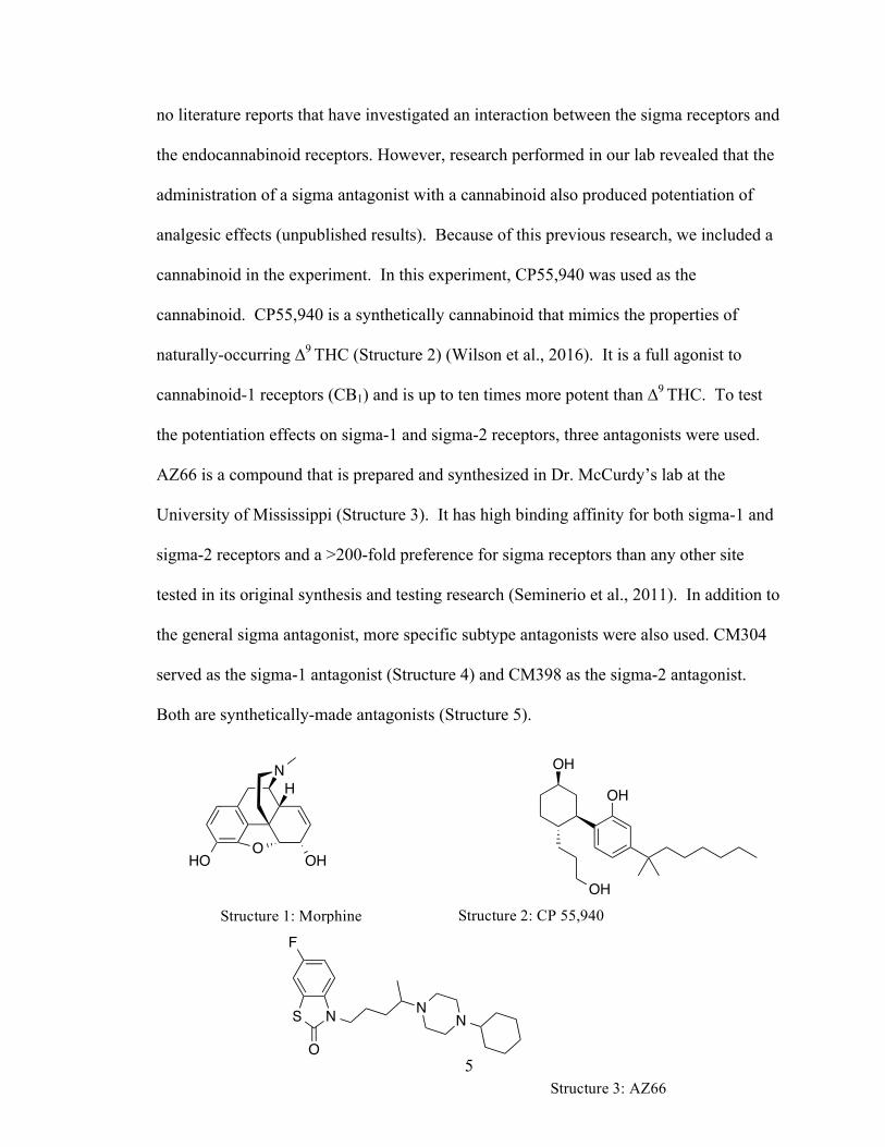
used to show effects that have already been described in previous literature (Structure 1).
Although the opioid system and the endocannabinoid systems are very similar, there are
5
no literature reports that have investigated an interaction between the sigma receptors and
the endocannabinoid receptors. However, research performed in our lab revealed that the
administration of a sigma antagonist with a cannabinoid also produced potentiation of
analgesic effects (unpublished results). Because of this previous research, we included a
cannabinoid in the experiment. In this experiment, CP55,940 was used as the
cannabinoid. CP55,940 is a synthetically cannabinoid that mimics the properties of
naturally-occurring ∆9 THC (Structure 2) (Wilson et al., 2016). It is a full agonist to
cannabinoid-1 receptors (CB1) and is up to ten times more potent than ∆9 THC. To test
the potentiation effects on sigma-1 and sigma-2 receptors, three antagonists were used.
AZ66 is a compound that is prepared and synthesized in Dr. McCurdy’s lab at the
University of Mississippi (Structure 3). It has high binding affinity for both sigma-1 and
sigma-2 receptors and a >200-fold preference for sigma receptors than any other site
tested in its original synthesis and testing research (Seminerio et al., 2011). In addition to
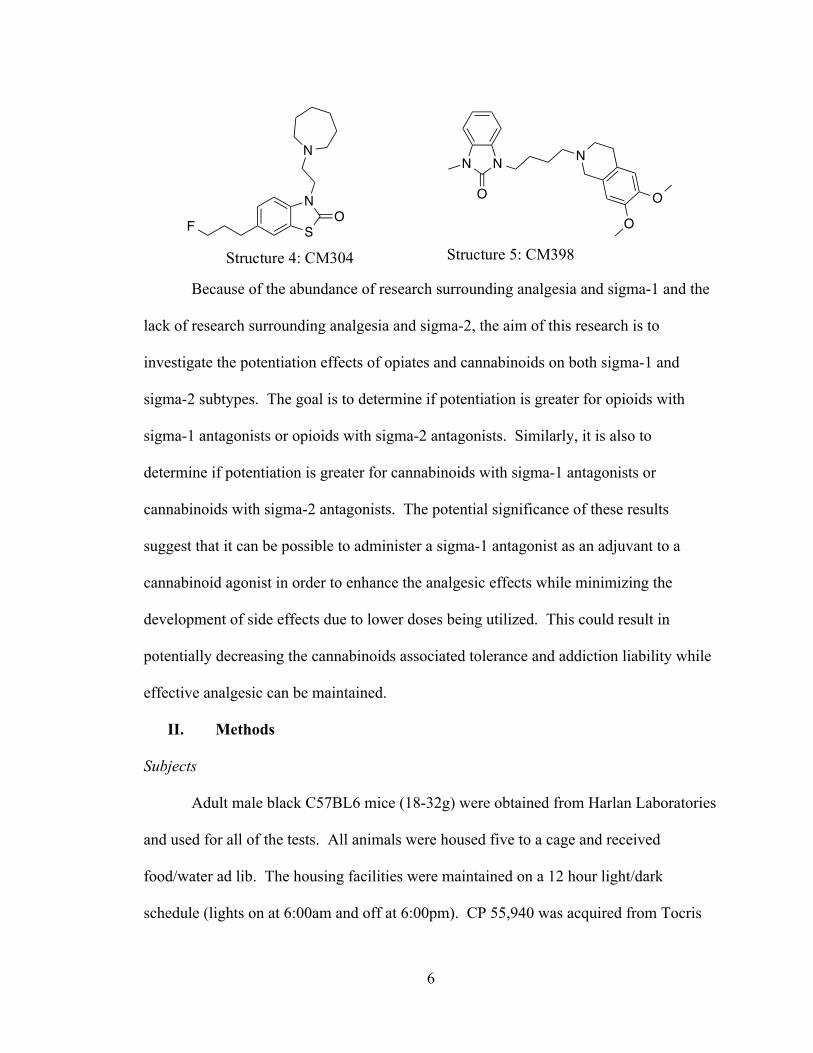
the general sigma antagonist, more specific subtype antagonists were also used. CM304
served as the sigma-1 antagonist (Structure 4) and CM398 as the sigma-2 antagonist.
Both are synthetically-made antagonists (Structure 5).
HO OH
NH
O
OH
OH
OH
S N
O
NN
FStructure 1: Morphine Structure 2: CP 55,940
Structure 3: AZ66
6
Because of the abundance of research surrounding analgesia and sigma-1 and the
lack of research surrounding analgesia and sigma-2, the aim of this research is to
investigate the potentiation effects of opiates and cannabinoids on both sigma-1 and
sigma-2 subtypes. The goal is to determine if potentiation is greater for opioids with
sigma-1 antagonists or opioids with sigma-2 antagonists. Similarly, it is also to
determine if potentiation is greater for cannabinoids with sigma-1 antagonists or
cannabinoids with sigma-2 antagonists. The potential significance of these results
suggest that it can be possible to administer a sigma-1 antagonist as an adjuvant to a
cannabinoid agonist in order to enhance the analgesic effects while minimizing the
development of side effects due to lower doses being utilized. This could result in
potentially decreasing the cannabinoids associated tolerance and addiction liability while
effective analgesic can be maintained.
II. Methods
Subjects
Adult male black C57BL6 mice (18-32g) were obtained from Harlan Laboratories
and used for all of the tests. All animals were housed five to a cage and received
food/water ad lib. The housing facilities were maintained on a 12 hour light/dark
schedule (lights on at 6:00am and off at 6:00pm). CP 55,940 was acquired from Tocris
S
N
F
N
O
N N
O
N
O
O
Structure 4: CM304 Structure 5: CM398
7
Bioscience (Bristol, United Kingdom). Morphine, Cremophor and Ethanol were obtained
from Sigma Aldrich (Bellefonte, PA). Lastly, CM304, CM398, and AZ66 were all
prepared and synthesized in Dr. McCurdy’s lab as a part of the Department of
BioMolecular Sciences Division of Medicinal Chemistry. All methods performed were
approved by the Institutional Animal Care and Use Committee (IACUC).
Drug Preparation
All drugs were dissolved according to the methods of Olson et al (1973). A
mixture of Ethanol, Cremophor, and Saline was prepared using a ratio of (1:1:18). Drugs
were completely dissolved into ethanol before adding Cremophor and saline. Drugs were
delivered to the animals using an intraperitoneal (i.p.) injection (Wilson et al., 2016).
Tetrad Assay
The mouse tetrad is a behavioral assay developed to characterize the biological
effects of cannabinoids and opiates using locomotor activity, nociception, changes in
body temperature, and catalepsy (Little et al., 1988). The assay has been well
documented to indicate that the typical effects of cannabinoids is decreased locomotion,
increased cataleptic activity, increased antinociception, and hypothermia (Pertwee et al.,
2008). Twenty-four hours prior to the start of the experiment the mice were acclimated
for 15 minute increments to the cold hotplate container and apparatus. On the
experimental day, the mice were brought into the experimental room and allowed to
acclimate to the room settings for 30 minutes and then to the locomotor chamber for 30
minutes (Wilson et al., 2016). Once the second thirty minute acclimation period was
over, baseline readings for supraspinal antinociception (hotplate), catalepsy, hypothermia,
and spinal antinociception (tail-flick) were recorded pre-injection.
8
In the hotplate assay, the subject was placed on a hotplate at 52°C inside of a
plastic cylinder, so that the subject was contained in one area. The timer was manually
started and then stopped once one of the cues was performed by the subject. These cues
included licking the back paw, moving the back paw to the side surface of the cylinder,
jumping, and rapidly tapping one of the back paws. Because mice lick their front paws
during grooming, only the activity of the back paws is marked as perception of pain.
Additionally, the cut-off time for this assay is 45 seconds to reduce the possibility of
tissue damage to the subject. The purpose of the hotplate is to measure the subject’s
perceived pain and the perceived peripheral pain analgesic effects of the drugs.
With the purpose of measuring the psychoactive effects of the drug, the catalepsy
test is the second test included in the tetrad. In this test, the subject’s front paws are
placed on a metal bar and his hind paws reside on the lower surface. Once the subject is
in this initial position, the timer is started and continues until the subject either jumps
onto the metal bar or lowers his front paws onto the lower surface. If the subject remains
in the initial position for more than five seconds, then it is considered cataleptic and
unaware of its surroundings. The cut-off time for this assay is three minutes. This
method makes it easy to determine the psychoactive effects of the drug on the subject.
The third test of the tetrad is hypothermia, or a measurement of the core body
temperature of the subject. The temperature is measured by inserting a temperature probe
into the subject’s rectum to note any changes in body temperature between pre- and post-
drug injection.
In the last test of the tetrad, the spinal antinociception properties were recorded in
the tail-flick assay. To gain these measurements, the subject was placed in a plastic
9
restrainer so that its tail was hanging out of the restrainer. The restrainer was then laid
down on the surface of the machine to ensure that the tail was also flat along the surface
of the machine. Once the subject had settled down into this position, the timer was
started and a high-energy beam of light was projected on the distal portion of the
subject’s tail. Once the subject moved its tail out of the path of the light beam, the light
and timer automatically shut off. This was repeated once more and the average of the
two trials was taken to ensure greater accuracy. To minimize potential tissue damage, the
cut-off time for this assay was 15 seconds. The purpose of the tail-flick assay is to
measure the spinal (reflex) analgesic effects of the administered drug.
Once the baseline readings were obtained for the tetrad, the animals were injected
i.p. with either the (1:1:18) vehicle, CP 55,940 (0.1, 0.25, 0.5mg/kg), morphine (2, 2.5, 5
mg/kg), CM304 (5, 10, 20 mg/kg), or CM398 (5, 10, 20 mg/kg). The animals were then
allowed to move around in their individual locomotor chamber (San Diego Instruments)
for 30 minutes. The locomotor chamber consists of a 16 x 16 beam ray that detected
movement of the animal. Breaking the photobeams was then quantified as a measure of
locomotor activity. The last 10 minutes of quantifying time was used for data analysis
(Wilson et al., 2016). Evaluating locomotor activity provided an insight to the sedative
effects of the drug. The larger the sedative effects of the drug, the fewer photobeams that
were broken.
Thirty minutes post-injection the subject was removed from the locomotor
chamber and again run through the tetrad assay. Hotplate latency, catalepsy, core body
temperature, and tail-flick latency were recorded.
10
Potentiation Studies
The purpose of the potentiation studies is to measure the analgesic effects of the
drug on the mice in the experiment. In these studies, only the hot plate assay and the tail-
flick assay were used (the same procedures as the ones described above). Again, the
analgesics of the mice were compared before and after the administration of the two
drugs. Twenty-four hours before the testing the mice were acclimated for fifteen minutes
to the hotplate surface. On the day of the experiment, the mice were brought into the lab
and allowed to acclimate for thirty minutes. After this thirty minute period, baseline
readings for supraspinal (hot plate) and spinal (tail-flick) nociception were measured.
Then, the mice were injected i.p. with the antagonist drug, which was either CM304 (20,
40 mg/kg), CM398 (20, 40 mg/kg), or AZ66 (20 mg/kg). The mice were placed back in
their cages for fifteen minutes, after which the mice were again injected i.p. with the
challenge drug, either 2 mg/kg morphine or 0.1 CP 55,940. Fifteen minutes after the
challenge drug injection, the hotplate and tail-flick latencies were recorded. The goal is
to see whether or not CM304, CM398, and/or AZ66 potentiated the effects of CP 55,940
and morphine.
Data Analysis
Data was shown as mean ± SEM. with each group having n=10 animals. Both
hotplate and tail-flick were expressed as percent maximum effect (%MPE=[(post-drug
latency-basal latency)/(cutoff latency-basal)]x 100 (Little et al., 1998). Statistical analysis
was performed using one way ANOVA preceded by the Dunnett’s post hoc test for
locomotor activity and Tukey post hoc test for hot plate, cataleptic effects, decrease in
11
0 2.5 CP 5 10 200
500
1000
1500
2000
2500
***
*
Dose (mg/kg)
Loco
mot
or A
ctiv
ity
0 2.5 CP 5 10 200
500
1000
1500
2000
2500
***
******
Dose (mg/kg)
Loco
mot
or A
ctiv
ity
Figure 1 Figure 2
rectal temperature and tail-flick to define significant different against the vehicle control
at p<0.05 for each specific time point (Wilson et al., 2016).
III. Results
Tetrad Assays
I. Locomotor Activity
Figures 1 and 2 demonstrate the locomotor activity dose response curves for CM304 and
CM398, respectively. Both graphs also illustrate the response when administered (i.p.)
the vehicle (1:1:18) and a 2.5 mg/kg dose of CP 55,940. The 2.5 mg/kg dose of CP
55,940 with both CM304 and CM398 produced a p value <0.001. For CM304, 5 mg/kg
generated a p value <0.05. Both 10 mg/kg, and 20 mg/kg doses of CM398 resulted in p
values <0.001. All of the above p values show statistical significance. While none of the
doses produce locomotor activity resembling the vehicle, all of the doses for CM304 and
for CM398 produce more locomotor activity than the 2.5 mg/kg (i.p.) CP 55,940 dose.
Additionally, CM398 appears to have a dose-dependent sedative effect. This has been
seen with other sigma-2 compounds and may be a behavior that is associated with sigma-
2 receptors, but this requires further investigation.
CM304 Locomotor CM398 Locomotor
Figures 1-2: Locomotor increase post-CM304 i.p. injection (Figure 1) and post-CM398 i.p. injection (Figure 2) as compared to the vehicle and to CP55,940.
12
Morphine Locomotor
Figure 4
(1:1:18) 0.1 0.25 0.5 0
500
1000
1500
2000
2500
***
Dose (mg/kg)
Loco
mot
or A
ctiv
ity
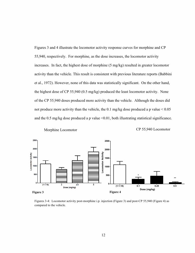
Figures 3 and 4 illustrate the locomotor activity response curves for morphine and CP
55,940, respectively. For morphine, as the dose increases, the locomotor activity
increases. In fact, the highest dose of morphine (5 mg/kg) resulted in greater locomotor
activity than the vehicle. This result is consistent with previous literature reports (Babbini
et al., 1972). However, none of this data was statistically significant. On the other hand,
the highest dose of CP 55,940 (0.5 mg/kg) produced the least locomotor activity. None
of the CP 55,940 doses produced more activity than the vehicle. Although the doses did
not produce more activity than the vehicle, the 0.1 mg/kg dose produced a p value < 0.05
and the 0.5 mg/kg dose produced a p value <0.01, both illustrating statistical significance.
Figures 3-4: Locomotor activity post-morphine i.p. injection (Figure 3) and post-CP 55,940 (Figure 4) as compared to the vehicle.
CP 55,940 Locomotor
Figure 3
13
0 CP 2.5 5 10 200
20
40
60
80
100
***
Dose (mg/kg)
Hot
plat
e La
tenc
y (%
MPE
)
***
0 CP 2.5 5 10 200
20
40
60
80
100 ***
Dose (mg/kg)
Hot
plat
e La
tenc
y (%
MPE
)
II. Hotplate Assay
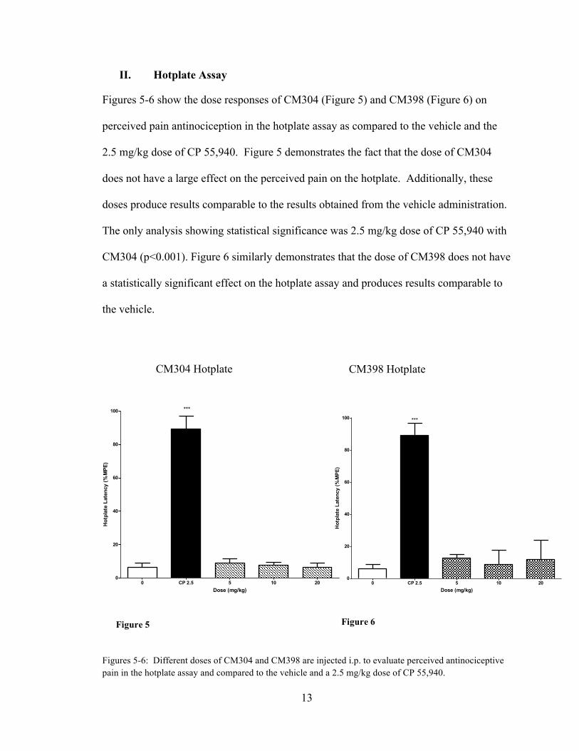
Figures 5-6 show the dose responses of CM304 (Figure 5) and CM398 (Figure 6) on
perceived pain antinociception in the hotplate assay as compared to the vehicle and the
2.5 mg/kg dose of CP 55,940. Figure 5 demonstrates the fact that the dose of CM304
does not have a large effect on the perceived pain on the hotplate. Additionally, these
doses produce results comparable to the results obtained from the vehicle administration.
The only analysis showing statistical significance was 2.5 mg/kg dose of CP 55,940 with
CM304 (p<0.001). Figure 6 similarly demonstrates that the dose of CM398 does not have
a statistically significant effect on the hotplate assay and produces results comparable to
the vehicle.
Figures 5-6: Different doses of CM304 and CM398 are injected i.p. to evaluate perceived antinociceptive pain in the hotplate assay and compared to the vehicle and a 2.5 mg/kg dose of CP 55,940.
CM304 Hotplate CM398 Hotplate
Figure 5 Figure 6
14
(1:1:18) 2 2.5 50
20
40
60
80
100
***
Dose (mg/kg)
Hot
plat
e La
tenc
y (%
MPE
)
(1:1:18) 0.1 0.25 0.5 0
20
40
60
80
100
***
***
***
Dose (mg/kg)
Hot
plat
e La
tenc
y (%
MPE
)
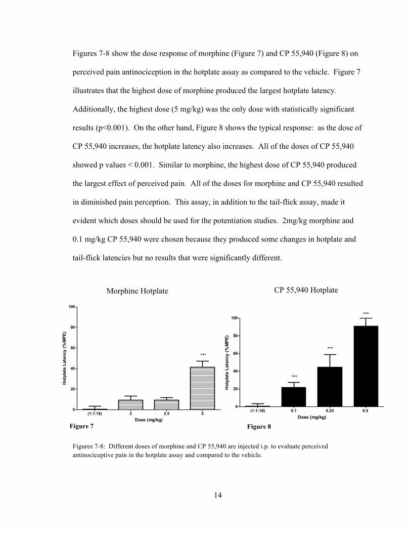
Figures 7-8 show the dose response of morphine (Figure 7) and CP 55,940 (Figure 8) on
perceived pain antinociception in the hotplate assay as compared to the vehicle. Figure 7
illustrates that the highest dose of morphine produced the largest hotplate latency.
Additionally, the highest dose (5 mg/kg) was the only dose with statistically significant
results (p<0.001). On the other hand, Figure 8 shows the typical response: as the dose of
CP 55,940 increases, the hotplate latency also increases. All of the doses of CP 55,940
showed p values < 0.001. Similar to morphine, the highest dose of CP 55,940 produced
the largest effect of perceived pain. All of the doses for morphine and CP 55,940 resulted
in diminished pain perception. This assay, in addition to the tail-flick assay, made it
evident which doses should be used for the potentiation studies. 2mg/kg morphine and
0.1 mg/kg CP 55,940 were chosen because they produced some changes in hotplate and
tail-flick latencies but no results that were significantly different.
Figures 7-8: Different doses of morphine and CP 55,940 are injected i.p. to evaluate perceived antinociceptive pain in the hotplate assay and compared to the vehicle.
Morphine Hotplate CP 55,940 Hotplate
Figure 7 Figure 8
15
0 CP 2.5 5 10 200
50
100
150
200
***
Dose (mg/kg)
Cat
alep
sy L
aten
cy (s
ec)
0 CP 2.5 5 10 200
50
100
150
200
***
Dose (mg/kg)
Cat
alep
sy L
aten
cy (s
ec)
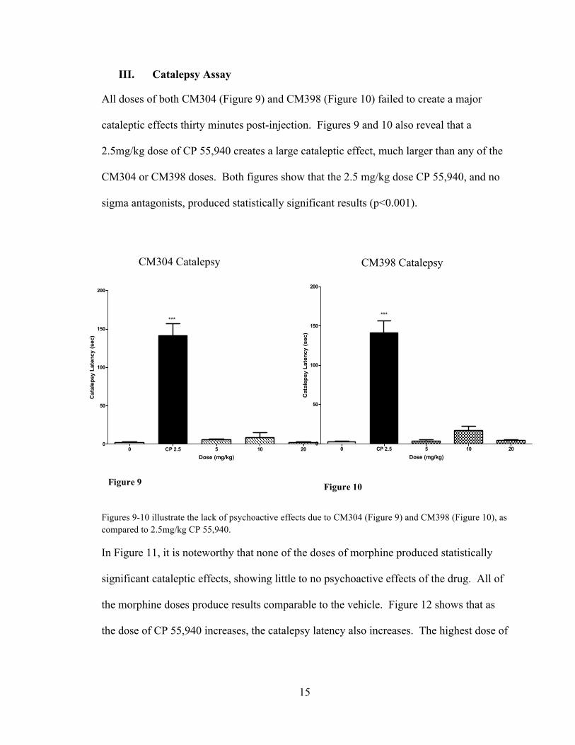
III. Catalepsy Assay
All doses of both CM304 (Figure 9) and CM398 (Figure 10) failed to create a major
cataleptic effects thirty minutes post-injection. Figures 9 and 10 also reveal that a
2.5mg/kg dose of CP 55,940 creates a large cataleptic effect, much larger than any of the
CM304 or CM398 doses. Both figures show that the 2.5 mg/kg dose CP 55,940, and no
sigma antagonists, produced statistically significant results (p<0.001).
Figures 9-10 illustrate the lack of psychoactive effects due to CM304 (Figure 9) and CM398 (Figure 10), as compared to 2.5mg/kg CP 55,940.
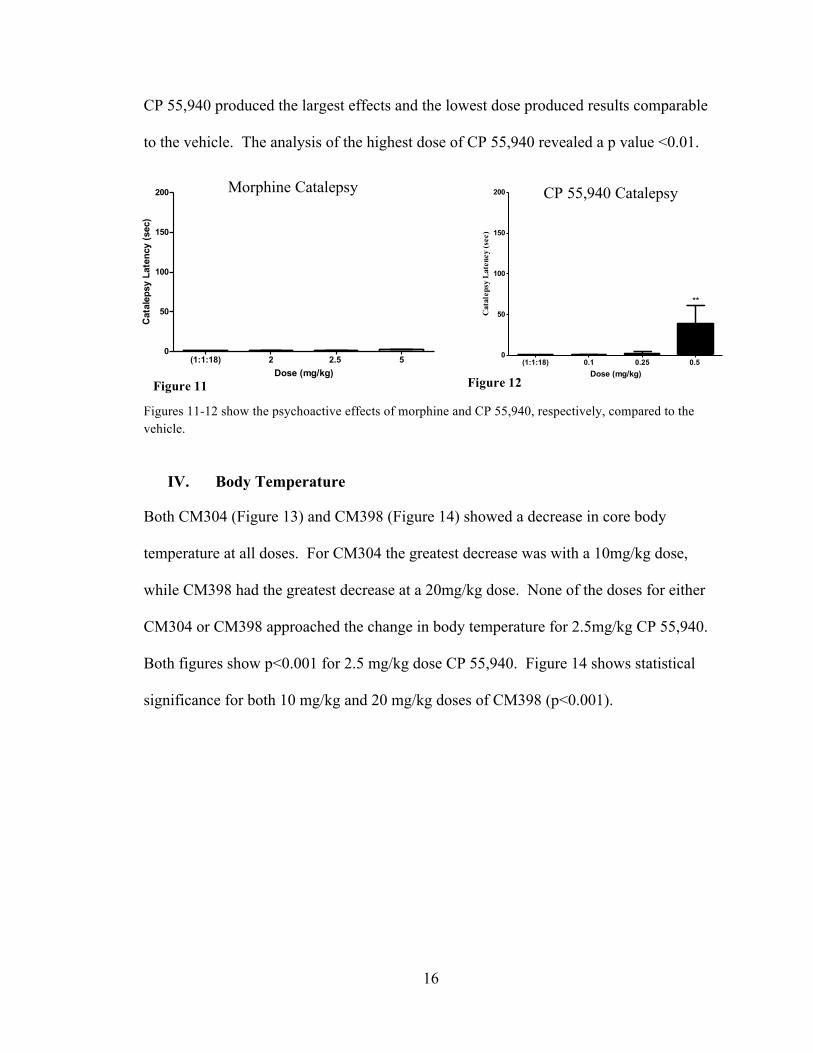
In Figure 11, it is noteworthy that none of the doses of morphine produced statistically
significant cataleptic effects, showing little to no psychoactive effects of the drug. All of
the morphine doses produce results comparable to the vehicle. Figure 12 shows that as
the dose of CP 55,940 increases, the catalepsy latency also increases. The highest dose of
CM304 Catalepsy CM398 Catalepsy
Figure 9 Figure 10
16
(1:1:18) 2 2.5 50
50
100
150
200
Dose (mg/kg)
Cat
alep
sy L
aten
cy (s
ec)
(1:1:18) 0.1 0.25 0.5 0
50
100
150
200
**
Dose (mg/kg)
Cat
alep
sy L
aten
cy (s
ec)
CP 55,940 produced the largest effects and the lowest dose produced results comparable
to the vehicle. The analysis of the highest dose of CP 55,940 revealed a p value <0.01.
Figures 11-12 show the psychoactive effects of morphine and CP 55,940, respectively, compared to the vehicle.
IV. Body Temperature
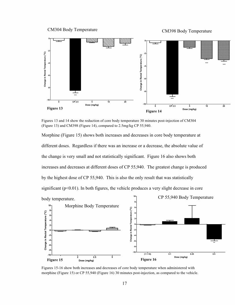
Both CM304 (Figure 13) and CM398 (Figure 14) showed a decrease in core body
temperature at all doses. For CM304 the greatest decrease was with a 10mg/kg dose,
while CM398 had the greatest decrease at a 20mg/kg dose. None of the doses for either
CM304 or CM398 approached the change in body temperature for 2.5mg/kg CP 55,940.
Both figures show p<0.001 for 2.5 mg/kg dose CP 55,940. Figure 14 shows statistical
significance for both 10 mg/kg and 20 mg/kg doses of CM398 (p<0.001).
Figure 11
Morphine Catalepsy CP 55,940 Catalepsy
Figure 12
17
0 CP 2.5 5 10 20-10
-8
-6
-4
-2
0
***
Dose (mg/kg)
Cha
nge
in R
ecta
l Tem
pera
ture
(o C)
0 CP 2.5 5 10 20-10
-8
-6
-4
-2
0
***
*** ***
Dose (mg/kg)
Cha
nge
in R
ecta
l Tem
pera
ture
(o C)
(1:1:18) 2 2.5 5-10
-8
-6
-4
-2
0
2
4
6
8
10
Dose (mg/kg)
Cha
nge
in R
ecta
l Tem
pera
ture
(o C)
(1:1:18) 0.1 0.25 0.5 -10
-8
-6
-4
-2
0
2
4
6
8
10
**
Dose (mg/kg)
Cha
nge
in R
ecta
l Tem
pera
ture
(o C)
Figures 13 and 14 show the reduction of core body temperature 30 minutes post-injection of CM304 (Figure 13) and CM398 (Figure 14), compared to 2.5mg/kg CP 55,940.
Morphine (Figure 15) shows both increases and decreases in core body temperature at
different doses. Regardless if there was an increase or a decrease, the absolute value of
the change is very small and not statistically significant. Figure 16 also shows both
increases and decreases at different doses of CP 55,940. The greatest change is produced
by the highest dose of CP 55,940. This is also the only result that was statistically
significant (p<0.01). In both figures, the vehicle produces a very slight decrease in core
body temperature.
Figures 15-16 show both increases and decreases of core body temperature when administered with morphine (Figure 15) or CP 55,940 (Figure 16) 30 minutes post-injection, as compared to the vehicle.
Figure 14
CM304 Body Temperature CM398 Body Temperature
Figure 13
CP 55,940 Body Temperature
Morphine Body Temperature
Figure 16 Figure 15
18
0 CP 2.5 5 10 20-50
0
50
100 ***
Dose (mg/kg)
Tail-
Flic
k La
tenc
y (%
MPE
)
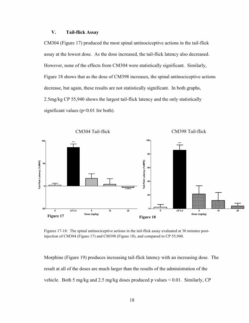
V. Tail-flick Assay
CM304 (Figure 17) produced the most spinal antinociceptive actions in the tail-flick
assay at the lowest dose. As the dose increased, the tail-flick latency also decreased.
However, none of the effects from CM304 were statistically significant. Similarly,
Figure 18 shows that as the dose of CM398 increases, the spinal antinociceptive actions
decrease, but again, these results are not statistically significant. In both graphs,
2.5mg/kg CP 55,940 shows the largest tail-flick latency and the only statistically
significant values (p<0.01 for both).
Figures 17-18: The spinal antinociceptive actions in the tail-flick assay evaluated at 30 minutes post-injection of CM304 (Figure 17) and CM398 (Figure 18), and compared to CP 55,940.
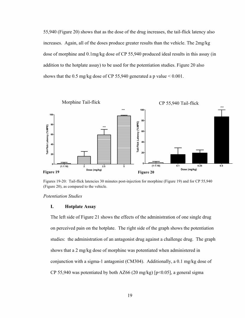
Morphine (Figure 19) produces increasing tail-flick latency with an increasing dose. The
result at all of the doses are much larger than the results of the administration of the
vehicle. Both 5 mg/kg and 2.5 mg/kg doses produced p values < 0.01. Similarly, CP
0 CP 2.5 5 10 200
20
40
60
80
100***
Dose (mg/kg)
Tail-
Flic
k La
tenc
y (%
MPE
)
Figure 17 Figure 18
CM398 Tail-flick CM304 Tail-flick
19
(1:1:18) 2 2.5 50
20
40
60
80
100
***
***
Dose (mg/kg)
Tail-
Flic
k La
tenc
y (%
MPE
)
(1:1:18) 0.1 0.25 0.5 0
20
40
60
80
100***
Dose (mg/kg)
Tail-
Flic
k La
tenc
y (%
MPE
)
55,940 (Figure 20) shows that as the dose of the drug increases, the tail-flick latency also
increases. Again, all of the doses produce greater results than the vehicle. The 2mg/kg
dose of morphine and 0.1mg/kg dose of CP 55,940 produced ideal results in this assay (in
addition to the hotplate assay) to be used for the potentiation studies. Figure 20 also
shows that the 0.5 mg/kg dose of CP 55,940 generated a p value < 0.001.
Figures 19-20: Tail-flick latencies 30 minutes post-injection for morphine (Figure 19) and for CP 55,940 (Figure 20), as compared to the vehicle.
Potentiation Studies
I. Hotplate Assay
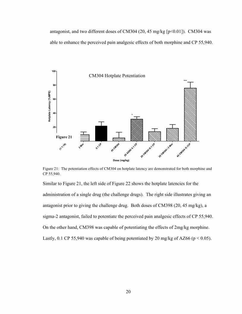
The left side of Figure 21 shows the effects of the administration of one single drug
on perceived pain on the hotplate. The right side of the graph shows the potentiation
studies: the administration of an antagonist drug against a challenge drug. The graph
shows that a 2 mg/kg dose of morphine was potentiated when administered in
conjunction with a sigma-1 antagonist (CM304). Additionally, a 0.1 mg/kg dose of
CP 55,940 was potentiated by both AZ66 (20 mg/kg) [p<0.05], a general sigma
CP 55,940 Tail-flick Morphine Tail-flick
Figure 19 Figure 20
20
(1:1:1
8)2 M
or
0.1 C
P
45 C
M304
20 A
Z66/ 0
.1 CP
20 C
M304 /
0.1 C
P
20 C
M304 /
2 Mor
45 C
M304 /
0.1CP
0
20
40
60
80
100
*
***
Dose (mg/kg)
Hot
plat
e La
tenc
y (%
MPE
)
antagonist, and two different doses of CM304 (20, 45 mg/kg [p<0.01]). CM304 was
able to enhance the perceived pain analgesic effects of both morphine and CP 55,940.
Figure 21: The potentiation effects of CM304 on hotplate latency are demonstrated for both morphine and CP 55,940.
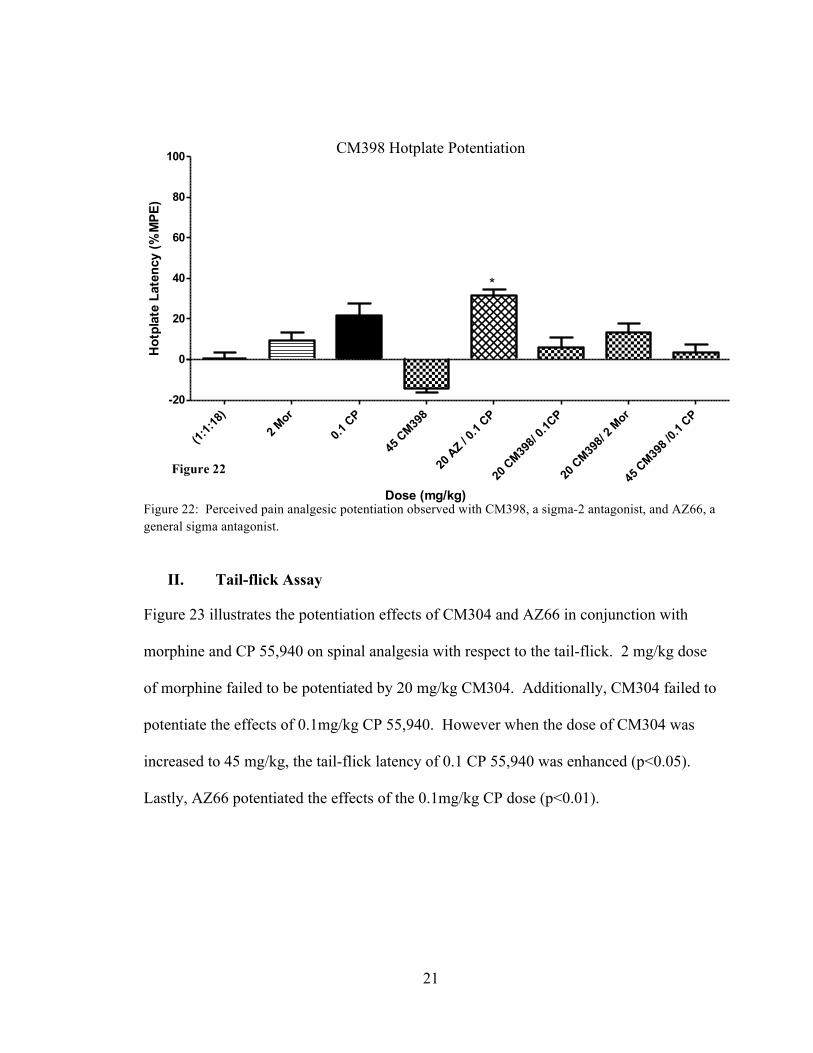
Similar to Figure 21, the left side of Figure 22 shows the hotplate latencies for the
administration of a single drug (the challenge drugs). The right side illustrates giving an
antagonist prior to giving the challenge drug. Both doses of CM398 (20, 45 mg/kg), a
sigma-2 antagonist, failed to potentiate the perceived pain analgesic effects of CP 55,940.
On the other hand, CM398 was capable of potentiating the effects of 2mg/kg morphine.
Lastly, 0.1 CP 55,940 was capable of being potentiated by 20 mg/kg of AZ66 (p < 0.05).
CM304 Hotplate Potentiation
Figure 21
21
(1:1:1
8)2 M
or
0.1 C
P
45 C
M398
20 A
Z / 0.1
CP
20 C
M398/
0.1CP
20 C
M398/
2 Mor
45 C
M398 /
0.1 C
P-20
0
20
40
60
80
100
*
Dose (mg/kg)
Hot
plat
e La
tenc
y (%
MPE
)
Figure 22: Perceived pain analgesic potentiation observed with CM398, a sigma-2 antagonist, and AZ66, a general sigma antagonist.
II. Tail-flick Assay
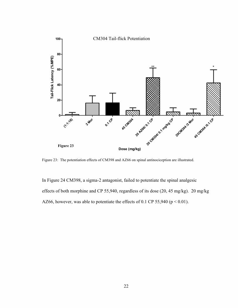
Figure 23 illustrates the potentiation effects of CM304 and AZ66 in conjunction with
morphine and CP 55,940 on spinal analgesia with respect to the tail-flick. 2 mg/kg dose
of morphine failed to be potentiated by 20 mg/kg CM304. Additionally, CM304 failed to
potentiate the effects of 0.1mg/kg CP 55,940. However when the dose of CM304 was
increased to 45 mg/kg, the tail-flick latency of 0.1 CP 55,940 was enhanced (p<0.05).
Lastly, AZ66 potentiated the effects of the 0.1mg/kg CP dose (p<0.01).
CM398 Hotplate Potentiation
Figure 22
22
(1:1:1
8)2 M
or
0.1 C
P
45 C
M304
20 A
Z66/ 0
.1 CP
20 C
M304/
0.1 m
g/kg C
P
20CM30
4 /2 M
or
45 C
M304 /
0.1 C
P0
20
40
60
80
100
***
Dose (mg/kg)
Tail-
Flic
k La
tenc
y (%
MPE
)
Figure 23: The potentiation effects of CM398 and AZ66 on spinal antinociception are illustrated.
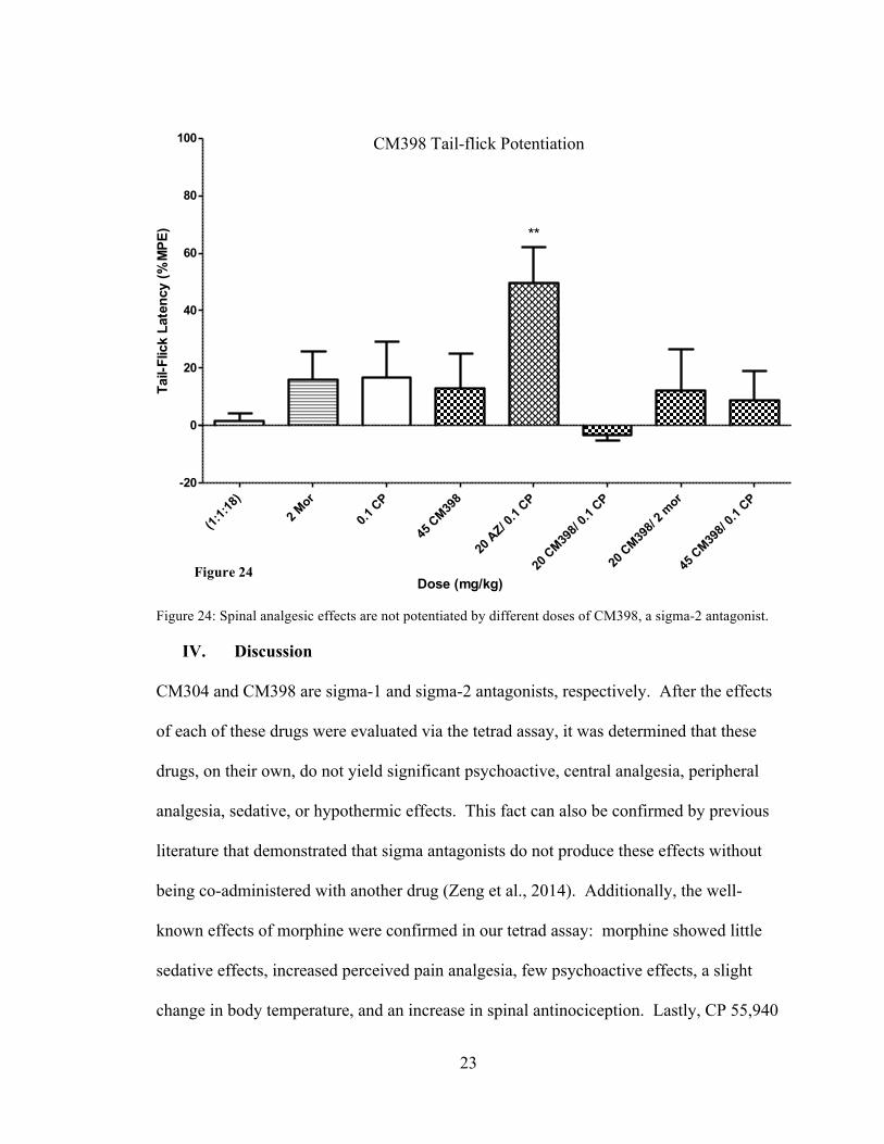
In Figure 24 CM398, a sigma-2 antagonist, failed to potentiate the spinal analgesic
effects of both morphine and CP 55,940, regardless of its dose (20, 45 mg/kg). 20 mg/kg
AZ66, however, was able to potentiate the effects of 0.1 CP 55,940 (p < 0.01).
CM304 Tail-flick Potentiation
Figure 23
23
(1:1:1
8)2 M
or
0.1 C
P
45 C
M398
20 A
Z/ 0.1
CP
20 C
M398/
0.1 C
P
20 C
M398/
2 mor
45 C
M398/
0.1 C
P-20
0
20
40
60
80
100
**
Dose (mg/kg)
Tail-
Flic
k La
tenc
y (%
MPE
)
Figure 24: Spinal analgesic effects are not potentiated by different doses of CM398, a sigma-2 antagonist.
IV. Discussion
CM304 and CM398 are sigma-1 and sigma-2 antagonists, respectively. After the effects
of each of these drugs were evaluated via the tetrad assay, it was determined that these
drugs, on their own, do not yield significant psychoactive, central analgesia, peripheral
analgesia, sedative, or hypothermic effects. This fact can also be confirmed by previous
literature that demonstrated that sigma antagonists do not produce these effects without
being co-administered with another drug (Zeng et al., 2014). Additionally, the well-
known effects of morphine were confirmed in our tetrad assay: morphine showed little
sedative effects, increased perceived pain analgesia, few psychoactive effects, a slight
change in body temperature, and an increase in spinal antinociception. Lastly, CP 55,940
Figure 24
CM398 Tail-flick Potentiation
24
brought about the expected high psychoactive and high sedative effects along with an
increase in central and peripheral analgesia (Melvin et al., 1993).
The 20 mg/kg dose of CM304 was able to potentiate the effects of morphine but
not the effects of CP 55,940 on the hotplate assay. Once the dose was increased to 45
mg/kg, CM304 could enhance the perceived pain analgesia. On the other hand, both
20mg/kg and 45 mg/kg of CM398 attenuated supraspinal analgesia. When examining the
antagonists’ effects on spinal analgesia, 20 mg/kg CM304 attenuated the effects of both
CP 55,940 and morphine. Once the dose was increased to 45 mg/kg, CM304 was able to
potentiate the effects of only CP 55,940. Dissimilarly, both doses of CM398 were
incapable of potentiating either morphine or CP 55,940. The general sigma antagonist,
AZ66, potentiated the effects of CP 55,940 for both the hotplate and the tail-flick assays.
These results demonstrate that administering a sigma-1 antagonist, as opposed to
a sigma-2 antagonist, in conjunction with either an opiate or a cannabinoid will potentiate
the effects of the challenge drug. This can serve as a potentially very important part of
future pain management research. Knowing that a sigma-1 antagonist instead of a sigma-
2 antagonist can potentiate the perceived pain analgesic effects of an opiate and a
cannabinoid can allow scientists to administer a more specific drug, providing researchers
with a greater understanding of the mechanism.
V. Conclusion For the first time, an interaction has been shown between the sigma receptors and
the endocannabinoid system. This opens a multitude of new areas of research. Beyond
the analgesic development that has begun here, research will be conducted to understand
what other behaviors may be modulated through sigma receptors. Additionally, the
major question that these results pose is whether or not the ability of sigma-1 to
25
potentiate the analgesic effects of the drug will also potentiate the psychoactive behavior
associated with cannabinoids. The interplay of these additional behaviors with the
endocannabinoid system can also be evaluated in future research. This research lays
important groundwork for the research of the future.
26
LIST OF REFERENCES
27
Maurice, Tangui, and Tsung-Ping Su. “The Pharmacology of Sigma-1 Receptors.” Pharmacology & Therapeutics 124.2 (2009): 195-206.
Zamanillo, Daniel, Luz Romero, Manuel Merlos, and José Miguel Vela. “Sigma 1 Receptor: A New Therapeutic Target for Pain.” European Journal of Pharmacology 716.1-3 (2013): 78-93.
Hanner M, Moebius FF, Flandorfer A, Knaus H-G, Striessnig J, Kempner E, Glossman H. Purification, molecular cloning, and expression of the mammalian σ1 binding site. Proceedings of the National Academy of Sciences USA 1996, 93:8072-8077.
Mei J and Pasterak GW. Molecular cloning and pharmacological characterization of the rat σ1 receptor. Biochemical Pharmacology 2001, 62:349-355.
Pan Y-X, Mei J, Xu J, Wan B-L, Zuckerman A, Pasternak GW. Cloning and characterization of a mouse σ1 receptor. J Neurochem 1998, 70: 2279-2285.
Seth P, Fei YJ, Li HW, Huang Wa, Leibach FH, Ganapathy V. Cloning and functional characterization of a σ receptor from rat brain. Journal of Neurochemistry 1998, 70:922-931.
Seth P, Leibach FH, Ganapathy V. Cloning and structural analysis of the cDNA and the gene encoding the murine type 1 σ receptor. Biochemical and Biophysical Research Communications 1997, 41:535-540.
Aydar E, Palmer CP, Klyachko VA, Jackson MB. The σ receptor as a ligand-regulated auxiliary potassium channel subunit. Neuron 2002, 34:399-410.
Jbilo O, Vidal H, Paul R, De Nys Nm Bensaid M, Silve S, Carayon P, Davi D, Galiegue S, Bourrie B, Guillemot J-C, Ferrara P, Loison G, Maffrand J-P, Le Fur G, Casellas P. Purification and characterization of the human SR 31747A-binding protein. A nuclear membrane protein related to yeast sterol isomerase. Journal of Biological Chemistry 1997, 272:27107-27115.
Matsomoto, Rae R., Wayne Darrell Bowen, and Tsung-Ping Su. “Table 1-2. Characteristics of σ1 and σ2 Receptors.” Sigma Receptors: Chemistry, Cell Biology and Clinical Implications. New York: Springer, 2007. 122. Print.
Chu U, Mavlyutov T, Chu ML, Yang H, Schulman A, Mesangeau C, McCurdy C, Guo LW, Ruoho A. “The Sigma-2 Receptor and Progesterone Receptor Membrane Component 1 are Different Binding Sites Derived From Independent Genes.” EBioMedicine 2 (2015): 1806-1813.
Zeng, Chenbo, Justin M. Rothfuss, Jun Zhang, Suwanna Vangveravong, Wenhua Chu, Shihong Li, Zhude Tu, Jinbin Xu, and Robert H. Mach. “Functional Assays to Define Agonists and Antagonists of the Sigma-2 Receptor.” Analytical Biochemistry 448 (2014): 68-74.
28
Matsumoto, Rae R., Wayne Darrell Bowen, and Tsung-Ping Su. “Chapter 16.” Sigma Receptors: Chemistry, Cell Biology and Clinical Implications. New York: Springer, 2007. 337. Print.
Sánchez-Fernández C, Montilla-García Á, González-Cano R, Nieto FR, Romero L, Artacho-Cordón A, Montes R, Fernández-Pastor B, Merlos M, Baeyens JM, Entrena JM, Cobos EJ. “Modulation of peripheral µ-opioid analgesia by σ1 receptors.” Journal of Pharmacology and Experimental Therapeutics, 348 (2014): 32-45.
Vidal-Torres, Alba, Beatriz De La Puente, Maria Rocasalbas, Clara Touriño, Simona Andreea Bura, Begoña Fernández-Pastor, Luz Romero, Xavier Codony, Daniel Zamanillo, Helmut Buschmann, Manuel Merlos, José Manuel Baeyens, Rafael Maldonado, and José Miguel Vela. “Sigma-1 Receptor Antagonism as Opioid Adjuvant Strategy: Ehancement of Opioid Antinociception without Increasing Adverse Effects.” European Journal of Pharmacology 711.1-3 (2013): 63-72.
Tseng LF, Hogan QH, Wu HE. “(+)-Morphine attenuates the (-)-morphine-produced tail-flick inhibition via the sigma-1 receptor in the mouse spinal cord.” Life Science, 89 (2011): 23-24.
Kim FJ, Kvalyshyn I, Burgman M, Neilan C, Chien CC, Pasternak GW. “Sigma 1 receptor modulation of G-protein-coupled receptor signaling: potentiation of opioid transduction independent from receptor binding.” Molecular Pharmacology, 77 (2010): 695-703.
Mei J, Pasternak GW. “Modulation of brainstem opiate analgesia in the rat by sigma 1 receptors: a microinjection study.” Journal of Pharmacology and Experimental Therapeutics, 3 (2007): 1278-85.
Mei J, Pasternak GW. “Sigma1 modulation of opioid analgesia in the mouse.” Journal of Pharmacology and Experimental Therapeutics, 3 (2002): 1070-4.
Sánchez-Fernández, Cristina, Francisco Rafael Nieto, Rafael González-Cano, Antonia ARtacho-Cordón, Lucía Romero, Ángeles Montilla-García, Daniel Zamanillo, José Manuel Baeyens, José Manuel Entrena, and Enrique José Cobos. “Potentiation of Morphine-induced Mechanical Antinociception by σ1 Receptor Inhibition: Role of Peripheral σ1 Receptors.” Neuropharmacology 70(2013): 348-58.Lui F, Ng KF. “Adjuvant analgesics in acute pain.” Expert Opinion on Pharmacotherapy, 3 (2011): 363-85.
Khan M, Walsh D, Brito-Dellan N. “Opioid and adjuvant analgesics: compared and contrasted.” American Journal of Hospice and Palliative Medicine, 5 (2011): 378-83.
Wilson LL, Morris J, Cutler SJ, McCurdy CR. Sigma Receptor Blockade Potentiates the Analgesic Effects of Cannabinoids. Marijuana and Cannabinoids: A Neuroscience
29
Research Summit. National Institutes of Health. NIH Campus, Bethesda, MD. March 22-23, 2016. Day #2 Poster #49
Seminerio, Michael J., Matthew J. Robson, Ahmed H. Abdelazeem, Christophe Mesangeau, Seshulatha Jamalapuram, Bonnie A. Avery, Christopher R. McCurdy, and Rae R. Matsumoto. “Synthesis and Pharmacological Characterization of a Novel Sigma Receptor Ligand with Improved Metabolic Stability and Antagonistic Effects Against Methamphetamine.” The AAPS Journal AAPS J 14.1 (2011): 43-51.
Olson, J.L., Makhani, M., Davis, K.H. and Wall, M.E. (1973) Preparation of Δ9-tetrahydrocannabinol for intravenous injection. Journal of Pharmacology. Pharmacol. 25, 344.
Little PJ, Compton DR, Johnson MR, Melvin LS, Martin BR. Pharmacology and stereoselectivity of structurally novel cannabinoids in mice. Journal of Pharmacology and Experimental Therapy. 1988;247:1046–1051
Pertwee, RG. “The diverse CB1 and CB2 receptor pharmacology of three plant cannabinoids: Δ9-tetrahydrocannabinol, cannabidiol, and Δ9-tetrahydrocannabivarin.” The British Journal of Pharmacology, 153 (2008): 199-215.
Babbini A, Davis WM. “Time-dose relationships for locomotor activity effects of morphine after acute or repeated treatment.” The British Journal of Pharmacology, 46 (1972): 213-224.
Melvin L, Milne G, Johnson MR, Subramaniam B, Wilken G, Howlett A. “Structure-Activity Relationships for Cannabinoid Receptor-Binding and Analgesic Activity: Studies of Bicyclic Cannabinoid Analogs.” Molecular Pharmacology, 44 (1993): 1008-1015.