To cite this article: Sandra Einarsson, Göran Laurell & Ylva Tiblom Ehrsson (2020) An explorativestudy on energy balance in patients with head and neck cancer, Nutrition and Cancer, 72:7,1191-1199, DOI: 10.1080/01635581.2019.1676454
To link to this article: https://doi.org/10.1080/01635581.2019.1676454
An explorative study on energy balance in patients with head andneck cancer
Sandra Einarssona , G€oran Laurellb , and Ylva Tiblom Ehrssonb
aDepartment of Food and Nutrition, Umeå University, Umeå, Sweden; bDepartment of Surgical Sciences, Section ofOtorhinolaryngology and Head & Neck Surgery, Uppsala University, Uppsala, Sweden
ABSTRACTBackground: Involuntary body weight loss in head and neck cancer is common. Fundamentalfor weight loss is an energy imbalance where total energy expenditure exceeds energy intake.Aim: To map energy intake and parameters of energy expenditure at the start of and afterradiotherapy, and their relation to weight change, body mass index, and immune markersin patients with head and neck cancer.Materials and Methods: Data from 20 patients on energy intake (24-hour dietary intakerecalls), total energy expenditure (SenseWear Armband Pro3), resting energy expenditure(indirect calorimetry), body weight, body mass index, and immune markers in serum(C-reactive protein and Interleukin-6) were collected at the start of and after radiotherapy(median 8mo, range 5–13).Results: No statistical significance was shown between the two measurement points forenergy intake or for the different parameters of energy expenditure. Median values forenergy balance were 0.93 and 0.96 for the start of treatment and follow-up, respectively.Twelve and 13 patients had a negative energy balance at the start of radiotherapy and atfollow-up, respectively.Conclusion: A negative energy balance was seen for the majority of patients, which stressesthe importance of nutritional treatment at the start of and after radiotherapy.
ARTICLE HISTORYReceived 8 May 2019Accepted 27 September 2019
Introduction
Patients with head and neck cancer (HNC) oftenexperience significant body weight loss during andafter treatment (1–3). Fundamental for this weightloss is an energy imbalance where total energyexpenditure exceeds energy intake (4).
There are many factors affecting energy intake inpatients with HNC. Tumor site might cause insufficientfood intake due to mechanical obstruction or pain (5),and treatment, i.e., radiotherapy (RT), surgery, and/oranticancer drugs might cause toxicities that affect foodintake both during and after treatment (6,7).
The primary components of energy expenditure arebasal energy expenditure and physical activity, both ofwhich can be altered in patients with cancer (4). Reducedenergy expenditure might be the result of reduced phys-ical activity because of reduced physical function (8) andfatigue (9). Basal energy expenditure can be elevated, nor-mal, or reduced (10,11), and this response might vary
among different cancer types (12) as well as among indi-viduals (13). More specifically, the inflammatory andmetabolic response due to a tumor and/or treatmentmight increase basal energy expenditure (4,14,15).
Weight loss is a central criterion for diagnosingpatients for malnutrition (16). Reduced fat-free mass hasbeen correlated in studies on HNC to reduced handgrip strength (17) and impaired physical performance(8,17). Involuntary weight loss has important clinicalimplications because it is related to the development ofmalnutrition-related complications. Thus, for the surveil-lance of patients with HNC it would be useful to estab-lish more knowledge on the relation between weightloss, body mass index (BMI), and energy balance.
Aim
The aim of the present study was to map energyintake and different parameters of energy expenditureat the start of and after radiotherapy and their relation
CONTACT Sandra Einarsson [email protected] Department of Food and Nutrition, Umeå University, SE-901 87 Umeå, Sweden.� 2019 The Author(s). Published with license by Taylor & Francis Group, LLC.This is an Open Access article distributed under the terms of the Creative Commons Attribution-NonCommercial-NoDerivatives License (http://creativecommons.org/licenses/by-nc-nd/4.0/), which permits non-commercial re-use, distribution, and reproduction in any medium, provided the original work is properly cited, and is not altered, transformed,or built upon in any way.
NUTRITION AND CANCER2020, VOL. 72, NO. 7, 1191–1199https://doi.org/10.1080/01635581.2019.1676454
to weight change, body mass index, and immunemarkers in patients with head and neck cancer.
Materials and methods
From 2010 to 2013, 20 patients were recruited from a ter-tiary care hospital in Sweden. Patients �18 years of agewith a newly diagnosed HNC planned for curative RTwere eligible and were consecutively asked to participatein the study by a research nurse. Exclusion criteria weredementia, physiological illness, or a social situation thatwould affect the possibility for the patient to completethe study. The patients received either RT (conventionalfractionation or accelerated fractionation) as a singlemodality treatment or RT followed by surgery.Treatment details for each patient are shown in Table 1.Nutritional treatment according to the hospital regimeswas given with the intent of keeping the patient in asteady energy state. None of the patients had tube feedingor parenteral nutrition at any of the measurement points.
Study subjects
All patients in the study cohort were men, and themedian age was 54.5 years (range 43–71 years).Detailed tumor characteristics for each patient areshown in Table 1 (16 oropharyngeal, two oral cavity,and two unknown primary).
Data collection
Patients were measured at the start of RT (before thestart or during the first week of RT) and with amedian follow-up of 8mo, (range 5–13mo). The
follow-up was chosen to capture the effect of latetreatment toxicities rather than acute and occurred ina time when patients returned to the hospital for amedical checkup. One patient (no. 5) did not com-plete the second measurements because of a palliativesituation. At both occasions, data on energy intake,energy expenditure, anthropometric measures, andimmune markers were gathered.
Measures of energy intake
Data for energy intake were collected during two week-days and one weekend (only on weekdays for n¼ 2)using 24-hour dietary intake recalls. First, patients gavewritten information on the food and beverages con-sumed. Second, a dietitian gathered detailed informa-tion about food selection and portion sizes through aface-to-face (first measurement) or a telephone (secondmeasurement) interview. Portion sizes were estimatedusing household measures and pictures for differentportion sizes and food types from the Swedish NationalFood Administration (18). These were sent to thepatients’ home before the telephone interview. Energyintake for each day was estimated using the softwareprogram Dietist XP version 3.2 (Kost och N€aringsdataAB), and the mean value was used in the analyses.
Measures of energy expenditure
Total energy expenditure was collected using a devicecalled the SenseWear Armband Pro3 (SWA, BodyMedia,Inc., Pittsburgh, PA, USA) (19). The armband is wornon the upper part of the right arm and uses sensors to
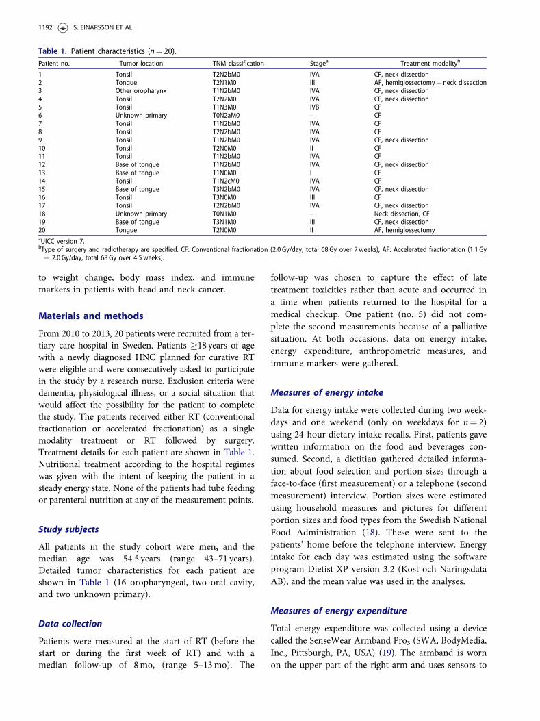
1 Tonsil T2N2bM0 IVA CF, neck dissection2 Tongue T2N1M0 III AF, hemiglossectomyþ neck dissection3 Other oropharynx T1N2bM0 IVA CF, neck dissection4 Tonsil T2N2M0 IVA CF, neck dissection5 Tonsil T1N3M0 IVB CF6 Unknown primary T0N2aM0 – CF7 Tonsil T1N2bM0 IVA CF8 Tonsil T2N2bM0 IVA CF9 Tonsil T1N2bM0 IVA CF, neck dissection10 Tonsil T2N0M0 II CF11 Tonsil T1N2bM0 IVA CF12 Base of tongue T1N2bM0 IVA CF, neck dissection13 Base of tongue T1N0M0 I CF14 Tonsil T1N2cM0 IVA CF15 Base of tongue T3N2bM0 IVA CF, neck dissection16 Tonsil T3N0M0 III CF17 Tonsil T2N2bM0 IVA CF, neck dissection18 Unknown primary T0N1M0 – Neck dissection, CF19 Base of tongue T3N1M0 III CF, neck dissection20 Tongue T2N0M0 II AF, hemiglossectomyaUICC version 7.bType of surgery and radiotherapy are specified. CF: Conventional fractionation (2.0 Gy/day, total 68 Gy over 7weeks), AF: Accelerated fractionation (1.1 Gyþ 2.0 Gy/day, total 68 Gy over 4.5 weeks).
1192 S. EINARSSON ET AL.
measure movement, heat flux, skin temperature, nearbody temperature, and galvanic skin response. The soft-ware program Interview Professional (version 6.1) esti-mates total energy expenditure from the SWA togetherwith information about the patient’s age, sex, height,weight, and whether the patient is a smoker or non-smoker and is right or left handed. Patients used theSWA during the same three days as the self-reported 24-h dietary intake recalls, and the mean value was used inthe analyses. For six patients, data from the SWA wereavailable for two days, and for one patient the totalenergy expenditure and energy intake were not meas-ured on the same days. The armband was taken off dur-ing RT or to avoid coming in contact with water.During this time, the software program calculated anestimation of energy expenditure corresponding to thepatients’ basal energy expenditure. Only days in whichthe armband was worn at least 20h, were used in theanalyses (missing, n¼ 4 days).
Energy balance was calculated as the energy intakedivided by the total energy expenditure. For values>1, patients were presumed to be in positive energybalance, whereas for values <1 the patients were pre-sumed to be in negative energy balance.
Resting energy expenditure was measured by indir-ect calorimetry (DeltratracTM II MBM 200). Patientshad fasted for four hours, and the measurements tookplace over 30mins, with the patient in a supine pos-ition (20). Physical activity level was calculated bysubtracting the resting energy expenditure from thetotal energy expenditure.
Anthropometric measures
Height and weight were measured using a wall stadi-ometer (to the nearest 0.1 cm) and an electronic scale(to the nearest 0.1 kg). The patients wore light cloth-ing and no shoes for the measurements. Weights atstart of RT and at follow-up were used to calculaterelative percentage weight loss. Weight loss of 5–10%was classified as moderate and weight loss of >10%was classified as severe (16), and a third group con-sisted of patients with weight gain, no change inweight, or little weight loss (<5%). Patients were alsodivided into groups based on their BMI (weight inkilograms divided by height in meters squared) asunderweight (BMI <20), normal weight (BMI 20–25),and overweight or obese (BMI >25) (16). For patientsover 70 years, BMI <22 was considered underweightand BMI between 22 and 27 was considered normal.Malnutrition was defined using the Global LeadershipInitiative on Malnutrition (GLIM) criteria (16), which
is a consensus from the global clinical nutrition com-munity on how to diagnose malnutrition. For thediagnosis of malnutrition, at least one phenotypic cri-terion and one etiologic criterion should be present,i.e., weight loss, low BMI, or reduced fat-free mass(phenotypic factors) and reduced food intake orinflammation (etiologic factors).
Blood samples
Blood samples (four-hour fasting values) were gath-ered for analyses of the immune markers C-reactiveprotein (CRP, ref <10mg/L) and Interleukin-6 (IL-6,ref <7 ng/L).
Ethical approval
The Regional Ethical Review Board in Umeå, Sweden(Dnr 2010-24-31), approved the study, and all patientssigned written informed consent forms.
Statistical analyses
Non-parametric tests were used due to the limitednumber of patients. For these statistical analyses, thedata software Statistical Package for the SocialSciences (SPSS) version 25.0 was used. The Wilcoxonsigned rank test was used to analyze the change inweight, BMI, total energy expenditure, resting energyexpenditure, energy intake, and physical activity levelbetween the two measured points. TheMann–Whitney U-test was used to analyze totalenergy expenditure in patients with CRP and/or IL-6above reference values compared to patients with CRPand IL-6 in the normal range. The correlationbetween energy balance and weight change percent orBMI was carried out using the Spearman rank test.All tests were two-sided, and a p-value � 0.05 wasconsidered statistically significant.
Results
Energy intake and energy expenditure
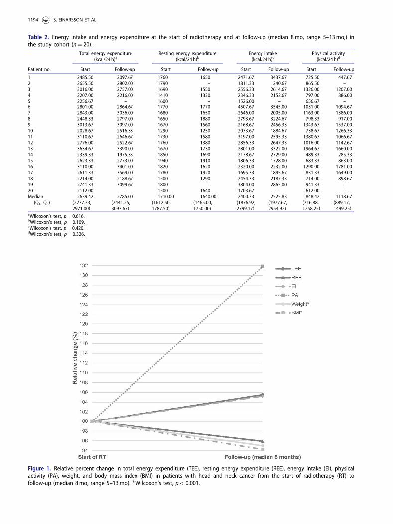
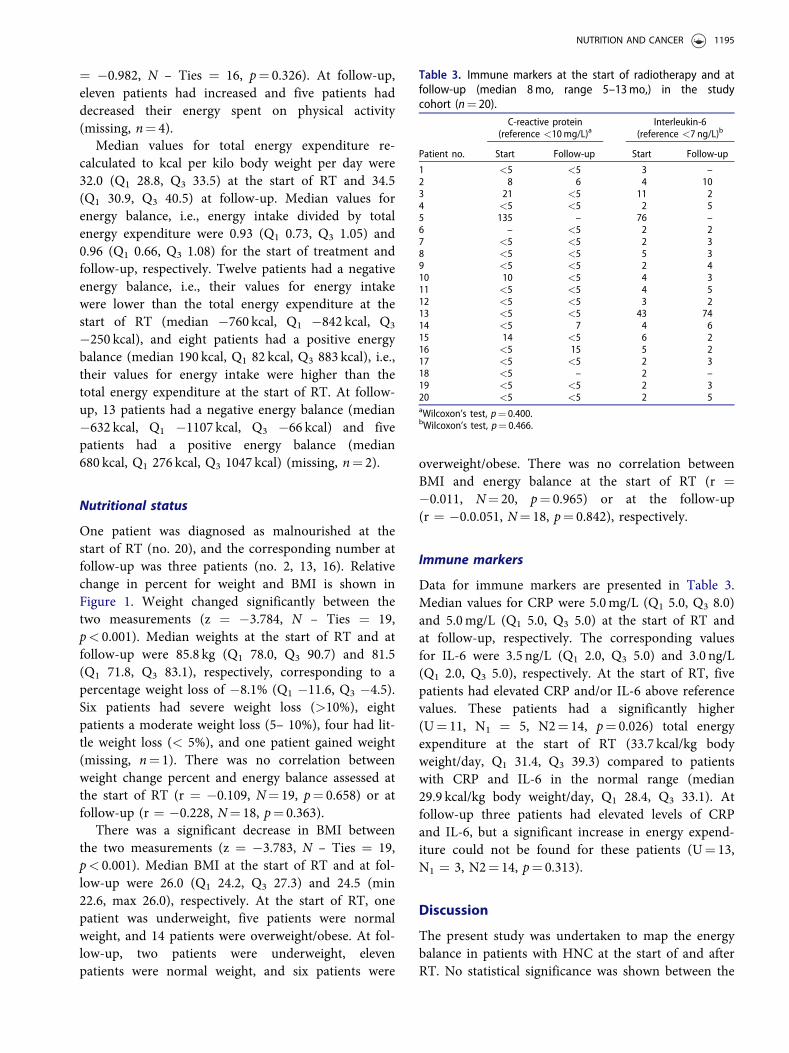
Data for energy intake and energy expenditure at startand follow-up are shown in Table 2, and relativechange in percent is shown in Figure 1. No statisticalsignificance was seen between the start of RT and fol-low-up for any of the variables: energy intake (z ¼�0.806, N – Ties ¼ 18, p¼ 0.420), total energyexpenditure (z ¼ �0.501, N – Ties ¼ 18, p¼ 0.616),resting energy expenditure (z ¼ �1.605, N – Ties ¼17, p¼ 0.109) and energy spent on physical activity (z
NUTRITION AND CANCER 1193
Table 2. Energy intake and energy expenditure at the start of radiotherapy and at follow-up (median 8mo, range 5–13mo,) inthe study cohort (n¼ 20).
Figure 1. Relative percent change in total energy expenditure (TEE), resting energy expenditure (REE), energy intake (EI), physicalactivity (PA), weight, and body mass index (BMI) in patients with head and neck cancer from the start of radiotherapy (RT) tofollow-up (median 8mo, range 5–13mo). �Wilcoxon’s test, p< 0.001.
1194 S. EINARSSON ET AL.
¼ �0.982, N – Ties ¼ 16, p¼ 0.326). At follow-up,eleven patients had increased and five patients haddecreased their energy spent on physical activity(missing, n¼ 4).
Median values for total energy expenditure re-calculated to kcal per kilo body weight per day were32.0 (Q1 28.8, Q3 33.5) at the start of RT and 34.5(Q1 30.9, Q3 40.5) at follow-up. Median values forenergy balance, i.e., energy intake divided by totalenergy expenditure were 0.93 (Q1 0.73, Q3 1.05) and0.96 (Q1 0.66, Q3 1.08) for the start of treatment andfollow-up, respectively. Twelve patients had a negativeenergy balance, i.e., their values for energy intakewere lower than the total energy expenditure at thestart of RT (median �760 kcal, Q1 �842 kcal, Q3
�250 kcal), and eight patients had a positive energybalance (median 190 kcal, Q1 82 kcal, Q3 883 kcal), i.e.,their values for energy intake were higher than thetotal energy expenditure at the start of RT. At follow-up, 13 patients had a negative energy balance (median�632 kcal, Q1 �1107 kcal, Q3 �66 kcal) and fivepatients had a positive energy balance (median680 kcal, Q1 276 kcal, Q3 1047 kcal) (missing, n¼ 2).
Nutritional status
One patient was diagnosed as malnourished at thestart of RT (no. 20), and the corresponding number atfollow-up was three patients (no. 2, 13, 16). Relativechange in percent for weight and BMI is shown inFigure 1. Weight changed significantly between thetwo measurements (z ¼ �3.784, N – Ties ¼ 19,p< 0.001). Median weights at the start of RT and atfollow-up were 85.8 kg (Q1 78.0, Q3 90.7) and 81.5(Q1 71.8, Q3 83.1), respectively, corresponding to apercentage weight loss of �8.1% (Q1 �11.6, Q3 �4.5).Six patients had severe weight loss (>10%), eightpatients a moderate weight loss (5– 10%), four had lit-tle weight loss (< 5%), and one patient gained weight(missing, n¼ 1). There was no correlation betweenweight change percent and energy balance assessed atthe start of RT (r ¼ �0.109, N¼ 19, p¼ 0.658) or atfollow-up (r ¼ �0.228, N¼ 18, p¼ 0.363).
There was a significant decrease in BMI betweenthe two measurements (z ¼ �3.783, N – Ties ¼ 19,p< 0.001). Median BMI at the start of RT and at fol-low-up were 26.0 (Q1 24.2, Q3 27.3) and 24.5 (min22.6, max 26.0), respectively. At the start of RT, onepatient was underweight, five patients were normalweight, and 14 patients were overweight/obese. At fol-low-up, two patients were underweight, elevenpatients were normal weight, and six patients were
overweight/obese. There was no correlation betweenBMI and energy balance at the start of RT (r ¼�0.011, N¼ 20, p¼ 0.965) or at the follow-up(r ¼ �0.0.051, N¼ 18, p¼ 0.842), respectively.
Immune markers
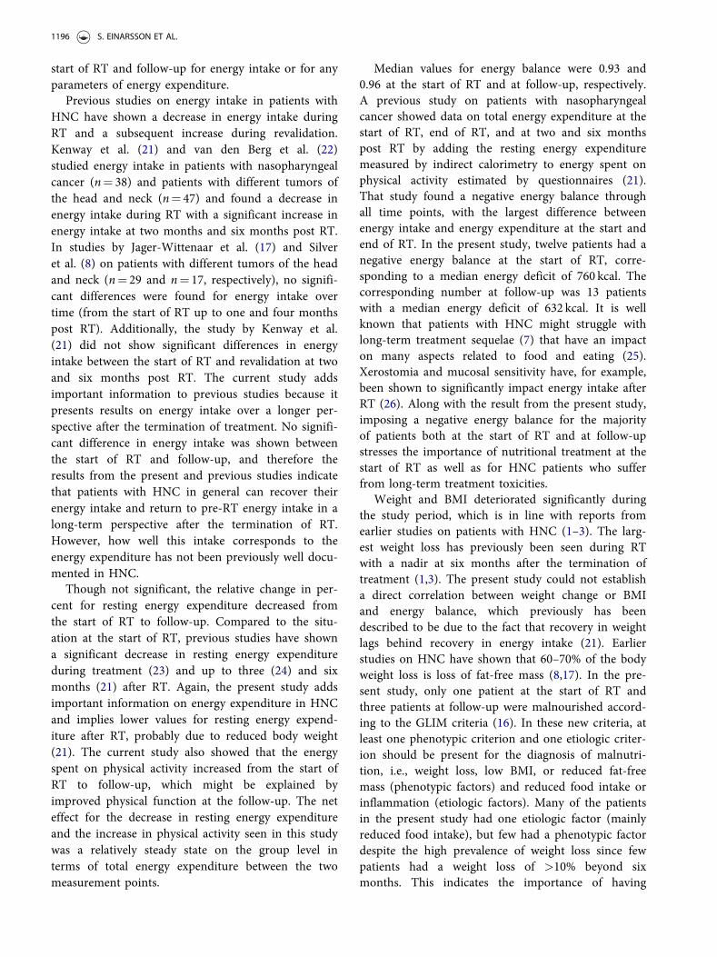
Data for immune markers are presented in Table 3.Median values for CRP were 5.0mg/L (Q1 5.0, Q3 8.0)and 5.0mg/L (Q1 5.0, Q3 5.0) at the start of RT andat follow-up, respectively. The corresponding valuesfor IL-6 were 3.5 ng/L (Q1 2.0, Q3 5.0) and 3.0 ng/L(Q1 2.0, Q3 5.0), respectively. At the start of RT, fivepatients had elevated CRP and/or IL-6 above referencevalues. These patients had a significantly higher(U¼ 11, N1 ¼ 5, N2¼ 14, p¼ 0.026) total energyexpenditure at the start of RT (33.7 kcal/kg bodyweight/day, Q1 31.4, Q3 39.3) compared to patientswith CRP and IL-6 in the normal range (median29.9 kcal/kg body weight/day, Q1 28.4, Q3 33.1). Atfollow-up three patients had elevated levels of CRPand IL-6, but a significant increase in energy expend-iture could not be found for these patients (U¼ 13,N1 ¼ 3, N2¼ 14, p¼ 0.313).
Discussion
The present study was undertaken to map the energybalance in patients with HNC at the start of and afterRT. No statistical significance was shown between the
Table 3. Immune markers at the start of radiotherapy and atfollow-up (median 8mo, range 5–13mo,) in the studycohort (n¼ 20).
start of RT and follow-up for energy intake or for anyparameters of energy expenditure.
Previous studies on energy intake in patients withHNC have shown a decrease in energy intake duringRT and a subsequent increase during revalidation.Kenway et al. (21) and van den Berg et al. (22)studied energy intake in patients with nasopharyngealcancer (n¼ 38) and patients with different tumors ofthe head and neck (n¼ 47) and found a decrease inenergy intake during RT with a significant increase inenergy intake at two months and six months post RT.In studies by Jager-Wittenaar et al. (17) and Silveret al. (8) on patients with different tumors of the headand neck (n¼ 29 and n¼ 17, respectively), no signifi-cant differences were found for energy intake overtime (from the start of RT up to one and four monthspost RT). Additionally, the study by Kenway et al.(21) did not show significant differences in energyintake between the start of RT and revalidation at twoand six months post RT. The current study addsimportant information to previous studies because itpresents results on energy intake over a longer per-spective after the termination of treatment. No signifi-cant difference in energy intake was shown betweenthe start of RT and follow-up, and therefore theresults from the present and previous studies indicatethat patients with HNC in general can recover theirenergy intake and return to pre-RT energy intake in along-term perspective after the termination of RT.However, how well this intake corresponds to theenergy expenditure has not been previously well docu-mented in HNC.
Though not significant, the relative change in per-cent for resting energy expenditure decreased fromthe start of RT to follow-up. Compared to the situ-ation at the start of RT, previous studies have showna significant decrease in resting energy expenditureduring treatment (23) and up to three (24) and sixmonths (21) after RT. Again, the present study addsimportant information on energy expenditure in HNCand implies lower values for resting energy expend-iture after RT, probably due to reduced body weight(21). The current study also showed that the energyspent on physical activity increased from the start ofRT to follow-up, which might be explained byimproved physical function at the follow-up. The neteffect for the decrease in resting energy expenditureand the increase in physical activity seen in this studywas a relatively steady state on the group level interms of total energy expenditure between the twomeasurement points.
Median values for energy balance were 0.93 and0.96 at the start of RT and at follow-up, respectively.A previous study on patients with nasopharyngealcancer showed data on total energy expenditure at thestart of RT, end of RT, and at two and six monthspost RT by adding the resting energy expendituremeasured by indirect calorimetry to energy spent onphysical activity estimated by questionnaires (21).That study found a negative energy balance throughall time points, with the largest difference betweenenergy intake and energy expenditure at the start andend of RT. In the present study, twelve patients had anegative energy balance at the start of RT, corre-sponding to a median energy deficit of 760 kcal. Thecorresponding number at follow-up was 13 patientswith a median energy deficit of 632 kcal. It is wellknown that patients with HNC might struggle withlong-term treatment sequelae (7) that have an impacton many aspects related to food and eating (25).Xerostomia and mucosal sensitivity have, for example,been shown to significantly impact energy intake afterRT (26). Along with the result from the present study,imposing a negative energy balance for the majorityof patients both at the start of RT and at follow-upstresses the importance of nutritional treatment at thestart of RT as well as for HNC patients who sufferfrom long-term treatment toxicities.
Weight and BMI deteriorated significantly duringthe study period, which is in line with reports fromearlier studies on patients with HNC (1–3). The larg-est weight loss has previously been seen during RTwith a nadir at six months after the termination oftreatment (1,3). The present study could not establisha direct correlation between weight change or BMIand energy balance, which previously has beendescribed to be due to the fact that recovery in weightlags behind recovery in energy intake (21). Earlierstudies on HNC have shown that 60–70% of the bodyweight loss is loss of fat-free mass (8,17). In the pre-sent study, only one patient at the start of RT andthree patients at follow-up were malnourished accord-ing to the GLIM criteria (16). In these new criteria, atleast one phenotypic criterion and one etiologic criter-ion should be present for the diagnosis of malnutri-tion, i.e., weight loss, low BMI, or reduced fat-freemass (phenotypic factors) and reduced food intake orinflammation (etiologic factors). Many of the patientsin the present study had one etiologic factor (mainlyreduced food intake), but few had a phenotypic factordespite the high prevalence of weight loss since fewpatients had a weight loss of >10% beyond sixmonths. This indicates the importance of having
1196 S. EINARSSON ET AL.
information on fat-free mass in order to be able todecipher the patients’ nutritional status further. Also,because fat-free mass is closely related to restingenergy expenditure (24), assessment of fat-free masswould have added valuable information to the presentstudy, enabling a more in-depth interpretation of therelationship between nutritional status and energy bal-ance in HNC.
The present study also showed that patients with ele-vated immune markers, i.e., CRP and/or IL-6 above ref-erence at the start of RT, had a significantly higher totalenergy expenditure compared to patients with values inthe normal range. The contribution of disease-relatedinflammation to the development of malnutrition isreported to be the increase of resting energy expenditureand muscle catabolism (16), and cancer diseases have ingeneral been stated to be associated with recurrent orchronic inflammation (15,16). Moreover, systemicinflammation might be induced in response to RT (14).To decipher if patients with HNC who present with ele-vated immune markers can be regarded as a risk groupfor malnutrition would therefore be an interestingapproach for future studies.
The level of evidence for energy requirements inpatients with cancer is low because few studies havemeasured total energy expenditure in patients withcancer, and all studies performed to date have all hadsmall study samples (27–32). Existing guidelines onenergy requirements for patients with cancer havebeen set to 25–30 kcal/kg body weight/day (33). Froma clinical perspective, treatment centers in Swedenoften use 30–35 kcal/kg body weight/day when calcu-lating energy requirements in patients with HNC. Aprevious study on patients with HNC showed a lossof body weight and fat-free mass after RT with intakes<35 kcal/kg/day (17). The results from the presentstudy showed that median values for total energyexpenditure were 32.0 kcal/kg body weight/day at thestart of RT and 34.5 kcal/kg body weight/day at fol-low-up. Hence, existing guidelines on energy require-ments for patients with cancer might be correct on agroup level at the start of RT but likely underestimateenergy requirements after RT in patients with HNC.The results from the present study were generatedfrom male patients exclusively. Women might have alower energy expenditure than men, which can mainlybe explained by differences in body compositionbetween men and women (34), and this should betaken into account when considering how the resultsfrom the present study might be applied in clin-ical practice.
To our knowledge, only three previous studies haveused SWA to measure energy expenditure in patientswith malignant disorders, i.e., studies on acute mye-logenous leukemia (n¼ 10 patients) (30), gastrointes-tinal cancer (n¼ 14 patients) (31), and gastrointestinalcancer (n¼ 6 patients) (32), thus making the presentstudy the first to use SWA in patients with HNC.SWA measures have been shown to correlate wellwith energy expenditure in healthy individuals withlow or moderate physical activity (19,35).Additionally, a small pilot study on patients withmalignant disorders has also showed promising valid-ity (30). In a study by Viggiani et al. (32), patientswith gastrointestinal cancer received nutritional coun-seling according to their measured total energyexpenditure, and because the patients remained weightstable throughout the course of treatment, the authorsconcluded that nutritional treatment could be basedon SWA measurements. However, studies are neededto further establish the accuracy of the SWA armbandin a clinical setting.
The present study includes data from a rather smallnumber of patients, and the results should therefore beinterpreted as hypothesis generating. Selection biasshould also be considered because patients whoaccepted participation might be in a better disease andnutritional state than patients who refrained. Oneimportant strength of this study is the extensive datacollection for each patient. The measure of total energyexpenditure has only been measured in few earlierstudies on patients with cancer (27–32). It is difficultto capture true day-to-day variation when collectingdata on total energy expenditure and energy intakeand the data from the present study consist of meanvalues from three subsequent days at the start of treat-ment and follow-up. Dietary intake methods have limi-tations of patient memory and that the results rest onwhat the patients want to convey about their eating.
In conclusion, the present study adds importantknowledge about nutritional surveillance in patientswith HNC because few studies have mapped energyintake and different parameters of energy expenditurein patients with cancer, especially including informa-tion on total energy expenditure. A negative energybalance was seen for the majority of patients both atthe start of RT and at follow-up, which stresses theimportance of nutritional treatment for patients withHNC both at the start of RT and for the patients whosuffer from long-term treatment toxicities. Also, cur-rent recommendations on energy requirements inpatients with cancer likely underestimate energyexpenditure on the group level in HNC after RT,
NUTRITION AND CANCER 1197
however larger studies on a heterogeneous patientcohort need to be conducted to establish the applic-ability in clinical practice.
Disclosure statement
The authors report no conflict of interest.
Funding
This work was supported by ALF grants from UmeåUniversity Hospital; The Swedish Cancer Society underGrant number CAN 2008/772 and number CAN 2015/363;The Kamprad Family Foundation for Entrepreneurship,Research & Charity under Grant number 20150003. Thanksto research nurse Inger Arnesj€o, specialist nurse BrithGranstr€om, and the participating patients.
1. Ottosson S, Zackrisson B, Kjell�en E, Nilsson P,Laurell G. Weight loss in patients with head and neckcancer during and after conventional and acceleratedradiotherapy. Acta Oncol. 2013;52(4):711–718.
2. Newman LA, Vieira F, Schwiezer V, Samant S, MurryT, Woodson G, Kumar P, Robbins KT. Eating andweight changes following chemoradiation therapy foradvanced head and neck cancer. Arch OtolaryngolHead Neck Surg. 1998;124(5):589–592.
3. Ehrsson YT, Langius-Ekl€of A, Laurell G. Nutritionalsurveillance and weight loss in head and neck cancerpatients. Support Care Cancer. 2012;20(4):757–765.
4. Purcell SA, Elliott SA, Baracos VE, Chu QSC, PradoCM. Key determinants of energy expenditure in can-cer and implications for clinical practice. Eur J ClinNutr. 2016;70(11):1230–1238.
5. List MA, Bilir SP. Functional outcomes in head andneck cancer. Semin Radiat Oncol. 2004;14(2):178–189.
6. Chasen MR, Bhargava R. A descriptive review of thefactors contributing to nutritional compromise inpatients with head and neck cancer. Support CareCancer. 2009;17(11):1345–1351.
7. Trotti A. Toxicity in head and neck cancer: a reviewof trends and issues. Int J Radiat Oncol Biol Phys.2000;47(1):1–12.
8. Silver HJ, Dietrich MS, Murphy BA. Changes in bodymass, energy balance, physical function, and inflam-matory state in patients with locally advanced headand neck cancer treated with concurrent chemoradia-tion after low-dose induction chemotherapy. HeadNeck. 2007;29(10):893–900.
10. Bosaeus I, Daneryd P, Svanberg E, Lundholm K.Dietary intake and resting energy expenditure in rela-tion to weight loss in unselected cancer patients. Int JCancer. 2001;93(3):380–383.
11. Cao D-X, Wu G-h, Zhang B, Quan Y-J, Wei J, Jin H,Jiang Y, Yang Z-A. Resting energy expenditure andbody composition in patients with newly detectedcancer. Clin Nutr. 2010;29(1):72–77.
12. Fredrix EWHM, Soeters PB, Wouters EFM,Deerenberg IM, von Meyenfeldt MF, Saris WHM.Energy balance in relation to cancer cachexia. ClinNutr. 1990;9(6):319–324.
13. Fredrix EWHM, Wouters EFM, Soeters PB, Van DerAalst ACJM, Kester ADM, Von Meyenfeldt MF, SarisWHM. Resting energy expenditure in patients withnon-small cell lung cancer. Cancer. 1991;68(7):1616–1621.
15. Diakos CI, Charles KA, McMillan DC, Clarke SJ.Cancer-related inflammation and treatment effective-ness. Lancet Oncol. 2014;15(11):e493–e503.
16. Cederholm T, Jensen G, Correia M, Gonzalez M,Fukushima R, et al. GLIM criteria for the diagnosis ofmalnutrition–A consensus report from the global clin-ical nutrition community. J Cachexia SarcopeniaMuscle. 2019;10:201–217.
17. Jager Wittenaar H, Dijkstra P, Vissink A, LangendijkJ, van der Laan B, et al. Changes in nutritional statusand dietary intake during and after head and neckcancer treatment. Head Neck. 2011;33:863–870.
18. Swedish National Food Administration [Internet].Sweden: Swedish National Food Administration; 2009[cited 2019 Jan 14]. Available from: https://www.livs-medelsverket.se/globalassets/publikationsdatabas/bro-schyrer/portionsguiden.pdf.
19. St-Onge M, Mignault D, Allison DB, Rabasa-LhoretR. Evaluation of a portable device to measure dailyenergy expenditure in free-living adults. Am J ClinNutr. 2007;85(3):742–749.
20. Compher C, Frankenfield D, Keim N, Roth-Yousey L.Best practice methods to apply to measurement ofresting metabolic rate in adults: a systematic review. JAm Diet Assoc. 2006;106(6):881–903.
21. Ng K, Leung SF, Johnson PJ, Woo J. Nutritional con-sequences of radiotherapy in nasopharynx cancerpatients. Nutr Cancer. 2004;49(2):156–161.
22. van den Berg MGA, Rasmussen-Conrad EL, GwasaraGM, Krabbe PFM, Naber AHJ, Merkx MA. A pro-spective study on weight loss and energy intake inpatients with head and neck cancer, during diagnosis,treatment and revalidation. Clin Nutr. 2006;25(5):765–772.
23. Garc�ıa-Peris P, Lozano MA, Velasco C, de La CuerdaC, Iriondo T, Bret�on I, Camblor M, Navarro C.Prospective study of resting energy expenditurechanges in head and neck cancer patients treated with
chemoradiotherapy measured by indirect calorimetry.Nutrition. 2005;21(11–12):1107–1112.
24. Langius JAE, Kruizenga HM, Uitdehaag BMJ,Langendijk JA, Doornaert P, Leemans CR, Weijs PJM.Resting energy expenditure in head and neck cancerpatients before and during radiotherapy. Clin Nutr.2012;31(4):549–554.
25. Einarsson S, Laurell G, Tiblom Ehrsson Y.Experiences and coping strategies related to food andeating up to two years after the termination of treat-ment in patients with head and neck cancer. Eur JCancer Care. 2019;28(2):e12964.
26. Ganzer H, Touger-Decker R, Parrott JS, Murphy BA,Epstein JB, Huhmann MB. Symptom burden in headand neck cancer: impact upon oral energy and proteinintake. Support Care Cancer. 2013;21(2):495–503.
27. Gibney E, Elia M, Jebb S, Murgatroyd P, Jennings G.Total energy expenditure in patients with small-celllung cancer: results of a validated study using thebicarbonate-urea method. Metabolism. 1997;46(12):1412–1417.
29. Moses A, Slater C, Preston T, Barber M, Fearon K.Reduced total energy expenditure and physical activity
in cachectic patients with pancreatic cancer can bemodulated by an energy and protein dense oral sup-plement enriched with n-3 fatty acids. Br J Cancer.2004;90(5):996–1202.
30. Cereda E, Turrini M, Ciapanna D, Marbello L,Pietrobelli A, Corradi E. Assessing energy expenditurein cancer patients: a pilot validation of a new wear-able device. JPEN J Parenter Enteral Nutr. 2007;31(6):502–507.
31. Bencini L, Di Leo A, Pozzessere D, Bozzetti F. Totalenergy expenditure in patients with advanced solidtumors: a preliminary report. Nutr Ther Metab. 2008;26:45–47.
32. Viggiani MT, Lorusso O, Natalizio F, Principi M, DiLeo A, Barone M. Influence of chemotherapy on totalenergy expenditure in patients with gastrointestinalcancer: a pilot study. Nutrition. 2017;42:7–11.
33. Arends J, Bachmann P, Baracos V, Barthelemy N,Bertz H, Bozzetti F, Fearon K, H€utterer E, Isenring E,Kaasa S, et al. ESPEN guidelines on nutrition in can-cer patients. Clin Nutr. 2017;36(1):11–48.
34. Westerterp KR. Control of energy expenditure inhumans. Eur J Clin Nutr. 2017;71(3):340–344.
35. Koehler K, Drenowatz C. Monitoring energy expend-iture using a multi-sensor device-applications andlimitations of the SenseWear armband in athletic pop-ulations. Front Physiol. 2017;8:983