doi: 10.1098/rsif.2007.0227, 1077-10924 2007 J. R. Soc. Interface
Tao Sun, Phil McMinn, Simon Coakley, Mike Holcombe, Rod Smallwood and Sheila MacNeil rules of keratinocyte colony formationAn integrated systems biology approach to understanding the
An integrated systems biology approach tounderstanding the rules of keratinocyte
colony formation
Tao Sun1, Phil McMinn2, Simon Coakley2, Mike Holcombe2,
Rod Smallwood2 and Sheila MacNeil1,*
1Department of Engineering Materials, and 2Department of Computer Science, University ofSheffield, Kroto Research Institute, Broad Lane, Sheffield S3 7HQ, UK
Closely coupled in vitro and in virtuomodels have been used to explore the self-organization ofnormal human keratinocytes (NHK). Although it can be observed experimentally, we lack thetools to explore many biological rules that govern NHK self-organization. An agent-basedcomputationalmodelwas developed, based on rules derived from literature,which predicts thedynamic multicellular morphogenesis of NHK and of a keratinocyte cell line (HaCat cells)under varying extracellular CaCC concentrations. The model enables in virtuo exploration ofthe relative importance of biological rules andwas used to test hypotheses in virtuowhichweresubsequently examined in vitro. Results indicated that cell–cell and cell–substrate adhesionswere critically important to NHK self-organization. In contrast, cell cycle length and thenumber of divisions that transit-amplifying cells could undergo proved non-critical to the finalorganization. Two further hypotheses, to explain the growth behaviour of HaCat cells, wereexplored in virtuo—an inability to differentiate and a differing sensitivity to extracellularcalcium. In vitro experimentation provided some support for both hypotheses. For NHKs, thepredictionwasmade that the position of stemcellswould influence the pattern of cellmigrationpost-wounding. This was then confirmed experimentally using a scratch wound model.
Keywords: computational modelling; keratinocyte; HaCat cell; calcium; wound healing;individual-based model
1. INTRODUCTION
In recent years, it has been proposed that thedevelopment of a complex tissue is crucially dependenton the coordination of relatively few mechanisms(Nishimura et al. 1999; Eglen & Willshaw 2002; Galleet al. 2005; Vespa et al. 2005). The explanation for thisis that many biological processes on the sub-cellularscale appear to interact at the cellular or the multi-cellular level (Aplin et al. 1999; Ingber 2003a,b).
Previously, we developed a cell-based computationalmodel of the growth characteristics of urothelial cells inmonolayer culture in low or physiological levels ofcalcium, which gives a qualitative fit to in vitro cellbehaviour (Walker et al. 2004a,b). Extension to normalhuman keratinocytes (NHK) and a keratinocyte cell line(HaCat) indicated that although thismodel can describethe effect of extracellular calcium on NHK proliferationand differentiation, it is not sufficient to simulate thecolony formation of keratinocytes and it also failed tomodel the behaviour of HaCat cells in response tochanging extracellular calcium (Walker et al. 2006).Theaim of this study was to further develop the model toexplore how NHK self-organize into an epithelium—
particularly how they form colonies. The motivation isthat, while there is growing knowledge of how individualcells respond from the genome through to the proteomeand metabolome, it is difficult for biologists to integrateall the information and regenerate a holistic view of theorganism (Rashbass 1996; Galle et al. 2005). Compu-tational modelling provides a powerful tool to handlethis complexity and can improve our understanding oftissue morphogenesis and pathogenesis (Rashbass 1996;Morel et al. 2001; Galle et al. 2005; Ponciano et al. 2005).
A modelling approach which is gathering popularitywith biologists is the use of agent-based models. Eachindividual cell is represented by a software agent. Inthis particular model, the software agents are a form ofcommunicating X-machine (Balanescu et al. 1999;Kefalas et al. 2003). Each agent has a rule set, basedon experimental cell biology, which determines thebehaviour of the agent and its interaction with itsneighbours. The interaction of a set of agents (which areequivalent to a cell population) can be used to modelthe organization of multicellular aggregates (Walkeret al. 2004a; Grabe & Neuber 2005). The model isimplemented using the FLexible Agent-based Model-ling Environment (FLAME) which is available athttp://www.flame.ac.uk. The attraction of thisapproach is that it is capable of connecting
1078 A systems biology approach for keratinocytes T. Sun et al.
rsif.royalsocietypublishing.orgDownloaded from
experimental results to fundamental principles ofbiological behaviour and can explore the influence of asingle parameter on the behaviour of a biologicalsystem, a scenario often unattainable in in vitro orin vivo experimentation. It is particularly useful foridentifying key parameters that play a central role indefining the overall behaviour of a biological system,and in this respect can lead to new andmore informativeexperiments (Olsson 2003; MacArthur et al. 2004;Drasdo & Hohme 2005; Zaman et al. 2005). It canopen new perspectives to process the huge amount ofdata provided by experiments and to separate genericprinciples of organization from cell-specific charac-teristics (Rashbass 1996).
With respect to the human epidermis, the knowledgebase is relatively mature. Normal human keratinocytesconstitute over 80% of the cells in interfollicularepidermis which is a rapidly renewing tissue (Holbrook1994). It is known that cells are shed at the skin surfaceand are replaced by the division in the basal layers ofthe epidermis, known as the germinative compartment(Webb et al. 2004). Evidence suggests that cells in thebasal layer are heterogeneous in type and have ahierarchical population structure (Laporte & Heenen1994; Jensen et al. 1999; Webb et al. 2004). Over thelast 15 years, a considerable volume of data about theregulation of epidermal stem cells and cell proliferationhave been collected. The approach for this study was touse areas of strong consensus in epithelial biologyliterature as the basis for the initial rule set forgoverning cellular behaviour. Thus, the behaviour ofthe software agents was based on generic rules for celldivision, migration, differentiation and signalling. Theinteraction of the software agents (in virtuo cells)—their emergent behaviour—describes the macroscopicmorphogenesis of NHK in vitro. The generic propertieswere then investigated by varying parameters and byselective in virtuo knockout of the mechanisms todescribe the growth behaviour of a transformedkeratinocyte cell line (HaCat cells). After validationof the in virtuo model by comparison with in vitrobehaviour, the model was then used in a predictivesense. For NHK, we hypothesized that the spatialdistribution of cells in mature colonies would influencethe pattern of wound healing if deliberate wounds(scratch wounds) were made in NHK cell cultures. ForHaCat cells, we explored two hypotheses that cellsbasically lacked the ability to self-regulate colonies anddifferentiate or that an insensitivity to extracellularcalcium might explain most (if not all) of the abnormaldifferentiation pattern of these cells. In vitro experi-ments proposed from these predictions were thencarried out to confirm or refute these predictions.
In §§§§2–5, we summarize the development of themodel, firstly describing the materials and methods ofin vitro experiment, introducing the concept of agent-based modelling, secondly the literature researchevidence on which the rule mechanisms for NHKbehaviour were based and thirdly how these wereapplied to the model. Results from this model both invirtuo and in vitro are then presented followed by adiscussion of these.
J. R. Soc. Interface (2007)
2. MATERIALS AND METHODS
2.1. Cell culture
The methodology of NHK isolation and culture andassessment of involucrin by immunofluorescencemicroscopywereasdescribedpreviously (Sun et al. 2005).
2.2. In vitro scratch wound assessment
Confluent NHK cells were cultured in low (0.09 mM)or physiological (2 mM) Ca CC media for varyingperiods of time in six-well tissue culture plates.Scratch wounds were then made across the centre ofeach well using a 600–800 mm wide sterile pipette tip.After removing debris by re-feeding the cells withfresh media, migration of cells into the denuded areaswas analysed by photographing and measuring thedistance between the two wound edges at differenttime points using a phase contrast microscope (Leica,Germany) and image analysis software (OPENLAB
v. 4.0.2 and VOLOCITY v. 3.0.2, Improvision, UK). Inorder to photograph the same field at each time point,the underside of culture dishes were labelled withletters on the bottom of each well to act as a referencepoint for scratch wound assessments.
Incorporation of bromodeoxyuridine (BrdU) into DNAduring the S phase of the cell cycle was used to show cellproliferation. Briefly, after incubation in BrdU labellingmedium (Roche) for 6 h at 378C, the cells were fixed inethanol and 50 mM glycine fixative (in 7 : 3 ratio) atK208C for 20–30 min. After removal of the fixativeand thoroughly washed thrice with PBS, cells wereincubated with anti-BrdU monoclonal antibodies(Roche, 1 : 300 dilution) for 30 min at 378C, fluor-escein-conjugated anti-mouse secondary antibodies(1 : 500 dilution) for 30 min at 378C, DAPI (to stainnuclei; 1 : 1000 dilution) for 15 min and washedthoroughly with PBS thrice after each incubation.
Epifluorescence images of immunostained cells weretaken with an ImageXpress (AXON, USA) at lexZ495 nm, lemZ515 nm (for FITC/BrdU, involucrinvisualization) and lexZ358 nm, lemZ461 nm (forDAPI/nuclei visualization).
3. DEVELOPMENT OF THE AGENT-BASEDMODEL
3.1. Introduction to agent-based modelling
Agent-based modelling is based on the behaviour ofindividual entities, their environment and the rules thatgovern their behaviour, rather than from a mathemat-ical description of the average behaviour of a collectionof entities. From the interaction of these basic entities,group behaviours emerge. It allows for a population ofheterogeneous agents (in this case a population of cellsmodelled using a form of communicating X-machines;Balanescu et al. 1999) with varying position and internalstate (Walker et al. 2004a; Grabe & Neuber 2005).
Figure 1. (a) Rules performed by each cell in each iteration.(b) Differentiation rules from stem cell (black) to TA cell(light grey). If the stem cell number goes above a thresholdand the number of stem cell contacts is relatively low(indicating it is on the edge of the niche), the stem celldifferentiates into a TA cell. (c) Differentiation rules from TAcell (light grey) to committed cell (dark grey). If a TA cellcannot find a stem cell (black) within a certain radius in threedimensions (out of the stem cell protection region), itcommits. (d ) The different radii used by stem cells (black)and TA cells (light grey) depend on the global factorsincluding exogenous calcium concentration. Distance con-stants used in differentiation rules are set according to thecalcium levels: in low CaCC environments, the distance wasassumed to be larger than the size of normal cell culturevessels; in physiological CaCC environments, the distancebased on the in vivo or in vitro data.
A systems biology approach for keratinocytes T. Sun et al. 1079
rsif.royalsocietypublishing.orgDownloaded from
An agent-based model is composed of two parts: theagents, in this case representing cells; and the environ-ment, here being the culture dish in which the cellsreside, along with global factors such as extracellularcalcium. Each cell was modelled as a non-deformablesphere of 20 mm in diameter, which was capable ofmigration, proliferation and differentiation. The cul-ture dish was modelled as a user-defined flat, squaresurface with ‘walls’. For the purposes of these experi-ments, the dish was 500 mm in dimension with a wallheight of 100 mm. The exogenous calcium level was setas variable in the range of 0.09–10.0 mM.
The model framework used was that of Coakley et al.(2006). Each cell is represented by an individual agent.Agents communicate (cell signalling) by reading andwriting to message lists. The process can be representedby the following pseudocode:
For each time-stepFor each agent
Read state and position of neighbouring agentsfrom message listUpdate state and position as determined byinternal rules and external signalsWrite new state and position to message list
EndEnd
The framework used, (FLAME, http://www.flame.ac.uk), provides automated tools for generating soft-ware in the language C which is highly efficient andcapable of being run on supercomputers if required.
Each iteration in this model represents a time-step of30 min. The rule sequence is as shown in figure 1a.Initially, agents (cells) output their location andtype (1Zstem cell; 2Ztransit amplifying (TA) cell;3Zcommitted cell; 4Zcorneocyte) to the message listsfor other cells to read. Each cell then performs rulesspecific to its own position in the cell cycle. Followingthis, cells decide whether to change to another cell typebased on the differentiation rules in the model. Cellsthen execute their migration and physical rules. Allrules are executed in the context of the agent’s owninternal state and its immediate environment asdiscovered through interrogation of the message lists.
Sections 3.2–3.7 describe the rules of the model ingreater detail.
3.2. Stem cells to corneocytes
Current literature supports the view that epithelialcells in skin exist in one of the four states (stem cells,TA cells, committed cells and corneocytes). Stem cellsare said to comprise approximately 5% of the humanadult epidermis with an indefinite lifespan and they arecapable of giving birth to stem or TA cells (Savill &Sherratt 2003; Okuyama et al. 2004; Webb et al. 2004).TA cells are estimated to be capable of dividing no morethan 3–5 times. Once they stop dividing, they arecategorized as post-mitotic or committed cells (Dover &Wright 1991; Jensen et al. 1999; Lowell et al. 2000;Okuyama et al. 2004; Kolly et al. 2005). Normallycommitted cells are referred to as undifferentiated
J. R. Soc. Interface (2007)
cells which have initiated, but not completed theirdifferentiation programme. These cells are found inthe basal layer and superbasal layer. Keratinocytes inthe spinous and granular layers of epidermis areprogressively more differentiated (Grabe & Neuber2005). In this research, the term ‘committed cell’ isused as an umbrella term to cover not just the post-mitotic cells in the basal layers but all the cells betweenTA cells and corneocytes at different differentiationstages. Therefore, the committed cells in the modelrepresent all the cells that are destined to move upwardsfrom the basal layer by a process that involves down-regulation of integrin expression and function, with-drawing from the cell cycle, undergoing terminaldifferentiation and finally desquamating at the skinsurface as corneocytes (Latkowski et al. 1995; Jensenet al. 1999; Zhu et al. 1999; Savill & Sherratt 2003).
1080 A systems biology approach for keratinocytes T. Sun et al.
rsif.royalsocietypublishing.orgDownloaded from
3.3. Application of skin cell division rules to themodel
Stem cells have an unlimited self-renewal capacity(Jensen et al. 1999; Potten & Booth 2002) and TA cellsare capable of division before being committed todifferentiation (Savill & Sherratt 2003). Accordingly,for modelling purposes, both cells were assumed toproliferate and re-enter the cell cycle after everypreceding birth was fully completed until they werecontact inhibited or received differentiation signals (asdiscussed in §§ 3.4 and 3.5). A cell will enter the restingphase (G0) if it becomes contact inhibited. The agentinterrogates the message lists to find the number ofneighbours in its immediate vicinity. If the number ofcells is more than x, then the cell enters G0. When thenumber of neighbouring cells falls below x again, the cellleaves G0 and restarts countdown to division until thecell stays in G0 longer than a certain period of time. Thenumber of neighbouring cells needed to trigger contactinhibition and initiate differentiation depends onthe cell type and exogenous CaCC level as discussedin §§ 3.4 and 3.5 on differentiation rules.
There are no conclusive, independent cell cyclemeasurements of stem and TA cells in humanepidermis. Measured cell cycle times vary between 30and 300 h, with the average being 60 h (Dover & Potten1983; Savill 2003). Although TA cells were reported todivide at a faster rate than stem cells in vivo, bothcells were reported to divide at the same rate in vitro(Savill & Sherratt 2003). In this model, the intrinsic cellcycle times designated to TA and stem cells were 30 h(60 iterations) and 60 h (120 iterations), respectively,which can be prolonged due to possible growth arrests.
3.4. Physical distribution and differentiation ofskin cells
Evidence suggests that stem cells aggregate to formpatches or clusters, which have a specific location withrespect to the epidermal–dermal junction (Jensen et al.1999; Lowell et al. 2000; Savill & Sherratt 2003). Thecell number of each stem cell niche has been estimatedto be at least 20, probably up to 40 (Asplund et al. 2001;Savill & Sherratt 2003). This regular distribution ofstem and TA cells is thought to be subject toautoregulation and can be recreated in cell culture(Jensen et al. 1999; Savill & Sherratt 2003).
Two cell adhesion molecules (CAM), b1 integrin andDelta 1, evolved in the autoregulation of the stem cellcolony, are currently used to locate stem cell niches.Stem cells express approximately twofold higher levelsof b1 integrin and Delta 1 than TA cells (Jensen et al.1999; Zhu et al. 1999; Lowell et al. 2000; Savill &Sherratt 2003). The high b1 integrin level and conse-quently strong adhesive characteristics of stem cells arethought to be required to prevent entry into the TA cellcompartment (Jensen et al. 1999; Zhu et al. 1999;Lowell et al. 2000). High Delta 1 expression has beenreported to protect stem cells by blocking Notchsignalling, enhancing cell–cell cohesiveness and indu-cing the cells on the colony edge to differentiate (Lowellet al. 2000; Savill & Sherratt 2003). Although epidermal
J. R. Soc. Interface (2007)
stem cell number is thought to be subject to auto-regulation, there is also evidence that environmentalfactors, specifically the composition of the basementmembrane (Aplin et al. 1999; Zhu et al. 1999) and thepresence of neighbouring keratinocytes (Lowell et al.2000; Okuyama et al. 2004) play important roles inestablishing and maintaining the patterned distri-bution of stem cells within the epidermal basal layer.The effect of different factors on cell pattern formationare also found in other cell types (Bhadriraju & Hansen2004; Nelson et al. 2005; Shraiman 2005).
Exit from the cell cycle and initiation of differen-tiation is thought to be under the control of manyfactors of which Notch 1 and c-Myc are key regulators(Zhu et al. 1999; Kolly et al. 2005). These are bothtriggered by cell confluence and are mutually exclusive(Kolly et al. 2005). At cell confluence, Notch 1 isactivated and c-Myc is inhibited and their cell–cellsignalling pathways are thought to trigger cell cycle exitfollowed by transcriptional upregulation of p21/p27and the onset of terminal differentiation (Kolly et al.2005). In vitro research also shows that there is arequirement for a certain level of extracellular (and byimplication intracellular) calcium to reinforce theinitiation of terminal differentiation and allow it toproceed to completion. Thus, the onset of terminaldifferentiation was observed to occur but failed tocomplete in low CaCC environments (Okuyama et al.2004; Kolly et al. 2005). Additionally, many otherfactors have been reported to trigger differentiation ofhuman and mouse keratinocytes, e.g. TGF-b, forcedexpression of PKC and detachment of cells from thesubstrate (Sakaguchi et al. 2003). Therefore, calcium istreated as one of the several factors to triggerkeratinocyte differentiation in this research, while inthe earlier model of Grabe & Neuber (2005) calcium isconsidered the key factor responsible for keratinocytedifferentiation.
3.5. Application of skin cell differentiation rulesto the model
The following differentiation mechanisms were expli-citly modelled: (i) contact inhibition (Galle et al. 2005),(ii) presence of differentiation/apoptosis signals, suchas ceramide (Kolettas et al. 2006), Fas (FasL; Ashkenaset al. 1996; Wang et al. 2004) and elevated extracellularCaCC concentration (Grabe & Neuber 2005), (iii)absence of protective signals (Ashkenas et al. 1996), (iv)simultaneous signals of growth and differentiation(King & Cidlowski 1995; Thompson 1995; Ashkenaset al. 1996), (v) loss of cell–matrix and or cell–cellcontacts (Adams & Watt 1990; Ashkenas et al. 1996;Assoian 1997; Santini et al. 2000; Grossmann et al.2001), and (vi) remaining in a quiescent state (G0) forlonger than a certain period of time as used by othermodels (Drasdo & Hohme 2005).
CAM-mediated cell–cell contact not only inhibits(Croix et al. 1998; Orford et al. 1999; Gizelda et al.2000) but also enhances cell proliferation (Bhatia et al.1999; Nelson & Chen 2002). Epithelial cells with strongcell–cell contacts were found to survive longer evenafter losing substrate contact (Santini et al. 2000).
Figure 2. Micrographs of modelled keratinocytes in (a) low and (b–d ) physiological CaCC environments, (b) shows an overviewof a colony, (c) the appearance of the basal layer and (d ) the appearance of the middle layer of cell for the same colony. Themodelled cells are stem cells (blue), TA cells (light green), committed cells (dark green) and corneocytes (brown). Fluorescentmicrographs of NHK cultured in (e) low and ( f ) physiological CaCC environments. The cells were stained with DAPI (blue) andpancytokeratin antibody (green). Scale bar, 100 mm.
A systems biology approach for keratinocytes T. Sun et al. 1081
rsif.royalsocietypublishing.orgDownloaded from
Therefore, confluence-regulated cell contact inhibitionor stimulation must be considered in a broaderbiological context (Scheffrahn et al. 2005; Walkeret al. 2005). In a low CaCC environment, individualcells are less stimulated as the result of weak cell–cellcontact, therefore, fewer neighbouring cells wereneeded to trigger contact inhibition. In a physiologicalCaCC environment, individual cells are stimulated bystrong cell–cell interactions and are assumed to requiremore neighbouring cells to become contact inhibited.
Stem cells are thought to be able to generate localniches by expressing specific basement membranecomponents (Korang et al. 1995; Smola et al. 1998;Fleischmajer et al. 2000) and higher levels of growthfactors and/or corresponding receptors (Morel et al.2001), which can stimulate and maintain the prolifer-ation of cells inside or close to the niches. Thisprotection effect is inversely proportional to thedistance between the affected cells and the nichesin three dimensions. Consequently, TA model cellsnear the niches are constantly undergoing division. Asthey escape the protective region, they are lessproliferative and start to differentiate, which isalso reinforced by differentiation or apoptosis signals
J. R. Soc. Interface (2007)
(Morel et al. 2001). The protection effect also dependson global factors, since large NHK colonies can beobtained in highly supplemented culture medium(Goulet et al. 1996). In this model, the protection effectwas assumed to be inversely proportional to theelevated CaCC level. In a low CaCC media, the longestprotection distance (assumed to be larger than the sizeof normal cell culture vessels) was applied as almost allthe cells were observed to behave as TA cells(Dalrymple et al. 2005).
The differentiation rules in the model centre on theformation of colonies. Through cell division, stem cellcolonies are formed from stem cells initially seeded by theuser. Stem cell colony size is subject to autoregulation(Jensen et al. 1999; Zhu et al. 1999; Lowell et al. 2000;Savill & Sherratt 2003). Based on the literature andexperiments, we assumed there was a bell-shape relation-ship between stem cell cluster size and exogenous CaCC
level (Asplund et al. 2001), with a maximum size atphysiological CaCC level (Sakaguchi et al. 2003).Consequently, when the cluster size is reached at acertainCaCC concentration, cells on the edge of the stemcell colony differentiate into TA cells, which give birth tofurther TA cells. This is implemented as follows. A stem
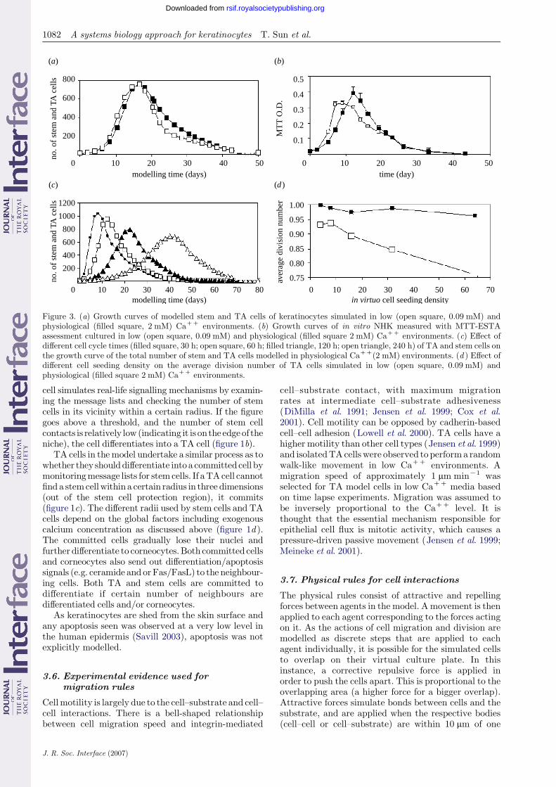
Figure 3. (a) Growth curves of modelled stem and TA cells of keratinocytes simulated in low (open square, 0.09 mM) andphysiological (filled square, 2 mM) CaCC environments. (b) Growth curves of in vitro NHK measured with MTT-ESTAassessment cultured in low (open square, 0.09 mM) and physiological (filled square 2 mM) CaCC environments. (c) Effect ofdifferent cell cycle times (filled square, 30 h; open square, 60 h; filled triangle, 120 h; open triangle, 240 h) of TA and stem cells onthe growth curve of the total number of stem and TA cells modelled in physiological CaCC(2 mM) environments. (d ) Effect ofdifferent cell seeding density on the average division number of TA cells simulated in low (open square, 0.09 mM) andphysiological (filled square 2 mM) CaCC environments.
1082 A systems biology approach for keratinocytes T. Sun et al.
rsif.royalsocietypublishing.orgDownloaded from
cell simulates real-life signalling mechanisms by examin-ing the message lists and checking the number of stemcells in its vicinity within a certain radius. If the figuregoes above a threshold, and the number of stem cellcontacts is relatively low (indicating it is on the edgeof theniche), the cell differentiates into a TA cell (figure 1b).
TA cells in themodel undertake a similar process as towhether they shoulddifferentiate intoa committedcell bymonitoringmessage lists for stemcells. If aTAcell cannotfind a stemcellwithin a certain radius in three dimensions(out of the stem cell protection region), it commits(figure 1c). The different radii used by stem cells and TAcells depend on the global factors including exogenouscalcium concentration as discussed above (figure 1d ).The committed cells gradually lose their nuclei andfurther differentiate to corneocytes.Both committed cellsand corneocytes also send out differentiation/apoptosissignals (e.g. ceramide and orFas/FasL) to the neighbour-ing cells. Both TA and stem cells are committed todifferentiate if certain number of neighbours aredifferentiated cells and/or corneocytes.
As keratinocytes are shed from the skin surface andany apoptosis seen was observed at a very low level inthe human epidermis (Savill 2003), apoptosis was notexplicitly modelled.
3.6. Experimental evidence used formigration rules
Cellmotility is largely due to the cell–substrate and cell–cell interactions. There is a bell-shaped relationshipbetween cell migration speed and integrin-mediated
J. R. Soc. Interface (2007)
cell–substrate contact, with maximum migrationrates at intermediate cell–substrate adhesiveness(DiMilla et al. 1991; Jensen et al. 1999; Cox et al.2001). Cell motility can be opposed by cadherin-basedcell–cell adhesion (Lowell et al. 2000). TA cells have ahigher motility than other cell types (Jensen et al. 1999)and isolatedTAcells were observed to performa randomwalk-like movement in low CaCC environments. Amigration speed of approximately 1 mm minK1 wasselected for TA model cells in low CaCC media basedon time lapse experiments. Migration was assumed tobe inversely proportional to the CaCC level. It isthought that the essential mechanism responsible forepithelial cell flux is mitotic activity, which causes apressure-driven passive movement (Jensen et al. 1999;Meineke et al. 2001).
3.7. Physical rules for cell interactions
The physical rules consist of attractive and repellingforces between agents in the model. Amovement is thenapplied to each agent corresponding to the forces actingon it. As the actions of cell migration and division aremodelled as discrete steps that are applied to eachagent individually, it is possible for the simulated cellsto overlap on their virtual culture plate. In thisinstance, a corrective repulsive force is applied inorder to push the cells apart. This is proportional to theoverlapping area (a higher force for a bigger overlap).Attractive forces simulate bonds between cells and thesubstrate, and are applied when the respective bodies(cell–cell or cell–substrate) are within 10 mm of one
Figure 4. Growth curves of (a) modelled HaCat cells and (b) in vitro HaCat cells measured with MTT-ESTA assessment thatsimulated or cultured in low (open square 0.09 mM) and physiological (filled square 2 mM) CaCC environments. (c) Totalnumber of modelled HaCat cells simulated in different CaCC environments. (d ) MTT-ESTA assessed overall cell viability ofHaCat cells (filled square) and NHKs (open square) cultured in different calcium environments.
A systems biology approach for keratinocytes T. Sun et al. 1083
rsif.royalsocietypublishing.orgDownloaded from
another. Bond strengths depend on the individual typeof cells involved and are related to the environmentalcalcium concentration (Lowell et al. 2000) via a sigmoidfunction (Baumgarter et al. 2000).
In lowCaCCmedia, cells (most of which areTA cells)have low cadherin-mediated cell–cell and integrin-basedcell–substrate bonds. In a physiological CaCC environ-ment, stem cells exhibit strong cell–cell and cell–substrate bonds due to high levels of Delta 1 andintegrins (Jensen et al. 1999; Lowell et al. 2000; Savill &Sherratt 2003). Committed cells and corneocytesin physiological CaCC form strong cell–cell and cell–sub-strate bonds through desmosomes and hemidesmosomes(Okuyama et al. 2004; Kolly et al. 2005) and age-relatedupregulated E-cadherin expression (Morel et al. 2001).TA cells have the lowest cell–cell or cell–substrate bondsdue to the low level of Delta 1, integrins and E-cadherin.Cells and neighbouring cell spheres (or substrates) formcontacts sharing a common, flat contact area. In thismodel, the cell–cell or cell–substrate adhesion wasdeliberately set within the range of 0–1, with a highernumber representing stronger bonds. This allowed for invirtuo exploration of the bond strengths on an empiricalbasis to simulate in vitro behaviour of cells.
The code for the model itself will be made availablefrom: http://www.flame.ac.uk.
4. RESULTS
4.1. Simulation of NHK dynamic multicellularmorphogenesis
As illustrated in figure 2a, in a low calcium environ-ment, following the rules described in the methods, the
J. R. Soc. Interface (2007)
stem cells divided symmetrically 2–3 times, thenasymmetrically to produce TA cells. TA cells divided,migrated randomly and distributed evenly over thesubstrate. After the culture achieved confluence, thecells withdrew from the cell cycle due to confluencetriggered contact inhibition and gradually lost viabi-lity. No stratification occurred as cells maintained highcell motility and only four neighbouring cells wereneeded to induce contact inhibition. In a physiologicalcalcium environment (as shown in figure 2b–d ), aheterogeneous cell composition and hierarchical popu-lation structure were reproduced by the model. Stemcells kept dividing symmetrically in compact clustersbefore the targeted colony size was obtained. Afterthat, the stem cells on the colony edge started to givebirth to a stem and a TA cell due to the clusterautoregulation mechanism proposed. TA cells near thestem cell niches were observed to proliferate, migrateand stratify actively. When TA cells escaped theinfluence of stem cells, they differentiated to committedcells and eventually changed to corneocytes. Due to thestrong cell–cell and cell–substrate adhesiveness, differ-entiated NHK cells formed a tight shell around TA andstem cell compartments as shown in figure 2b. Figure 2cdepicts the basal layer for the colonies seen in figure 2b,and figure 2d a middle layer of cells in the same colony(The model allows the colonies to stratify for up to fourlayers). The differentiation of TA and stem cells wasalso initiated by their neighbouring committed cellsthrough synthesis of differentiation signals (e.g. cer-amide and Fas/FasL) and/or establishment of localconfluence. (Please see the website http://www.dcs.shef.ac.uk/wphil/epiresources for movie clips of
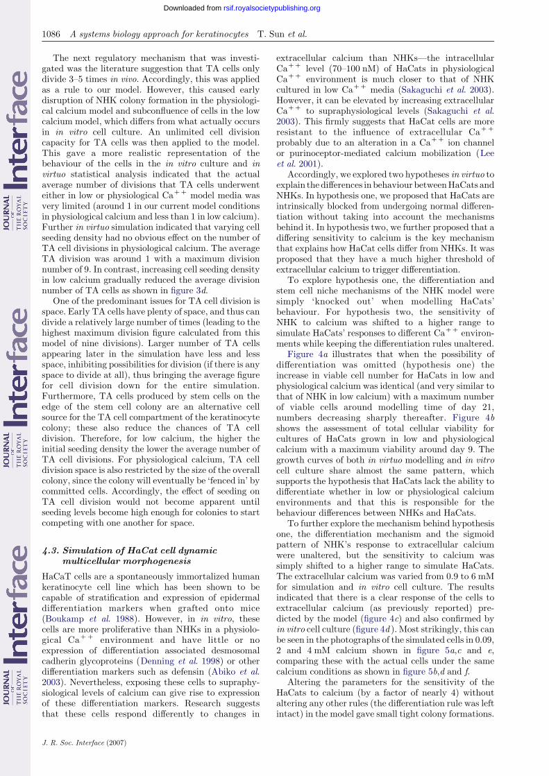
Figure 5. Micrographs of modelled HaCat cells (blue) in (a) low (0.09 mM), (c) physiological (2 mM) and (e) high (4 mM) CaCC
environments. Fluorescentmicrographs ofHaCat cells cultured in (b) low (0.09 mM), (d ) physiological (2 mM) and ( f ) high (4 mM)CaCC environments. The HaCat cells were stained with DAPI (blue) and pancytokeratin antibody (green). Scale bar, 100 mm.
1084 A systems biology approach for keratinocytes T. Sun et al.
rsif.royalsocietypublishing.orgDownloaded from
modelling of keratinocyte colony formation in low andphysiological CaCC.)
Comparing this simulation of keratinocytebehaviour to the behaviour of the cells in vitro, it wasfound that NHK cells proliferated, migrated activelyand distributed evenly throughout the tissue culturesurface in a low calcium medium (0.09 mM) asillustrated in figure 2e, whereas compact self-limitingcolonies were found in the physiological calcium(2 mM) environment as shown in figure 2f.
Figure 3a shows the total number of modelled TAcells and stem cells when simulated in low andphysiological calcium for up to six weeks. Normally,skin cells are only cultured for one to two weeks priorto experimentation or clinical use. With deliberatelonger term culture of cells both the in virtuo modeland the in vitro data (figure 3b) showed an initialincrease in the total viable cell population followed bya much slower decrease in cell number. In the model,the decreasing cell number after the peak value is dueto loss of stem cells and TA cells to becomecommitted cells and corneocytes (ultimately corneo-cytes would be shed from the skin surface henceare not counted in this model). In vitro it was foundthat cells reduced in volume and detached from theculture surface with time—almost certainly following
J. R. Soc. Interface (2007)
irreversible differentiation. Thus, the initial modelpredicted the overall pattern of cell number versustime in vitro.
However, the timing of the modelled increase didnot accurately simulate the in vitro increase anddecrease seen for cells either in low or physiologicalcalcium. In low calcium levels, viable cell numberpeaked at around day 7–8 and then subsequentlydecreased. In physiological calcium level, the greatestnumber of viable cells was achieved at around day 12.To adjust the model to simulate the biological data,the average cell cycle length can be adjusted as shownin figure 3c.
4.2. Identification of key regulation mechanisms
Once we had established an initial model that roughlydescribed the behaviour of the cells in vitro both in lowand physiological calcium, the model was then used totest the relative importance of some of the literature-derived rules on which the model was based. Firstly, theeffect of different cell–cell and cell–substrate adhesive-ness on NHK pattern formation was tested in the agent-based model. Weak stem cell–cell bonds resulted indisruption of the stem cell clusters and intermingling ofstem, TA and committed cells. A pattern of colony
Figure 6. Micrographs of modelled NHK cells in physiological calcium in the bottom layer of (a) a scratch wound immediatelypost created in virtuo by removing the cells in the strip, the arrows show the position of stem cell compartments and (b) partlyhealed wound 370 iterations after scratch with slow and quick healing areas predicted by the agent-based model, the arrows showthe quick healing areas, (c) scratch wound immediately post transplanted with stem cells in virtuo and (d ) partly healed wound370 iterations after transplanted with stem cells. Phase contrast micrographs of (e, g) scratch wounds created in confluent NHKcells cultured in physiological CaCC environments, ( f ) quick healing area 24 h after wounding, (h) slow healing area 7 days postwounding. Scale bar, 100 mm.
A systems biology approach for keratinocytes T. Sun et al. 1085
rsif.royalsocietypublishing.orgDownloaded from
formation was only created when more cohesive bondswere applied between stem cells than between theirdaughter TA cells. Strong stem cell–substrateinteractions were important to keep the stem cellclusters on the substrate. Weak cell–substrate bondsinduced the detachment of stem cell niches from thesubstrate. The crucial roles of cell–cell and cell–substrate bonds of committed cells and corneocyteswere also identified by in virtuo simulation. These
J. R. Soc. Interface (2007)
‘failed’ simulations are not shown. In contrast, asalready stated, figure 3c shows the impact of in virtuotesting different cell cycle times of stem and TA cells inphysiological CaCC media. However, not shown butimportant to note, varying cell cycle times had noobvious effects on the overall NHK macroscopicmorphogenesis produced using this model (which wasessentially similar to that seen in figure 2b) the influencewas solely on the time taken to achieve this pattern.
1086 A systems biology approach for keratinocytes T. Sun et al.
rsif.royalsocietypublishing.orgDownloaded from
The next regulatory mechanism that was investi-gated was the literature suggestion that TA cells onlydivide 3–5 times in vivo. Accordingly, this was appliedas a rule to our model. However, this caused earlydisruption of NHK colony formation in the physiologi-cal calcium model and subconfluence of cells in the lowcalcium model, which differs from what actually occursin in vitro cell culture. An unlimited cell divisioncapacity for TA cells was then applied to the model.This gave a more realistic representation of thebehaviour of the cells in the in vitro culture and invirtuo statistical analysis indicated that the actualaverage number of divisions that TA cells underwenteither in low or physiological CaCC model media wasvery limited (around 1 in our current model conditionsin physiological calcium and less than 1 in low calcium).Further in virtuo simulation indicated that varying cellseeding density had no obvious effect on the number ofTA cell divisions in physiological calcium. The averageTA division was around 1 with a maximum divisionnumber of 9. In contrast, increasing cell seeding densityin low calcium gradually reduced the average divisionnumber of TA cells as shown in figure 3d.
One of the predominant issues for TA cell division isspace. Early TA cells have plenty of space, and thus candivide a relatively large number of times (leading to thehighest maximum division figure calculated from thismodel of nine divisions). Larger number of TA cellsappearing later in the simulation have less and lessspace, inhibiting possibilities for division (if there is anyspace to divide at all), thus bringing the average figurefor cell division down for the entire simulation.Furthermore, TA cells produced by stem cells on theedge of the stem cell colony are an alternative cellsource for the TA cell compartment of the keratinocytecolony; these also reduce the chances of TA celldivision. Therefore, for low calcium, the higher theinitial seeding density the lower the average number ofTA cell divisions. For physiological calcium, TA celldivision space is also restricted by the size of the overallcolony, since the colony will eventually be ‘fenced in’ bycommitted cells. Accordingly, the effect of seeding onTA cell division would not become apparent untilseeding levels become high enough for colonies to startcompeting with one another for space.
4.3. Simulation of HaCat cell dynamicmulticellular morphogenesis
HaCaT cells are a spontaneously immortalized humankeratinocyte cell line which has been shown to becapable of stratification and expression of epidermaldifferentiation markers when grafted onto mice(Boukamp et al. 1988). However, in in vitro, thesecells are more proliferative than NHKs in a physiolo-gical CaCC environment and have little or noexpression of differentiation associated desmosomalcadherin glycoproteins (Denning et al. 1998) or otherdifferentiation markers such as defensin (Abiko et al.2003). Nevertheless, exposing these cells to supraphy-siological levels of calcium can give rise to expressionof these differentiation markers. Research suggeststhat these cells respond differently to changes in
J. R. Soc. Interface (2007)
extracellular calcium than NHKs—the intracellularCaCC level (70–100 nM) of HaCats in physiologicalCaCC environment is much closer to that of NHKcultured in low CaCC media (Sakaguchi et al. 2003).However, it can be elevated by increasing extracellularCaCC to supraphysiological levels (Sakaguchi et al.2003). This firmly suggests that HaCat cells are moreresistant to the influence of extracellular CaCC
probably due to an alteration in a CaCC ion channelor purinoceptor-mediated calcium mobilization (Leeet al. 2001).
Accordingly, we explored two hypotheses in virtuo toexplain thedifferences in behaviour betweenHaCats andNHKs. In hypothesis one, we proposed that HaCats areintrinsically blocked from undergoing normal differen-tiation without taking into account the mechanismsbehind it. In hypothesis two, we further proposed that adiffering sensitivity to calcium is the key mechanismthat explains how HaCat cells differ from NHKs. It wasproposed that they have a much higher threshold ofextracellular calcium to trigger differentiation.
To explore hypothesis one, the differentiation andstem cell niche mechanisms of the NHK model weresimply ‘knocked out’ when modelling HaCats’behaviour. For hypothesis two, the sensitivity ofNHK to calcium was shifted to a higher range tosimulate HaCats’ responses to different CaCC environ-ments while keeping the differentiation rules unaltered.
Figure 4a illustrates that when the possibility ofdifferentiation was omitted (hypothesis one) theincrease in viable cell number for HaCats in low andphysiological calcium was identical (and very similar tothat of NHK in low calcium) with a maximum numberof viable cells around modelling time of day 21,numbers decreasing sharply thereafter. Figure 4bshows the assessment of total cellular viability forcultures of HaCats grown in low and physiologicalcalcium with a maximum viability around day 9. Thegrowth curves of both in virtuo modelling and in vitrocell culture share almost the same pattern, whichsupports the hypothesis that HaCats lack the ability todifferentiate whether in low or physiological calciumenvironments and that this is responsible for thebehaviour differences between NHKs and HaCats.
To further explore the mechanism behind hypothesisone, the differentiation mechanism and the sigmoidpattern of NHK’s response to extracellular calciumwere unaltered, but the sensitivity to calcium wassimply shifted to a higher range to simulate HaCats.The extracellular calcium was varied from 0.9 to 6 mMfor simulation and in vitro cell culture. The resultsindicated that there is a clear response of the cells toextracellular calcium (as previously reported) pre-dicted by the model (figure 4c) and also confirmed byin vitro cell culture (figure 4d ). Most strikingly, this canbe seen in the photographs of the simulated cells in 0.09,2 and 4 mM calcium shown in figure 5a,c and e,comparing these with the actual cells under the samecalcium conditions as shown in figure 5b,d and f.
Altering the parameters for the sensitivity of theHaCats to calcium (by a factor of nearly 4) withoutaltering any other rules (the differentiation rule was leftintact) in the model gave small tight colony formations.
A systems biology approach for keratinocytes T. Sun et al. 1087
rsif.royalsocietypublishing.orgDownloaded from
This simulated the actual colonies obtained at supra-physiological calcium which appeared to be very self-limiting with limited multilayering. Thus, some, albeitabnormal, self-regulation of colony formation was seenat supraphysiological extracellular calcium (please seethe website http://www.dcs.shef.ac.uk/wphil/epire-sources for movie clips of modelling of HaCat cells indifferent CaCC environments), indicating that alteredcalcium sensitivity only partially contributed to thebehaviour of HaCat cells. Thus, the data suggestHaCats lack the ability to form NHK type coloniesin vitro even when supraphysiologically high levels ofcalcium are present, but further investigation showsthere is some remaining response to calcium in thesecells as they do achieve increased cell–cell binding atrelatively high levels of extracellular calcium.
4.4. Prediction and validation of scratch woundhealing in two-dimensional cell culture
We then used the in virtuomodel for the NHKs to makepredictions on how cells would respond to wounding.A simple scratch wound was simulated by removing allof the modelled NHK cells located within a strip (300–400 mm wide) in the centre of confluent modelled cellsunder different CaCC environments. In a low CaCC
media, rapid wound healing was achieved by active cellmigration and subsequent cell proliferation (data notshown). Cells moved individually and achievedcomplete re-epithelialization within 1–2 days of wound-ing. In contrast, in physiological CaCC media, cellsmigrated as a coherent sheet.
The model predicted that injuries close to stem orTA cell compartments would heal quickly, whereasvery slow wound closure would be achieved in areascontaining only committed cells and/or corneocytes asillustrated in figure 6a,b. The model also predicted thatthe wound healing process could be accelerated byseeding a small number of stem cells in the denudedarea as shown in figure 6c,d. (Please see the websitehttp://www.dcs.shef.ac.uk/wphil/epiresources formovie clips of modelling of keratinocyte wound healingin low and physiological CaCC.)
In vitro scratch wound-healing experiments werethen designed based on these predictions. NHK cells inlow CaCC were observed to migrate freely into thedenuded area to close the wound within 24 h in the lowCaCC media (results not shown). In physiologicalCaCC, however, the wound edge was observed tomigrate as a contiguous sheet with significantlydifferent rates: in some areas rapid wound healing wasobserved within 24 h (figure 6e, f ), while in other areasthere was no sign of complete healing even after 7 daysof culture (figure 6g,h). The rapidly healing areas wereidentified as germinative compartments with high ratesof BrdU incorporation (indicative of proliferation)(figure 7a,b) and low levels of involucrin expression(indicative of differentiation; figure 7c). In contrast, theslow or non-healing areas were identified as committedcell compartments with low rates of BrdU incorpor-ation (figure 7d,e) and high levels of involucrinexpression as illustrated in figure 7f.
J. R. Soc. Interface (2007)
5. DISCUSSION
The regulation of epidermal homeostasis involves acomplex interplay between different generic and geneticmechanisms, making it difficult to investigate except byfocusing on separate discrete aspects of the biology. Inagent-based computational model, individual cells arerepresented by autonomous software agents, whichexecute a set of rules according to cell internal statesand immediate environments. This has the advantage ofcreating a global view of themacroscopicmorphogenesisofNHKcells in virtuo, allowing the testing of hypothesesand designing of new and informative experiments.
Previously, we developed a cell-based computationalmodel for urothelial cells in monolayer culture (Walkeret al. 2004a,b). However, extension of this model to NHKand HaCat cell demonstrated some of its limitations(Walker et al. 2006) suggesting it did not include sufficientbiological rules. Another agent-based model has recentlybeen developed to simulate and predict epidermalmorphology, tissue kinetics and the two-dimensionalflow of calcium ions (Grabe & Neuber 2005). In thismodel, the keratinocyte differentiation mechanism wasassumed to be solely based on the calcium levels.
Our aim in the current study was to develop andvalidate a model of epidermal cell social behaviour,which would be accessible for use by cell biologistsinterested in questions of epidermal organization andhomeostasis. In establishing this agent-based model, wefound we had to change initial assumptions aboutkeratinocyte TA cell division and the length of cell cyclein different calciummedia. We then tested two differenthypotheses about how the growth of the HaCatkeratinocyte cell line differed from NHKs. Twohypotheses were proposed: the first was that HaCatsare intrinsically incapable of normal colony formationirrespective of the calcium concentration and thesecond was that these cells had an altered (abnormallyso) calcium sensitivity. Both hypotheses were sup-ported—the first to a greater extent than the second.This suggests that cells fail to differentiate for reasonswhich include but are not fully explained by an alteredsensitivity to calcium. In this research, we alsopredicated and validated a position-dependent scratchwound healing pattern of keratinocytes in monolayerculture. The development of the model is described inthis study and access to this software model can beobtained via our website as mentioned earlier.
In brief, we suggest that the initial validation of themodel seems promising and has quickly led to thegeneration of hypotheses for exploration. The hypotheseswedescribe are onlya fewof the questions that cannowbetackled using this model.
Firstly, to summarize the main outcomes of themodel, we found that cell–cell and cell–substratemediated contacts were crucially important for thecolony-forming pattern of NHK cells in physiologicalcalcium. These results, while important, are asexpected. Other outcomes were less predictable—thebasic model rules simulated the initial increase and thendecrease in total viable cell number seen when cellswere grown for up to six weeks. However, it wasnecessary to alter the cell cycle length of the cells to
Figure 7. Fluorescent micrographs of NHK in physiological calcium in a scratch wound model with (a) cell nuclei stained withDAPI (blue), (b) cells incorporating BrdU and (c) cells which expressed involucrin in quick healing areas. In contrast, (d ) showscell nuclei stained with DAPI and (e) cells failing to incorporate BrdU ( f ) cells expressing involucrin in a slow healing area withinthe same culture. Scale bar, 100 mm.
1088 A systems biology approach for keratinocytes T. Sun et al.
rsif.royalsocietypublishing.orgDownloaded from
reflect the more rapid or slow increase in cell number.The next finding was that the literature suggests thatTA cells can divide only 3–5 times (Dover & Wright1991; Jensen et al. 1999; Lowell et al. 2000; Okuyamaet al. 2004; Kolly et al. 2005). Data from the modelhowever suggested that the limited division ability ofTA cells was not an intrinsic cell property of the cellsbut rather a statistical phenomenon. In the model, TAcells were allowed to divide without any limitation.However, it became apparent that whereas some cells(those TA cells arising from stem cells early in colonyformation) might divide up to a maximum number ofnine times, other TA cells did not divide at all (largelydue to space limitations in low calcium or to parameterslimiting the size of the colonies in physiologicalcalcium). Thus, the model showed that the averageTA cell division time was around 1 in physiological
J. R. Soc. Interface (2007)
calcium and less than 1 in low calcium under currentsimulation conditions. These data strongly suggest thatTA cells divide according to ‘need’ or opportunity,rather than it being an inherent property of the cell.
Another interesting finding related to the length ofthe cell cycle. The lack of methods to identify andmeasure cell cycle times of TA cells and stem cells isfrustrating for biologists but here, interestingly, inves-tigation in virtuo showed that varying the cell cycletimes of stem or TA cells had no obvious effects onmacroscopic morphogenesis. Essentially, different cycletimes had no overall effect on the final structure of thecolonies, they simply affected the length of timerequired to achieve this structure.
The model was then extended to see if we couldsimulate the behaviour of the HaCat keratinocyte cellline. HaCat cells are a useful keratinocyte cell line in
A systems biology approach for keratinocytes T. Sun et al. 1089
rsif.royalsocietypublishing.orgDownloaded from
that they behave as though they are undifferentiatedTA cells even in physiological CaCC environments.Thus, they are easy to expand and culture withoutgoing into terminal differentiation. However, theliterature suggests that these cells have at least somecapacity for terminal differentiation when they areimplanted in vivo on nude mice (Boukamp et al. 1988).Also previous work has suggested that one of theirmajor differences to NHK cells is their sensitivity toextracellular calcium (Sakaguchi et al. 2003). Asdiscussed earlier, calcium is clearly key to helpinginitiate differentiation (although not the only factor)but crucial to allow differentiation to complete inNHKs. Thus, if for whatever reason the intracellularcalcium of HaCats does not rise to the permissive levelsrequired for differentiation, then this could explaintheir behaviour to a large extent.
Two alternative hypotheses to explain HaCat cellin vitro behaviour were explored: an intrinsic abnorm-ality in their ability to differentiate and an abnormalityin their calcium regulation. The first was simulated byknocking out mechanisms for differentiation and stemcell niche autoregulation. This then produced an invirtuo model which simulated the behaviour of theHaCats in physiological calcium. The growth curves ofboth in virtuo modelling and in vitro cell culture sharealmost the same pattern, which supports thehypothesis. The second hypothesis that themechanisms for differentiation and stem cell nicheautoregulation would start to function if HaCat cellswere exposed to a superphysiological level of extra-cellular calcium was then explored. The in vitroexperimentation revealed that if cultured in sufficientlyhigh calcium media HaCat cell proliferation decreasedand cells began to form compact colonies. However,these were still clearly and visibly different to NHKcolonies (no stratification of cells in these very tightHaCaT colonies) but nevertheless this hypothesis wassupported to a certain extent. We suggest that themodelling supports cells having a clear abnormality intheir calcium responsiveness (as already supported bythe current literature) but also some additional defectin their ability to differentiate beyond this.
The in virtuo model for colony formation in NHKswas then tested further by making a wound (equivalentto a scratch wound) in a monolayer culture of NHKs.The modelling revealed that active cell migration andsubsequent cell proliferation contributed to the rapidwound closure in a low CaCC media, whereas inphysiological calcium media wound healing was verydependant on the position of the wound with respect tothe nearest stem cells or TA cells. At this stage, themodel was used to design in vitro scratch woundexperiments to examine to what extent this hypothesiswould hold true. We were able to show, within the samescratch wound, areas of rapid wound healing and areasof slow wound healing. Rapid healing turned out to beattributable to actively proliferating keratinocytes(germinative compartments) and areas of very slowwound healing were associated with cells in the sameculture showing low levels of proliferation and rela-tively high levels of differentiation. We suggest thisconfirms the prediction from the in virtuo modelling.
J. R. Soc. Interface (2007)
In tissue development, cells actively change theirbehaviour and properties, as a consequence of internaldecisions due to ‘rules’ that are encoded in the geneticinformation of each cell. These have been selectedthroughout evolution and are influenced by theimmediate environments. Hence, a model that permitsprediction of individual cells or multicellular behaviourshould combine a description of a cell with a descrip-tion of the rules that dictate the change of itsbehaviour or parameters (Baker et al. 1998; Hogeweg2000a,b; Drasdo & Hohme 2005; Galle et al. 2005;Ponciano et al. 2005). This goes beyond the workdescribed in this study in which we have a model thatdescribes the spatial growth pattern of keratinocytesdetermined by the relationship between individual cellsevolving with time. At this stage of development, themodel does not incorporate detailed intracellularinformation, nor does it deal with the cells’ responsesto a wide range of external agents (but, in principle,any rule in the model could be replaced by a model ofthe mechanism underlying the rule). The model doesallow us to begin to see organization of cells in threedimensions (in physiological calcium) and most impor-tantly allows one to test different hypotheses of howcells will respond to basic features of cell manipulationin vitro.
Now that this basic model has been established,there are significant further challenges to be taken onin terms of looking at the interaction of the keratino-cytes with stromal fibroblasts and then progressivelyadding in more details wherever the model indicatesparticular parameters are particularly important (suchas in cell–cell adhesion for example). However, themodel at this stage can be used to explore many of thequestions which keratinocyte biologists currentlyanswer at an empirical level. Thus, it should now bepossible to predict why certain seeding densities ofkeratinocytes will get a better expansion of cells thanothers, why passaging cells at a certain density andafter a certain length of culture yield more prolifera-tive cells than under other conditions and how tomanipulate the extracellular calcium environment toget best value from biopsies of patient cells for clinicalexpansion.
In summary, in this study, we have described a novelcomputational model of keratinocyte colony formation.Since the model treats cells as individual entities it canbe tightly coupled to experimental work, enablinghypotheses to be readily generated and tested. Ourstudy shows that this synergy between computationaland experimental models has the potential to become apowerful tool for understanding how cells organize intotissue
We gratefully acknowledge financial support from EPSRC(UK) for this research.
REFERENCES
Abiko, Y., Nishimura, M., Kusano, K., Yamazaki, M.,Arakawa, T., Takuma, T. & Kaku, T. 2003 Upregulatedexpression of human b defensin-1 and -3 mRNA during
1090 A systems biology approach for keratinocytes T. Sun et al.
rsif.royalsocietypublishing.orgDownloaded from
differentiation of keratinocyte immortalized cell lines,HaCaT and PHK16-0b. J. Dermatol. Sci. 31, 225–228.(doi:10.1016/S0923-1811(03)00007-0)
Adams, J. C. & Watt, F. M. 1990 Changes in keratinocyteadhe sion during terminal differentiation: reduction infibronectin binding precedes alpha 5 beta 1 integrin lossfrom the cell surface. Cell 63, 425–435. (doi:10.1016/0092-8674(90)90175-E)
Aplin, A. E., Howe, A. K. & Juliano, R. L. 1999 Cell adhesionmolecules, signal transduction and cell growth. Curr.Opin. Cell Biol. 11, 737–744. (doi:10.1016/S0955-0674(99)00045-9)
Ashkenas, J., Muschler, J. & Bissell, M. J. 1996 Theextracellular matrix in epithelial biology: shared moleculesand common themes in distant phyla. Dev. Biol. 180,433–444. (doi:10.1006/dbio.1996.0317)
Asplund, A., Guo, Z., Hu, X., Wassberg, C. & Ponten, F. 2001Mosaic pattern of maternal and paternal keratinocyteclones in normal human epidermis revealed by analysis ofX-chromosome inactivation. J. Invest. Dermatol. 117,128–131. (doi:10.1046/j.0022-202x.2001.01385.x)
Assoian, R. K. 1997 Anchorage-dependent cell cycle pro-gression. J. Cell Biol. 136, 1–4. (doi:10.1083/jcb.136.1.1)
Baker, C. T. H., Bocharov, G. A., Paul, C. A. H. & Rihan,F. A. 1998 Modelling and analysis of time-lags in somebasic patterns of cell proliferation. J. Math. Biol. 37,341–371. (doi:10.1007/s002850050133)
Balanescu, T., Cowling, A. J., Georgescu, H., Gheorghe, M.,Holcombe, M. & Vertan, C. 1999 Communicating streamX-machines systems are no more than X-machines.J. Univ. Comput. Sci. 5, 494–507.
Baumgarter, W., Hinterdorfer, P., Ness, W., Raab, A.,Vestweber, D., Schindler, H. & Drenckhahn, D. 2000Cadherin interaction probed by atomic force microscopy.Proc. Natl Acad. Sci. USA 97, 4005–4010. (doi:10.1073/pnas.070052697)
Bhadriraju, K. & Hansen, L. K. 2004 Extracellular matrix-dependent myosin dynamics during G1-S phase cell cycleprogression in hepatocytes. Exp. Cell Res. 300, 259–271.(doi:10.1016/j.yexcr.2004.06.033)
Bhatia, S. N., Balis, U. J., Yarmush, M. L. & Toner, M. 1999Effect of cell–cell interactions in preservation of cellularphenotype: cocultivation of hepatocytes and nonparench-ymal cells. FASEB J. 13, 1883–1900.
Boukamp, P., Petrussevska, R. T., Breitkreutz, D., Hornung,J., Markham, A. & Fusenig, N. E. 1988 Normalkeratinization in a spontaneously immortalized aneuploidhuman keratinocyte cell line. J. Cell Biol. 106, 761–771.(doi:10.1083/jcb.106.3.761)
Coakley, S., Smallwood, R. & Holcombe, M. 2006 UsingX-machines as a formal basis for describing agents inagent-based modeling. In Proc. Agent-Directed Simulation(ADS ’06) Conference, 2–6 April.
Cox, E. A., Sastry, S. K. & Huttenlocher, A. 2001 Integrin-mediated adhesion regulates cell polarity and membraneprotrusion through Rho family of GTPases.Mol. Biol. Cell12, 265–277.
Croix, B. S., Sheehan, C., Rak, J. W., Florenes, V. A.,Slingerland, J. M. & Kerbel, R. S. 1998 E-Cadherindepen-dent growth suppression is mediated by the cyclin-dependent kinase inhibitor p27 KIPI. J. Cell Biol. 142,557–571. (doi:10.1083/jcb.142.2.557)
Dalrymple, S., Antony, L., Xu, Y., Uzgare, A. R., Arnold,J. T., Savaugeot, J., Sokoll, L. J., Marzo, A. M. D. &Isaacs, J. T. 2005 Role of Notch-1 and E -Cadherin in thedifferential response to calcium in culturing normal versusmalignant prostate cells. Cancer Res. 65, 9269–9279.(doi:10.1158/0008-5472.CAN-04-3989)
J. R. Soc. Interface (2007)
Denning, M. F., Guy, S. G., Ellerbroek, S. M., Norvell, S. M.,Kowalczyk, A. P. & Green, K. J. 1998 The expression ofdesmoglein isoforms in cultured human keratinocytes isregulated by calcium, serum, and protein kinase C. Exp.Cell Res. 239, 50–59. (doi:10.1006/excr.1997.3890)
DiMilla, P. A., Barbee, K. & Lauffenburger, D. A. 1991Mathematical model for the effects of adhesion andmechanics on cell migration speed. Biophys. J. 60, 15–37.
Dover, R. & Potten, C. S. 1983 Cell cycle kinetics of culturedhuman epidermal keratinocytes. J. Invest. Dermatol. 80,423–429. (doi:10.1111/1523-1747.ep12555494)
Dover, R. &Wright, N. A. 1991 The cell proliferation kineticsof the epidermis. In Physiology, biochemistry and molecu-lar biology of the skin (ed. L. A. Goldsmith), pp. 239–265.Oxford, UK: Oxford University Press.
Drasdo, D. & Hohme, S. 2005 A single-cell-based model oftumor growth in vitro: monolayers and spheroids. Phys.Biol. 2, 133–147. (doi:10.1088/1478-3975/2/3/001)
Eglen, S. J. & Willshaw, D. J. 2002 Influence of cell fatemechanisms upon retinal mosaic formation: a modellingstudy. Development 129, 5399–5408. (doi:10.1242/dev.00118)
Fleischmajer, R. et al. 2000 Differential expression of laminina chains during proliferative and differentiation stages in amodel for skin morphogenesis. Matrix Biol. 19, 637–647.(doi:10.1016/S0945-053X(00)00092-5)
Galle, J., Loeffler, M. &Drasdo, D. 2005Modeling the effect ofderegulated proliferation and apoptosis on the growthdynamics of epithelial cell populations in vitro. Biophys. J.88, 62–75. (doi:10.1529/biophysj.104.041459)
Gizelda, T. B., Wieser, R., Bunge, R. P., Margitich, I. S., Katz,J., Olson, L. & Wood, P. M. 2000 Density dependentregulation of human Schwann cell proliferation. Glia 30,165–177. (doi:10.1002/(SICI)1098-1136(200004)30:2!165::AID-GLIA6O3.0.CO;2-L)
Goulet, F., Poitras, A., Rouabhia, M., Cusson, D., Germain,L. & Auger, F. A. 1996 Stimulation of human keratinocyteproliferation through growth factor exchanges with dermalfibroblasts in vitro. Burns 22, 107–112. (doi:10.1016/0305-4179(95)00098-4)
Grabe, N. & Neuber, K. 2005 A multicellular systems biologymodel predicts epidermal morphology, kinetics and Ca 2Cflow. Bioinformatics 21, 3541–3547. (doi:10.1093/bioinfor-matics/bti585)
Grossmann, J., Walther, K., Artinger, M., Kiessling, S. &Scholmerich, J. 2001 Apoptotic signaling during initiationof detachment-induced apoptosis (“Anoikis”) of primaryhuman intestinal epithelial cells. Cell Growth Differ. 12,147–155.
Hogeweg, P. 2000a Evolving mechanisms of morphogenesis:on the interplay, between differential adhesion and celldifferentiation. J. Theor. Biol. 203, 317–333. (doi:10.1006/jtbi.2000.1087)
Hogeweg, P. 2000b Shapes in the shadow: evolutionarydynamics of morphogenesis. Artif. Life 6, 85–101.(doi:10.1162/106454600568339)
Holbrook, K. A. 1994 Ultrastructure of the epidermis. In Thekeratinocyte handbook (eds I. M. Leigh, B. Lane & F. M.Watt), pp. 3–39. London, UK: Cambridge UniversityPress.
Ingber, D. E. 2003a Tensegrity I. Cell structure andhierarchical systems biology. J. Cell Sci. 116, 1157–1173.(doi:10.1242/jcs.00359)
Ingber, D. E. 2003b Tensegrity II. How structural networksinfluence cellular information processing networks. J. CellSci. 116, 1397–1408. (doi:10.1242/jcs.00360)
Jensen, U. B., Lowell, S. & Watt, F. M. 1999 The spatialrelationship between stem cells and their progeny in the
A systems biology approach for keratinocytes T. Sun et al. 1091
rsif.royalsocietypublishing.orgDownloaded from
basal layer of human epidermis: a new view based onwhole-mount labelling and lineage analysis. Development126, 2409–2418.
Kefalas, P., Holcombe, M., Eleftherakis, G. & Gheorghe, M.2003 A formal method for the development of agent-basedsystems. In Intelligent agent software engineering (ed.V. Plekhavona), pp. 68–98. Hershey, PA: Idea GroupPublishing.
King, K. L. & Cidlowski, J. A. 1995 Cell cycle and apoptosis:common pathways to life and death. J. Cell. Biochem. 58,175–180. (doi:10.1002/jcb.240580206)
Kolettas, E., Skoufos, I., Kontargiris, E., Markopoulou, S.,Tzavaras, T. & Gonos, E. S. 2006 Bcl-2 but notclusterin/apolipoprotein J protected human diploid fibro-blasts and immortalized keratinocytes from ceramide-induced apoptosis: role of p53 in the ceramide response.Arch. Biochem. Biophys. 445, 184–195. (doi:10.1016/j.abb.2005.10.006)
Kolly, C., Suter, M. M. &Muller, E. J. 2005 Proliferation, cellcycle exit, and onset of terminal differentiation in culturedkeratinocytes: pre-programmed pathways in control ofC-Myc and Notch 1 prevail over extracellular calciumsignals. J. Invest. Dermatol. 124, 1014–1025. (doi:10.1111/j.0022-202X.2005.23655.x)
Korang, K., Christiano, A. M., Uitto, J. & Mauviel, A. 1995Differerential cytokine modulation of the genes LAMA3,LAMB3, and LAMC2, encoding the constitutive poly-peptides, a3, b3, and g2, of human laminin 5 in epidermalkeratinocytes. FEBS Lett. 368, 556–558. (doi:10.1016/0014-5793(95)00740-Z)
Laporte, M. & Heenen, M. 1994 The heterogeneity of thegerminative compartment in human epidermis and itsimplications in pathogenesis. Dermatology 189, 340.
Latkowski, J. A. M., Freedberg, I. M. & Blumenberg, M. 1995Keratinocyte growth factor and keratin gene regulation.J. Dermatol. Sci. 9, 36–44. (doi:10.1016/0923-1811(94)00350-N)
Lee, W. K., Choi, S. W., Lee, H. R., Lee, E. J., Lee, K. H. &Kim, H. O. 2001 Purinoceptor-mediated calcium mobil-ization and proliferation in HaCaT keratinocytes.J. Dermatol. Sci. 25, 97–105. (doi:10.1016/S0923-1811(00)00117-1)
Lowell, S., Jones, P., Roux, I. L., Dune, J. &Watt, F. M. 2000Stimulation of human epidermal differentiation by Delta-Notch signalling at the boundaries of stem-cell clusters.Curr. Biol. 10, 491–500. (doi:10.1016/S0960-9822(00)00451-6)
MacArthur, B. D., Please, C. P., Taylor, M. & Oreffo,R. O. C. 2004 Mathematical modelling of skeletal repair.Biochem. Biophys. Res. Commun. 313, 825–833. (doi:10.1016/j.bbrc.2003.11.171)
Meineke, F. A., Potten, C. S. & Loeffler, M. 2001 Cellmigration and organization in the intestinal crypt using alattice-free model. Cell Prolif. 34, 253–266. (doi:10.1046/j.0960-7722.2001.00216.x)
Morel, D., Marcelpoil, R. & Brugal, G. 2001 A proliferationcontrol network model: the simulation of two-dimensionalepithelial homeostasis. Acta Biotheoretica 49, 219–234.(doi:10.1023/A:1014201805222)
Nelson, C. M. & Chen, C. S. 2002 Cell–cell signaling by directcontact increases cell proliferation via a PI3K-dependentsignal. FEBS Lett. 514, 238–242. (doi:10.1016/S0014-5793(02)02370-0)
Nelson, C. M., Jean, R. P., Tan, J. L., Liu, W. F., Sniadecki,N. J., Spector, A. A. & Chen, C. S. 2005 Emergent patternsof growth controlled by multicellular form and mechanics.Proc. Natl Acad. Sci. USA 102, 11 594–11 599. (doi:10.1073/pnas.0502575102)
J. R. Soc. Interface (2007)
Nishimura, E. K., Yoshida, H., Kunisada, T. & Nishikawa,S. I. 1999 Regulation of E- and P-cadherin expressioncorrelated with melanocyte migration and diversification.Dev. Biol. 215, 155–166. (doi:10.1006/dbio.1999.9478)
Okuyama, R., LeFort, K. & Dotto, G. P. 2004 A dynamicmodel of keratinocyte stem cell renewal and differen-tiation: role of the p21WAF1/Cip1 and Notch 1 signalingpathways. J. Invest. Dermatol. Symp. Proc. 9, 248–252.(doi:10.1111/j.1087-0024.2004.09308.x)
Olsson, L. 2003 Cell migration, pattern formation and cell fateduring head development in lungfishes and amphibians.Theory Biosci. 122, 252–265. (doi:10.1078/1431-7613-00086)
Orford, K., Orford, C. C. & Byers, S. W. 1999 Exogenousexpression of beta-catenin regulates contact inhibition,anchorage-independent growth, anoikis and radiation-induced cell cycle arrest. J. Cell Biol. 146, 855–867.(doi:10.1083/jcb.146.4.855)
Ponciano, J. M., Vandecasteele, F. P. J., Hess, T. F., Forney,L. J., Crawford, R. L. & Joyce, P. 2005 Use of stochasticmodels to assess the effect of environmental factors onmicrobial growth. Appl. Environ. Microbiol. 71,2355–2364. (doi:10.1128/AEM.71.5.2355-2364.2005)
Potten, C. S. & Booth, C. 2002 Keratinocyte stem cells: acommentary. J. Invest. Dermatol. 119, 888–899. (doi:10.1046/j.1523-1747.2002.00020.x)
Rashbass, J. 1996 Modelling tissues on the computer. TrendsCell Biol. 6, 280–281. (doi:10.1016/0962-8924(96)60028-2)
Sakaguchi, M., Miyazaki, M., Takaishi, M., Sakaguchi, Y.,Makino, E., Kataoka, N., Yamada, H., Namba, M. & Huh,N. 2003 S100C/A11 is a key mediator of Ca2C inducedgrowth inhibition of human epidermal keratinocytes.J. Cell Biol. 163, 825–835. (doi:10.1083/jcb.200304017)
Santini, M. T., Rainaldi, G. & Indovina, P. L. 2000 Apoptosis,cell adhesion and extracellular matrix in the three-dimensional growth of multicellular tumor spheroids.Crit. Rev. Oncol. Hematol. 36, 75–87.
Savill, N. J. 2003 Mathematical models of hierarchicallystructured cell populations under equilibrium with appli-cation to the epidermis. Cell Prolif. 36, 1–26. (doi:10.1046/j.1365-2184.2003.00257.x)
Savill, N. J. & Sherratt, J. A. 2003 Control of epidermal stemcell clusters by Notch-mediated lateral induction. Dev.Biol. 258, 141–153. (doi:10.1016/S0012-1606(03)00107-6)
Scheffrahn, I., Singer, B. B., Sigmundsson, K., Lucka, L. &Obrink, B. 2005 Control of density-dependent, cell state-specific signal transduction by the cell adhesion moleculeCEACAM1, and its influence on cell cycle regulation. Exp.Cell Res. 307, 427–435. (doi:10.1016/j.yexcr.2005.03.030)
Shraiman, B. I. 2005 Mechanical feedback as a possibleregulator of tissue growth. Proc. Natl Acad. Sci. USA 102,3318–3323. (doi:10.1073/pnas.0404782102)
Smola, H., Stark, H.-J., Thiekotter, G., Mirancea, N., Krieg,T. & Fusenig, N. E. 1998 Dynamics of basementmembrane formation by keratinocyte–fibroblast interac-tions in organotypic skin culture. Exp. Cell Res. 239,399–410. (doi:10.1006/excr.1997.3910)
Sun, T., Mai, S., Haycock, J., Ryan, A. & MacNeil, S. 2005Self-organisation of skin cells in 3D-electrospun poly-styrene scaffolds. Tissue Eng. 11, 1023–1033. (doi:10.1089/ten.2005.11.1023)
Thompson, C. B. 1995 Apoptosis in the pathogenesis andtreatment of disease. Science 267, 1456–1462. (doi:10.1126/science.7878464)
Vespa, A., DSouza, S. J. A. & Dagnino, L. 2005 A novel rolefor integrin-linked kinase in epithelial sheet morpho-genesis. Mol. Biol. Cell 16, 4084–4095. (doi:10.1091/mbc.E05-02-0087)
1092 A systems biology approach for keratinocytes T. Sun et al.
rsif.royalsocietypublishing.orgDownloaded from
Walker, D. C., Southgate, J., Hill, G., Holcombe, M., Hose,D. R., Wood, S. M., MacNeil, S. & Smallwood, R. H. 2004aThe epitheliome: agent-based modelling of the socialbehaviour of cells. BioSystems 76, 89–100. (doi:10.1016/j.biosystems.2004.05.025)
Walker, D. C., Hill, G., Wood, S. M., Smallwood, R. H. &Southgate, J. 2004b Agent-based computational modelingof wounded epithelial cell monolayers. IEEE Trans.Nanobiosci. 3, 153–163. (doi:10.1109/TNB.2004.833680)
Walker, J. L., Fournier, A. K. & Assoian, R. K. 2005Regulation of growth factor signaling and cell cycleprogression by cell adhesion and adhesion-dependentchanges in cellular tension. Cytokine Growth Factor Rev.16, 395–405. (doi:10.1016/j.cytogfr.2005.03.003)
Walker, D., Sun, T., MacNeil, S. & Smallwood, R. 2006Modelling the effect of exogenous calcium on keratinocyteand HaCat cell proliferation and differentiation using anagent-based computational paradigm. Tissue Eng. 12,2301–2309. (doi:10.1089/ten.2006.12.2301)
J. R. Soc. Interface (2007)
Wang, X., Bregegere, F., Soroka, Y., Kayat, A.,Redziniak, G. & Milner, Y. 2004 Enhancement ofFas-mediated apoptosis in aging human keratinocytes.Mech. Aging Dev. 125, 237–249. (doi:10.1016/j.mad.2003.12.007)
Webb, A., Li, A. & Kaur, P. 2004 Location and phenotype ofhuman adult keratinocyte stem cells of the skin. Differen-tiation 72, 387–395. (doi:10.1111/j.1432-0436.2004.07208005.x)
Zaman, M. H., Kamm, R. D., Matsudaira, P. &Lauffenburger, D. A. 2005 Computational model for cellmigration in three-dimensional matrices. Biophys. J. 89,1389–1397. (doi:10.1529/biophysj.105.060723)
Zhu, A. J., Haase, I. & Watt, F. M. 1999 Signaling via b1integrins and mitogen activated protein kinasedetermines human epidermal stem cell fate in vitro.Proc. Natl Acad. Sci. USA 96, 6728–6733. (doi:10.1073/pnas.96.12.6728)