42
DR.LAVANYA FROM THE UNIT OF DR.M.S.VISWANATHAN PEDIATRIC HEPATOLOGY AND GASTROENTEOLOGY APOLLO CHILDREN’S HOSPITAL AN INTERESTING CASE OF LIVER FAILURE

DR.LAVANYA
FROM THE UNIT OF DR.M.S.VISWANATHAN
PEDIATRIC HEPATOLOGY AND GASTROENTEOLOGY
APOLLO CHILDREN’S HOSPITAL
AN INTERESTING CASE OF LIVER FAILURE

HISTORY
• Ms. A, a 9 year old girl,
• First born of non consanguineous marriage
• From Tripura
• Intermittent fever for 15 days and yellowish discoloration of the sclera for 10 days
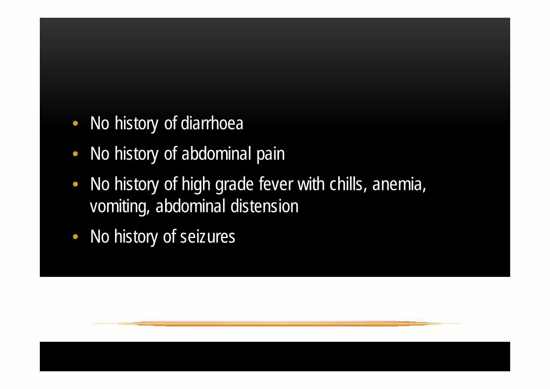
• No history of diarrhoea
• No history of abdominal pain
• No history of high grade fever with chills, anemia, vomiting, abdominal distension
• No history of seizures

• No history of TB contact
• No history of encephalopathy/ sleep disturbances
• No history of GI bleeds(hemetemesis/ melena)
• No history of edema

• No history of any treatment(herbal supplements) given outside
• No history of recent blood transfusion, needle stick injury

• She was the first born of non consanguineous parents
• No family history of liver diseases/ deaths.

• Antenatal period / Perinatal period was uneventful
• Vaccination as per the national immunisation program

ON EXAMINATION
• Alert, GCS-15/15
• Icteric
• No stigmata of chronic liver disease

• Liver palpable 4cm below the right costal margin, firm, smooth surface, margins well felt
• Spleen palpable 1 cm below the left costal margin
• No KF ring (s/b ophthalmologist)
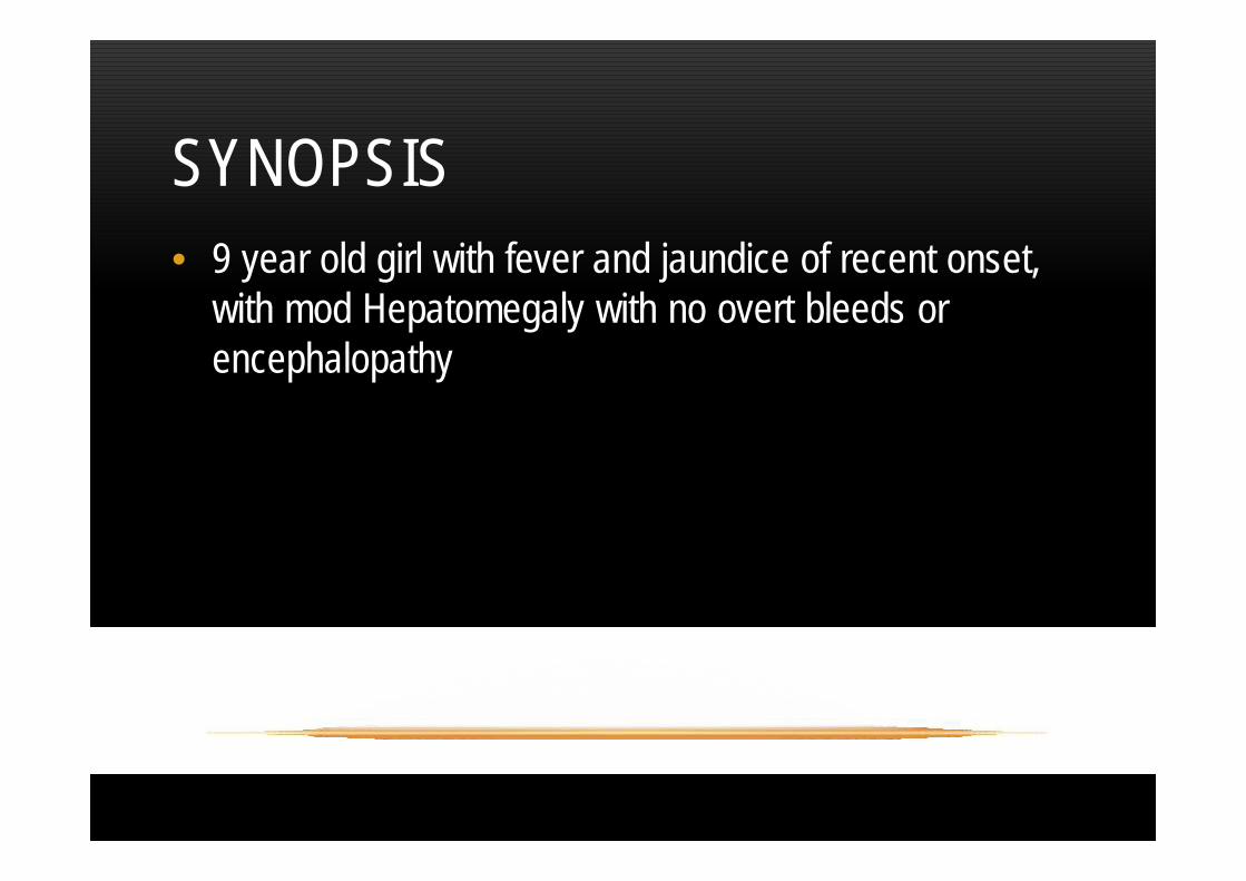
SYNOPSIS
• 9 year old girl with fever and jaundice of recent onset, with mod Hepatomegaly with no overt bleeds or encephalopathy
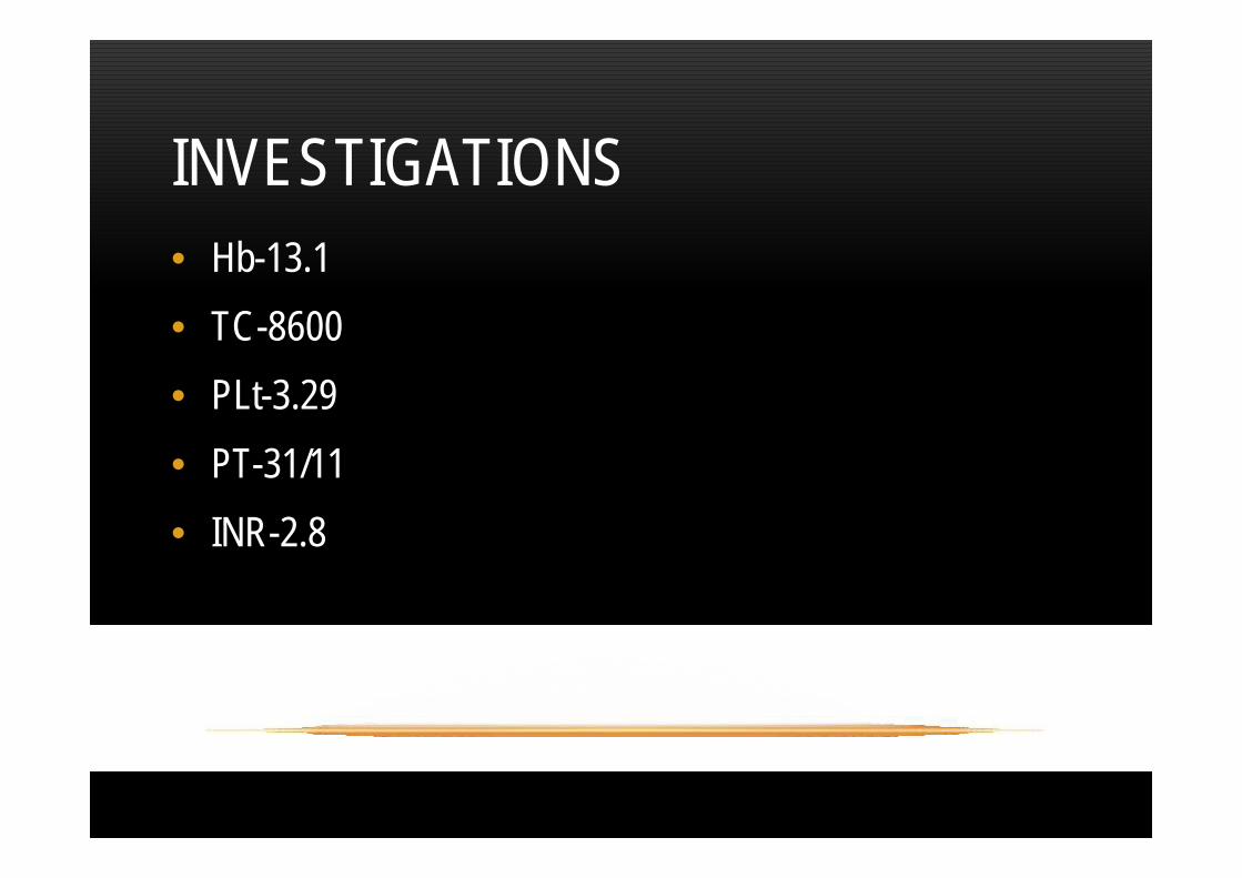
INVESTIGATIONS
• Hb-13.1
• TC-8600
• PLt-3.29
• PT-31/11
• INR-2.8

LIVER FUNCTION• Total bili - 26.5 (upto1mg/dl)
• Direct bili - 18.2 (upto0.2mg/dl)
• Total protein - 6 (6-8g/dl)
• Albumin - 3.6 (3.8-5.4g/dl)
• SGOT - 975 (15-55U/L)
• SGPT - 725 (5-45U/L)
• ALP - 376 (<300U/L)
• GGT - 28 (5-32)
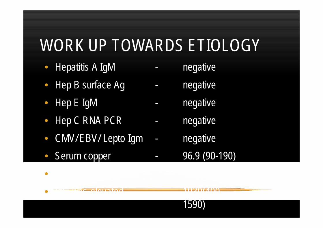
WORK UP TOWARDS ETIOLOGY• Hepatitis A IgM - negative
• Hep B surface Ag - negative
• Hep E IgM - negative
• Hep C RNA PCR - negative
• CMV/ EBV/ Lepto Igm - negative
• Serum copper - 96.9 (90-190)
• Serum ceruloplasmin - 32 (20-60)
• IgG was elevated - 1930(400-1590)

• USG abdomen – mildly enlarged liver, prominence with thickening of intra hepatic biliary radicles in both lobes. Multiple, enlarged peripancreatic and periportal LN noted, s/o active hepatitis
• Bilateral renal pelvic prominence, rest normal

• Cultures - no growth
• ANA - negative
• ANCA - negative
• ASMA - negative
• Liver biopsy - deferred in view of high PT /INR

THE DIAGNOSTIC INVESTIGATION??
Liver
Immunity

• DCT - positive
• Anti LKM - positive
• Anti LC-1 - positive
• Anti M2-PDH - negative
• Anti SLA - negative

DIAGNOSIS
• AUTO IMMUNE HEPATITIS

DEFINITION
chronic hepatic inflammatory process
• elevated serum aminotransaminase concentrations
• liver associated serum autoantibodies
• hypergammaglobulinemia

EPIDEMIOLOGY
§ INCIDENCE: 1.9 cases per 100,000 persons per yr
§ PREVALENCE: 16.9 cases per 100,000 persons per yr
§ Females account for 70% of cases, 50% £ 40 years
§ Cause of chronic liver disease: 11-23%
§ AIH accounts for 2.6% and 5.9% of liver transplants in Europe and U.S. respectively

NATURAL HISTORY
§ Severe disease (untreated)
• 40% die within 6 months of diagnosis
• 40% of survivors develop cirrhosis
• 54% of cirrhotics develop varices within 2 years of diagnosis of cirrhosis
• 20% of patients with varices will bleed

PATHOGENESIS
AIH
AutoantigensImmuno-
regulatory
Geneticfactors
Triggeringfactors

§ Genetic factors
• Antigen presentation/immunocyte activation
• DRB1 encodes for MHC II antigen binding grooves (antigen presentation to T cells)
§ Triggering factors
• Infections (HAV, HBV, HCV, HSV, EBV, measles)?
• Medications (ABX, statins, NSAIDs etc.)?
• Toxins?
• Molecular mimicry?

§ Antibody-dependent cellular cytotoxicity
• Antibodies directed against ASGPR
• Suppressor T cell defect
• Binding of NK cell to antigen-antibody complex followed by hepatocyte destruction
§ Cell-mediated cytotoxicity
• IL-12 and IL-2 released
• Aberrant display of MHC class II
• CD8 T cell destruction of hepatocyte

Biochemical Histological
Gamma globulinAutoantibody
Interface hepatitisPortal plasma cell

§ Gender
§ AP/AST, ALT ratio
§ Serum globulins/IgG
§ ANA, ASMA, LKM-1
§ AMA positive
§ Viral serologies
§ Drug history/Alcohol intake
§ Liver histology
§ Other autoimmune diseases
§ HLA DR3/DR4
§ Response to therapy
INTERNATIONAL AUTO IMMUNE HEPATITIS GROUP CRITERIA
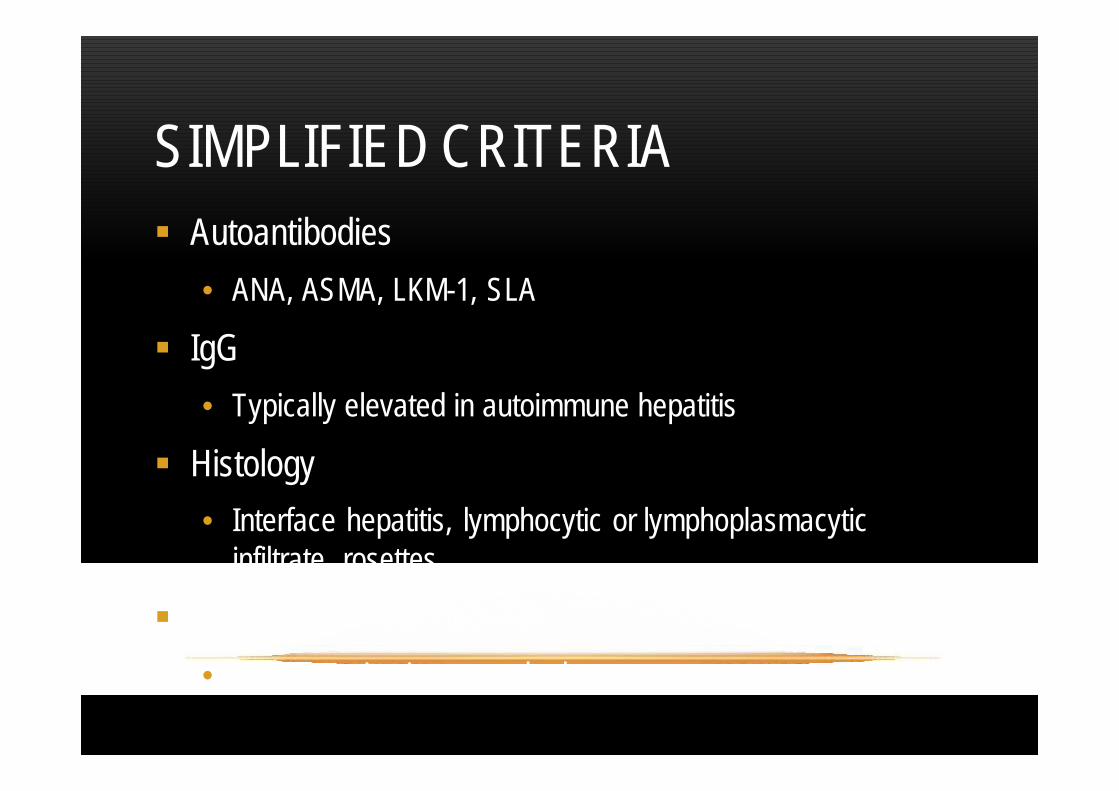
SIMPLIFIED CRITERIA
§ Autoantibodies
• ANA, ASMA, LKM-1, SLA
§ IgG
• Typically elevated in autoimmune hepatitis
§ Histology
• Interface hepatitis, lymphocytic or lymphoplasmacyticinfiltrate, rosettes
§ Exclusion of viral hepatitis
• Hepatotropic viruses and others

AIH TYPE 1
§ Age: 10-20yrs
§ Female: 78%
§ g-globulin elevation: marked
§ Autoantigen: asialoglycoprotein receptor?
§ Autoantibodies: ANA, ASMA
• Others: pANCA, actin, ASGPR, SLA/LP
§ HLA: A1-B8-DR3 or HLA DR4 serotypes
§ Extrahepatic autoimmune disease: 15-40%

AIH TYPE 2
§ Age: 2-14 years
§ Female: 90%
§ g-globulin elevation: Mild
§ Autoantigen: CYP450 IID6
§ Autoantibodies: LKM-1
• Others: LC-1, SLA/LP
§ Extrahepatic autoimmune disease: 40%
§ Severity: more severe than type 1?

HISTOLOGY§ Piecemeal necrosis (interface hepatitis)
§ Panacinar inflammation or collapse
§ Lymphoplasmacytic infiltrates
§ Eosinophils
§ Rosette formation
§ Fibrosis or cirrhosis
§ Absence of portal lymphoid aggregates and steatosis
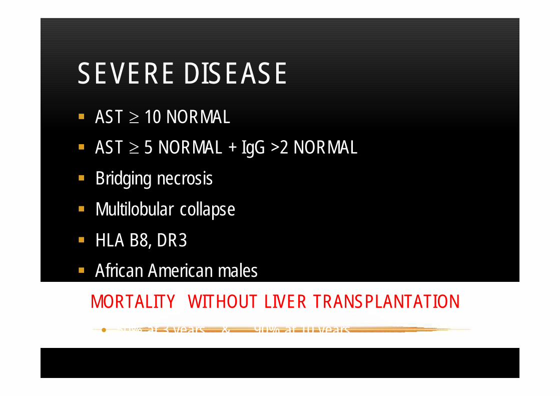
SEVERE DISEASE
§ AST ³ 10 NORMAL
§ AST ³ 5 NORMAL + IgG >2 NORMAL
§ Bridging necrosis
§ Multilobular collapse
§ HLA B8, DR3
§ African American males
MORTALITY WITHOUT LIVER TRANSPLANTATION
• 50% at 3 years & 90% at 10 years

HITOLOGY AND PROGNOSIS
§ Interface hepatitis
• 17% risk of cirrhosis at 5 years
• Normal survival
§ Bridging or multilobular necrosis
• 82% risk of cirrhosis at 5 years
• 45% 5-year mortality
§ Cirrhosis
• 58% 5-year mortality
TREATMENT OPTIONS
IMMUNOSUPPRESANTS
• STEROIDS
• AZATHIOPRINE

• Prednisone
• initial dose of 1-2 mg/kg/24 hr - UNTIL aminotransferase values return to less than twice the upper limit of normal.
• The dose should then be lowered in 5 mg decrements
• over 2-4 mo until a maintenance dose of 0.1-0.3 mg/kg/24 hr is achieved.

SECOND LINE AGENTS (CENTRE BASED PROTOCOL)
• Mycophenolate
• Cyclosporine
• Tacrolimus
• Budesonide
• Methotrexate
• Cyclophosphamide

REMISSION
• Disappearance of symptoms
• Normalization or near normalization of AST to < 2 normal
• IgG and bilirubin: normal
• Minimal or no hepatic inflammation
• 65% and 80% of patients within 18 months and 3 yrs of initiation of Rx respectively
• 10 year survival: 90%

TREATMENT END POINTS
• Disease remission
• Relapse after treatment withdrawal
• Treatment failure
• Incomplete response
• Drug toxicity

LIVER TRANSPLANTATION
• In End-stage liver disease
• In Fulminant liver disease
• Results• 5 yr pt and graft survival: 80-90%
• Recurrence: 15-40%
• High rates of rejection
COURSE OF MS A.
• Clinically well
• Started on 2mg/kg prednisolone – slowly tapered and is on 0.2mg/kg/day dose
• On follow up for 9 months now.
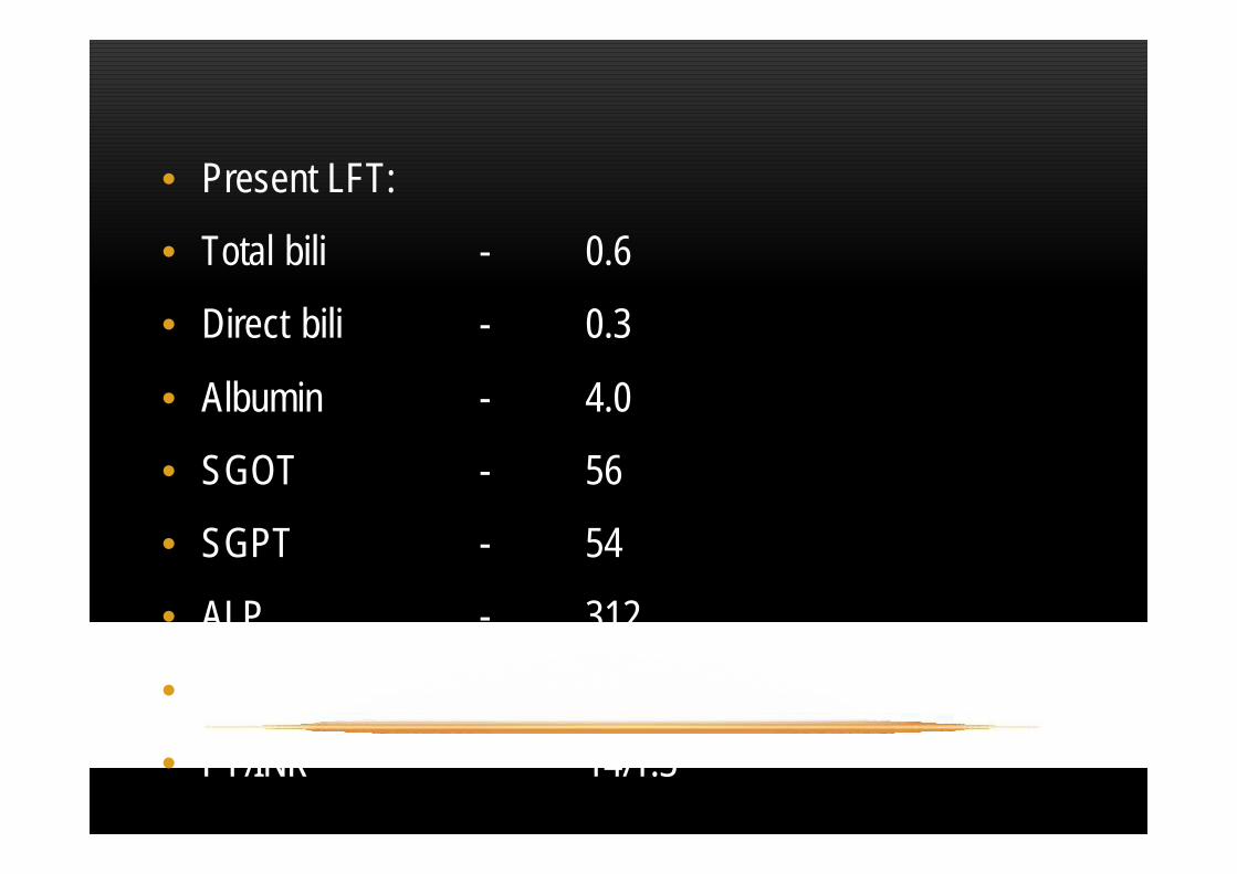
• Present LFT:
• Total bili - 0.6
• Direct bili - 0.3
• Albumin - 4.0
• SGOT - 56
• SGPT - 54
• ALP - 312
• GGT - 32
• PT/INR - 14/1.3
TAKE HOME MESSAGE
• Clinical presentation is variable
• Diagnosis based upon LFTs, serology, gamma
globulins, and histology
• Early diagnosis is crucial (death if undiagnosed)
• Timely referral to the specialist (esp aculte liver failure
or coag not correcting with vitamin k)
• Immunosuppressive therapy is the mainstay of
treatment

THANK YOU


