An introduction to the Globus Pallidus in schizophrenia
An introduction to the Globus Pallidus in schizophreniaDr Matthew Williams
Visiting researcher, Rob Steiner Unit, Hammersmith Hospital, London W12 0NN
Received – 30 August 2017; Accepted – 10 October 2017
A B S T R A C T
The Globus Pallidus is a bilateral grey matter nucleus found at the medial part of the basal ganglia. Whilst a single physical structure it is split into two distinct functional nuclei, which are in adjacent and complementary regulatory circuits involving the striato-thalamic and striato-tegmental systems. The connections of the Globus Pallidus make it a critical structure in controlling both motor systems and midbrain dopamine systems, potentially having a key role in controlling dopamine-regulated psychosis in schizophrenia and other disorders. Imaging studies have shown contradictory findings in Globus Pallidus size and shape, and it is unclear to what extent this is due to disease burden or antipsychotic medication. Whilst the Globus Pallidus is a little-studied structure in schizophrenia research but has important roles in striatal, thalamic and dopaminergic regulation, and could potentially be a key part in further development of drug therapies.
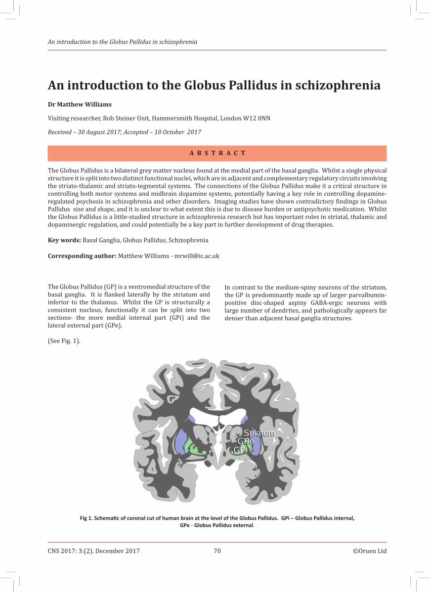
The Globus Pallidus (GP) is a ventromedial structure of the basal ganglia. It is flanked laterally by the striatum and inferior to the thalamus. Whilst the GP is structurally a consistent nucleus, functionally it can be split into two sections- the more medial internal part (GPi) and the lateral external part (GPe). (See Fig. 1).
In contrast to the medium-spiny neurons of the striatum, the GP is predominantly made up of larger parvalbumin-positive disc-shaped aspiny GABA-ergic neurons with large number of dendrites, and pathologically appears far denser than adjacent basal ganglia structures.
Fig 1. Schematic of coronal cut of human brain at the level of the Globus Pallidus. GPi – Globus Pallidus internal, GPe - Globus Pallidus external.
An introduction to the Globus Pallidus in schizophrenia
The GPe, structurally lying immediately medial to the striatum, predominantly receives inputs directly from the striatum by means of a pronounced GABA-ergic pathway, making up the majority of synaptic nuclei within this structure. The primary output of the GPe is composed of GABA-ergic projections to the Sub-Thalamic Nucleus (STN), located inferior of the GPi. In turn, the STN has a substantial glutamatergic projection to the GPi, tracing an indirect pathway from the striatum via GPe.
The GPi is found more medial than the GPe but still receives a similar direct GABA-ergic inhibitory cluster of projections directly from the striatum. The GPi has two major outputs, the first to the thalamus via two pathways. The lenticular fasciculus is one output pathway which projects via the internal capsule while the ansa lenticularis projects ventrally around the internal capsule. These meet at the thalamic fasciculus and terminate in the ventral anterior and ventrolateral parts of the thalamus, which in turn project excitatory glutamatergic pathways to the premotor and frontal cerebral cortices, areas involved in cognition, planning and initiation of movement. This feedback pathway is involved in regulating the level of excitation in the premotor cortex, and thus giving a mechanism for basal-thalamic-cortical regulation of movement. The second major output is a cluster of glutamatergic outputs to the reticulata region of the Substantia Nigra, which is
Fig. 2. Circuit diagram of the major connections of the GP. Blue – inhibitory pathways, Red – excitatory pathways. STN – Sub-Thalamic Nucleus, SNr – Substantia Nigra pars reticulata, GABA – Gamma-Amino-Butyric Acid, GLU – Glutamate.
suggested to regulate nigral dopamine release, and thus provide a feedback loop for the nigrostriatal tract. Various experimental manipulations, including microdialysis in rodent and PET in primate models, have shown a role of the GP and STN in regulating somatodendritic dopamine release in the SN under normal and experimental conditions (Cobb and Abercrombie 2003; Perez-Costas, Melendez-Ferro and Roberts 2010), and probabilistic computation has added to the anatomical argument that the GP is the critical regulatory step from striatal output to the thalamus (Csernansky and Cronenwett 2008).
The returning cortical input to the striatum activates inhibitory neurons to the GP, decreasing activity in the tonically-active neurons in the GP which then decrease the inhibitory action on ventral anterior and ventrolateral parts of the thalamus. The conclusion of this pathway is therefore disinhibition of thalamic excitation of the cerebral cortex, and thus greater activation of the cortex. In the case of the motor cortex activation of the direct pathway would increase the ease of initiation and ease of movement. The GP is also connected with structures involved in reward circuitry such as the nucleus accumbens, habenula, and the ventral tegmental area, although further research is required to identify the GP specific role in these circuits (Hong and Hikosaka 2008, Miller et al. 2006). A schematic of the primary GP pathways is shown in Fig. 2.
An introduction to the Globus Pallidus in schizophrenia
Structural MRI imaging studies have shown conflicting results, with studies often showing no overall change in size in the GP in schizophrenia, but a change in shape, whilst others suggest that GP size is changed. One analysis of basal ganglia relative size and shape suggests that the GP was significantly larger overall, with another study showing increased GP size and altered shape in schizophrenia on the right side only, a lateral change which seems unique within the basal ganglia. Enlargement of basal ganglia structures has typically been found to be related to antipsychotic medication, although the study finding the lateralised effect found no such effect of medication taken at time of study (Mamah et al. 2007, Womer et al. 2014, Hutcheson et al. 2014, Shenton et al. 2001).
The size increase in GP in schizophrenia has been suggested to be related to the total psychotic symptoms. One study showing GP enlargement in schizophrenia with psychosis also showed GP shrinkage in schizophrenia cases without psychosis, with the GP smaller even than unaffected controls (Womer et al. 2014). Dysfunctional interhemispheric connections of the GP has been prosed as the primary site for cognitive disturbance in first-episode schizophrenia measuring negative symptoms and cognitive impairment in functional MRI examination (Mwansisya et al. 2013).
Despite the intriguing imaging results and the key role the GP plays in regulating striatonigral and striatothalamic circuits, direct neuropathological investigation of the GP are thin on the ground in schizophrenia. One landmark study has reported that the GPi decreased in volume by 20% whilst the GPe was not changed when systematically examined in sections through the structure using histological staining (Bogerts, Meertz and Schonfeldt-Bausch 1985). Interestingly, one early study suggested increased iron, termed ‘mineralization’, of the GP in schizophrenia, although follow-up studies failed to replicate this effect (Stevens 1982, Casanova et al. 1990a, Casanova, Waldman and Kleinman 1990b). As has previously been mentioned, animal studies using microdialysis have revealed that the striatopallidal pathway from to both GP nuclei regulates Nigral and VTA dopaminergic cells. Although so far direct investigation of binding potential change of D2 receptor in the pallidus using PET show inconclusive results in both schizophrenic patients and high-risk individuals (O’Connor 2001, Suridjan et al. 2013).
However high-risk individuals have shown more interesting results in other studies. Teenagers at high-risk of developing schizophrenia have left GP size increased by a small but significant average of 1% compared to non-high-risk controls, with more a pronounced increase with age. This suggests that the increased size may predate schizophrenia first-onset and GP enlargement may not be down entirely to antipsychotic action (Dougherty et al. 2012).In a similar finding, voxel-based morphometry examination with structural MRI has shown increased grey matter in
the GP in first-degree relatives of schizophrenia patients, as well as those patients themselves (Oertel-Knöchel et al. 2012). GP deformation was also observed in unaffected relatives of schizophrenia patients, albeit to a lesser degree than those suffering with schizophrenia (Womer et al. 2014).
Although the role of the GP in modulating striatal output to the thalamus and midbrain has good supporting evidence, the GP’s role in other, less prominent circuits is not well understood. There have been few good quality studies to investigate the cognitive- and movement-related role of this structure in both normal controls and schizophrenia patients, and a woeful lack of direct examination at the cellular level using any type of pathological techniques. Additionally, the problems of interpreting the results we have obtained are compounded by the known effects of antipsychotic medication. This makes the GP a prime candidate for further examination in schizophrenia from both a basic neuroscientific stance and in future drug development.
REFERENCES.Bogerts, B., E. Meertz & R. Schonfeldt-Bausch (1985) Basal ganglia and
limbic system pathology in schizophrenia. A morphometric study of brain volume and shrinkage. Arch Gen Psychiatry, 42, 784-91.
Casanova, M. F., C. M. Prasad, I. Waldman, B. Illowsky, B. Stein, D. R. Weinberger & J. B. Kleinman (1990a) No difference in basal ganglia mineralization between schizophrenic and nonschizophrenic patients: a quantitative computerized tomographic study. Biol Psychiatry, 27, 138-42.
Casanova, M. F., I. N. Waldman & J. E. Kleinman (1990b) A postmortem quantitative study of iron in the globus pallidus of schizophrenic patients. Biol Psychiatry, 27, 143-9.
Cobb, W. S. & E. D. Abercrombie (2003) Relative involvement of globus pallidus and subthalamic nucleus in the regulation of somatodendritic dopamine release in substantia nigra is dopamine-dependent. Neuroscience, 119, 777-86.
Csernansky, J. G. & W. J. Cronenwett (2008) Neural networks in schizophrenia. Am J Psychiatry, 165, 937-9.
Dougherty, M. K., H. Gu, J. Bizzell, S. Ramsey, G. Gerig, D. O. Perkins & A. Belger (2012) Differences in subcortical structures in young adolescents at familial risk for schizophrenia: a preliminary study. Psychiatry Res, 204, 68-74.
Hong, S. & O. Hikosaka (2008) The globus pallidus sends reward-related signals to the lateral habenula. Neuron, 60, 720-9.
Hutcheson, N. L., D. G. Clark, M. S. Bolding, D. M. White & A. C. Lahti (2014) Basal ganglia volume in unmedicated patients with schizophrenia is associated with treatment response to antipsychotic medication. Psychiatry Res, 221, 6-12.
Mamah, D., L. Wang, D. Barch, G. A. de Erausquin, M. Gado & J. G. Csernansky (2007) Structural analysis of the basal ganglia in schizophrenia. Schizophr Res, 89, 59-71.
Miller, J. M., S. R. Vorel, A. J. Tranguch, E. T. Kenny, P. Mazzoni, W. G. van Gorp & H. D. Kleber (2006) Anhedonia after a selective bilateral lesion of the globus pallidus. Am J Psychiatry, 163, 786-8.
Mwansisya, T. E., Z. Wang, H. Tao, H. Zhang, A. Hu, S. Guo & Z. Liu (2013) The diminished interhemispheric connectivity correlates with negative symptoms and cognitive impairment in first-episode schizophrenia. Schizophr Res, 150, 144-50.
O’Connor, W. T. (2001) Functional neuroanatomy of the ventral striopallidal GABA pathway. New sites of intervention in the treatment of schizophrenia. J Neurosci Methods, 109, 31-9.
Oertel-Knöchel, V., C. Knöchel, S. Matura, A. Rotarska-Jagiela, J. Magerkurth, D. Prvulovic, C. Haenschel, H. Hampel & D. E. Linden (2012) Cortical-basal ganglia imbalance in schizophrenia patients and unaffected first-degree relatives. Schizophr Res, 138, 120-7.
An introduction to the Globus Pallidus in schizophrenia
Perez-Costas, E., M. Melendez-Ferro & R. C. Roberts (2010) Basal ganglia pathology in schizophrenia: dopamine connections and anomalies. J Neurochem, 113, 287-302.
Shenton, M. E., C. C. Dickey, M. Frumin & R. W. McCarley (2001) A review of MRI findings in schizophrenia. Schizophr Res, 49, 1-52.
Stevens, J. R. (1982) Neuropathology of schizophrenia. Arch Gen Psychiatry, 39, 1131-9.
Suridjan, I., P. Rusjan, J. Addington, A. A. Wilson, S. Houle & R. Mizrahi (2013) Dopamine D2 and D3 binding in people at clinical high risk for schizophrenia, antipsychotic-naive patients and healthy controls while performing a cognitive task. J Psychiatry Neurosci, 38, 98-106.
Womer, F. Y., L. Wang, K. I. Alpert, M. J. Smith, J. G. Csernansky, D. M. Barch & D. Mamah (2014) Basal ganglia and thalamic morphology in schizophrenia and bipolar disorder. Psychiatry Res, 223, 75-83.


![Globus Pallidus Internus Deep Brain Stimulation for …downloads.hindawi.com/journals/crinm/2017/2165905.pdfEffective 9mo. PersistentHHinoffstimulation Gotoetal.[7],2010 1 78/f N/A](https://static.documents.pub/doc/80x56/5e3e2094a4093825cd258b9d/globus-pallidus-internus-deep-brain-stimulation-for-effective-9mo-persistenthhinoffstimulation.jpg)


