An objective measure for the assessmentand management of fluid shifts in acutemajor burnsPippa Kenworthy1,3,6,7* , Michael Phillips5, Tiffany L. Grisbrook1,2, William Gibson6, Fiona M. Wood1,3
and Dale W. Edgar1,3,4
Abstract
Background: Major burns are life threatening. Fluid resuscitation is required for survival to maintain intravascularvolumes and prevent hypovolemic shock. Bioimpedance spectroscopy (BIS) has been recognised as a potentialmethod of monitoring fluid shifts after burn and in other disease states. The aims of this study were to examine thereliability of BIS across different dressing conditions and electrode positions, establish the influence of Acticoat™ onBIS variable measures and determine the validity of whole-body BIS to assess net fluid shift in the presence ofmoderate to major burns.
Methods: An observational longitudinal cohort study was conducted from December 2014 to February 2016.Patients with over 15% total body surface area (TBSA) burns and injury less than 48 h were enrolled in the study.BIS triplicate measures were collected in an open wound and with an ActicoatTM dressing (at 5 half hour intervals).Standard and alternate electrode placements were utilised for the reliability analysis and standard placement onlyfor determining the validity of BIS in moderate to major burns. The ImpediMde SFB7 was used to collect whole-body and segmental BIS measures. Stata statistical software, release 14 was utilised to analyse all results. Descriptiveanalyses were performed and were reported using the means and standard deviations (SD).
Results: BIS-repeated measures established BIS raw resistance (R), and predicted volume variables were reliable inany condition (intra-class correlation coefficient (ICC) 0.996–0.999, 95% confidence intervals (CI) 0.996–0.999) without asystematic difference. Acticoat™ dressings significantly influenced all BIS-predicted volumes (p≤ 0.01) as determined bymultilevel mixed effects (MLME) linear regression analysis. Validity of BIS was demonstrated by resistance variablessignificantly decreasing with increasing net ionic fluid shift and increased TBSA (severity of injury) and calculated fluidvolumes increasing with increasing net fluid shift and TBSA. BIS resistance also decreased with time as oedemareduced. For clinical use, a calculator was developed to adjust BIS variables when an Acticoat™ dressing is in situ,thus facilitating BIS variable change estimates in real time, with dressings intact.
Conclusion: BIS may be used clinically to monitor fluid volume change in major acute burns.
* Correspondence: [email protected] Wood Foundation, Fiona Stanley Hospital, Perth, Western Australia,Australia3Burns Service of Western Australia, Fiona Stanley Hospital, Perth, WesternAustralia, AustraliaFull list of author information is available at the end of the article
BackgroundLarge fluid shifts and local and distant tissue swellingare features of burn injuries. Swelling hampers burnwound healing and the volume created is directly relatedto the size and depth of the burn [1]. Major burnsgreater than 15–20% total body surface area (TBSA)with a depth of partial to full thickness result in both alocal and systemic inflammatory response [2, 3]. Thiscan be a life-threatening scenario which requires formalfluid resuscitation. Acute burn fluid resuscitation is vitalin decreasing patient morbidity and mortality in the first24–48 h of injury but can contribute to already largeamounts of oedema [4].Despite the importance of fluid resuscitation in the early
management of traumatic burn injuries, there is currentlyno single, simple, non-invasive and accurate outcomemeasure which can assist clinicians to titrate fluid volumesin acute burns or monitor the effect of treatments onswelling. Thus, the objective, timely adjustment of fluidresuscitation is challenging, particularly when patients arenot supported by critical care and invasive monitoring.This research investigates the accuracy of bioimpedancespectroscopy (BIS) in monitoring whole-body fluid vol-ume and oedema change in moderate to large acuteburns.There has been little advancement in the area of burn
fluid resuscitation over the last 30 years [4] and in recenttimes, there has been a trend to over resuscitate patients[5, 6], necessitating a descriptor known as fluid creep. Ex-cess fluid can contribute to burn wound progression, lead-ing to complications such as peripheral and abdominalcompartment syndromes, pulmonary oedema and periph-eral tissue oedema. Any one or a combination of these willaffect patient recovery and increase medical costs and islikely to increase patient length of stay [3, 7–10].Fluid resuscitation formulas such as the Parkland
and Brookes are used to instigate intravenous (IV)fluid rates but are guidelines only, and fluid mustthen be titrated according to particular endpoints ofresuscitation [11–13]. The most commonly used out-come measure for fluid therapy is urine output, withthe aim to maintain a rate of 30–50 ml per hour foran average-sized man while preserving haemodynamicproperties such as oxygen saturation and blood pres-sure [5, 14]. There are other objective measures toguide volume titration however they are invasive andnot without limitations [6, 14, 15].BIS has historically been used in healthy populations
to measure body composition. However, in the last20 years, it has gained increasing popularity in clinicalpopulations and is now commonly used to measure armlymphoedema post breast surgery [16] and dry weight inhaemodialysis patients [17, 18]. BIS has demonstratedsensitivity, high reliability (repeatability) of measures in a
number of clinical areas [19]. The method has also beenvalidated (determined credible) in both healthy and clin-ical populations against magnetic resonance imaging(MRI) and bromide and potassium dilution techniques,which are considered gold standard in the assessment offluid compartment volumes and lean body mass (LBM)[20–23]. It can investigate the body’s physiological pa-rameters such as extracellular fluid (ECF), intracellularfluid (ICF) and total body fluid (TBF). It achieves thisby passing a small alternating current, over a numberof frequencies (4–1000 kHz), through the tissues andfluid compartments of the body via electrodes on in-tact skin. It provides instantaneous measures of resist-ance (R) and reactance (capacitive resistance (Xc)).Resistance is the opposition to flow of an electriccurrent, is reflective of the body’s water compart-ments and is inversely proportional to fluid volumeand therefore oedema [24, 25]. Capacitance is thedelay in the passage of current through the cell mem-branes and tissue interfaces [25]. The current flow isfrequency (Hz) dependent and varies according to thecomposition of the body [26]. Resistances at zero(R0) and infinite frequencies (Rinf ) (considered idealmeasurement frequencies) are estimated utilising theCole-Cole plot embedded in the BIS software, due tothe constraints of using a direct or very high fre-quency alternating current in humans [27]. The R0
and Rinf [25] are representative of ECF and TBF re-spectively. Resistance (Ri) of the ICF is extrapolatedusing the other raw variable data. At low frequencies,the current can penetrate the ECF only, and at highfrequencies, it passes through both the ECF and ICFmeasuring TBF.The ability of BIS to quantify individual body fluid
compartments, the ease of use and non-invasive naturehas led to a small number of papers examining its use inthe burn population. Miller et al. [28] and Zdolsek et al.[29] were able to determine the development of oedemapost burn injury but each study lacked power and nei-ther was able to provide statistical conclusions regardingthe reliability of BIS in the burns populace. In 2009,Edgar et al. demonstrated whole-body bioimpedancespectroscopy was a reliable means of quantifying real-time oedema shifts in patients with burns less than 30%TBSA across numerous dressing conditions [30]. How-ever, the study only had six participants with burnsgreater than 15% TBSA and was therefore inconclusivein this subset of patients. Further, each study utilisedstandard whole-body electrode positions only and it isunknown whether alternate electrode positions, for bothwhole-body and limb segmental BIS, are reliable in thisparticular population. Grisbrook et al. (2015) investi-gated whether alternate electrode configuration BISmeasurements were interchangeable with standard
Kenworthy et al. Burns & Trauma (2018) 6:3 Page 2 of 12
electrode configurations in the healthy population butreliability was not determined [31]. In Edgar et al’s(2009) study [30], it was also apparent that the dressingcondition affected the sensitivity of the BIS results.Bioimpedance measures were found to be less sensitivein older dressings (> 8 h old) than in an open wound ornew dressing condition.Dressing-type may pose a further challenge in the
assessment of fluid shifts by BIS. Acticoat™ (Smith &Nephew) is an antimicrobial dressing, composed ofnanocrystalline silver particles [32]. It is the standarddressing used in the first 48 h of burn care, and asindicated after, in the Burn Service of WesternAustralia (BSWA). Understanding that BIS measuresthe resistance of the body’s tissues and inter-compartmental fluid volumes by introducing a lowamplitude electrical current into the body, it wouldnot be unexpected that Acticoat™ may affect the BISmeasures. Silver is a highly conductive material, andsuch dressings release ionic silver species and are ap-plied in a wet condition. Both the silver ions and wetcondition would therefore be expected to reduce theBIS resistance measured, thus potentially limiting theuse of monitoring fluid shifts with BIS in acute burnspatients.To extend Edgar et al.’s (2009) [30] reliability study
and on the premise that BIS can reliably quantify tissuefluid, it was hypothesised BIS would provide a methodfor real-time accurate measures of fluid shifts in theacute major burn. The study aimed to examine the reli-ability with respect to dressing condition and electrodeposition, investigate the influence of Acticoat™ on BISvariable outputs and determine the validity of whole-body BIS to assess net fluid shift in the presence of mod-erate to major burns, greater than 15% TBSA.
MethodsParticipantsAn observational longitudinal cohort study was con-ducted from December 2014 to February 2016. Patientswere recruited into the study if they were over 18 yearsold and receiving formal fluid resuscitation and had aflame and/or scald burn, and the injury was less than48 h old. The BSWA medical team instigates fluid resus-citation for partial to deep thickness burns greater than15% TBSA (modified however based on each individualsclinical presentation and nutritional status at admission)and uses Ringer’s Lactate (crystalloid) solution with vol-umes initially determined by the modified Parkland’sformula. Fluid volumes were titrated to maintain anadequate urine output of 0.5–1.0 ml/kg/h for the first36–48 h after burn injury. Participants were excludedfrom the research if they had hand and/or feet burnsprecluding placement of standard whole-body electrode
placement and body mass index (BMI) ≤ 15 and ≥ 40 kg/m2 (manufacturer’s guidelines) and if they met ImpedimedSFB7 (ImpediMed, Brisbane, Queensland, Australia) man-ufacturer’s contraindications which includes pregnant orbreast-feeding patients, patients with surgical implants,cardiac pacemakers and/or are on electronic life supportdevices (ventilated patients).Burn inpatients were recruited initially from the Burn
Unit at Royal Perth Hospital (RPH) and then at FionaStanley Hospital (FSH) due to the transition of theadult care of the BSWA to the new FSH. There was nochange to the study protocol or equipment used in thestudy.
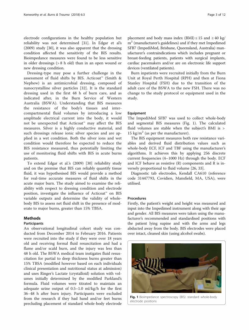
EquipmentThe ImpediMed SFB7 was used to collect whole-bodyand segmental BIS measures (Fig. 1). The calculatedfluid volumes are stable when the subject’s BMI is >15 kg/m2 (as per the manufacturer).The BIS equipment measures both raw resistance vari-
ables and derived fluid distribution values such aswhole-body ECF, ICF and TBF using the manufacturer’salgorithms. It achieves this by applying 256 discretecurrent frequencies (4–1000 Hz) through the body. ECFand ICF behave as resistive (R) components and R is in-versely proportional to fluid volume [26, 33].Diagnostic tab electrodes, Kendall CA610 (reference
ProceduresFirstly, the patient’s weight and height was measured andinput into the Impedimed instrument along with their ageand gender. All BIS measures were taken using the manu-facturer’s recommended and standardised positions withthe patient lying supine and with the arms and legsabducted away from the body. BIS electrodes were placedover intact, cleaned skin (using alcohol swabs).
Fig. 1 Bioimpedance spectroscopy (BIS): standard whole-bodyelectrode positions
Kenworthy et al. Burns & Trauma (2018) 6:3 Page 3 of 12
Electrode configurationsStandardised tetrapolar electrode placements (EP) wereutilised [25, 34], and alternate electrode configurationswere placed based on the theory of equi-potentials (seeCornish et al. [34] for further details of equipotentialpoints) and were placed as per Grisbrook et al. [31].Electrodes were placed on intact skin only. Participantswith bilateral hand or foot injuries which precluded theapplication of standardised electrode placements wereexcluded. Bioimpedance measures were taken on theright side of the body unless precluded by wounds, thenthe left side was utilised. The location of their woundsdetermined whether all other electrode placements (seg-mental) could be used and measured.BIS measures were taken in triplicate in an open
wound (time point 0 (T0)) and in the new Acticoat™dressing condition at five half-hour intervals (T1–T5)after the baseline measure, i.e. five measures in total(Fig. 2). The time between T0 and T1 was recorded, asthis was unable to be standardised. Standard and alter-nate whole-body, upper limb segmental and lower limbsegmental BIS measures were taken at T0–T1. Standardwhole-body EP’s only were utilised at T2–T5 (Fig. 2).Burn wounds often prevent electrodes being applied inthe standard position; therefore, alternative whole-bodyand limb segment electrode positions were utilised asable at T0–T1 and their reliability investigated. The datato determine the validity of alternate electrode place-ment has been analysed separately [35]. The segmentalmeasures were included in the reliability analysis only.The effect of Acticoat™ on whole-body BIS resultswas determined from T0-T1 BIS readings. Electrodesremained in situ between triplicate measures where
possible, unless prohibited by dressing changes or ad-hesive loss.Net fluid shift was recorded between each time point
(T1–T5), in conjunction with the BIS measures. Netfluid shift was calculated by subtracting urine outputand other bodily fluid output recorded (e.g. emesis) fromfluid intake (IV and oral fluids and food).The researcher was blinded to all BIS measurements
as only a file name was viewed and recorded, not theactual BIS values.
Data analysisStata statistical software, release 14 (StataCorp LP 2014,College Station, TX), was utilised to analyse all results.Descriptive analyses were performed and were reportedusing the means and standard deviations (SD).
ReliabilityA three-level nested mixed effects linear regression wasperformed to examine the reliability of the BIS triplicatemeasures, taking into account random effects of con-founders of electrode position, time and dressing condi-tion. The multilevel mixed effects (MLME) linearregression also explored whether there was a significantwithin-session difference between the triplicate measuresfor each of the BIS variables. Reliability is presented asthe intra-class correlation coefficient (ICC) (acceptable,0.75–0.89, excellent ≥ 0.9) [36], variance indicated by95% confidence intervals (CI) and systematic biasbetween within session trial measures (p < 0.05 consid-ered significant). All BIS triplicate measures were usedin the analysis.
Fig. 2 Consort diagram-flow diagram of data collection process. ICU Intensive care unit, BIS, Bioimpedance spectroscopy
Kenworthy et al. Burns & Trauma (2018) 6:3 Page 4 of 12
Analysis was completed using the MLME model as itcan account for random effects from individuals andresponses within individuals [37]. It is a robust methodproviding hierarchical analysis, adjusting for nested obser-vations of measures for each individual and giving the mostprecise and least biased estimates of treatment effects.Prior to interpreting the results of the MLME, severalassumptions were evaluated, confirming that each variablein the regression was approximately normally distributed.
Factors influencing BIS readingsThe effect of dressing condition, %TBSA and initial TBFon the BIS whole-body variables only was determined byMLME linear regression. A separate model was per-formed for each BIS variable. The interaction betweenActicoat™ and %TBSA and their influence on the BISvariables was also examined. The whole-body standardand alternate electrode placement BIS variable outputswere grouped together for use in the analysis for the effectof Acticoat™ and %TBSA. T0 (open wound) and T1 (newActicoat™ dressing) were used only.
ValidityValidity was determined using a series of MLME linear re-gression models including the data with the Acticoat™dressing condition only, and whole-body standard elec-trode placement (T1–T5) and alternate electrode place-ment (T1) only. The final model was produced bycompleting step-wise, backward elimination of predictorvariables on each of the dependent BIS variables. The finalmodel included %TBSA, time, net fluid shift and initialTBF volume. Initial TBF volume was derived from themean of the TBF measured with an open wound usingstandard tetrapolar whole-body electrode placement assingle-frequency bioimpedance analysis has been shownto measure TBF accurately in burns patients with nodressings [38]. This provided a baseline total body volume(L). A correlation matrix was performed to determine therelationship between initial TBF, weight and height andthe skewness-kurtosis test demonstrated that they wereeach normally distributed.Change scores or calculated difference of the BIS vari-
ables between time points (e.g. R0 at T1–R0 at T2) werenot used in the validity analysis, as the calculation of achange score requires measurement of the outcometwice, and in practice, it is proposed that it is more effi-cient to use a (single) change from baseline measure-ment to derive outcomes. In addition by not analysingchange (difference) data, the additive effect of the ran-dom errors is potentially reduced [39].
CalculatorA calculator was developed to estimate the net fluid shiftbetween consecutive BIS measures, when an Acticoat™
dressing is in place. Algorithms, for calculation of esti-mated fluid volumes were developed incorporating thesignificant and influential variables (on BIS variables)from the MLME models.
ResultsTwenty-one patients, 7 females and 14 males, wererecruited post burn injury. One patient had an incom-plete set of fluid recordings and two patients only hadrepeated measures completed four times in the newActicoat™ dressing condition. The mean net fluid shift(SD) at each time point, separated by ~ 30 min for T1–T5, were as follows: T1 174.72 ml (533.18), T2189.15 ml (164.23), T3 204.00 ml (135.37), T4 141.48 ml(253.25) and T5 123.20 ml (114.33). The average timebetween T0–T1 (SD) was 67 min (31). The mean TBF(SD) of patients on initial assessment was 46.06 L (9.71).Other patient data are presented in Table 1.
ReliabilityBIS triplicate measures were reliable within any elec-trode position, dressing condition and over time.Table 2 presents that BIS was a reliable measure inall circumstances, as confirmed by the ICC’s. Therewere no significant differences between the estimatedmeans of within session triplicate trial measures foreach of the BIS variables (i.e. no systematic bias)(Table 2). Final numbers included in each EP analysiswere whole body standard (WBS) (n = 21), whole bodyalternate (WBA) (n = 18), upper limb standard (ULS)(n = 14), upper limb alternate (ULA) (n = 14), lowerlimb standard (LLS) (n = 15), lower limb alternate(LLA) (n = 14).The means and CI for each of the BIS variables for the
standard whole-body electrode placement and timepoint are presented in Table 3.
Factors influencing BIS readingsThe regression analysis demonstrated Acticoat™ had a sig-nificant effect on the raw variables Ri and Rinf (but not R0)and on all the calculated variables (ECF, ICF, TBF) inwhole-body BIS (Table 4). The resistance variables re-duced between 182.22 and 23.87 Ω for Ri and Rinf, and thecalculated volumes were increased by 31.00–67.23 L whenan Acticoat™ dressing was in place, compared to the openwound condition.
Table 1 Patient data (n = 21)
%TBSA Age(years)
Recruitment postburn injury (h)
Height(cm)
Weight(kg)
24 (13) range12–80
36.4(13.5)
25 (11) 172.2 77.4(16.3)
Values presented as means (SD) ± rangeTBSA total body surface area
Kenworthy et al. Burns & Trauma (2018) 6:3 Page 5 of 12
There was no evidence of an effect of TBSA on any of theBIS variables (Table 4). However, there was a statistically sig-nificant interaction (p < 0.01) between %TBSA and Acticoat™for all BIS variables, raw and calculated. When an Acticoat™dressing was in place and for every 1% increase in %TBSAR0 decreased by 4.68 Ω, Ri by 17.98 Ω and Rinf by 3.96 Ω.This results in a divergence away from the open wound Rvalues as %TBSA increases. ECF, ICF and TBF volumes allincreased with greater %TBSA when an Acticoat™ dressingwas in place also resulting in divergence away from the openwound fluid volumes as %TBSA increased (Table 4).
As expected, there was a strong positive correlationbetween initial TBF and weight, with a correlationcoefficient (r) of 0.83 (p < 0.01). There was also amoderate positive correlation between initial TBF andheight, r = 0.67 (p < 0.01). Initial TBF was thereforeincluded in the model, and height omitted, to reducecollinearity. Initial TBF was included in preference toBMI as it was determined to be a more robust indica-tor of a person’s size as the random error was re-duced when compared to BMI (as it is one variablecompared to two (height and weight)). Initial TBF issignificantly associated with all BIS variables. Forevery 1 L increase in initial TBF, R0 decreased by5.71 Ω (p < 0.01), Ri decreased by 32.52 Ω (p < 0.01)and Rinf decreased by 5.30 Ω (p < 0.01). All estimatedfluid volumes increased (ECF 0.93 L, ICF 1.08 L, TBF2.02 L) with every 1 L increase in initial TBF.Algorithms were developed to correct for the effect of
Acticoat™ for the BIS variables. They are as follows:Corrected ECF =measured ECF with Acticoat dress-
ing – (− 59.02 + (time since dressing applied × 1.38)+ (initial measured ECF × 2.69))Corrected ICF =measured ICF with Acticoat dress-
ValidityBIS resistance and fluid volume variables were analysedto determine BIS validity. The MLME linear regressionunivariate analysis, in the Acticoat™ dressing conditiononly, showed R0, Ri and Rinf significantly changed withtime (Table 5). Compared to T1 (new Acticoat™ dress-ing), for every minute increase in time, R0 decreased by0.40 Ω (p < 0.01), Ri decreased 2.51 Ω (p < 0.01) and Rinf
ICC intraclass correlation coefficient, R0 resistance at zero frequency, Riintracellular resistance, Rinf resistance at infinite frequency, ECF extracellularfluid, ICF intracellular fluid, TBF total body fluid, BIS bioimpedancespectroscopy, CI confidence intervals*Each BIS measure coefficient is in reference to measure 1 of thetriplicate measures
Table 3 BIS variable values for the standard whole-body electrode placement and time point
BISvariableat WBS
Time point
T0 T1 T2 T3 T4 T5
R0 (ohms) 498.77(467.17–530.37)
351.94(295.56–408.32)
366.70(314.94–418.45)
371.18(319.50–422.86)
371.76(322.20–422.33)
401.01(348.18–453.84)
Ri (ohms) 1412.47(1225.51–1599.42)
715.75(505.83–925.68)
715.51(536.09894.93)
721.81(546.31–897.31)
713.41(541.38–885.44)
798.52(611.02–986.02)
Rinf(ohms)
361.89(337.57–386.20)
226.58(183.50–269.67)
234.35(195.19–273.52)
237.45(198.23–276.67)
238.65(200.24–277.06)
261.95(220.50–303.40)
ECF (L) 20.76(17.56–23.97)
34.77(14.00–55.54)
32.50(13.22–51.78)
31.93(14.21–49.66)
31.50(15.07–47.92)
24.84(10.44–39.25)
ICF (L) 25.26(21.62–28.91)
48.47(27.74–69.21)
46.97(27.11–66.83)
46.71(27.15–66.27)
46.18(27.20–65.16)
37.80(21.38–54.23)
TBF (L) 46.03(39.67–52.38)
83.16(43.11–123.20)
79.48(41.84–117.12)
78.53(42.85–114.20)
77.65(43.18–112.11)
62.65(33.67–91.63)
Values presented as means (confidence intervals)BIS bioimpedance spectroscopy, WBS standard whole-body electrode position, R0 resistance at zero frequency, Ri intracellular resistance, Rinf resistance at infinitefrequency, ECF extracellular fluid, ICF intracellular fluid, TBF total body fluid, T0 initial BIS measurement with no dressing, T1 first BIS measure with new Acticoat™dressing, T2–T5 BIS measures taken at half hourly intervals
Kenworthy et al. Burns & Trauma (2018) 6:3 Page 6 of 12
decreased 0.40 Ω (p < 0.01). The BIS-calculated fluid vol-umes ICF and TBF were also significantly associatedwith time, increasing by 60 and 20 ml for every minuteincrease in time (p < 0.01). ECF was not significantly as-sociated with time.The regression analyses demonstrated all resistance
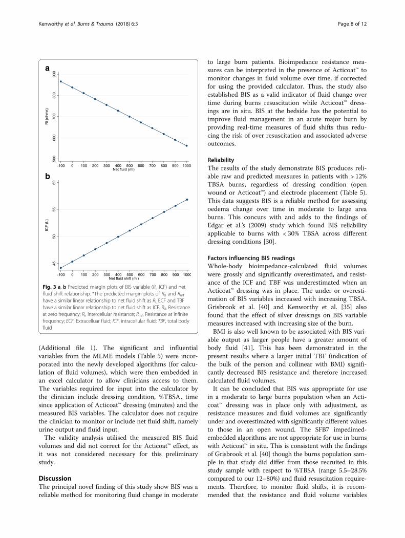
values significantly decreased with increasing net fluidvolume in a linear relationship (Table 5, Fig. 3a). Netfluid volume was significantly associated with ICF andTBF BIS fluid volume change, increasing with increasingnet fluid shift (Fig. 3b). All BIS variables were signifi-cantly associated with % TBSA. For every 1% increase inTBSA, R0 decreased by 5.09 Ω, Ri decreased 8.85 Ω andRinf decreased 3.25 Ω. Fluid volumes increased between1.20–2.77 L with every 1% increase in TBSA (p < 0.01)(Table 5).
Two individuals who had large negative fluid shifts> 850 ml across a single time point were removedfrom the analysis after the stepwise analysis foundthat they significantly altered the results of the finalmodel. Leaving these patients in the analysis wouldhave resulted in a non-homogenous sample. Itappears a large loss of fluid volume compromises theinterpretation of BIS measures. Both patients sufferedloss of large volumes of ionic fluid due to emesiswhich likely altered the measured BIS resistance [27].When a patient’s initial TBF increased by 1 L, R0
decreased by 5.78 Ω (p < 0.01), Ri decreased 28.79 Ω(p < 0.01) and Rinf decreased 5.31 Ω (p < 0.01).
CalculatorA calculator was developed to estimate the net fluid shiftbetween consecutive BIS measures, accounting fordressing condition, %TBSA and time since dressing
Table 4 Predictor variable effects on whole-body BIS variablesfor determining the effect of Acticoat™
Acticoat™ is in reference to an open woundR0 resistance at zero frequency, Ri intracellular resistance, Rinf resistance atinfinite frequency, ECF extracellular fluid, ICF intracellular fluid, TBF total bodyfluid, TBSA total body surface area, BIS bioimpedance spectroscopy*p ≤ 0.05#Interaction term
Table 5 Univariate analysis of variable correlation on whole-body BIS measures
R0 resistance at zero frequency, Ri intracellular resistance, Rinf resistance atinfinite frequency, ECF extracellular fluid, ICF intracellular fluid, TBF total bodyfluid, TBSA total body surface area, BIS bioimpedance spectroscopy*p ≤ 0.05
Kenworthy et al. Burns & Trauma (2018) 6:3 Page 7 of 12
(Additional file 1). The significant and influentialvariables from the MLME models (Table 5) were incor-porated into the newly developed algorithms (for calcu-lation of fluid volumes), which were then embedded inan excel calculator to allow clinicians access to them.The variables required for input into the calculator bythe clinician include dressing condition, %TBSA, timesince application of Acticoat™ dressing (minutes) and themeasured BIS variables. The calculator does not requirethe clinician to monitor or include net fluid shift, namelyurine output and fluid input.The validity analysis utilised the measured BIS fluid
volumes and did not correct for the Acticoat™ effect, asit was not considered necessary for this preliminarystudy.
DiscussionThe principal novel finding of this study show BIS was areliable method for monitoring fluid change in moderate
to large burn patients. Bioimpedance resistance mea-sures can be interpreted in the presence of Acticoat™ tomonitor changes in fluid volume over time, if correctedfor using the provided calculator. Thus, the study alsoestablished BIS as a valid indicator of fluid change overtime during burns resuscitation while Acticoat™ dress-ings are in situ. BIS at the bedside has the potential toimprove fluid management in an acute major burn byproviding real-time measures of fluid shifts thus redu-cing the risk of over resuscitation and associated adverseoutcomes.
ReliabilityThe results of the study demonstrate BIS produces reli-able raw and predicted measures in patients with > 12%TBSA burns, regardless of dressing condition (openwound or Acticoat™) and electrode placement (Table 5).This data suggests BIS is a reliable method for assessingoedema change over time in moderate to large areaburns. This concurs with and adds to the findings ofEdgar et al.’s (2009) study which found BIS reliabilityapplicable to burns with < 30% TBSA across differentdressing conditions [30].
Factors influencing BIS readingsWhole-body bioimpedance-calculated fluid volumeswere grossly and significantly overestimated, and resist-ance of the ICF and TBF was underestimated when anActicoat™ dressing was in place. The under or overesti-mation of BIS variables increased with increasing TBSA.Grisbrook et al. [40] and Kenworthy et al. [35] alsofound that the effect of silver dressings on BIS variablemeasures increased with increasing size of the burn.BMI is also well known to be associated with BIS vari-
able output as larger people have a greater amount ofbody fluid [41]. This has been demonstrated in thepresent results where a larger initial TBF (indication ofthe bulk of the person and collinear with BMI) signifi-cantly decreased BIS resistance and therefore increasedcalculated fluid volumes.It can be concluded that BIS was appropriate for use
in a moderate to large burns population when an Acti-coat™ dressing was in place only with adjustment, asresistance measures and fluid volumes are significantlyunder and overestimated with significantly different valuesto those in an open wound. The SFB7 impedimed-embedded algorithms are not appropriate for use in burnswith Acticoat™ in situ. This is consistent with the findingsof Grisbrook et al. [40] though the burns population sam-ple in that study did differ from those recruited in thisstudy sample with respect to %TBSA (range 5.5–28.5%compared to our 12–80%) and fluid resuscitation require-ments. Therefore, to monitor fluid shifts, it is recom-mended that the resistance and fluid volume variables
a
b
Fig. 3 a, b Predicted margin plots of BIS variable (Ri, ICF) and netfluid shift relationship. *The predicted margin plots of R0 and Rinfhave a similar linear relationship to net fluid shift as Ri. ECF and TBFhave a similar linear relationship to net fluid shift as ICF. R0, Resistanceat zero frequency; Ri, Intercellular resistance; Rinf, Resistance at infinitefrequency; ECF, Extracelluar fluid; ICF, intracellular fluid; TBF, total bodyfluid
Kenworthy et al. Burns & Trauma (2018) 6:3 Page 8 of 12
measured when an Acticoat™ dressing, in situ, be cor-rected using the provided calculator.
ValidityThe present results show that BIS is a valid indicator offluid volume change over time in moderate to large burnresuscitation with TBSA, time, net fluid shift and initialTBF all significantly associated with BIS resistance andcalculated fluid volumes. For clinically interpretableresults, the measured BIS variables need to be adjustedusing the provided calculator if Acticoat™ is in place.Time was significantly associated with resistance vari-
ables, with an increase in time decreasing all estimatedresistances and increasing ICF and TBF volumes. Thismay be explained by a combination of factors includingthe time since dressing application, the effect of Acti-coat™ and the amount of fluid resuscitation adminis-tered. Firstly, over time the Acticoat™ dressing depositsmore silver ions into the wound, therefore decreasingthe raw resistance values and in turn increasing the‘equivalent’ fluid volumes as calculated by BIS-embedded algorithms [42]. Secondly, the total mean vol-ume of fluid resuscitation over time increased, thus in-creasing all inter-compartmental fluid volumes andconsequentially decreasing the associated estimated re-sistance values. Although ECF was not associated withtime, the p value (0.15) is arguably low enough to acceptthat a clinical relationship may exist despite a small sam-ple. In contrast, the embedded algorithm of analysis mayexplain why ECF is not associated with time in thispopulation (each algorithm has different constants forestimating the individual fluid compartments [43]).However, R0, the equivalent resistance of ECF signifi-cantly changed with time, suggesting fluid volumechange in the extracellular compartment is associatedwith time.It is known that BIS resistance is inversely propor-
tional to fluid volume [22, 24]. The results of this studysupport this. Bioimpedance variables and net fluid shiftwere found to have a negative inverse linear relationshipwith resistance and as expected, calculated fluid volumesa positive linear relationship (Fig. 3) provided that thenet fluid shift (at each half hour measure) was greaterthan 100 ml. There were two patients who had a large(> 850 ml) negative fluid shift, both noted to have emesisduring the single measurement period, and thus, thesedata were excluded from the analysis, as they wereassumed to have an altered, uncorrected physiological(ionic) state at the time of measurement and thus, sig-nificantly differed from others in the sample. It appearsthat a large loss of fluid consequentially affects the fol-lowing repeated BIS measures (within at least the follow-ing 2 h). It is proposed that not only was the volumechange a contributor to the difficulty in interpretation of
the BIS measures but also the loss of electrolytes fromthe gut following emesis. The emesis could have alteredthe whole-body fluid ionic state for a short period untilit was corrected by the body systems. Bioimpedance re-sistance is inversely proportional to fluid volume andelectrolyte concentration. Therefore, significant changesin the ionic status of the fluid or tissues measured willalter the BIS raw variables and render the machine-embedded algorithms for calculated volumes invalid. Cli-nicians are advised not to use BIS measures in theperiod after an episode of emesis [27]. Further, the re-sults suggest the BIS measure is only sensitive to fluidlosses ≤ 100 ml per half hour in the burns resuscitationperiod. The sensitivity of the BIS measure for fluid lossesgreater than 100 ml and less than 850 ml cannot be pre-dicted as the patient cohort did not experience losses inthis range.
CalculatorOn the basis of the results a calculator was developed toimprove the clinical utility of BIS in burns resuscitationpatients at the bedside. It adjusts for the Acticoat™ effectand provides an estimated change in BIS resistance andfluid volumes between consecutive BIS measurements,hence allowing fluids to be titrated accordingly. It hasbeen established however that BIS is reliable and valid inthe open wound condition. Therefore, BIS can be uti-lised without variable adjustment when no dressings arein place.
Clinical practice recommendationsOptimum fluid resuscitation requires maintenance of theintracellular volume with minimal expansion (extravasa-tion) of the extracellular volume. The results of this studyindicate that using the relationship or pattern between R0or ECF and Ri or ICF is a non-invasive, interpretablemethod of monitoring or titrating fluid resuscitation. Astabilised Ri or ICF volume, over time, equal to or greaterthan the normal range (ICF 22.9–25 L) [24] represents afluid resuscitation target. Fluid volumes should then betitrated to maintain R0 or ECF at a steady state whilst con-tinuing to preserve Ri or ICF at the target volume. IdeallyECF volumes would be maintained as close to normal (orthe average for a healthy person) as possible (13.2–15.3 L).However due to the body’s systemic “leaky vessel” inflam-matory response to a major burn injury, with extravasa-tion of fluid into the extracellular space, volumes within5–10% of these norms would be a suggested acceptabletarget range [44, 45]. In postoperative surgical patients,fluid overload has been defined as > 15% of preoperativefluid volume [44] and in haemodialysis patients reachingECF volumes within one to two litres of normal values isdeemed acceptable [46]. An example of how to titratefluids: If Ri or ICF is stable and the change values of R0 or
Kenworthy et al. Burns & Trauma (2018) 6:3 Page 9 of 12
ECF continue to increase, the fluid administered is addingto the extracellular compartment (swelling) rather thanpreferentially maintaining the intracellular compartment.Infused fluid volumes therefore need to be reduced if Ri(ICF) is stable and R0 (ECF) is trending upward. However,in a recent study, intracellular volume actually decreased(~ 0.8 L over 70 min) upon rapid infusion of intravenousfluid (~ 2 L in ~ 60 min) into healthy male volunteers [47].It was suggested that the infusion of fluid was responsiblefor the increase in ECF. The fluid administered in thisstudy was < 500 ml/h; therefore, it is difficult to concludewhether this may have the same effect. It however doessuggest potentially accepting an ICF volume of ~ 1 L lessthan the average volumes when considering titrating fluidas above. For greater sensitivity to change, at this time,this study suggests that it is more advantageous to use thechange in BIS raw resistance values (adjusted in the pres-ence of Acticoat™) rather than the calculated volumes as itremoves the need for specific predictive equations andeliminates the need for height and weight measures [48].There are a growing number of studies suggesting thatraw BIS variables may be more useful in predicting clinicaloutcomes [49, 50]. BIS raw variables may also be able toindicate changes associated with cell membrane damageand cell wall integrity [50].Further work is required to increase the confidence
and promote greater utility of this sensitive measureover standard haemodynamic monitoring. In contrast,urine output, a ‘quasi’ measure of fluid shifts and whole-body perfusion [8] has been suggested to lag behind theactual events of hypoperfusion by up to 2 h [51, 52].Bioimpedance also removes the need to rely heavily oninitial fluid volume calculations such as the Parkland orBrooke’s. This proves to be highly useful out in the fieldwith paramedics and in isolated country hospitals whereclinician’s burns experience may be limited and whereWestern Australia’s vastness means it is not uncommonfor people to travel greater than 8 h to be admitted to atertiary hospital.
Future researchAdditional research is warranted in evaluating the effectof other silver and non-silver dressings such as sulfadia-zine and hydrocolloids, in moderate to large burns to in-crease the utility of BIS across burns services.Further, consideration may need to be given of the
type of resuscitation fluid (e.g. crystalloids versus col-loids) in future studies as BIS electrical conductivity isaffected by electrolyte concentration. This may thereforeinfluence BIS variable measurements. Electrical andchemical burn injuries may also influence or change theionic state of the tissue. Thus, future research should in-clude these modes of injury.
Ideally, BIS would be able to be used on burns patientson life support or mechanical ventilation however fur-ther study needs to be done to determine whether elec-tronic equipment interferes with the BIS instrument.Several studies have been conducted in intensive careunits however they did not stipulate whether ventilatedpatients were included [53, 54].
ConclusionsIn moderate to large burn patients, BIS is a reliable andvalid method of oedema change. The Acticoat™ dressingssignificantly alter the BIS raw outputs. To allow clinicalinterpretation of BIS, measures must be adjusted for silverdressings.
Additional files
Additional file 1: Acticoat calculator for oedema—excel spreadsheet.(XLSX 13 kb)
AcknowledgementsThe authors would like to acknowledge Chevron for their financialcontribution towards the research salaries of Kenworthy, Grisbrook andEdgar. The authors would like to acknowledge the University of Notre Damefor their support of Kenworthy’s higher degree research studies.
FundingThis study was supported by the Fiona Wood Foundation, Western Australia.
Availability of data and materialsThe datasets used and/or analysed during the current study are availablefrom the corresponding author on reasonable request.
Authors’ contributionsThe authors submit that they have all made substantial contributions toconception, design, drafting and final approval of this paper. DE and FWinstigated the project and MP primarily undertook the data analysis. PK andTG conducted the majority of data collection and database management. PKdrafted the manuscript and all authors contributed to and approved the finalversion.
Ethics approval and consent to participateThis study was approved by RPH Ethics Committee (EC 2011/028), FSHResearch Governance Committee (2014 106) and The University of NotreDame, Australia Human Research Ethics Committee (014139F). Consent ofparticipants was gained prior to inclusion in the study.
Consent for publicationNot applicable.
Competing interestsThe authors declare that they have no competing interests.
Author details1Fiona Wood Foundation, Fiona Stanley Hospital, Perth, Western Australia,Australia. 2School of Physiotherapy and Exercise Science, Curtin University,Perth, Western Australia, Australia. 3Burns Service of Western Australia, FionaStanley Hospital, Perth, Western Australia, Australia. 4Burn Injury ResearchNode, Notre Dame University, Fremantle, Western Australia, Australia. 5HarryPerkins Institute of Medical Research, The University of Western Australia,Perth, Western Australia, Australia. 6School of Physiotherapy, Notre DameUniversity, Fremantle, Western Australia, Australia. 7Adult State Burns Service,Fiona Stanley Hospital, Murdoch Drive, Murdoch, Western Australia 6150,Australia.
Kenworthy et al. Burns & Trauma (2018) 6:3 Page 10 of 12
Received: 6 September 2017 Accepted: 19 December 2017
References1. Tiwari VK. Burn wound: how it differs from other wounds? Indian J Plast
Surg. 2012;45:364–73.2. Greenhalgh D. Burn resuscitation. J Burn Care Res. 2007;28:1–11.3. Saffle JR. The phenomenon of “fluid creep” in acute burn resuscitation. J
Burn Care Res. 2007;28:382–95.4. Alvarado R, Chung KK, Cancio LC, Wolf SE. Burn resuscitation. Burns. 2009;35:
4–14.5. Pruitt B. Protection from excessive resuscitation: “pushing the pendulum
back”. The J Trauma. 2000;49:567–8.6. Mitchell KB, Khalil E, Brennan A, Shao H, Leah ARNE, Yurt RW, et al. New
management strategy for fluid resuscitation: quantifying volume in the first48 hours after burn injury. J Burn Care Res. 2013;34:196–202.
7. Singh V, Devgan L, Bhat S, Milner SM. The pathogenesis of burn woundconversion. Ann Plast Surg. 2007;59:109–15.
8. Hayek S, Ibrahim A, Abu Sittah G, Atiyeh B. Burn resuscitation: is itstraightforward or a challenge? Ann Burns Fire Disasters. 2011;24:17–21.
9. Tricklebank S. Modern trends in fluid therapy for burns. Burns. 2009;35:757–67.
10. Klein MB, Hayden D, Elson C, Nathens AB, Gamelli RL, Gibran NS, et al. Theassociation between fluid administration and outcome following majorburn. Ann Surg. 2007;245:622–8.
11. Cartotto R, Zhou A. Fluid creep: the pendulum hasn’t swung back yet! JBurn Care Res. 2010;31:551–8.
12. Dulhunty JM, Boots RJ, Rudd MJ, Muller MJ, Lipman J. Increased fluidresuscitation can lead to adverse outcomes in major-burn injured patients,but low mortality is achievable. Burns. 2008;34:1090–7.
13. Fodor L, Ramon Y, Shoshani O, Rissin Y, Ullmann Y. Controversies in fluidresuscitation for burn management: literature review and our experience.Injury, Int J Care Injured. 2006;37:374–9.
14. Cancio L, Lundy JB, Sheridan RL. Evolving changes in the management ofburns and environmental injuries. Surg Clin N Am. 2012;92:959–86.
15. Chung K, Blackbourne LH, Wolf SE, White CE, Renz E, Cancio L, et al.Evolution of burn resuscitation in Operation Iraqi Freedom. J Burn Care Res.2006;27:1–6.
16. Cornish BH, Chapman M, Hirst C, Mirolo B, Bunce IH, Ward LC, et al. Earlydiagnosis of lymphedema using multiple frequency bioimpedance.Lymphology. 2001;34:2–11.
17. Vine SM, Painter PL, Kuskowski MA, Earthman CP. Bioimpedancespectroscopy for the estimation of fat-free mass in end-stage renal disease.E Spen Eur E J Clin Nutr Metab. 2011;6:1–6.
18. Mialich MS, Sicchieri JMF, Jordao Junior AA. Analysis of bodycomposition—a critical review of the use of bioelectrical impedanceanalysis. Int J Clin Nutr. 2014;2:1–10.
19. Ward L: Is BIS ready for prime time as the gold standard measure? 2009.20. Anderson L, Erceg D, Schroeder E. Utility of multi-frequency bioelectrical
impedance compared to deuterium dilution for assessment of total bodywater. Nutr Diet. 2015;72:183–9.
21. van marken Lichtenbelt WD, Westerterp KR, Wouters L, Luijendzjk SC.Validation of bioelectrical-impedance measurements as a method toestimate body-water compartments. Am J Clin Nutrition. 1994;60(2):159–66.
22. Janssen I, Heymsfield SB, Baumgartner RN, Ross R. Estimation of skeletalmuscle mass by bioelectrical impedance analysis. J Appl Physiol. 2000;89:465–71.
23. Armstrong LE, Kenefick RW, Castellani JW, Riebe D, Kavouras SA, Kuznicki JT,et al. Bioimpedance spectroscopy technique: intra-, extracellular, and totalbody water. Med Sci Sports Exerc. 1997;29:1657–63.
24. Malbrain ML, Huygh J, Dabrowski W, De Waele JJ, Staelens A, Wauters J. Theuse of bio-electrical impedance analysis (BIA) to guide fluid management,resuscitation and deresuscitation in critically ill patients: a bench-to-bedsidereview. Anaesthesiol Intensive Ther. 2014;46:381–91.
25. Kyle U, Bosaues I, De Lorenzo A, Durenberg P, Elia M, Gomez JM, et al.Bioelectrical impedance analysis—part II: review of principles and methods.Clin Nutr. 2004;23:1226–43.
26. Gaw R, Box R, Cornish BH. Bioimpedance in the assessment of unilaterallymphedema of a limb: the optimal frequency. Lymphat Res Biol.2011;9:93–9.
27. Kyle U, Bosaues I, De Lorenzo A, Durenberg P, Elia M, Gomez JM, et al.Bioelectrical impedance analysis-part I: review of principles and methods.Clin Nutr. 2004;23:1226–43.
28. Miller S, Carlson R, Fegelman E, Quinones J, Finley R. Comparison of TotalBody Water Analysis:Bioelectrical Impedance Analysis Versus the TitratedMethod. J Burn Care Rehabil. 1999;20:363–6.
29. Zdolsek HJ, Lindahl OA, Angquist KA, Sjoberg F. Non-Invasive Assessment ofIntercompartmental Fluid Shifts in Burn Victims. Burns. 1998;24(3):233–40.
30. Edgar D, Briffa K, Cole J, Tan MH, Khoo B, Goh J, et al. Measurement ofacute edema shifts in human burn survivors—the reliability and sensitivityof bioimpedence spectroscopy as an objective clinical measure. J Burn CareRes. 2009;30:818–23.
31. Grisbrook TL, Kenworthy P, Phillips M, Gittings PM, Wood FM, Edgar DW.Alternate electrode placement for whole body and segmentalbioimpedance spectroscopy. Physiol Meas. 2015;36:2189–201.
32. Fong J, Wood F. Nanocrystalline silver dressings in wound management: areview. Int J Nanomedicine. 2006;1:441–9.
33. Kekonen A. Bioimpedance measurement device for chronic wound healingmonitoring. Tampere: Tampere University of Technology, Science; 2013.
34. Cornish BH, Jacobs A, Thomas BJ, Ward LC. Optimizing electrode sites forsegmental bioimpedance measurements. Physiol Meas. 1999;20:241–50.
35. Kenworthy P, Grisbrook TL, Phillips M, Gibson W, Wood F, Edgar D.Addressing the barriers to bioimpedance spectroscopy use in major burns:alternate electrode placement. J Burn Care Res. 2017; In Press
37. Cheng J, Edwards LJ, Maldonado-Molina MM, Komro KA, Muller KE. Reallongitudinal data analysis for real people: building a good enough mixedmodel. Stat Med. 2010;29:504–20.
38. Zdolsek HJ, Lindahl OA, Angquist KA, Sjoberg F. Non-invasive assessment ofintercompartmental fluid shifts in burn victims. Burns. 1998;24:233–40.
39. Cochrane Handbook for Systematic Reviews of Interventions [http://handbook.cochrane.org/chapter_9/9_4_5_2_meta_analysis_of_change_scores.htm]. Accessed Feb 2017.
40. Grisbrook TL, Kenworthy P, Phillips M, Wood FM, Edgar DW.Nanaocrystalline silver dressings influence bioimpedance spectroscopymeasurements in burns patients. Burns. 2016;42(7):1548–55.
41. Lukaski HC, Johnson PE, Bolonchuk WW, Lykken GI. Assessment of fat-freemass using bioelectrical impedance measurements of the human body. AmJ Clin Nutr. 1985;41:810–7.
42. Guidelines for use of Nanocrystalline Silver Dressing-Acticoat™. Departmentof Health WA ed. Perth: Health Networks Branch, Department of Health,Western Australia; 2011.
43. Ward LC, Isenring E, Dyer JM, Kagawa M, Essex T. Resistivity coefficients forbody composition analysis using bioimpedance spectroscopy: effects ofbody dominance and mixture theory algorithm. Physiol Meas. 2015;36:1529–49.
44. Ernstbrunner M, Kostner L, Kimberger O, Wabel P, Säemann M,Markstallar K, et al. Bioimpedance spectroscopy for assessment ofvolume status in patients before and after general anaesthesia.PLoS One. 2014;9(10):e111139.
45. Earthman C, Traughber D, Dobratz J, Howell W. Bioimpedance spectroscopyfor clinical assessment of fluid distribution and body cell mass. Nutr ClinPract. 2007;22:389–405.
46. Tattersall J. Bioimpedance analysis in dialysis: state of the art and what wecan expect. Blood Purif. 2009;27:70–4.
47. Ernstbrunner M, Kabon B, Zotti O, Zeitlinger M, Berner C, Hinterholzer G, etal. Intravenous fluid challenge decreases intracellular volume: abioimpedance spectroscopy-based crossover study in healthy volunteers.Sci Rep. 2017;7:9644.
48. Haverkort EB, Reijven PLM, Binnekade JM, de van der Schueren MA,Earthman CP, Gouma DJ, et al. Bioelectrical impedance analysis to estimatebody composition in surgical and oncological patients: a systematic review.Eur J Clin Nutr. 2015;69:3–13.
49. Lukaski H, Moore M. Bioelectrical impedance assessment of wound healing.J Diabetes Sci Technol. 2012;6:209–12.
50. Slotwinski R, Saragat B, Cabras S, Rinaldi A, Marini E. Raw impedance dataanalysis in severe ill patients with sepsis. Fluids. 2013;2:168–70.
51. Jaskille AD, Jeng JC, Sokolich JC, Lunsford P, Jordan MH. Repetitiveischemia-reperfusion injury: a plausible mechanism for documented clinicalburn-depth progression after thermal injury. J Burn Care Res. 2007;28:13–20.
Kenworthy et al. Burns & Trauma (2018) 6:3 Page 11 of 12
52. Jeng JC, Jaskille AD, Lunsford PM, Jordan MH. Improved markers for burnwound perfusion in the severely burned patient: the role for tissue andgastric PCO2. J Burn Care Res. 2008;29:49–55.
53. Basso F, Berdin G, Virzi GM, Mason G, Piccinni P, Day S, et al. Fluidmanagement in the intensive care unit: bioelectrical impedance vectoranalysis as a tool to assess hydration status and optimal fluid balance incritically ill patients. Blood Purif. 2013;36:192–9.
54. Lee Y, Kwon O, Shin CS, Lee SM. Use of bioelectrical impedance analysis forthe assessment of nutritional status in critically ill patients. Clin Nutr Res.2015;4:32–40.
• We accept pre-submission inquiries
• Our selector tool helps you to find the most relevant journal
• We provide round the clock customer support
• Convenient online submission
• Thorough peer review
• Inclusion in PubMed and all major indexing services
• Maximum visibility for your research
Submit your manuscript atwww.biomedcentral.com/submit
Submit your next manuscript to BioMed Central and we will help you at every step:
Kenworthy et al. Burns & Trauma (2018) 6:3 Page 12 of 12