61 Caso Clínico / Radiological Case Report Correspondência Alexandre Gomes Martins Batista Rua dos almocreves, nº16, Aldeia Nova da Azoia 2970-085 Sesimbra e-mail: [email protected]1 - Hospital José Joaquim Fernandes, Beja 2 - Instituto Português de Oncologia de Lisboa Francisco Gentil, Lisboa Serviço de Radiologia do Instituto Português de Oncologia de Lisboa Francisco Gentil Diretor: Dr. José Venâncio Recebido a 16/07/2014 Aceite a 13/09/2014 POST-MENOPAUSAL METRORRHAGIA – AN OVARIAN THECOMA PRESENTATION METRORRAGIA PÓS-MENOPAUSA – UMA APRESENTAÇÃO DE TECOMA OVÁRICO Alexandre Batista 1 , Teresa Margarida Cunha 2 Resumo Os tecomas são tumores raros do ovário, do grupo dos tumores dos cordões sexuais, de natureza sólida e frequentemente unilaterais. Têm maior incidência no período pós- menopausa e normalmente são silenciosos. Quando sintomáticos traduzem-se por dor pélvica e metrorragia (condicionada pela habitual natureza produtora de estrogénios do tumor). Podem ser concomitantes a síndrome de Meigs e/ou de Golin-Goltz e associarem- se a transformação benigna ou maligna do endométrio. Embora a ecografia possa ser inespecífica neste contexto, uma avaliação multiparamétrica abrangente em ressonância magnética, incluindo por estudo dinâmico e com ponderação em difusão, permite frequentemente orientar de modo favorável a marcha diagnóstica. Apresentamos um caso raro de tecoma do ovário, com espessamento associado do endométrio, avaliado por ecografia ginecológica por vias supra-púbica e transvaginal bem como tomografia computorizada e ressonância magnética, confirmado cirurgicamente. Tratou-se de uma examinada caucasiana de 61 anos de idade, apresentando-se com metrorragia pós- menopáusica, sem outros sintomas nem contexto familiar relevante. Procedeu-se, a este propósito, a uma revisão da literatura focada no diagnóstico multimodal diferencial, apresentação clínica, tratamento e prognóstico destes tumores. Palavras-chave Tecoma; Ovário; Ecografia; Tomografia Computadorizada; Ressonância Magnética. Abstract Thecomas represent rare, solid sex-cord stromal ovarian tumors, often unilateral, asymptomatic and occurring in postmenopausal patients. When symptomatic, they most commonly present with pelvic pain and metrorrhagia (due to their frequent estrogenic releasing nature). Thecomas can occur concomitantly with Meigs and/or Golin-Goltz syndrome and may also be associated with benign or malign endometrial transformation. Although gynecologic transabdominal and transvaginal ultrasound can be quite unspecific in this particular solid ovarian tumor presentation, magnetic resonance imaging including diffusion and dynamic data can frequently suggest the diagnosis and significantly facilitate the diagnostic work-up. We report a rare case of ovarian thecoma, with concomitant endometrial thickening, demonstrated by gynecologic transvaginal ultrasonography, computed tomography and magnetic resonance contrasted imaging, surgically confirmed. The patient was a 61 years old caucasian female presenting with postmenopausal metrorrhagia, without other associated symptoms nor family medical context. On this regard, we performed a review of the literature, focused on multimodal differential diagnosis imaging, clinical presentation, treatment and prognostic of this pathological finding. Key-words Thecoma; Ovary; Ultrasound; Computed Tomography; Magnetic Resonance. ACTA RADIOLÓGICA PORTUGUESA Setembro-Dezembro 2014 nº 103 Volume XXVI 61-65 Clinical history 61 years old caucasian female patient presenting with postmenopausal metrorrhagia for a period of four months, without other associated symptoms and with no relevant family medical context. The patient was not on hormone replacement therapy. Physical examination was unremarkable, with no evident abdominal or pelvic masses, nor localized pain. Laboratory findings included an elevated CA 125 value (86,7 U/ml), normal CEA (0,9 ng/ml) and CA 19.9 (11,8 U/ml), and normal hemoglobin (13,2 g/dL) and hematocrit (37,8%). Image findings Outside our Institution the patient underwent gynecologic transabdominal and transvaginal ultrasonography (TVUS) that revealed a solid right ovarian mass, heterogeneous, mostly hypoechoic, measuring 108 x 71 mm (Fig. 1-A). Concomitantly
Transcript
61
Caso Clínico / Radiological Case Report
Correspondência
Alexandre Gomes Martins BatistaRua dos almocreves, nº16,Aldeia Nova da Azoia2970-085 Sesimbrae-mail: [email protected]
1 - Hospital José Joaquim Fernandes, Beja 2 -Instituto Português de Oncologia de LisboaFrancisco Gentil, LisboaServiço de Radiologia do Instituto Portuguêsde Oncologia de Lisboa Francisco GentilDiretor: Dr. José Venâncio
Recebido a 16/07/2014Aceite a 13/09/2014
POST-MENOPAUSAL METRORRHAGIA – AN OVARIANTHECOMA PRESENTATION
METRORRAGIA PÓS-MENOPAUSA – UMA APRESENTAÇÃO DETECOMA OVÁRICO
Alexandre Batista1, Teresa Margarida Cunha2
Resumo
Os tecomas são tumores raros do ovário, dogrupo dos tumores dos cordões sexuais, denatureza sólida e frequentemente unilaterais.Têm maior incidência no período pós-menopausa e normalmente são silenciosos.Quando sintomáticos traduzem-se por dorpélvica e metrorragia (condicionada pelahabitual natureza produtora de estrogénios dotumor). Podem ser concomitantes a síndromede Meigs e/ou de Golin-Goltz e associarem-se a transformação benigna ou maligna doendométrio. Embora a ecografia possa serinespecífica neste contexto, uma avaliaçãomultiparamétrica abrangente em ressonânciamagnética, incluindo por estudo dinâmico ecom ponderação em difusão, permitefrequentemente orientar de modo favorável amarcha diagnóstica.Apresentamos um caso raro de tecoma doovário, com espessamento associado doendométrio, avaliado por ecografiaginecológica por vias supra-púbica etransvaginal bem como tomografiacomputorizada e ressonância magnética,confirmado cirurgicamente. Tratou-se de umaexaminada caucasiana de 61 anos de idade,apresentando-se com metrorragia pós-menopáusica, sem outros sintomas nemcontexto familiar relevante. Procedeu-se, a estepropósito, a uma revisão da literatura focadano diagnóstico multimodal diferencial,apresentação clínica, tratamento e prognósticodestes tumores.
Thecomas represent rare, solid sex-cord stromalovarian tumors, often unilateral, asymptomaticand occurring in postmenopausal patients.When symptomatic, they most commonlypresent with pelvic pain and metrorrhagia (dueto their frequent estrogenic releasing nature).Thecomas can occur concomitantly with Meigsand/or Golin-Goltz syndrome and may alsobe associated with benign or malignendometrial transformation. Althoughgynecologic transabdominal and transvaginalultrasound can be quite unspecific in thisparticular solid ovarian tumor presentation,magnetic resonance imaging includingdiffusion and dynamic data can frequentlysuggest the diagnosis and significantly facilitatethe diagnostic work-up.We report a rare case of ovarian thecoma, withconcomitant endometrial thickening,demonstrated by gynecologic transvaginalultrasonography, computed tomography andmagnetic resonance contrasted imaging,surgically confirmed. The patient was a 61 yearsold caucasian female presenting withpostmenopausal metrorrhagia, without otherassociated symptoms nor family medicalcontext. On this regard, we performed a reviewof the literature, focused on multimodaldifferential diagnosis imaging, clinicalpresentation, treatment and prognostic of thispathological finding.
Key-words
Thecoma; Ovary; Ultrasound; ComputedTomography; Magnetic Resonance.
61 years old caucasian female patient presenting withpostmenopausal metrorrhagia for a period of four months,without other associated symptoms and with no relevant familymedical context. The patient was not on hormone replacementtherapy. Physical examination was unremarkable, with no evidentabdominal or pelvic masses, nor localized pain. Laboratoryfindings included an elevated CA 125 value (86,7 U/ml), normal
CEA (0,9 ng/ml) and CA 19.9 (11,8 U/ml), and normalhemoglobin (13,2 g/dL) and hematocrit (37,8%).
Image findings
Outside our Institution the patient underwent gynecologictransabdominal and transvaginal ultrasonography (TVUS) thatrevealed a solid right ovarian mass, heterogeneous, mostlyhypoechoic, measuring 108 x 71 mm (Fig. 1-A). Concomitantly
62
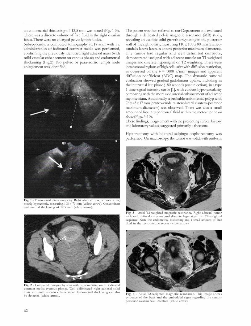
The patient was then referred to our Department and evaluatedthrough a dedicated pelvic magnetic resonance (MR) study,revealing an exofitic solid growth originating in the posteriorwall of the right ovary, measuring 110 x 100 x 80 mm (craneo-caudal x latero-lateral x antero-posterior maximum diameters).The tumor had regular and well delimited contours,demonstrated isosignal with adjacent muscle on T1 weightedimages and discrete hypersignal on T2 weighting. There wereintratumoral regions of high cellularity with diffusion restriction,as observed on the b = 1000 s/mm2 images and apparentdiffusion coefficient (ADC) map. The dynamic tumoralevaluation showed gradual gadolinium uptake, including inthe interstitial late phase (180 seconds post injection), in a type1 time-signal intensity curve [1], with evident hypovascularitycomparing with the more avid arterial enhancement of adjacentmyometrium. Additionally, a probable endometrial polyp with76 x 43 x 17 mm (craneo-caudal x latero-lateral x antero-posteriormaximum diameters) was observed. There was also a smallamount of free intraperitoneal fluid within the recto-uterine culde sac (Figs. 3-10).These findings, in agreement with the presenting clinical historyand laboratory values, suggested primarily a thecoma.
Hysterectomy with bilateral salpingo-oophorectomy wasperformed. On macroscopy, the tumor was solid, with uniform
an endometrial thickening of 12,5 mm was noted (Fig. 1-B).There was a discrete volume of free fluid in the right ovarianfossa. There were no enlarged pelvic lymph nodes.Subsequently, a computed tomography (CT) scan with i.v.administration of iodinated contrast media was performed,confirming the previously identified right adnexal mass (withmild vascular enhancement on venous phase) and endometrialthickening (Fig.2). No pelvic or para-aortic lymph nodeenlargement was identified.
A
B
Fig. 1 - Transvaginal ultrasonography. Right adnexal mass, heterogeneous,mostly hypoechoic, measuring 108 x 71 mm (yellow arrow). Concomitantendometrial thickening of 12,5 mm (white arrow).
Fig. 2 - Computed tomography scan with i.v. administration of iodinatedcontrast media (venous phase). Well delimitated right adnexal solidmass with mild vascular enhancement. Endometrial thickening can alsobe detected (white arrow).
Fig. 3 - Axial T2-weighted magnetic resonance. Right adnexal tumorwith well defined contours and discrete hypersignal on T2-weightedsequence. Note the endometrial thickening and a small amount of freefluid in the recto-uterine recess (white arrow).
Fig. 4 - Axial T2-weighted magnetic resonance. This image showsevidence of the beak and the embedded signs regarding the tumor-posterior ovarian wall interface (white arrow).
63
texture (excluding a few cystic spaces), well-circumscribed, andmeasured 120 x 110 x 70 mm. Sectional analysis of the tumorrevealed a yellow and fascicular mass (Fig.11). The tumor hadmore than 80% of its cellular content positive for alpha-inhibinand a low proliferation index (<10%). There were twoendometrial polyps (40 and 30 mm of maximum diameter).The pathological findings were consistent with thecoma.
Discussion
Thecomas represent rare, solid sex-cord stromal ovarian tumors,accounting for approximately 0.5 – 1% of all primary ovarian
A
B
Fig. 5 - Coronal (A) and sagital (B) T2-weighted magnetic resonancedepicting normal left ovary appearance (white arrow).
Fig. 6 - Axial T1-weighted magnetic resonance. The mass demonstratesisosignal with adjacent muscle on T1-weighted sequence.
Fig. 7 - Fat-suppressed axial T1-weighted MR image after gadoliniumadministration (arterial phase). There is little mass enhancementcomparing to myometrium.
Fig. 8 - Fat-suppressed axial T1-weighted MR image after gadoliniumadministration (intersticial phase). Gradual gadolinium uptake withdiscrete enhancement in the interstitial late phase (180 seconds postinjection), in a type 1 time-signal intensity curve, revealing the lesion´sfibrin rich territory.
Fig. 9 - Fat-suppressed axial T1-weighted MR image after gadoliniumadministration. Type 1 time-signal intensity curve, commonly seen inbenign lesions.
64
ovarian medulla and should be considered apart form fibromas,which originate from the cortex [4]. Although beinguncommon, they represent the most frequent solid primaryovarian tumor [5].Thecomas of the ovary can present with pleural effusion orascites (Meigs syndrome) and may also be associated with basalcell nevus Golin-Goltz syndrome (large bilateral fibrotic ovariantumors, basal cell carcinomas of the skin, odontogenickeratocysts and other abnormalities) [6], though they usuallyare asymptomatic, and when symptomatic, they are mostcommonly manifested by pelvic pain and metromenorrhagia(due to their frequent estrogen releasing nature) [7,8]. Estrogenstimulation by a pre-existing thecoma may also induce thedevelopment of endometrial hyperplasia and endometrialpolyps and presumably induce mesenchymal and mixedepithelial / mesenchymal uterine tumors, namely adenosarcoma[9, 10].They are often unilateral and occur in postmenopausal patientsbut can develop in younger patients (mean ages in the fifth andsixth decades) [11, 12]. As most adnexal tumors do, thelaboratory workup on a thecoma presentation can reflect elevationof particular tumor markers, such as CA-125 [13].Histologically thecomas are similar to theca interna cells of theovary and have a mixed lipid and colagenic composition, thelater derived from spindle, oval or round cells [6].The management approach to thecomas is surgical in largerlesions, with excellent prognosis as they are mainly benigntumors [14].TVUS is commonly the first choice imaging modality forsuspected pelvic tumor, due to its easy access and safety profile,nonetheless being often non specific. Thecomas usually presentas a solid mass or, if larger, predominantly solid with few cysticareas, being the solid component iso or hypoecogenic in relationwith adjacent stroma, frequently with significant posteriorattenuation, without identifiable tumoral calcification. On colorDoppler evaluation thecomas usually have neglectable vascularity[9, 15]. On CT evaluation tumor attenuation is often similar toadjacent myometrium, with evident hypovascularity on arterialphase enhancement study, sometimes being evident progressivedelayed contrast uptake due to fibrin rich tumoral areas [11].This same fibrous tumoral component is responsible for thelow T1-weighted and very low T2-weighted MR signal(excluding scattered areas of high signal intensity correspondingto cystic or edematous change) [5, 11]. If fatty elements arepresent, they can be identified as hyperintense on T1-weightedand translate in a decreased signal intensity on selective fat-saturation or out-of-phase gradient echo sequences [9]. Ondiffusion-weighted imaging (DWI), most thecomas displayintermediate signal similar to that of myometrium, attributableto their cellular content of fibroblasts and thecal cells, but lowerthan the usually observed in malignant, high cellular ovariantumors. The ADC of thecomas and other adnexal masses hasno described relevant difference, although the ADC of thecomashas been reported to be significantly lower than that ofleyomiomas [16].The differential diagnosis of ovarian thecoma primarilyencompasses masses with a fibrous component, as uterineleyomioma, Brenner tumor and mature cystic teratoma [17].When considering uterine leyomioma, the most common pitfalllies in broad ligament leyomioma and pedunculated leyomioma.However, their origin can usually be traced, namely due to the
Fig. 11 - Tumor macroscopic surface section. Two sections of the rightoophorectomy specimen revealed a yellowish tone and fascicular solidmass.
neoplasms [2]. Ovarian sex cord tumors are those who arisefrom granulosa, theca, Leydig, Sertoli or stromal fibroblast cells[3]. Notwithstanding sometimes having an interspersed fibrouscomponent, thecomas are presumably originated from the
A
B
Fig. 10 - Diffusion-weighted MR image. Intratumoral areas of highcellularity with diffusion restriction, as observed in theb = 1000 s/mm2 images (A) and apparent diffusion coefficient (ADC)map (B), revealing the tumor´s mixed theca cells and fibrouscomposition.
65
bridging vascular sign [18], that clarifies their feeding by uterinearteries, opposed to thecomas, supplied by ovarian arteries orovarian branches of uterine arteries [17].Brenner tumours are usually very small (<2 cm) and benignepithelial ovarian lesions, mostly composed of fibrous densetissue and urothelium-like transitional cells. They mostcommonly present as a multilocular cystic tumor or a smallerpredominantly solid mass. Their dense fibrous stroma has lowsignal on T2-weighted MR imaging, and an expected gradualgadolinium uptake. Usually they comprise extensive amorphouscalcifications [19]. Their small size compared to the medianthecoma size of approximately 13 cm, more prominentcalcifications and extensive cystic component usually allows thedifferential diagnosis [9].Mature teratomas most frequently occur in pre-menopausalwomen, and present as a unilocular cystic tumour (in 88% ofthe cases), filled with sebaceous content and lined withepithelium. Due to their origin in two or more germinal layers,they can contain hair and teeth, that when present are commonlyencompassed by a wall protuberance (the Rokitansky nodule ordermoid plug) [20]. The most common imaging US featuresfor mature teratoma are the presence of a cystic lesion with adensely echogenic shadowing mural nodule (dermoid plug); an
ecogenic (sebaceous) mass with very strong posterior attenuation(“tip of the iceberg” sign) and a dermoid mesh, with multiplethin echogenic lines (hairs) passing within the cyst. On CT andMRI imaging, macroscopic fat within a cyst, with or withoutmural calcification, is diagnostic. Less common monodermaldermoids include struma ovarii (with a predominance of thyroidaltissue) and carcinoid [21].
Our patient presented with a predominantly solid right adnexalmass, hypoechoic on ultrasound, with regular and definedcontours and no evident vascularization on color Doppler. OnMRI interrogation the tumor showed to be originated fromthe posterior right ovarian wall, had gradual intersticialenhancement, revealing its fibrous component, and smallpockets of cellularity in DWI. There was no accompanyingsignificant ascites, peritoneal lesions or pelvic enlarged lymphnodes. There was also an endometrial polyp. These findings, ina postmenopausal metrorrhagia context, and in conjunctionwith the described clinical and laboratory data suggested primarilya thecoma, with mixed theca cells and fibrous composition andexpected estrogenic endometrial effect.The lesion was surgically confirmed as thecoma.
References1 - Thomassin-Naggara, I.; Daraï, E.; Cuenod, C. A.; Rouzier, R.; Callard,P.; Bazot, M. - Dynamic contrast-enhanced magnetic resonance imaging: a useful toolfor characterizing ovarian epithelial tumors. J Magn Reson Imaging, 2008,28(1):111-20.2 - Chen, V. W.; Ruiz, B.; Killeen, J. L.; Coté, T. R.; Wu, X. C.; Correa, C. N.- Pathology and classification of ovarian tumors. Cancer, 2003, 97:2631-42.3 - Wilkinson, N.; Osborn, S.; Young, R. H. - Sex cord stromal tumours of theovary: A review highlighting recent advances. Diagn Histopathol, 2008, 14(8):388-400.4 - Nocito, A. L.; Sarancone, S.; Bacchi, C.; Tellez, T. - Ovarian thecoma:Clinicopathological analysis of 50 cases. Ann Diagn Pathol, 2008, 12:12-6.5 - Troiano, R. N.; Lazzarini, K. M.; Scoutt, L. M.; Lange, R. C.; Flynn, S. D.;McCarthy, S. - Fibroma and fibrothecoma of the ovary: MR imaging findings.Radiology, 1997, 204:795-8.6 - Scully, R. E.; Young, R. H.; Clement, P. B. - Atlas of Tumor Pathology,Tumors of the Ovary, Maldeveloped Gonads, Fallopian Tube, and Broad Ligament.Third series, Fascicle 23. Washington, DC: Armed Forces Institute ofPathology, 1998.7 - Sivanesaratnam, V.; Dutta, R.; Jayalakshmi, P. - Ovarian fibroma: clinical andhistopathological characteristics. Int J Gynaecol Obstet, 1990, 33:243-7.8 - Leung, S. W.; Yuen, P. M. - Ovarian fibroma: a review on the clinicalcharacteristics, diagnostic difficulties, and management options of 23 cases. GynecolObstet Invest, 2006, 62:1-6.9 - Hricak, H. - Diagnostic imaging: gynecology, 1st ed. Salt Lake City, UT,Amirsys/Elsevier, 2007, 7:28-31.10 - Nomura, K.; Aizawa, S.; Ushigome, S. - Adenosarcoma of the uterinecorpus associated with ovarian thecoma. Pathol Int, 2001 Sep, 51(9):735-8.11 - Bazot, M.; Ghossain, M. A.; Buy, J. N. et al. - Fibrothecomas of the ovary:CT and US findings. J Comput Assist Tomogr, 1993, 17:754-9.
12 - Chechia, A.; Attia, L.; Temime, R. B.; Maklouf, T.; Koubaa, A. - Incidence,clinical analysis and management of ovarian fibromas and fibrothecomas. Am JObstet Gynecol., 2008, 199(5):473. e1-4.13 - Takemori, M.; Nishimura, R.; Hasegawa, K. - Ovarian thecoma withascites and high serum levels of CA125. Arch Gynecol Obstet, 2000 Jul,264(1):42-4.14 - Jung, S. E.; Rha, S. E.; Lee, J. M. et al. - CT and MRI findings of sex cord-stromal tumor of the ovary. AJR Am J Roentgenol, 2005, 185(1):207-15.15 - Atri, M.; Nazarnia, S.; Bret, P. M.; Aldis, A. E.; Kintzen, G.; Reinhold,C. - Endovaginal sonographic appearance of benign ovarian masses. Radiographics,1994, 14(4):747-60, discussion 761-2.16 - Zhang, H.; Zhang, G. F.; Wang, T. P. - Value of 3.0 T diffusion-weightedimaging in discriminating thecoma and fibrothecoma from other adnexal solid masses.Journal of ovarian research, 2013, 6:58.17 - Seung, E.; Lee, J. M.; Rha, S. E.; Byun, J. Y.; Jung, J. I.; Hahn, S. T. - CTand MR Imaging of Ovarian Tumors with Emphasis on Differential Diagnosis.RadioGraphics, 2002, 22:1305-25.18 - Kim, J. C.; Kim, S. S.; Park, J. Y. - Bridging vascular sign” in the MRdiagnosis of exophytic uterine leiomyoma. J Comput Assist Tomogr, 2000 Jan-Feb, 24(1):57-60.19 - Moon, W. J.; Koh, B. H.; Kim, S. K. et al. - Brenner tumor of the ovary: CTand MR findings. J Comput Assist Tomogr, 2000, 24:72-6.20 - Comerci, J. T. Jr; Licciardi, F.; Bergh, P. A.; Gregori, C.; Breen, J. L. -Mature cystic teratoma: a clinicopathologic evaluation of 517 cases and review of theliterature. Obstet Gynecol, 1994, 84:22-8.21 - Outwater, E. K.; Siegelman, E. S.; Hunt, J. L. - Ovarian teratomas: tumortypes and imaging characteristics. RadioGraphics, 2001, 21:475-90.