An Unconventional Zwitterionic Bolaamphiphile Containing PEG asSpacer Chain: Surface Tension and Self-Assembly BehaviorRita Ghosh and Joykrishna Dey*
Department of Chemistry, Indian Institute of Technology, Kharagpur 721 302, India
*S Supporting Information
ABSTRACT: Monolayer lipid membrane formation based onself-assembly of bolaamphiphiles containing hydrophobicspacer are well-established in the literature, but monolayervesicle formation by so-called hydrophilic poly(ethyleneglycol) (PEG) spacer has not been reported to date. Here, anovel L-cysteine-derived bolaamphiphile with PEG as spacerhas been developed and characterized. The interfacialproperties and the solution behavior of the amphiphile wereinvestigated in pH 7.0 at 25 °C. The self-assembly propertiesof the bolaamphiphile in aqueous buffer were investigated byusing different techniques, such as surface tensiometry,fluorescence spectroscopy, UV−vis spectroscopy, isothermaltitration calorimetry, dynamic light scattering, transmissionelectron microscopy, and atomic force microscopy. Surprisingly, despite having so-called polar spacer in between two polar headgroups, it exhibits formation of microstructures in aqueous buffer as well as in water at 25 °C. The molecule undergoes self-organization leading to the formation of monolayer vesicles with hydrodynamic diameters between 100 and 250 nm in a widerange of concentration. The thermodynamic parameters clearly suggest that the aggregate formation is mainly driven by thehydrophobic effect. The monolayer vesicles were found to form at a very low concentration (≥0.63 mM) and within a wide pHrange (2−10). The vesicles exhibit excellent shelf life at physiological temperature.
1. INTRODUCTION
Conventional bolaamphiphiles are two-headed bipolar amphi-philes consisting of two hydrophilic head groups separated by ahydrophobic spacer.1−6 The two hydrophilic head groups in abolaamphiphile can be charged or uncharged and can even beidentical to or different from each other. On solid surfacebolamphiphiles are known to self-associate into mono-layers.7−11 On the other hand, bolaamphiphiles form differenttypes of self-assemblies, including micelle, monolayer lipidmembrane (MLM), vesicles, tubes, helical structures, multi-layers, and nanofibers in aqueous solution.12−19 However, it hasbeen found that the basic thermodynamic properties ofbolaamphiphiles, including critical micelle concentration(cmc) and micellization free energy, are not just twice that ofthe corresponding conventional surfactant having just half of itslength.Bolaamphiphiles have been shown to have enough potential
in effective drug as well as gene delivery. Therefore, they arenow widely employed in formulating stable nanocarriersystems. In fact, there are many reports on chemical propertiesof bolaamphiphiles and their effects on self-assemblybehavior.20−22 Thus, after 30 years of initial development,bolaamphiphiles have now become a new field of research toformulation chemists and biologists. Bolaamphiphiles can alsoserve as important structural blocks of micelles, vesicles, andother nanostructures for biomedical applications as depicted by
archaeosomes. A number of reports on bolaamphiphiles depictthat they can be used as the potential candidates for theformulation of nanovehicles for targeted delivery of drugs andgenes. Extensive research is also going on to assess their safetyprofile to establish them as safe excipients for pharmaceuticalapplications.Despite numerous reports on the aggregation behavior and
biomedical applications of bolaamphiphiles containing hydro-carbon chains, steroids, porphyrenes, etc., until now there is nota single report on bolaamphiphile bearing poly(ethylene glycol)(PEG) as spacer. We all know about the versatile uses of PEGin biomedical and pharmaceutical applications.23,24 Since PEGis flexible in nature, nonreactive, less toxic, and water-soluble, itshows versatile properties. Moreover, very high water solubilityof PEG at moderate temperatures leads to its enormousapplications in drug delivery, biomedical applications, surfactantchemistry, and osmotic stress methods.25 Indeed, PEGs havinglow molecular weight (Mn < 1500 Da) are generally consideredhydrophilic.26 However, recently, work from this laboratory hasdemonstrated self-assembly formation PEG-containing single-tailed surfactants where PEG itself acts as the hydrophobictail.27−30 In this work, we were interested in developing a first-
Received: June 4, 2017Revised: July 12, 2017Published: July 13, 2017
in-class bolaamphiphile containing PEG backbone as thespacer. It will be very interesting to see whether the PEG-containing bolaamphiphile can form aggregates in solution. Ifyes, then it would be interesting to see what kind of aggregatethey will form in aqueous solution.Therefore, in this work, we have developed a pH-sensitive
zwitterionic bolaamphiphile, poly(ethylene glycol) di(propionylcysteine) (PEGDPC) (see Chart 1 for structure), bearing PEG
as spacer and L-cysteine as the polar head groups. It wasintended to examine if there is any micelle formation by thisnew class of amphiphile. Zwitterionic bolaamphiphile with PEGspacer is advantageous because the amphiphile is pH-sensitive.Also, since both PEGs and L-cysteine are biocompatible andeco-friendly, their self-assembled structures in aqueous mediumcan have potential applications in drug delivery. Therefore, self-assembly properties of this amphiphile were investigated in pH7.0 buffer at 25 °C. The cmc of the amphiphile andmicropolarity and microviscosity of the nanostructures wereestimated by the fluorescence probe technique. The energeticsof the self-assembly process was investigated by isothermaltitration calorimetry (ITC). The standard free energy change(ΔG°m) and standard enthalpy change (ΔH°m) of micellizationwere measured to understand the nature of molecularinteractions. The size and shape of the aggregates weredetermined by using methods, including dynamic lightscattering (DLS), transmission electron microscopy (TEM),and atomic force microscopy (AFM). The stability of theaggregates with respect to aging, concentration, solution pH,and temperature was examined.
naphthylamine (NPN, 98%), pyrene (Py, 99%), 1,6-diphenyl-1,3,5-hexatriene (DPH, 98%), and D2O (99.6 atom % D) were obtainedfrom Sigma-Aldrich (Bangalore, India). For further purification, thefluorescent probes were recrystallized from acetone−ethanol mixturetwo times before use. Fluorescence excitation spectra of the probeswere recorded to confirm purity. L-Cysteine was obtained from LabChem (Kolkata, India) and was used without further purification.Sodium dihydrogen phosphate (NaH2PO4) and disodium monohy-drogen phosphate (Na2HPO4) obtained from SRL (Mumbai, India)were of analytical grade. Methanol and triethylamine (TEA, 98%)purchased from Merck (Mumbai, India) were further dried for use insynthesis. Milli-Q water (18 MΩ cm) was used for the preparation ofaqueous solutions.The amphiphile PEGDPC was synthesized following a method
reported earlier by our group.29,30 The details of synthetic procedureare available in the Supporting information.
NMR Measurements. All 1D (1H and 13C) and 2D NMR spectrawere recorded on a Bruker (600 MHz) NMR spectrometer. D2Osolvent was used as the chemical shift reference for mode locking.
Surface Tension Measurements. Surface tension (γ) measure-ments were carried out with a surface tensiometer (GBX 3S, France)applying the Du Nuoy ring method. Before each measurement, theinstrument was calibrated by measuring the surface tension of Milli-Qwater (18 MΩ cm). The γ value was measured by successive additionof aliquots of PEGDPC stock solution to a 10 mL phosphate buffer(pH 7.0) solution. Before each measurement the solution wasequilibrated for 10 min at 25 °C. Each measurement was repeatedthree times to minimize any error. A JULABO MC water-circulatingbath with a temperature accuracy of ±0.1 °C was used for temperaturecontrol.
Steady-State Fluorescence Measurements. The fluorescencespectra of NPN and DPH probes were recorded on a PerkinElmer LS-55 luminescence spectrometer equipped with a temperature-controlledcell holder. A SPEX Fluorolog-3 (FL3-11, USA) spectrophotometerwas used to measure fluorescence emission spectra of Py. Forfluorescence measurements surfactant solutions of required concen-trations were prepared in pH 7.0 buffer and were equilibrated forabout 30 min prior to measurement. The final probe concentrations(Py and DPH) were kept at 1 μM. Py solutions were excited at 335nm, and emission spectra were recorded in the wavelength range of350−500 nm using excitation and emission slit widths of 3 and 1 nm,respectively. For fluorescence titration using NPN probe, a saturatedsolution of NPN in pH 7.0 buffer was used. NPN solutions wereexcited at 340 nm, and the emission spectra were measured usingexcitation slit width of 2.5 nm and emission slit width of 2.5−10 nm,depending upon sample concentration. The temperature of the cuvetteholder was controlled by a Thermo Neslab RTE-7 circulating bath.
Fluorescence Anisotropy Measurements. Steady-state fluo-rescence anisotropy (r) of DPH was measured by the PerkinElmer LS-55 luminescence spectrometer equipped with a polarization accessorythat uses the L-format instrumental configuration. A Thermo NeslabRTE-7 circulating bath was used for temperature control of themagnetically stirred cuvette holder. The anisotropy was calculatedemploying the equation
= − +r I GI I GI( )/( 2 )VV VH VV VH (1)
where IVV and IVH are the fluorescence intensities when the emissionpolarizer is oriented parallel and perpendicular to the excitationpolarizer, and G (= IHV/IHH) is the instrumental grating factor. Thesoftware supplied by the manufacturer automatically determined the Gfactor and r. In all measurements, the r value was recorded over anintegration time of 10 s, and an average of five readings was acceptedas the r value. A stock solution of 1 mM DPH was prepared in superdry methanol. The final concentration of DPH was maintained at 1μM by addition of an aliquot of the stock solution. Variabletemperature anisotropy measurements were performed in thetemperature range 25−75 °C. The sample was excited at 350 nm,and the emission intensity was followed at 450 nm using excitation andemission slit width of 2.5 nm and 2.5−10.0 nm, respectively. A 430 nmemission cutoff filter was employed to reduce the effect of scatteredand stray radiation. The fluorescence measurements started 30 minafter sample preparation.
Time-Resolved Fluorescence Measurements. The fluores-cence lifetime (τf) of DPH probe was measured by an OpticalBuilding Blocks Corporation Easylife instrument that uses a 380 nmdiode laser. The time-resolved decay curves were analyzed by single-exponential or biexponential iterative fitting program. Best fitting wasjudged by the χ2 value (0.8−1.2) and by the residual plot.
Determination of Microviscosity. The fluorescence lifetime (τf)and fluorescence anisotropy (r) of DPH probe were used to determinethe microviscosity (ηm) of the aggregates. The ηm value was calculatedusing Debye−Stokes−Einstein relation:
η τ= kT v/m R h (2)
Chart 1. (A) Molecular Structure of the Amphiphile(PEGDPC) and (B) Energy-Minimized Structure of theAmphiphile in Solution Phase (Water) (Gray: C; White: H;Red: O; Blue: N; Yellow: S)
where vh is the hydrodynamic volume (313 Å3)31 of the DPH probe.The τR was calculated using Perrin’s equation:
τ τ= − −r r( / 1)R f 01 (3)
where r0 (= 0.362)32 is the steady-state fluorescence anisotropy ofDPH in a highly viscous solvent and τf is the measured fluorescencelifetime of DPH in surfactant solution.Dynamic Light Scattering. The hydrodynamic diameter (dh) of
the aggregates in aqueous media was measured with a Zetasizer NanoZS (Malvern Instrument Lab, Malvern, U.K.) dynamic light scattering(DLS) spectrometer equipped with a He−Ne laser operated at 4 mWat λ0 = 632.8 nm. The scattering intensity was measured at θ = 173° tothe incident beam. Surfactant solutions were prepared in pH 7.0 buffer.Before each measurement the solution was filtered through MilliporeMillex syringe filter (Triton free, 0.22 μm). For all light scatteringmeasurements, the temperature was set at 25 °C, and the sample wasequilibrated inside the DLS optical system chamber for 10 min beforethe measurement started. The data acquisition was carried out for atleast 100 counts, and each experiment was repeated three times.Zeta-Potential Measurements. The surface zeta (ζ)-potential of
the aggregates was determined using the same Zetasizer Nano ZS(Malvern Instrument Lab, Malvern, UK) at 25 °C. For each sample, anaverage of the values of three successive measurements was noted.Transmission Electron Microscopy (TEM). High-resolution
transmission electron micrographs of the specimens were taken onHRTEM (JEOL-JEM 2100, Japan) operating at 200 kV. For samplepreparation, 4 μL of surfactant solution was dropped on to a 400 meshcarbon-coated copper grid and allowed to stand for 1 min. The excesssolution was then blotted with a tissue paper, and the grid was air-dried. The specimens were kept in vacuum desiccators until beforemeasurement. Each measurement was repeated at least three times tocheck the reproducibility.For cryo-TEM measurements, 3 and 10 mM PEGDPC solutions
were prepared in pH 7.0 buffer. Specimen preparation was done in acontrolled environment vitrification system (CEVS). The specimenswere prepared in a chamber at 100% relative humidity (Cryoplunge 3)in order to keep surfactant concentration fixed. 5 μL of the samplesolution was applied via the side port of the Vitrobot directly onto thecarbon-coated side of the Formvar carbon-coated perforated polymerfilm TEM grid held by tweezers inside the chamber by using a pipet.The excess solution was then blotted with a filter paper wrapped on ametal trip to thin the drop into a film the thickness of which was lessthan 300 nm. The grid was then plunged into liquid ethane at itsfreezing point (−170 °C) cooled by liquid nitrogen. Until imaging, thevitrified specimens were kept under liquid nitrogen, and thetemperature was maintained at −170 °C. Cryo-specimens wereimaged in JEM-2100F transmission electron microscope (JEOL,Japan), operated at 200 kV, using a Gatan 655 (Gatan, Pleasanton,CA) cooling holder and transfer station. Specimens were equilibratedin the microscope below −170 °C. To nullify the electron beamradiation damage, the specimens were investigated under low-doseintensity. The images were recorded at a nominal under focus toenhance phase contrast and were acquired digitally by CCD cameras(Gatan, Pleasanton, CA), using the Digital Micrograph software(Gatan UK, Abingdon, UK).Atomic Force Microscopy (AFM). AFM measurements were
conducted by a Nanoscope IIIA from Digital Instruments in tappingmode under ambient conditions. For the sample preparation, one dropof 10 mM PEGDPC solution was placed on a clean mica surface. Thespecimen was then dried in air overnight.Gauss View Analysis. To get the energy-minimized monomeric
structures of the amphiphile in solution state (water), the theoreticalstudy was performed. All the calculations were done using theGaussian 09 software package.33 The geometries were optimized withthe spin-unrestricted formalism using B3LYP functional and 3-21G*basis set. The polarized continuum model (PCM) was used to modelthe solvation effects of water.Isothermal Titration Calorimetry (ITC). The thermometric
measurements were performed by use of an isothermal titration
calorimeter (Microcal iTC200, U.S.A.) at 25 °C. For the measurements,concentrated surfactant stock solution (5 mM) was taken in amicrosyringe of capacity 40 μL and was added in multiple steps to pH7.0 buffer kept in the calorimeter cell of capacity 200 μL underconstant stirring conditions. The stirring speed was set at 400 rpm.The reference cell contained pH 7.0 buffer. The thermogram of theheats of dilution by stepwise addition of the surfactant solution wasrecorded. Each run was performed twice to check reproducibility. Thecalculation of heat changes was performed with the help of ITCsoftware provided by the manufacturer.
3. RESULTS AND DISCUSSIONSurface Tension. Amphiphilic molecules are known to self-
organize at the air/water interface which is indicated by thereduction of γ value of water. The surface tension of theaqueous solutions of PEGDPC at different concentrations wasmeasured at pH 7.0 and 25 °C. The decrease of γ value uponincrease of PEGDPC concentration (Cs) (Figure 1) suggests
amphiphilic character and spontaneous adsorption of PEGDPCmolecules at the interface. The surface activity of an amphiphileas usually measured by the pC20 value (−log(concentration ofthe amphiphile required to lower γ value of water by 20 units))was observed to be ∼2.3. Since for good surfactants the pC20value is ≥3, PEGDPC can be considered as a reasonably goodsurfactant.34,35 Interestingly, unlike conventional surfactants,the plot does not show any break followed by a plateau in theinvestigated concentration range. However, a dent in the curvecan be seen at Cs of about 0.5 mM, which may be taken as thecmc value. The reduction of γ value at higher concentrations(>cmc) can be attributed to formation of larger aggregates.Similar behavior has already been reported in the literature forother surfactants.29,30
Self-Assembly Studies. NPN has been extensively used asan efficient fluorescent probe as it shows a large spectral shiftalong with a huge increase of intensity upon incorporation intothe hydrophobic microdomains of the self-assembled micro-structures.36 In the presence of PEGDPC the fluorescenceemission maximum (λmax) of NPN exhibits a 55 nm blue-shiftand about 10 times enhancement of fluorescence intensityrelative to that in pH 7.0 buffer (Figure 2a). The large blue-shiftof λmax of NPN suggests its encapsulation within less polarenvironment of the microstructures formed by PEGDPCmolecules in aqueous solution. In addition, the enhancementof the fluorescence intensity indicates a viscous microenviron-ment of the NPN probe. The plot showing variation of thespectral shift Δλ (= λmax(water) − λmax(surfactant)) of theNPN probe with Cs is depicted in Figure 2b. The sigmoid plotcorresponding to a two-state process clearly suggests existence
Figure 1. Plot of surface tension (γ) versus log Cs (M) of PEGDPC inpH 7.0 at 25 °C.
of equilibrium between surfactant monomers and self-assembled aggregates. The cmc value (0.60 ± 0.04 mM)obtained from the onset of rise of Δλ is almost equal to thevalue obtained from surface tension plot.The microenvironment of the aggregates was also inves-
tigated using Py probe to estimate micropolarity and thereby tohave an idea about the nature of the aggregates. The ratio of theintensities of first (I1, 374 nm) and third (I3, 384 nm) vibronicbands in the Py fluorescence spectrum is usually used as anindex of apparent polarity of the medium.37 The fluorescenceemission spectra of Py measured in pH 7.0 buffer at different Csare depicted in Figure S4. In pure buffer medium, the I1/I3 ratiohas a value of 1.81. But the ratio falls off with increasing Cs, andthe limiting value reaches to 1.48 ± 0.03 at Cs = 20 mM,indicating formation of microstructures with micropolarity lessthan that of water. However, this micropolarity index is higherthan that of aggregates formed by the single PEG tail surfactantwith the L-cysteine head group.29 The micropolarity valuecorresponds to the polarity of ethylene glycol.38 In pH 3.0buffer, however, the decrease of the I1/I3 ratio is much larger,and the limiting value (1.38 ± 0.04) is reached at 20 mMPEGDPC. That is, when the head groups become cationic(−NH3
+) in nature, the microenvironment of the aggregatesbecomes less polar. This suggests that the Py or NPN probesare solubilized within the microdomains of the aggregatesconstituted by the PEG spacer of the amphiphile.Further, in order to investigate whether there is any
structural change of the aggregates with increase of PEGDPC
concentration, steady-state fluorescence anisotropy (r) of DPHprobe was measured. As reported in the literature, the rigidityof the microenvironments of aggregates is manifested by the rvalue of DPH probe.32,39 A high r value (>0.14) indicatesviscous microenvironment. Thus, the high r value (0.185 ±0.003) of DPH in 20 mM PEGDPC solution indicates that themicroenvironment of the probe molecules is very rigid and isconstituted by tight packing of PEG chains.40 The micro-viscosity (ηm) was calculated from the steady-state r value(0.185) and fluorescence lifetime (τf = 5.28 ± 0.10 ns) of theDPH probe using the Debye−Stokes−Einstein relation.32 Theηm value thus obtained is ca. 70.14 mPa·s, which is much higherthan those of conventional surfactant micelles.31 The plot(Figure S5) showing variation of r value with the increase of Csshows an increase of ηm of the aggregates with the increase ofCs above the cmc. This can be attributed to formation ofaggregates with core constituted by tighter packing of thePEGDPC molecules.On the basis of the results of fluorescence probe studies, we
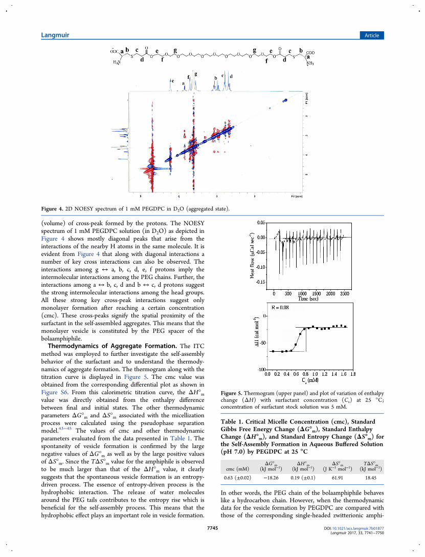
propose vesicle formation by the amphiphiles as shown by thecartoon pictures depicted in Figure 3. The vesicle wall isconstituted by the monolayer arrangement of the PEGDPCmolecules. The constitution of vesicle membrane was alsoconfirmed by 2D NOESY analysis. The NOESY analysis revealsthrough-space interactions among the protons of closeproximity in a molecule as well as in a complex ofmolecules.41,42 The distance of two specific protons (1H−1H)in a NOESY spectrum predominantly controls the intensity
Figure 2. (a) Representative fluorescence emission spectra of NPN in pH 7.0 buffer in the presence of different concentrations of PEGDPC (Cs).(b) Plot of variation of spectral shift (Δλ) of NPN probe in phosphate buffer (20 mM, pH 7.0) with the change in Cs at 25 °C.
Figure 3. Schematic representation of monolayer vesicle structure formed by the monomers.
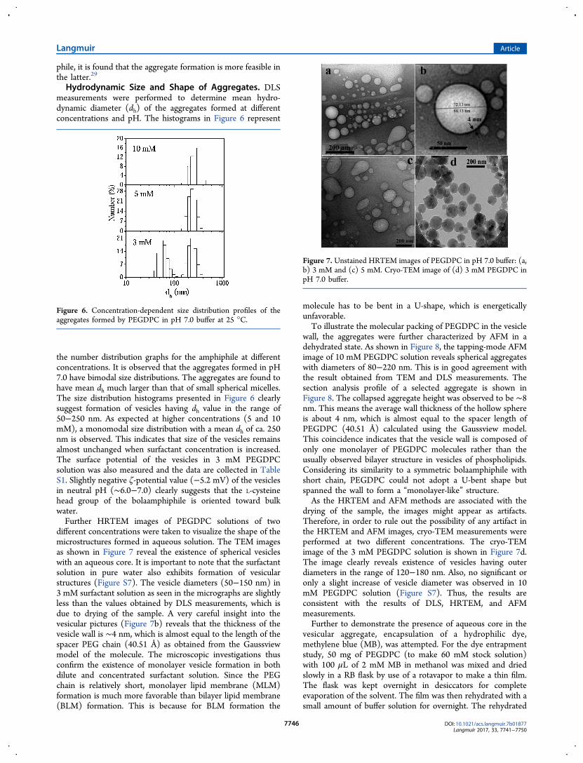
(volume) of cross-peak formed by the protons. The NOESYspectrum of 1 mM PEGDPC solution (in D2O) as depicted inFigure 4 shows mostly diagonal peaks that arise from theinteractions of the nearby H atoms in the same molecule. It isevident from Figure 4 that along with diagonal interactions anumber of key cross interactions can also be observed. Theinteractions among g ↔ a, b, c, d, e, f protons imply theintermolecular interactions among the PEG chains. Further, theinteractions among a ↔ b, c, d and b ↔ c, d protons suggestthe strong intermolecular interactions among the head groups.All these strong key cross-peak interactions suggest onlymonolayer formation after reaching a certain concentration(cmc). These cross-peaks signify the spatial proximity of thesurfactant in the self-assembled aggregates. This means that themonolayer vesicle is constituted by the PEG spacer of thebolaamphiphile.Thermodynamics of Aggregate Formation. The ITC
method was employed to further investigate the self-assemblybehavior of the surfactant and to understand the thermody-namics of aggregate formation. The thermogram along with thetitration curve is displayed in Figure 5. The cmc value wasobtained from the corresponding differential plot as shown inFigure S6. From this calorimetric titration curve, the ΔH°mvalue was directly obtained from the enthalpy differencebetween final and initial states. The other thermodynamicparameters ΔG°m and ΔS°m associated with the micellizationprocess were calculated using the pseudophase separationmodel.43−45 The values of cmc and other thermodynamicparameters evaluated from the data presented in Table 1. Thespontaneity of vesicle formation is confirmed by the largenegative values of ΔG°m as well as by the large positive valuesof ΔS°m. Since the TΔS°m value for the amphiphile is observedto be much larger than that of the ΔH°m value, it clearlysuggests that the spontaneous vesicle formation is an entropy-driven process. The essence of entropy-driven process is thehydrophobic interaction. The release of water moleculesaround the PEG tails contributes to the entropy rise which isbeneficial for the self-assembly process. This means that thehydrophobic effect plays an important role in vesicle formation.
In other words, the PEG chain of the bolaamphiphile behaveslike a hydrocarbon chain. However, when the thermodynamicdata for the vesicle formation by PEGDPC are compared withthose of the corresponding single-headed zwitterionic amphi-
Figure 4. 2D NOESY spectrum of 1 mM PEGDPC in D2O (aggregated state).
Figure 5. Thermogram (upper panel) and plot of variation of enthalpychange (ΔH) with surfactant concentration (Cs) at 25 °C;concentration of surfactant stock solution was 5 mM.
Table 1. Critical Micelle Concentration (cmc), StandardGibbs Free Energy Change (ΔG°m), Standard EnthalpyChange (ΔH°m), and Standard Entropy Change (ΔS°m) forthe Self-Assembly Formation in Aqueous Buffered Solution(pH 7.0) by PEGDPC at 25 °C
phile, it is found that the aggregate formation is more feasible inthe latter.29
Hydrodynamic Size and Shape of Aggregates. DLSmeasurements were performed to determine mean hydro-dynamic diameter (dh) of the aggregates formed at differentconcentrations and pH. The histograms in Figure 6 represent
the number distribution graphs for the amphiphile at differentconcentrations. It is observed that the aggregates formed in pH7.0 have bimodal size distributions. The aggregates are found tohave mean dh much larger than that of small spherical micelles.The size distribution histograms presented in Figure 6 clearlysuggest formation of vesicles having dh value in the range of50−250 nm. As expected at higher concentrations (5 and 10mM), a monomodal size distribution with a mean dh of ca. 250nm is observed. This indicates that size of the vesicles remainsalmost unchanged when surfactant concentration is increased.The surface potential of the vesicles in 3 mM PEGDPCsolution was also measured and the data are collected in TableS1. Slightly negative ζ-potential value (−5.2 mV) of the vesiclesin neutral pH (∼6.0−7.0) clearly suggests that the L-cysteinehead group of the bolaamphiphile is oriented toward bulkwater.Further HRTEM images of PEGDPC solutions of two
different concentrations were taken to visualize the shape of themicrostructures formed in aqueous solution. The TEM imagesas shown in Figure 7 reveal the existence of spherical vesicleswith an aqueous core. It is important to note that the surfactantsolution in pure water also exhibits formation of vesicularstructures (Figure S7). The vesicle diameters (50−150 nm) in3 mM surfactant solution as seen in the micrographs are slightlyless than the values obtained by DLS measurements, which isdue to drying of the sample. A very careful insight into thevesicular pictures (Figure 7b) reveals that the thickness of thevesicle wall is ∼4 nm, which is almost equal to the length of thespacer PEG chain (40.51 Å) as obtained from the Gaussviewmodel of the molecule. The microscopic investigations thusconfirm the existence of monolayer vesicle formation in bothdilute and concentrated surfactant solution. Since the PEGchain is relatively short, monolayer lipid membrane (MLM)formation is much more favorable than bilayer lipid membrane(BLM) formation. This is because for BLM formation the
molecule has to be bent in a U-shape, which is energeticallyunfavorable.To illustrate the molecular packing of PEGDPC in the vesicle
wall, the aggregates were further characterized by AFM in adehydrated state. As shown in Figure 8, the tapping-mode AFMimage of 10 mM PEGDPC solution reveals spherical aggregateswith diameters of 80−220 nm. This is in good agreement withthe result obtained from TEM and DLS measurements. Thesection analysis profile of a selected aggregate is shown inFigure 8. The collapsed aggregate height was observed to be ∼8nm. This means the average wall thickness of the hollow sphereis about 4 nm, which is almost equal to the spacer length ofPEGDPC (40.51 Å) calculated using the Gaussview model.This coincidence indicates that the vesicle wall is composed ofonly one monolayer of PEGDPC molecules rather than theusually observed bilayer structure in vesicles of phospholipids.Considering its similarity to a symmetric bolaamphiphile withshort chain, PEGDPC could not adopt a U-bent shape butspanned the wall to form a “monolayer-like” structure.As the HRTEM and AFM methods are associated with the
drying of the sample, the images might appear as artifacts.Therefore, in order to rule out the possibility of any artifact inthe HRTEM and AFM images, cryo-TEM measurements wereperformed at two different concentrations. The cryo-TEMimage of the 3 mM PEGDPC solution is shown in Figure 7d.The image clearly reveals existence of vesicles having outerdiameters in the range of 120−180 nm. Also, no significant oronly a slight increase of vesicle diameter was observed in 10mM PEGDPC solution (Figure S7). Thus, the results areconsistent with the results of DLS, HRTEM, and AFMmeasurements.Further to demonstrate the presence of aqueous core in the
vesicular aggregate, encapsulation of a hydrophilic dye,methylene blue (MB), was attempted. For the dye entrapmentstudy, 50 mg of PEGDPC (to make 60 mM stock solution)with 100 μL of 2 mM MB in methanol was mixed and driedslowly in a RB flask by use of a rotavapor to make a thin film.The flask was kept overnight in desiccators for completeevaporation of the solvent. The film was then rehydrated with asmall amount of buffer solution for overnight. The rehydrated
Figure 6. Concentration-dependent size distribution profiles of theaggregates formed by PEGDPC in pH 7.0 buffer at 25 °C.
Figure 7. Unstained HRTEM images of PEGDPC in pH 7.0 buffer: (a,b) 3 mM and (c) 5 mM. Cryo-TEM image of (d) 3 mM PEGDPC inpH 7.0 buffer.
suspension was vortexed for 30 min followed by dilution withpH 7.0 buffer to attain 0.1 mM MB (λmax = 665 nm). Theresulting solution (2 mL) was then loaded into a columnpacked with Sephadex G-75 (25 cm height and 1.2 cmdiameter) pre-equilibrated in the eluting buffer. The elutionwas carried out using pH 7.0 buffer, and the eluent wascollected in 2 mL fraction each. The vesicular suspensionseluted just after the void volume. However, the filtration wascontinued to gel filter all the free MB dye. The absorbance forall the fractions was measured at 665 nm. The data are plottedagainst the elution volume as shown in Figure S8. Theencapsulation was confirmed by the characteristic UV spectrumof MB in water. The peak with low absorbance corresponds tothe vesicle entrapped dye molecule, whereas the peak of veryhigh absorbance corresponds to the free dye. It was found thatapproximately 1.75% of the total dye was entrapped into a smallinitial portion containing vesicles. This study clearly demon-strates that the vesicles contain inner aqueous pool which canentrap water-soluble drugs.Stability of the Vesicles. Temperature has an important
factor of self-assembly formation, and it also affects the size andshape of self-assembled aggregate. Since the microenvironmentof the vesicle membrane formed by the PEG spacer is slightlypolar in character, it can be concluded that the PEG chains inthe membrane are partially hydrated. Thus, heating of thePEGDPC solution is expected to cause dehydration of the PEG
chains and hence change the membrane rigidity. Thefluorescence anisotropy of DPH probe was used to determinethe phase transition, if any, of the vesicle membrane. Figure 9adepicts the variation of r with temperature. As seen, the r valuedecreases with the increase of temperature, but the limitingvalue of r lies in the vesicular range, even at 80 °C. Theexistence of vesicle phase at 80 °C was confirmed by the TEMimage depicted in the inset of Figure 9a. This suggests that thePEG chains become more fluid and hence leaky at elevatedtemperatures. This is due to weakening of the hydrophobicinteractions caused by the thermal motion among PEG chains.The temperature corresponding to the inflection point of thecurves can be taken as the melting or phase transitiontemperature, Tm, of the membrane. The high meltingtemperature, 48 °C for PEGDPC, clearly suggests that thevesicles are quite stable at the physiological temperature (37°C).Since the surfactant headgroup is zwitterionic in nature at pH
∼6.0−7.0, the stability of the vesicles was studied by varying pHof the medium. The pH-stability measurement was carried outby measuring r of DPH probe in surfactant solutions ofdifferent pH. The pH of the solution containing 20 mMPEGDPC was varied from 2 to 12 at 25 °C. Each solution wasincubated for 30 min prior to the measurement. Figure 9bshows the variation of r values with the change in pH of thesurfactant solution. It is observed that both decrease and
Figure 8. (a) AFM height image, (b) the energy-minimized structure of PEGDPC in solution phase (water) showing the spacer length, (c) AFMimage with scale bar, and (d) 3D sectional analysis of PEGDPC solution (10 mM, pH 7.0) on mica.
Figure 9. Plots of variation of fluorescence anisotropy (r) of DPH as a function of (a) temperature (inset: TEM image at 80 °C) and (b) pH.
increase of solution pH result in a decrease of r value and showa maximum at pH ∼6.0−7.0. This suggests that the MLMbecomes less rigid at both lower and higher pHs as a result ofweakening of the packing of the PEG units. The weak packingof the PEG units results from increased electrostatic repulsionbetween anionic or cationic head groups of the amphiphiles atthe surface. This is associated with the change in ionizationequilibrium with pH. The pKa values for the proton transferequilibria were obtained from the inflection point of the pHtitration curves. The pKa values thus obtained are 4.8 and 8.4for cation−zwitterion and zwitterion−anion equilibrium,respectively. The pI value calculated from the respective pKavalue is ∼6.6, which means that at around neutral pH thesurfactants remain in the zwitterionic form and the aggregatesexist either as uncharged or weakly charged entities.In order to investigate the change in vesicle structure with
pH, DLS measurements were performed with the PEGDPCsolutions of varying pH. The existence of large aggregates inhigher as well as in lower pH is confirmed by the sizedistribution histograms (Figure S9) of the correspondingsolution. The size distribution histograms show that withincrease or decrease in pH from 7.0 the aggregate sizedecreases. The aggregate size is highest in pH 7.0 and isconsistent with the zwitterionic headgroup of the surfactant atpH 7.0. The existence of vesicles in both lower and higher pHsis demonstrated by the TEM picture (Figure S10) of therespective solution. Indeed, spontaneous vesicle formation wasfound at pH 3.0 and 10.0. However, the vesicle size becomessmaller in comparison to that in neutral pH, which is consistentwith the size distribution histograms in Figure S9. This is due tothe cationic and anionic nature of the amphiphile at acidic andbasic pH, respectively. In both cationic and anionic forms,electrostatic repulsion among the ionic head groups results in aformation of smaller vesicles.However, the vesicles were found to be stable enough at all
the pHs. The stability of the aggregates is manifested by thesurface ζ-potential. A high positive or negative ζ-potential valuesuggests repulsive interaction among particles and hence theirstability against flocculation or coagulation, whereas a low ζ-potential value indicates that the dispersion tends to collapseleading to flocculation or coagulation. Therefore, ζ values of thevesicles in 3 mM PEGDPC were measured at different pHs.The data are listed in Table S1. The results clearly indicate thatat pH 3.0 the surface charge is positive and at pH 10.0 thesurface charge is negative, whereas at around pH 6.0−7.0, thesurface charge is slightly negative. This means the vesicles arestable in a wide range of pH. Since the zwitterionic vesicles ataround neutral pH have tendencies to either grow in size orcoagulate producing precipitates, turbidity (τ) measurement ofthe surfactant solution (3 mM, pH 7.0) was monitored over aperiod of two months at 400 nm to ensure vesicle stability.However, the plot in Figure S11 shows only a slight increase inturbidity for the surfactant solution and can be attributed to thedevelopment of the vesicles. This is supported by the sizedistribution profiles (Figure S12) of the surfactant solution atdifferent time intervals. The monolayer vesicles in 3 mMPEGDPC solution are found to be highly stable even after twomonths. The excellent stability of the vesicles makes thempotential candidate for drug delivery applications.To investigate whether there is any effect of salt on the
stability of monolayer vesicles, fluorescence anisotropy of DPHin vesicle solution of PEGDPC was measured at different NaClconcentrations. The plot in Figure S13 shows only a slight
increase of r value with the rise of [NaCl]. The slight increaseof r value of DPH at [NaCl] = 200 mM suggests that themonolayer vesicles remain unaffected even in the presence ofmoderately high salt concentration. The size distributionhistogram of 5 mM PEGDPC in the presence of 200 mMNaCl has been depicted as inset in Figure S13. As seen, the sizeof the monolayer vesicles also remains unaltered even at highNaCl concentration.
4. CONCLUSIONS
In summary, in this work, a novel L-cysteine-derivedbolaamphiphile (PEGDPC) with PEG as spacer was developedand characterized. The solution behavior of the amphiphile wasinvestigated in different pHs and temperatures. Despite havingso-called polar PEG spacer, the molecule exhibits a reasonablygood surface activity in water. Different techniques includingfluorescence spectroscopy, DLS, TEM, and AFM confirmformation of monolayer vesicles by the amphiphilic moleculesin neutral, acidic, and basic pHs. The surfactant monomersorganize themselves to form monolayer vesicle in very dilute aswell as in concentrated solution. To the best of our knowledge,this is the first report on vesicle formation by a bolaamphiphilecontaining PEG as spacer chain. The thermodynamic datasuggest that the driving force for vesicle formation is ahydrophobic effect. The monolayer vesicles were observed tobe fairly stable with respect to increase of surfactantconcentration and temperature. But, the hydrodynamic size ofthe monolayer vesicles is found to decrease with both decreaseand increase of solution pH. The vesicle stability underphysiological condition suggests that they can have potentialuse in drug delivery applications.
■ ASSOCIATED CONTENT
*S Supporting InformationThe Supporting Information is available free of charge on theACS Publications website at DOI: 10.1021/acs.lang-muir.7b01877.
Details of synthetic procedure, FT-IR, 1H NMR, and 13CNMR spectra and chemical identification of thesynthesized amphiphiles, representative fluorescenceemission spectra of Py, concentration-dependent aniso-tropy change, MB entrapment, pH-dependent DLS, andHRTEM and cryo-TEM images, salt effect and variationof turbidity with aging (PDF)
ORCIDJoykrishna Dey: 0000-0001-8357-5560NotesThe authors declare no competing financial interest.
■ ACKNOWLEDGMENTS
The authors thank Indian Institute of Technology, Kharagpur,for financial support of this work. We are thankful to Prof.Subhas Chandra Kundu for the DLS and ζ-potential measure-ments.
■ REFERENCES(1) Fuhrhop, H. J.; David, H. H.; Mathieu, J.; Liman, U.; Winter, J.H.; Boekema, E. Bolaamphiphiles and Monolayer Lipid MembranesMade from 1,6,19,24-Tetraoxa-3,21-cyclohexatriacontadiene−2,5,20,23−tetrone. J. Am. Chem. Soc. 1986, 108, 1785−1791.(2) Bohme, P.; Hicke, H.-G.; Boettcher, C.; Fuhrhop, H. J. Reactiveand Rigid Monolayers of Bisaroyl Azide Diamide Bolaamphiphiles onPolyacrylonitrile Surfaces. J. Am. Chem. Soc. 1995, 117, 5824−5828.(3) Escamilla, G. H.; Newkome, G. R. Bolaamphiphiles: From GolfBalls to Fibers. Angew. Chem., Int. Ed. Engl. 1994, 33 (19), 1937−1940.(4) Fuhrhop, H. J.; Fritsch, D. Bolaamphiphiles Form Ultrathin,Porous and Unsymmetric Monolayer Lipid Membranes. Acc. Chem.Res. 1986, 19 (5), 130−137.(5) Mao, G.; Tsao, Y. H.; Tirrell, M.; Davis, T. H.; Hessel, V.; vanEsch, J.; Ringsdorf, H. Monolayers of Bolaform Amphiphiles:Influence of Alkyl Chain Length and Counterions. Langmuir 1994,10 (11), 4174−4184.(6) Fuhrhop, H. J.; Wang, T. Bolaamphiphiles. Chem. Rev. 2004, 104,2901−2937.(7) Kunitake, T.; Okahata, Y. Formation of Stable MonolayerMembranes and Related Structures in Dilute Aqueous Solution fromTwo-headed Ammonium Amphiphiles. J. Am. Chem. Soc. 1979, 101,5231−5234.(8) Newkome, R.; Lin, X. F.; Chen, Y. X.; Escamilla, G. H. Two-Directional Cascade Polymer Synthesis: Effects of Core Variation. J.Org. Chem. 1993, 58, 3123−3129.(9) Fuhrhop, J. H.; Spiroski, D.; Boettcher, C. Molecular MonolayerRods and Tubules Made of.Alpha.-(L-lysine),.omega.-(amino) Bo-laamphiphiles. J. Am. Chem. Soc. 1993, 115, 1600−1601.(10) Shimizu, T.; Masuda, M. Stereochemical Effect of Even−OddConnecting Links on Supramolecular Assemblies Made of 1-Glucosamide Bolaamphiphiles. J. Am. Chem. Soc. 1997, 119, 2812−2818.(11) Roussel, M.; Lognone, V.; Plusquellec, D.; Benvegnu, T.Monolayer Lipid Membrane-Forming Dissymmetrical Bolaamphi-philes Derived from Alginate Oligosaccharides. Chem. Commun.2006, 34, 3622−3624.(12) Guilbot, J.; Benvegnu, T.; Legros, N.; Plusquellec, D.; Dedieu,J.-C.; Gulik, A. Efficient Synthesis of Unsymmetrical Bolaamphiphilesfor Spontaneous Formation of Vesicles and Disks with a Trans-membrane Organization. Langmuir 2001, 17, 613−618.(13) Shimizu, T.; Iwaura, R.; Masuda, M.; Hanada, T.; Yase, K.Internucleobase-Interaction-Directed Self-Assembly of Nanofibersfrom Homo- and Heteroditopic 1,ω-Nucleobase Bolaamphiphiles. J.Am. Chem. Soc. 2001, 123, 5947−5955.(14) Iwaura, R.; Yoshida, K.; Masuda, M.; Yase, K.; Shimizu, T.Spontaneous Fiber Formation and Hydrogelation of NucleotideBolaamphiphiles. Chem. Mater. 2002, 14, 3047−3053.(15) Yin, S.; Song, B.; Liu, G.; Wang, Z.; Zhang, X. Self-Organizationof Polymerizable Bolaamphiphiles Bearing Diacetylene MesogenicGroup. Langmuir 2007, 23, 5936−5941.(16) Kobayashi, H.; Amaike, M.; Jung, J. H.; Friggeri, A.; Shinkai, S.;Reinhoudt, D. N. Organogel or Polymer Gel; Facilitated Gelation of aSugar-Based Organic Gel by the Addition of a Boronic Acid-AppendedPolymer. Chem. Commun. 2001, 1038−1039.(17) Shimizu, T.; Kogiso, M.; Masuda, M. Noncovalent Formation ofPolyglycine II-Type Structure by Hexagonal Self-Assembly of LinearPolymolecular Chains. J. Am. Chem. Soc. 1997, 119, 6209−6210.(18) Shimizu, T.; Kogiso, M.; Masuda, M. Vesicle Assembly inMicrotubes. Nature 1996, 383, 487−488.(19) Claussen, C. R.; Rabatic, M. B.; Stupp, I. S. Aqueous Self-Assembly of Unsymmetric Peptide Bolaamphiphiles into Nanofiberswith Hydrophilic Cores and Surfaces. J. Am. Chem. Soc. 2003, 125,12680−12681.(20) Ambrosi, M.; Fratini, E.; Alfredsson, V.; Ninham, W. B.; Giorgi,R.; Lo Nostro, P.; Baglioni, P. Nanotubes from a Vitamin C-BasedBolaamphiphile. J. Am. Chem. Soc. 2006, 128 (22), 7209−7214.
(21) Ray, S.; Das, A. K.; Banerjee, A. Self-Assembly Fibrillar NetworkGels of Simple Surfactants in Organic Solvents. Chem. Mater. 2007, 19(7), 1633−1639.(22) Fariya, M.; Jain, A.; Dhawan, V.; Shah, S.; Nagarsenker, S. M.Bolaamphiphiles: A Pharmaceutical Review. Adv. Pharm. Bull. 2015, 4(2), 483−491.(23) Knop, K.; Hoogenboom, R.; Fischer, D.; Schubert, S. U.Poly(ethylene glycol) in Drug Delivery: Pros and Cons as well asPotential Alternatives. Angew. Chem., Int. Ed. 2010, 49, 6288−6308.(24) Svenson, S.; Tomalia, A. D. Dendrimers in BiomedicalApplications–Reflections on the Field. Adv. Drug Delivery Rev. 2005,57, 2106−2129.(25) Tasaki, K. Poly(oxyethylene)−Water Interactions: A MolecularDynamics Study. J. Am. Chem. Soc. 1996, 118, 8459−8469.(26) Elbert, D. L.; Hubbell, J. A. Surface Treatments of Polymer forBiocompatibility. Annu. Rev. Mater. Sci. 1996, 26, 365−394.(27) Dey, J.; Shrivastava, S. Can Molecules with Anionic Head andPoly(ethylene glycol) methyl ether Tail Self-assemble in Water? ASurface tension, Fluorescence probe, Light scattering, and Trans-mission Electron Mmicroscopic Investigation. Soft Matter 2012, 8,1305−1308.(28) Dey, J.; Shrivastava, S. Physicochemical Characterization andSelf-Assembly Studies on Cationic Surfactants bearing mPEG Tail.Langmuir 2012, 28, 17247−17255.(29) Ghosh, R.; Dey, J. Vesicle Formation by L-Cysteine-DerivedUnconventional Single- Tailed Amphiphiles in Water: A Fluorescence,Microscopy, and Calorimetric Investigation. Langmuir 2014, 30,13516−13524.(30) Ghosh, R.; Dey, J. Aggregation Behavior of Poly(ethyleneglycol) Chain-Containing Anionic Amphiphiles: Thermodynamic,Spectroscopic and Microscopic Studies. J. Colloid Interface Sci. 2015,451, 53−62.(31) Roy, S.; Mohanty, A.; Dey, J. Microviscosity of Bilayermembranes of some N-Acylamino Acid Surfactants determined byFluorescence Probe Method. Chem. Phys. Lett. 2005, 414, 23−27.(32) Lakowicz, J. R. Principles of Fluorescence Spectroscopy; PlenumPress: New York, 1983; p 132.(33) Frisch, M. J.; Trucks, G. W.; Schlegel, H. B.; Scuseria, G. E.;Robb, M. A.; Cheeseman, J. R.; Scalmani, G.; Barone, V.; Mennucci,B.; Petersson, G. A. Gaussian 09, revision B.01; Gaussian, Inc.:Wallingford, CT, 2010.(34) Rosen, J. M.; Mathias, H. J.; Davenport, L. Aberrant AggregationBehavior in Cationic Gemini Surfactants Investigated by SurfaceTension, Interfacial Tension, and Fluorescence Methods. Langmuir1999, 15, 7340−7346.(35) Rosen, M.; Li, F.; Morrall, S. W.; Versteeg, D. J. TheRelationship between the Interfacial Properties of Surfactants andTheir Toxicity to Aquatic Organisms. Environ. Sci. Technol. 2001, 35,954−959.(36) Ghosh, R.; Dey, J. Vesicle-to-Micelle Transition in AqueousSolutions of L-Cysteine- Derived Carboxylate Surfactants ContainingBoth Hydrocarbon and Poly(ethylene glycol) Tails. Langmuir 2017,33, 543−552.(37) Kalyanasundaram, K.; Thomas, J. K. Environmental Effects onVibronic Band Intensities in Pyrene Monomer Fluorescence and TheirApplication in Studies of Micellar Systems. J. Am. Chem. Soc. 1977, 99,2039−2044.(38) Kalyansundaram, K. Photophysics of Microheterogeneous Systems;Academic Press: New York, 1988.(39) Zana, R.; In, M.; Levy, H.; Duportail, G. Alkanediyl-α,ω-bis(dimethylalkylammonium bromide). 7. Fluorescence ProbingStudies of Micelle Micropolarity and Microviscosity. Langmuir 1997,13, 5552−5557.(40) Laskar, P.; Dey, J.; Ghosh, K. S. Evaluation of ZwitterionicPolymersomes Spontaneously Formed by pH-Sensitive and Biocom-patible PEG Based Random Copolymers as Drug Delivery Systems.Colloids Surf., B 2016, 139, 107−116.(41) Tao, W.; Liu, Y.; Jiang, B.; Yu, S.; Huang, W.; Zhou, Y.; Yan, D.A Linear-Hyperbranched Supramolecular Amphiphile and Its Self-
Assembly into Vesicles with Great Ductility. J. Am. Chem. Soc. 2012,134, 762−764.(42) Napoli, A.; Valentini, M.; Tirelli, N.; Muller, M.; Hubbell, A. J.Oxidation-Responsive Polymeric Vesicles. Nat. Mater. 2004, 3, 183−189.(43) Garidel, P.; Hildebrand, A.; Neubert, R.; Blume, A.Thermodynamic Characterization of Bile Salt Aggregation as aFunction of Temperature and Ionic Strength Using IsothermalTitration Calorimetry. Langmuir 2000, 16, 5267−5275.(44) Majhi, P.; Moulik, S. Energetics of Micellization: Reassessmentby a High-Sensitivity Titration Microcalorimeter. Langmuir 1998, 14,3986−3990.(45) Bhattacharya, S.; Haldar, J. Thermodynamics of Micellization ofMultiheaded Single-Chain Cationic Surfactants. Langmuir 2004, 20,7940−7947.