Analysis of the surface labelling characteristics of human spermatozoa and the interaction with anti-sperm antibodies R. J. Aitken, M. J. Hulme, C. J. Henderson, T. B. Hargreave and A. Ross MRC Units of Reproductive Biology and ^Clinical and Population Cytogenetics and %Department of Urology, University of Edinburgh, U.K. Summary. Washed ejaculated human spermatozoa were surface labelled with 125I, using solid phase (iodogen) or enzymic (lactoperoxidase) methods, while membrane components possessing terminal galactose or galactosamine residues were labelled with the galactose oxidase\p=n-\sodium [3H]borohydride technique. All three procedures revealed the presence of 2 major labelled surface components. The first comprised a broad band of radioactivity migrating just behind the ion front on SDS-PAGE, which could be extracted with chloroform and methanol, suggesting a lipid-like composition. The second fraction exhibited properties consistent with a major glycoprotein component of the human sperm plasma membrane, giving a peak of radioactivity with Mr = 20 000, within which a discrete doublet of bands (Mr = 17 000 and 19 000) could be resolved by autoradiography. A more detailed analysis of the labelled protein frac- tion after TCA precipitation revealed a number of other surface components, the major ones of which exhibited Mr values of 30 000, 45 000, 66 000, 115 000000 and 160 000. Western blot analysis was then used to determine whether any of the surface com- ponents described above interacted with the \g=g\-globulin fraction of antisera obtained from patients exhibiting idiopathic autoimmunity against sperm antigens. Using a purified membrane preparation as the target, antibodies were detected against numer- ous high molecular weight bands with Mr values similar to the major components of the human sperm surface (35 000, 45 000, 66 000, 90 000 and 150 000). The nature of the antigens targeted by these antisera did not correlate with the ability of the latter to stimulate or suppress sperm\p=n-\oocyte fusion. Introduction The biological functions of the mammalian spermatozoon depend heavily upon the properties of the plasma membrane, which must not only control the passage of ions into the interior of the cell (Bradley & Forrester, 1980; Ashraf et al., 1982; Breitbart & Rubinstein, 1983) but must also interact with constituents of the female genital tract in order to activate those mechanisms responsible for capacitation and the induction of the acrosome reaction (O'Rand, 1977; Ebenshade & Clegg, 1980; Voglmayr et al., 1980). The sperm plasma membrane is also involved in two inde¬ pendent cell recognition events associated with the process of fertilization. The first involves the attachment of the spermatozoon to the surface of the zona pellucida, as a result of modifications to the sperm surface which take place in the epididymis (Saling, 1982; Shur & Hall, 1982). The second event concerns the capacity of the equatorial segment of the sperm head to recognize and fuse with *Address for reprints: Dr R. J. Aitken, MRC Unit of Reproductive Biology, Centre for Reproductive Biology, 37 Chalmers Street, Edinburgh EH3 9EW, U.K. Downloaded from Bioscientifica.com at 03/23/2022 02:01:27PM via free access
Transcript
Analysis of the surface labelling characteristics of humanspermatozoa and the interaction with anti-sperm
antibodiesR. J. Aitken, M. J. Hulme, C. J. Henderson, T. B. Hargreave and A. Ross
MRC Units of Reproductive Biology and ^Clinical and Population Cytogenetics and%Department of Urology, University ofEdinburgh, U.K.
Summary. Washed ejaculated human spermatozoa were surface labelled with 125I,using solid phase (iodogen) or enzymic (lactoperoxidase) methods, while membranecomponents possessing terminal galactose or galactosamine residues were labelled withthe galactose oxidase\p=n-\sodium[3H]borohydride technique. All three proceduresrevealed the presence of 2 major labelled surface components. The first comprised a
broad band of radioactivity migrating just behind the ion front on SDS-PAGE, whichcould be extracted with chloroform and methanol, suggesting a lipid-like composition.The second fraction exhibited properties consistent with a major glycoproteincomponent of the human sperm plasma membrane, giving a peak of radioactivity withMr = 20 000, within which a discrete doublet of bands (Mr = 17 000 and 19 000) couldbe resolved by autoradiography. A more detailed analysis of the labelled protein frac-tion after TCA precipitation revealed a number of other surface components, the majorones of which exhibited Mr values of 30 000, 45 000, 66 000, 115000000 and 160 000.
Western blot analysis was then used to determine whether any of the surface com-
ponents described above interacted with the \g=g\-globulinfraction of antisera obtainedfrom patients exhibiting idiopathic autoimmunity against sperm antigens. Using a
purified membrane preparation as the target, antibodies were detected against numer-
ous high molecular weight bands with Mr values similar to the major components ofthe human sperm surface (35 000, 45 000, 66 000, 90 000 and 150 000). The nature ofthe antigens targeted by these antisera did not correlate with the ability of the latter tostimulate or suppress sperm\p=n-\oocytefusion.
Introduction
The biological functions of the mammalian spermatozoon depend heavily upon the properties ofthe plasma membrane, which must not only control the passage of ions into the interior of the cell(Bradley & Forrester, 1980; Ashraf et al., 1982; Breitbart & Rubinstein, 1983) but must alsointeract with constituents of the female genital tract in order to activate those mechanismsresponsible for capacitation and the induction of the acrosome reaction (O'Rand, 1977; Ebenshade& Clegg, 1980; Voglmayr et al., 1980). The sperm plasma membrane is also involved in two inde¬pendent cell recognition events associated with the process of fertilization. The first involves theattachment of the spermatozoon to the surface of the zona pellucida, as a result of modifications tothe sperm surface which take place in the epididymis (Saling, 1982; Shur & Hall, 1982). The secondevent concerns the capacity of the equatorial segment of the sperm head to recognize and fuse with
*Address for reprints: Dr R. J. Aitken, MRC Unit of Reproductive Biology, Centre for Reproductive Biology, 37Chalmers Street, Edinburgh EH3 9EW, U.K.
Downloaded from Bioscientifica.com at 03/23/2022 02:01:27PMvia free access
the vitelline membrane of the oocyte, as a result of changes in the sperm plasma membrane whichoccur concomitant with the acrosome reaction (Yanagimachi, 1981).
In a clinical context, analysis of the human sperm plasma membrane is of importance in boththe development of new approaches to immunocontraception (Aitken, 1982) and in understandingthe nature and origins of idiopathic autoimmunity directed against sperm antigens (Poulsen &Hjort, 1981; Lee et ai, 1982; Naaby-Hansen & Bjerrum, 1985).
Despite the fundamental and clinical importance of this research area, there have been very fewformal studies concerning the composition of the human sperm surface and the nature of thecomponents targeted by autoantibodies capable of disrupting human sperm function (Young &Goodman, 1980; Dor et al., 1981). In the present study we have sought to extend our knowledge ofthis area by using a variety of membrane isolation and surface labelling techniques to determine thenature and orientation of the major constituents of the human sperm plasma membrane. Westernblot analysis was then used to determine which of these membrane components interact with anti-sperm autoantibodies, previously characterized for their ability to influence human sperm functionin vitro.
Materials and Methods
Semen donation. Human spermatozoa were collected from healthy donors exhibiting normal semen profiles(>20 106 spermatozoa/ml, >40% progressive motility and >40% normal morphology) into sterile plastic con¬tainers. After allowing at least 30 min for liquefaction to occur the spermatozoa were separated from seminal plasmaby 3 cycles of centrifugation and resuspension in 8-ml volumes of Medium BWW (Biggers et ai, 1971). The finalsperm pellet was resuspended in 1 ml Medium BWW, analysed to determine the concentration of spermatozoa usingan improved Neubauer haemocytometer and finally diluted to a concentration of 20 IO6 cells/ml.
Anti-sperm antibodies. Antisera were obtained from patients exhibiting infertility associated with idiopathicautoimmunity against sperm antigens. The Tray Agglutination Test (TAT) was used to detect the presence of anti-sperm antibodies in samples of heat-inactivated (56°C for 30 min) blood serum. The assay was performed accordingto the protocol described by Friberg (1974) with the minor modifications introduced by Jager et al. (1978). Seriallydiluted heat-inactivated samples were incubated with donor spermatozoa in microchamber trays under paraffin oil at37°C and the results were read after 2 h using an inverted microscope.
The presence of antisperm antibodies was also assessed using immunobeads coated with anti-human lgG or IgA(Biorad Laboratories, Watford, Herts). To perform this assay 200 µ of a donor sperm suspension (10 106 cells/ml)were incubated with 20 µ heat-inactivated antiserum for 3 h; 5 µ of this preparation were then mixed with 5 µ of thewashed immunobeads on a microscope slide and incubated for 10 min before examination by phase-contrastmicroscopy.
Sperm function tests. The ability of human spermatozoa, obtained from 12 individual donors, to recognize andfuse with the vitelline membrane of the oocyte was assessed using the zona-free hamster egg penetration test(Yanagimachi et ai, 1976) following the induction of the acrosome reaction wih A23187 (Aitken et al., 1984). The -globulin fraction from each autoimmune antiserum was isolated by sodium sulphate precipitation (Aitken et al.,1982) to yield preparations containing 5 mg -globulin/ml. These preparations were then incubated at a dilution of1:10 with spermatozoa (20 106/ml) for 1 h before the addition of A23187, prepared as an aqueous suspension ofthe Ca2 + /Mg2T salt (0026 mg/ml; Aitken et ai, 1984). After a further incubation period of 2 h the spermatozoa werepelleted by centrifugation at 500 g for 5 min and resuspended in Medium BWW at a concentration of 10 106/ml. Atthis point in the procedure, the percentage of motile spermatozoa was estimated with the aid of a grid on an eye-piecegraticule. Zona-free hamster oocytes were then introduced into these sperm suspensions and after 3 h they were scoredfor the presence of decondensing sperm heads with an attached or closely associated tail. Control incubationscontained the same concentration of -globulin from non-immune donors.
The influence of the antisera on the ability of human spermatozoa to penetrate human cervical mucus was assessedusing the procedure described by Katz et al. (1980) as modified by Aitken et al. (1985).
Isolation ofmembrane vesicles. Sperm samples were prepared as described above and resuspended in 1 mM-EDTAOpH 5-0) containing a protease inhibitor, 1 mM-phenylmethyl sulphonyl fluoride (PMSF; Sigma, MO, U.S.A.). Thesperm membranes were detached using an ultrasonicator (Soniprep, MSE, Crawley, Sussex, U.K.) using 3 10-secbursts, with a probe amplitude of 3 µ , keeping the tube on ice between bursts to minimize heating effects. Thissonication protocol was established experimentally using several different combinations of probe amplitude anddurations of treatment, assessing the degree of plasma membrane removal and cell disruption by electron microscopy.
The ultrasonicated suspension was centrifuged at 500 g to pellet the intact spermatozoa and the supernatant was
again centrifuged at 3000g to remove any residual cells. The supernatant was removed and centrifuged at 100 000 gfor 1 h on an ultracentrifuge (Sorvall OTD-50 Du Pont Instruments, Stevenage, Herts, U.K.) to yield a crude mem¬brane preparation. After the method of Gillis et al. (1978) for the preparation of human sperm plasma membranes.
Downloaded from Bioscientifica.com at 03/23/2022 02:01:27PMvia free access
the crude membrane pellet was resuspended in 0-25 M-sucrose and layered onto a discontinuous sucrose gradient(1-57 M, 1-3 M, 10m sucrose) and centrifuged at 100 000g for 2h. The membrane fraction was collected as a discretelayer at the 10 M-sucrose interface, diluted in Dulbecco's phosphate-buffered saline (PBS; Flow Labs, Irvine, U.K.)and again centrifuged at 100 000 g to pellet the membranes. According to Gillls et al. (1978) the membrane fractioncollected from the 1 -0 M-sucrose interface derives from the plasma membrane in view of its ultrastructural appearance,sedimentation rate, high 5'-nucleotidase and low succinic dehydrogenase activities. We have also found that thisfraction is enriched in NADPH oxidase activity, which is indicative of the plasma membrane. These preparationswere stored at
—
20°C before solubilization in sample buffer for sodium dodecyl sulphate-polyacrylamide gelelectrophoresis (SDS-PAGE).
The ultrastructural appearance of the sperm pellets and isolated membrane fractions was evaluated after fixationwith 5% glutaraldehyde in cacodylate buffer and post-fixation in 1% osmium tetroxide.
Surface labelling of intact spermatozoa. To identify the externally orientated components of the human spermplasma membrane, various surface labelling procedures were used with intact cells.
Exposed tyrosine residues were iodinated with the solid-phase reagent l,3,4,6-tetrachloro-3a,6a-diphenylglyco-luril (Iodogen: Pierce Chemical Company, Rockford, IL, U.S.A.), according to the method of Fraker & Speck (1978)and Markwell & Fox (1978). Typically, 107 spermatozoa in 100 µ PBS were added to a glass tube plated with 100 µgIodogen, and exposed to 500 µ Na125I (Amersham, Bucks, U.K.) for 10 min, agitating the tube occasionally. Thereaction was stopped by the addition of excess 0-25 M-KI and the iodinated spermatozoa were centrifuged (500 g) andwashed 3 times in PBS to remove unlabelled iodine. The surface label was solubilized by the addition of 0-5 ml 1%Triton X-100, containing 10 mM-Tris-HCl, 1 mM-EDTA, and 1 mM-PMSF (pH 80) for 30 min at 4°C. The residualsperm pellet was removed by centrifugation, and the supernatant containing the surface label was stored at 4°C beforeanalysis by gel electrophoresis.
Enzymic iodination of the sperm surface was achieved with lactoperoxidase and adapted from the methoddescribed by Young & Goodman (1980), using 107 spermatozoa and 100 µg lactoperoxidase/ml, activated by theaddition of 10 20 µ samples of 9 10~5% (v/v) hydrogen peroxide at 15-sec intervals. The reaction was stoppedby the addition of excess 0-25 M-KI and the labelled sperm surface was solubilized as described above.
Surface labelling of terminal galactose or galactosamine residues was achieved by the procedure described byGahmberg & Hakomori (1973) and Steck & Dawson (1974). In brief, 1 ml of 107 spermatozoa in PBS was treated with40 i.u. galactose oxidase (Sigma, MO, U.S.A.) and incubated for 30 min at 37°C, after which 0-5 mCi NaB[3H]4(Amersham, Bucks, U.K.) was added, and the mixture was incubated for a further 20 min at 37°C. The reaction was
terminated by the addition of excess PBS, the labelled preparations were washed to remove excess NaB[3H]4 andsolubilized as before. To account for non-specific incorporation of tritium, galactose oxidase was omitted fromcontrol incubations of spermatozoa.
Label fractionation. To remove any radiolabelled lipid or glycolipid components from the above preparations(Teuscher et al., 1982; Klinefelter & Hamilton, 1985) the 125I labelled material was fractionated by the addition of 4volumes of chloroform/methanol mixture (2:1 v/v) to one volume of solubilized label in a glass tube, mixingthoroughly. After the mixture had settled, precipitated protein was removed from the interface between the chloro¬form and methanol and rewashed with the same solvent mixture. The chloroform phases were combined, dried downunder nitrogen, and both this material and the precipitate were independently solubilized in sample buffer beforeSDS-PAGE.
The proteins were also precipitated by the addition of 40% ice-cold trichloroacetic acid (TCA) to the solubilizedlabel to give a final concentration of 10% TCA in the mixture. After incubation for 30 min at 4°C, the precipitatecontaining the labelled proteins/glycoproteins was pelleted by centrifugation at 500 g for 20 min at 4°C. The precipi¬tate was rewashed in 10% TCA, centrifuged again and finally washed with an ethanohether mixture (1:1, v/v). Theorganic phase was dried down as before and both this and the precipitate fraction were prepared for electrophoresis.Protein determinations were carried out using the technique of Lowry et al. (1951).
SDS-polyacrylamide gel electrophoresis (SDS-PAGE). SDS-PAGE was performed according to the method ofLaemmli (1970) using a Protein 1 electrophoresis cell (Bio Rad Labs, Watford, U.K.). The stacking gel was composedof 3-5% acrylamide, while the resolving gel contained 10% or 12% acrylamide. Labelled, or unlabelled samples, andknown molecular weight standards were run under reducing conditions, using 2-mercaptoethanol (5%, v/v) as thereducing agent, and bromophenol blue as indicator. Electrophoresis was performed at a constant current of 60 mAthrough the stacking gel, then at 30 mA through the resolving gel, for 2-3 h, after which the gels were silver stained(Biorad, Watford, Herts, U.K.). For the radiolabelling experiments the sample tracks were carefully cut up into 2 mm
sections, and for the 125I-labelled material, the individual slices were counted in a multihead gamma counter (NuclearEnterprises NE 1600) in 63 11 mm plastic tubes (Sarstedt, Leicester, U.K.). The tritiated gel slices had first to besolubilized with the tissue solubilizer NCS (Amersham, Bucks, U.K.), each slice being incubated with 10 ml of a 10%NCS solution for 2 h at 50°C. For certain experiments gels containing the iodinated samples were silver stained beforeautoradiography ( X -Omat-AR film: Kodak, Manchester, U.K.).
Immunoblotting. The Western blotting experiments were carried out according to the method of Towbin et al.(1979) using a Trans Blot cell and a 160/1-6 Model power pack (Bio-rad, Watford, Herts, U.K.). The transfer bufferused was 25 mM-Tris, 192 mM-glycine, pH 8-3, with the nitrocellulose paper placed anodal to the gels. Typically, thetransfers were performed for 3 h at 60 V and 0-25 A.
The antigens used in these experiments (20-50 µg/track) were either TCA precipitates of purified sperm membrane
Downloaded from Bioscientifica.com at 03/23/2022 02:01:27PMvia free access
vesicles or whole sperm samples treated with 1% deoxycholate (BDH, Glasgow, U.K.) for 1 h at 4°C and then TCAprecipitated as previously described. These materials were produced from samples provided by a randomly selectedpanel of 60 donors exhibiting normal semen profiles. The purpose of the deoxycholate treatment was to solubilize notonly the sperm plasma membrane, but also to release internal antigens, to investigate whether the auto-antibodieswere, in part, directed against such components. For these studies the -globulin fractions from control and auto¬immune antisera were used at a concentration of 0T7mg/ml in Tris-buffered saline (20mM-Tris, 500mM-NaCl, 3%gelatin, pH 7-5).
Results
Surface labelling studies
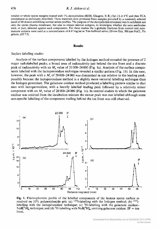
Analysis of the surface components labelled by the Iodogen method revealed the presence of 2major radiolabelled peaks; a broad area of radioactivity just behind the ion front and a discretepeak of radioactivity with an Mr value of 20 000-24 000 (Fig. la). Analysis of the surface compo¬nents labelled with the lactoperoxidase technique revealed a similar pattern (Fig. lb). In this case,however, the peak with a M, of 20 000-24 000 was diminished in size relative to the leading peak,possibly because the lactoperoxidase method is a slightly more vectorial labelling technique thanthe Iodogen procedure. The galactose oxidase method produced a labelling pattern similar to thatseen with lactoperoxidase, with a heavily labelled leading peak followed by a relatively minorcomponent with an M, value of 20 000-24 000 (Fig. lc). In control studies in which the galactoseoxidase was omitted from the incubation mixture the minor peak was not labelled although some
non-specific labelling of the component trailing behind the ion front was still observed.
22 000 IF
(O
2
b 0
X
,2w
(d)
2
96 112 0 16Distance migrated (mm)
80
IF
96
Fig. 1. Electrophoretic profile of the labelled components of the human sperm surface asresolved on 10% polyacrylamide gels, (a) 125I-labelling with the Iodogen method; (b) 125I-labelling with the lactoperoxidase technique; (c) 3H-labelling with the galactose oxidase-NaB[3H]4 technique; and (d) 3H-labelling with NaB[3H]4 omitting galactose oxidase. IF = ionfront.
Downloaded from Bioscientifica.com at 03/23/2022 02:01:27PMvia free access
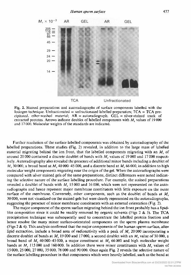
Fig. 2. Stained preparations and autoradiographs of surface components labelled with theIodogen technique. Unfractionated = unfractionated labelled preparation; TCA = TCA pre¬cipitated, ether-washed material; AR = autoradiograph; GEL = silver-stained track ofextracted proteins. Arrows indicate doublet of labelled components with Mr values of 19 000and 17 000. Molecular weights of the standards are indicated.
Further resolution of the surface labelled components was obtained by autoradiography of thelabelled preparations. These studies (Fig. 2) revealed, in addition to the large mass of labelledmaterial migrating behind the ion front, that the labelled components migrating with an ofaround 20 000 contained a discrete doublet of bands with Mr values of 19 000 and 17 000 respect¬ively. Autoradiography also revealed the presence of additional minor bands including a doublet ofMT 30 000, a broad band at Mr 40 000-45 000, and a discrete band at Mr 66 000, in addition to highmolecular weight components migrating near the origin of the gel. When the autoradiographs were
compared with silver stained gels of the same preparations, distinct differences were noted indicat¬ing the selective nature of the surface labelling procedure. For example, the stained preparationsrevealed a doublet of bands with Mr 15 000 and 16 000, which were not represented on the auto¬radiographs and hence represent major membrane constituents with little exposure on the outersurface of the membrane. Conversely, other components, such as the doublet of bands at MT30 000, were not visualized on the stained gels but were clearly represented on the autoradiographs,suggesting the presence of minor membrane constituents with an external orientation (Fig. 2).
The major component of the sperm surface migrating behind the ion front probably has a lipid-like composition since it could be readily removed by organic solvents (Figs 2 & 3). The TCAprecipitation technique was subsequently used to concentrate the labelled protein fraction andhence resolve the many minor surface-orientated components on the sperm plasma membrane(Figs 2 & 4). This analysis confirmed that the major components of the human sperm surface, afterlipid extraction, include a broad area of radioactivity with a peak of Mr 20 000 incorporating adiscrete doublet of bands at 19 000 and 17 000, a second doublet with an MT value of 30 000, a
broad band of Mr 40 000-45 000, a major constituent at M, 66 000 and high molecular weightbands at MT 115 000 and 160 000. In addition there were minor constituents with MT values of15 000, 25 000, 27 000, 35 000, 78 000 and 90 000. Once again Fig. 2 reveals the selective nature ofthe surface labelling procedure in that components which were heavily labelled, such as the band at
Downloaded from Bioscientifica.com at 03/23/2022 02:01:27PMvia free access
24- (a) Total label
66 000
I
0J
24X
Ecio 16
ß-
(b) Organicphase
0J |""r""t' I.V.I"
(c) Precipitate j
20 000 IF
-i-1-1-1-1
li \
I11"!1"*?
-1-1-r-1-1-1 · l r-0 20 40 60 80 100 120
Distance migrated (mm)
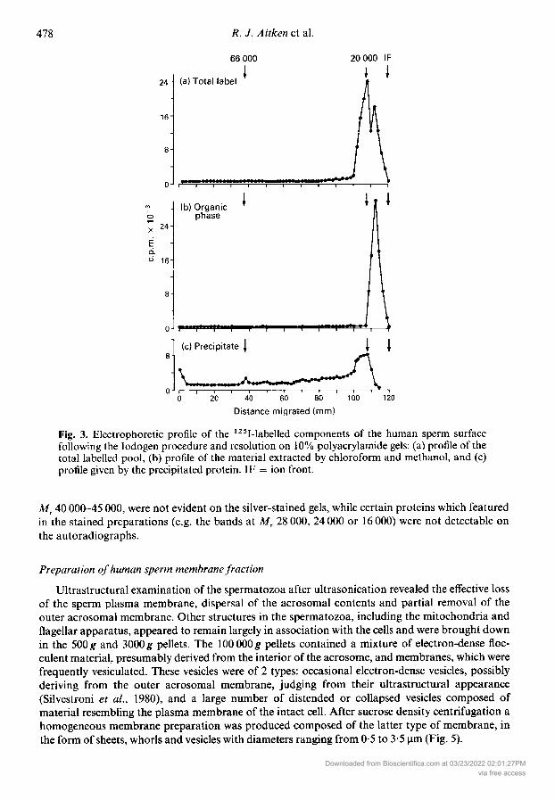
Fig. 3. Electrophoretic profile of the 125I-labelled components of the human sperm surfacefollowing the Iodogen procedure and resolution on 10% polyacrylamide gels: (a) profile of thetotal labelled pool, (b) profile of the material extracted by chloroform and methanol, and (c)profile given by the precipitated protein. IF = ion front.
MT 40 000-45 000, were not evident on the silver-stained gels, while certain proteins which featuredin the stained preparations (e.g. the bands at 28 000, 24 000 or 16 000) were not detectable on
the autoradiographs.
Preparation ofhuman sperm membrane fractionUltrastructural examination of the spermatozoa after ultrasonication revealed the effective loss
of the sperm plasma membrane, dispersal of the acrosomal contents and partial removal of theouter acrosomal membrane. Other structures in the spermatozoa, including the mitochondria andflagellar apparatus, appeared to remain largely in association with the cells and were brought downin the 500g and 3000g pellets. The 100 000g pellets contained a mixture of electron-dense floc-culent material, presumably derived from the interior of the acrosome, and membranes, which were
frequently vesiculated. These vesicles were of 2 types: occasional electron-dense vesicles, possiblyderiving from the outer acrosomal membrane, judging from their ultrastructural appearance(Silvestroni et ai, 1980), and a large number of distended or collapsed vesicles composed ofmaterial resembling the plasma membrane of the intact cell. After sucrose density centrifugation a
homogeneous membrane preparation was produced composed of the latter type of membrane, inthe form of sheets, whorls and vesicles with diameters ranging from 0-5 to 3-5 µ (Fig. 5).
Downloaded from Bioscientifica.com at 03/23/2022 02:01:27PMvia free access
90 78 66IH
40 35302725 18W HI
IFI
16-
12-
40 60 80Distance migrated (mm)
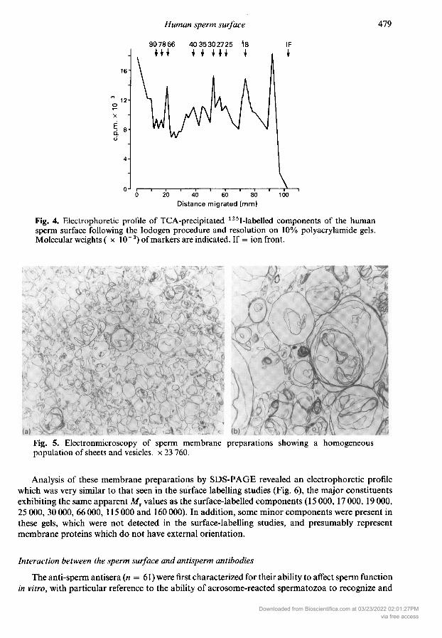
Fig. 4. Electrophoretic profile of TCA-precipitated 125I-labelled components of the humansperm surface following the Iodogen procedure and resolution on 10% polyacrylamide gels.Molecular weights ( IO"3) of markers are indicated. If = ion front.
Fig. 5. Electronmicroscopy of sperm membrane preparations showing a homogeneouspopulation of sheets and vesicles, 23 760.
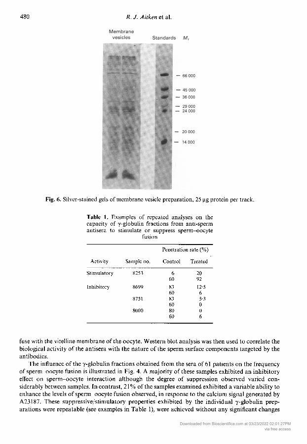
Analysis of these membrane preparations by SDS-PAGE revealed an electrophoretic profilewhich was very similar to that seen in the surface labelling studies (Fig. 6), the major constituentsexhibiting the same apparent values as the surface-labelled components (15 000, 17 000, 19 000,25 000, 30 000, 66 000, 115 000 and 160 000). In addition, some minor components were present inthese gels, which were not detected in the surface-labelling studies, and presumably representmembrane proteins which do not have external orientation.
Interaction between the sperm surface and antisperm antibodies
The anti-sperm antisera ( = 61) were first characterized for their ability to affect sperm functionin vitro, with particular reference to the ability of acrosome-reacted spermatozoa to recognize and
Downloaded from Bioscientifica.com at 03/23/2022 02:01:27PMvia free access
Fig. 6. Silver-stained gels of membrane vesicle preparation, 25 µg protein per track.
Table 1. Examples of repeated analyses on thecapacity of -globulin fractions from anti-spermantisera to stimulate or suppress sperm-oocyte
fusion
Activity Sample no.
Penetration rate (%)Control Treated
Stimulatory
Inhibitory
8253
8699
8751
8000
660836083608060
209212-565-3006
fuse with the vitelline membrane of the oocyte. Western blot analysis was then used to correlate thebiological activity of the antisera with the nature of the sperm surface components targeted by theantibodies.
The influence of the -globulin fractions obtained from the sera of 61 patients on the frequencyof sperm-oocyte fusion is illustrated in Fig. 4. A majority of these samples exhibited an inhibitoryeffect on sperm-oocyte interaction although the degree of suppression observed varied con¬
siderably between samples. In contrast, 21 % of the samples examined exhibited a variable ability toenhance the levels of sperm-oocyte fusion observed, in response to the calcium signal generated byA23187. These suppressive/stimulatory properties exhibited by the individual -globulin prep¬arations were repeatable (see examples in Table 1), were achieved without any significant changes
Downloaded from Bioscientifica.com at 03/23/2022 02:01:27PMvia free access
300i
200
1:10000
1:1000
1:10-"
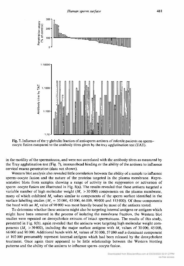
Fig. 7. Influence of the -globulin fraction of anti-sperm antisera of infertile patients on sperm-oocyte fusion compared to the antibody titres given by the tray agglutination test (TAT).
in the motility of the spermatozoa, and were not correlated with the antibody titres as measured bythe Tray agglutination test (Fig. 7), immunobead binding or the ability of the antisera to influencecervical mucus penetration (data not shown).
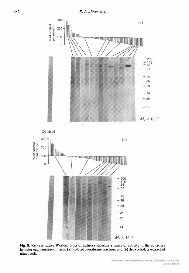
Western blot analysis also revealed little correlation between the ability of a sample to influencesperm-oocyte fusion and the nature of the proteins targeted in the plasma membrane. Repre¬sentative blots from samples showing a range of activity in the suppression or activation ofsperm-oocyte fusion are illustrated in Fig. 8(a). The results revealed that these antisera targeted avariable number of high molecular weight (Mr > 30 000) components on the plasma membrane,many of which exhibited Mr values similar to components of the sperm surface identified in thesurface labelling studies (Mr = 35 000, 45 000, 66 000, 90 000 and 115 000). Of these componentsthe band with an Mr value of 90 000 was most heavily bound by most of the antisera tested.
To determine whether these antisera might also be targeting internal antigens or antigens whichmight have been removed in the process of isolating the membrane fraction, the Western blotstudies were repeated on deoxycholate extracts of intact spermatozoa. The results of this study,presented in Fig. 8(b), again revealed that the antisera were targeting high molecular weight com¬
ponents (Mr > 30 000), including the major surface antigens with Mr values of 30 000, 45 000,66 000 and 90 000. Additional bands with Mr values of 50 000, 57 000 and a dominant componentat 102 000 presumably represent internal antigens which had been released by the deoxycholatetreatment. Once again there appeared to be little relationship between the Western blottingpatterns and the ability of the antisera to influence sperm-oocyte fusion.
Downloaded from Bioscientifica.com at 03/23/2022 02:01:27PMvia free access
Fig. 8. Representative Western blots of antisera showing a range of activity in the zona-freehamster egg penetration tests: (a) isolated membrane fraction, and (b) deoxycholate extract ofintact cells.
Downloaded from Bioscientifica.com at 03/23/2022 02:01:27PMvia free access
Discussion
The surface labelling patterns obtained with human spermatozoa in this study are very similar tothose observed in the rat by Klinefelter & Hamilton (1985) in revealing 2 major peaks of radio¬activity, one with an Mr of around 23 000 and a second migrating just behind the ion front. Thisleading peak was extractable with chloroform and methanol or ether, suggesting a lipid-like consti¬tution. The equivalent fraction on the rat sperm plasma membrane has been definitively identifiedas a glycolipid (Klinefelter & Hamilton, 1985) while a major antigen on the surface of guinea pigspermatozoa has also been shown to possess properties consistent with a glycolipid (Teuscher et ai,1982). The second peak with an Mx value of 20-24 000 could be precipitated with TCA and was
specifically labelled with galactose oxidase-NaB[3H]4 treatment, suggesting that this component isglycoprotein. The Mr 24 000 component on the rat sperm surface is also labelled with this techniqueand has been identified as an rx-lactalbumin-like glycoprotein, which is normally present on thesurface of spermatozoa from the cauda but not the caput epididymidis (Klinefelter & Hamilton,1985).
The function of a-lactalbumin on the surface of the mammalian spermatozoon is currentlyunknown although it has been speculated that this complex is involved in modifying the carbo¬hydrate structure of the sperm surface (Hamilton, 1981), possibly conferring upon the spermatozoathe ability to recognize the zona pellucida. Recent studies by O'Rand et al. (1985) have shown thatheat-solubilized, iodinated human zonae pellucidae bind specifically to two major peptides derivedfrom human spermatozoa with Mr values of 17 000 and 18 000, together with a minor componentat M, = 15 000. It is possible that these components correspond to the major doublet of bandsobserved in the surface labelling studies with M, values of 17 000 and 19 000, together with theminor band at 15 000. The small difference in molecular weights is possibly due to differences in theconditions used for electrophoresis or microheterogeneity in the molecular weights due to differentdegrees of glycosylation (Hamilton, 1981).
The only other formal study of the surface labelling patterns given by human spermatozoa was
undertaken by Young & Goodman (1980). The electrophoretic profile described by these workers,with 5 even peaks of radioactivity with molecular weights ranging from Mr = 20 000 to
MT = 92 000 appears quite unlike the patterns described in this paper and at present we cannotexplain these differences. Although the molecular weights ascribed to the major constituents of thehuman sperm surface by Young & Goodman (1980) do resemble some of the values presented here(the bands with Mr values of 92 000, 72 000, 46 000, 30 000 and 20 000 described by these authorscould be equated with the components at 115 000,66 000,40-45 000, 30 000 and 19 000 identified inthis study) the distribution of radioactivity is different and none of the minor components describedby us were recorded.
The isolation of a membrane-rich fraction from human spermatozoa was achieved following a
protocol previously described by Gillis et ai (1978), with the exception that short bursts of ultra-sonication rather than nitrogen cavitation were used to disrupt the spermatozoa. The end resultappears to be similar, however, with the isolation of a homogeneous population of vesicles from thesucrose gradients exhibiting ultrastructural features, and an electrophoretic profile, characteristicof the plasma membrane.
These membrane preparations were subsequently used to examine the nature of the surfaceantigens targeted by anti-sperm antisera, which had previously been assessed for their ability toinfluence sperm-oocyte fusion in vitro. The antisera revealed an unexpected ability to stimulate orinhibit the levels of sperm-oocyte fusion observed after the induction of the acrosome reaction withA23187. We have previously reported (Dor et ai, 1981) that antisera from the World HealthOrganization's serum bank exhibit a variable capacity to suppress the levels of sperm-oocyte fusionobserved in a system lacking A23187. In contrast, Bronson et ai (1981) have recorded the stimu¬lation of gamete fusion when anti-sperm antisera were added to their zona-free hamster egg system.The findings reported in this study resolve these differences by indicating that populations of both
Downloaded from Bioscientifica.com at 03/23/2022 02:01:27PMvia free access
inhibitory and stimulatory antisera exist within the cohort of patients exhibiting idiopathic auto¬immunity. We have also observed the presence of stimulatory and inhibitory antibodies in theseminal plasma of patients exhibiting autoimmunity following vasovasostomy (data not pre¬sented). The existence of inhibitory antisera was anticipated and presumably reflects the presence ofantibodies capable of masking those surface determinants on the acrosome-reacted human sperm¬atozoon capable of recognizing and initiating fusion with the vitelline membrane of the oocyte. Thepresence of stimulatory antibodies is more difficult to explain. One exciting possibility is that -globulin preparations exhibiting this activity contain antibodies which are able to cross-link, andthence activate, receptor sites on the human sperm surface which are capable of initiating thosebiochemical events normally responsible for inducing membrane fusion and which, in physiologicalcircumstances, interact with complementary receptors on the zona surface (Singer et ai, 1985). Theability of anti-receptor antibodies to induce cell activation in other cell types is well established;antibodies against the insulin receptor, for example, are known to induce the spectrum ofbiochemical changes associated with the action of insulin itself (Zick et ai, 1984).
The ability of the -globulin preparations to stimulate or inhibit sperm-oocyte fusion bore no
relationship to any of the other biological and immunological properties measured for thesesamples. When the isolated membrane fraction was used as substrate, most samples appeared toexhibit a preferential affinity for a band exhibiting an MT value of 90 000 corresponding to one ofthe minor components of the sperm surface identified in the surface labelling studies. Analysis ofthe sperm antigens binding to antisera from the World Health Organization's serum bank by Lee etai (1983) also recorded a high frequency of samples containing antibodies directed against a 90 000molecular weight component. The presence of a high proportion of antisera possessing antibodiesagainst surface components exhibiting Mr values of around 35 000 and 45 000 is also in keepingwith previous studies (Poulson & Hjort, 1981; Czuppon et ai, 1981; Naaby-Hansen & Bjerrum,1985), although the 14 000 molecular weight antigen targeted by the antisera examined byLehmann et al. (1985) was not observed in our study. The lack of a direct correlation betweenthe nature of the bands targeted and the ability of a given antiserum to enhance or suppresssperm-oocyte fusion may simply reflect the fact that these antisera contain a complex mixture ofantibodies with differing, possibly conflicting, biological activities, some of which have not beenassessed by the bioassays used in this study. Such studies emphasize that ascribing defined biologi¬cal roles to specific membrane components will only be possible when an appropriate range ofmonoclonal antibodies has been prepared against the major antigens presented on the surface ofhuman spermatozoon.
We thank Ms Jane Clarkson and Mr David Richardson for their invaluable assistance in thecourse of this project.
References
Aitken, R.J. (1982) The contraceptive potential of anti-sperm antibodies. In Progress towards a Male Contra¬ceptive, pp. 109-134. Eds S. L. Jeffcoate& M. Sandier.John Wiley & Sons, Chichester.
Aitken, R.J., Holme, E., Richardson, D.W. & Hulme, M.(1982) Properties of intact and univalent (Fab) anti¬bodies raised against isolated, solubilized, mousezonae pellucidae. /. Reprod. Fert. 66, 327-334.
Aitken, R.J., Ross, ., Hargreave, T., Richardson, D. &Best, F. (1984) Analysis of human sperm function fol¬lowing exposure to the ionophore A23187. Compari¬son of normospermic and oligozoospermic men. /.Andro!. 5, 321-329.
Aitken, R.J., Surton, M., Warner, P. & Richardson, D.W.(1985) Relationship between the movement charac-
teristics of human spermatozoa and their ability topenetrate cervical mucus and zona-free hamsteroocytes. J. Reprod. Fert. 73, 441^149.
Ashraf, M., Peterson, R.N. & Russell, L.D. (1982)Activity and location of cation-dependent ATPaseson the plasma membrane of boar spermatozoa.Biochem. Biophys. Res. Commun. 107, 1273-1278.
Biggers, J.D., Whitten, W.K. & Whittingham, D.G.(1971) The culture of mouse embryos in vitro. InMethods in Mammalian Embryology, pp. 86-116. Ed.J. C. Daniel, Jr. Freeman, San Francisco.
Bradley, M.P. & Forrester, I.T. (1980) A (Ca2+ + Mg2 + )ATPase and active Ca2+ transport in the plasmamembranes isolated from ram sperm flagella. CellCalcium 1, 381-390.
Downloaded from Bioscientifica.com at 03/23/2022 02:01:27PMvia free access
Breitbart, H. & Rubinstein, S. (1983) Ca transport by bullspermatozoa plasma membranes. Biochim. Biophys.Acta 732,464-468.
Branson. R., Cooper, G. & Rosenfeld, D. ( 1981 ) Ability ofantibody-bound human sperm to penetrate zona-freehamster ova in vitro. Feri. Steril. 36, 778-783.
Czuppon, A.B., Mettler, L., Schauer, R. & Pawassarat, V.(1981) Purification of a human spermatozoal antigen.Hoppe-Seyler's Z. Physiol. Chem. 362, 963-968.
Dor, J., Rudak, E. & Aitken, R.J. (1981) Antispermantibodies: their effect on the process of fertilizationstudied in vitro. Fert. Steril. 35, 535-541.
Ebenshade, K. & Clegg, E.D. (1980) Surface proteins ofejaculated porcine sperm and sperm incubated in theuterus. Biol. Reprod. 23, 530-537.
Fraker, P.J. & Speck, J.C. (1978) Protein and cellmembrane iodinations with a sparingly solublechloroamide, l,3,4,6-tetrachloro3,6-diphenylglyco-luril. Biochem. Biophys. Res. Commun. 80, 849-857.
Friberg, J. (1974) A simple and sensitive micro-methodfor demonstration of sperm agglutinating antibodiesin serum from infertile men and women. Ada obstet,gynaecol. scand. (Suppl.) 36, 21-29.
Gahmberg, CG. & Hakomori. S. (1973) External labell¬ing of cell surface galactose and galactosamine inglycolipid and glycoprotein of human erythrocytes.J. biol. Chem. 248,4311^1317.
Gillis, G., Peterson, R. & Russell, L. (1978) Isolation andcharacterization of membrane vesicles from humanand boar spermatozoa: methods using nitrogen cavi-tation and ionophore induced vesiculation. Prep.Biochem. 8, 363-378.
Hamilton, D.W. (1981) Evidence for a-lactalbumin-likeactivity in reproductive tract fluids of the male rat.Biol. Reprod. 25, 385-392.
Jager, S., Kremer, J. & van Slochteren-Draaisma, T.(1978) A simple method of screening for antispermantibodies in the human male. Int. J. Fértil. 23,12-21.
Katz, D.F., Overstreet, J.W. & Hanson, F.W. (1980) Anew quantitative test for sperm penetration intocervical mucus. Fert. Steril. 33, 179-186.
Klinefelter, G.R. & Hamilton, D.W. (1985) Synthesis andsecretion of proteins by perifused caput epididymaltubules and association of secreted proteins withspermatozoa. Biol. Reprod. 33, 1017-1027.
Laemmli, U.K. (1970) Cleavage of structural proteinsduring the assembly of the head of bacteriophage T4.Nature, Lond. 227, 680-685.
Lee, C-Y.G., Huang, Y-S., Hu, P-C, Gomel, V. &Menge, A.C. (1982) Analysis of sperm antigens bysodium dodecyl sulfate gel/protein blot radioimmunobinding method. Analyt. Biochem. 123, 14-22.
Lee, C-Y.G., Lum, V., Wong, E., Menge, A.C. & Huang,Y-S. (1983) Identification of human sperm antigensto antisperm antibodies. Am. J. Reprod. Immunol. 3,183-187.
Lehmann, D., Temminck, B., Da Rugna, D., Leibundgut, . & Muller, . (1985) Blot-immunobinding test forthe detection of anti-sperm antibodies. J. Reprod.Immunol. 8, 329-336.
Lowry, O.H., Rosebrough, N.J., Farr, A.L. & Randall,R.J. (1951) Protein measurements with the Folin-phenol reagent. J. biol. Chem. 193, 265-275.
Mark well, M.A.K. & Fox, CF. (1978) Surface specific
iodination of membrane proteins of viruses andeucaryotic cells using l,3,4,6,-tetrachloro-3,6-diphe-nylglycoluril. Biochemistry, N.Y. 17,4807^1817.
Naaby-Hansen, S. & Bjerrum, O.J. (1985) Auto- andiso-antigens of human spermatozoa detected byimmunoblotting with human sera after SDS-PAGE.J. Reprod. Immunol. 7, 41-57.
O'Rand, M.G. (1977) Restriction of a sperm surface anti¬gen's mobility during capacitation. Devi Biol. 55,260-270.
O'Rand, M.G., Matthews, J.E., Welch, J.E. & Fisher,S.J. (1985) Identification of zona binding proteinsof rabbit, pig, human and mouse spermatozoa onnitrocellulose blots. J. exp. Zool. 235,423—428.
Poulsen, F. & Hjort, T. (1981) Identification of auto anti¬gens of the human sperm membrane. J. clin. lab.Immunol. 6, 69-74.
Saling, P.M. (1982) Development of the ability to bind tothe zona pellucida during epididymal maturation:reversible immobilization of mouse spermatozoa bylanthanum. Biol. Reprod. 26, 429-436.
Shur, B.D. & Hall, N.G. (1982) A role for mouse spermsurface galactosyl transferase in sperm binding to theegg zona pellucida. /. Cell Biol. 95, 574-579.
Silvestroni, L., Sartori, C, Modesti, A. & Frajese, G.(1980) Preparation of pure fractions of human spermmembranes. Archs Androl. 4, 221-230.
Singer, S.L., Lambert, II., Overstreet, J.W., Yanagimachi,R. & Hanson, F.W. (1985) The kinetics of humansperm binding to the human zona pellucida andzona-free hamster oocyte in vitro. Gamete Res. 12,29-39.
Steck, T.L. & Dawson, G. (1974) Topographical distri¬bution of complex carbohydrates in the erythrocytemembrane. J. biol. Chem. 249, 2135-2142.
Towbin, H., Stachelin, T. & Gordon, J. (1979) Electro¬phoretic transfer of proteins from polyacrylamidegels to nitrocellulose sheets: procedure and some
applications. Proc. natn. Acad. Sci. U.S.A. 76,4350-4354.
Voglmayr, J.K., Fairbanks, G., Jackowitz, M.A. &Colella, J.R. (1980) Post-testicular developmentalchanges in the ram sperm cell surface and theirrelationship to luminal fluid proteins of the repro¬ductive tract. Biol. Reprod. 22, 655-667.
Yanagimachi, R. (1981) Mechanisms of fertilization inmammals. In Fertilization and Embryonic Develop¬ment in Vitro, pp. 81-182. Eds L. Mastroianni, Jr &J. D. Biggers. Plenum, New York.
Yanagimachi, R., Yanagimachi, H. & Rogers, B.J. (1976)The use of zona-free animal ova as a test system forthe assessment of fertilizing capacity of humanspermatozoa. Biol. Reprod. 15,471-476.
Zick, Y., Rees-Jones, R.W., Taylor, SUL, Gorden, P. &Roth, J. (1984) The role of antireceptor antibodies instimulatory phosphorylation of the insulin receptor.J. biol. Chem. 259, 4396-4400.
Received 2 October 1986
Downloaded from Bioscientifica.com at 03/23/2022 02:01:27PMvia free access