Analysis of Lung Surfactant Model Systems with Time-of-FlightSecondary Ion Mass Spectrometry
Nikolaus Bourdos,* Felix Kollmer,† Alfred Benninghoven,† Michaela Ross,* Manfred Sieber,* andHans-Joachim Galla**Institut fur Biochemie and †Physikalisches Institut, Westfalische Wilhelms-Universitat, D-48149 Munster, Germany
ABSTRACT An often-used model lung surfactant containing dipalmitoylphosphatidylcholine (DPPC), dipalmitoylphosphati-dylglycerol (DPPG), and the surfactant protein C (SP-C) was analyzed as Langmuir-Blodgett film by spatially resolvedtime-of-flight secondary ion mass spectrometry (TOF-SIMS) to directly visualize the formation and composition of domains.Binary lipid and lipid/SP-C systems were probed for comparison. TOF-SIMS spectra revealed positive secondary ions (SI)characteristic for DPPC and SP-C, but not for DPPG. SI mapping results in images with domain structures in DPPC/DPPGand DPPG/SP-C, but not in DPPC/SP-C films. We are able to distinguish between the fluid and condensed areas probablydue to a matrix effect. These findings correspond with other imaging techniques, fluorescence light microscopy (FLM),scanning force microscopy (SFM), and silver decoration. The ternary mixture DPPC/DPPG/SP-C transferred from the collapseregion exhibited SP-C-rich domains surrounding pure lipid areas. The results obtained are in full accordance with our earlierSFM picture of layered protrusions that serve as a compressed reservoir for surfactant material during expansion. Our studydemonstrates once more that SP-C plays a unique role in the respiration process.
INTRODUCTION
The lung surfactant is a complex mixture of lipids, proteins,and carbohydrates (Harwood, 1987; King and Clements,1972; Shelly et al., 1984) lining out the hypophase of thealveolar epithelium and maintaining lung stability by reduc-ing the surface retractive force. The surface tension wasreported to be very small in vivo (Schu¨rch et al., 1976) oreven zero (Cochrane and Revak, 1991). According to thesurfactant monolayer theory (Clements, 1962) this is at-tained by almost pure monolayers of dipalmitoylphosphati-dylcholine (DPPC) (Bangham, 1979), where non-DPPCcomponents are supposed to be squeezed out from themonolayer at compression/exhalation and respread into themonolayer at expansion/inhalation. This theory, however, isquestioned by Scarpelli and Mautone (1994), stating that itis incompatible with regional lung function.
Four lung surfactant proteins (SP) are known: the hydro-philic SP-A and SP-D, and the hydrophobic SP-B and SP-C(Johansson et al., 1994a, Johansson and Curstedt, 1997).SP-C is a small amphipathic peptide of 33 to 35 amino acidresidues depending on the species, with a largea-helicalportion composed mainly of valine and leucine residues,making it extremely hydrophobic (Johansson et al., 1994b).The flexible and disordered amino terminal domain includestwo palmitoyl residues in the human peptide. SP-B andSP-C are probably responsible for many of the surfaceproperties of the lung surfactant, such as promoting the
insertion of phospholipids into monolayers and enabling theadsorption of surfactant material from vesicles to the air/water interface or to a preexisting monolayer (Oosterlaken-Dijksterhuis et al., 1991a, b; Pe`rez-Gil et al., 1992b; Wanget al., 1995, 1996a, b). In film balance measurements onmixed phospholipid films with SP-B and SP-C, exclusion ofproteins or proteins associated with phospholipids from themonolayer was found (Taneva and Keough, 1994a–d). Thisexclusion is obviously accomplished by formation of mul-tilayer structures, either observed directly with imaging tech-niques or derived from other measurements (Amrein et al.,1997; Galla et al., 1998; Liu, 1997; Post et al., 1995; Schu¨rchet al., 1995; Sen et al., 1988; Tchoreloff et al., 1991; vonNahmen et al., 1997a, b). It is reported that the excludedmaterial remains adsorbed to the monolayer as multilayerstacks representing a reservoir from which the excluded ma-terial becomes reinserted into the monolayer upon expansion.A complete reinsertion of excluded material is already accom-plished by SP-C alone, as was shown in a compression/expan-sion cycle for a mixture of DPPC, dipalmitoylphosphatidyl-glycerol (DPPG), and SP-C (Galla et al., 1998).
Multilayer stacks in such a film are formed when theisotherm turns into the plateau region at'50 mN/m (seeFig. 1). In our laboratory their existence and the topologywere derived by ellipsometry (Post et al., 1995), from theintensity distribution in fluorescence micrographs (vonNahmen et al., 1997b), and by scanning force microscopy(SFM) (Amrein et al., 1997; von Nahmen et al., 1997a).Protrusions have also been found recently in a surfactantfilm containing SP-B and SP-C, but far below the plateaupoint (Kruger et al., 1999). Although highly valuable tovisualize lateral domain formation in surfactant model sys-tems, neither fluorescence light microscopy (FLM) (Discheret al., 1996; Horowitz et al., 1992; Pe`rez-Gil et al., 1992a;Nag et al., 1996, 1997; von Nahmen et al., 1997b) nor SFM
Received for publication 5 November 1999 and in final form 29 March2000.
357Biophysical Journal Volume 79 July 2000 357–369
(Amrein et al., 1997; Panaiotov et al., 1996; von Nahmen et al.,1997a) are able to yield laterally resolved chemical informa-tion, because normal SFM imaging is caused by a topography-modulated signal, whereas FLM contrast depends on the lateraldistribution of a dye, determined by its lipid solubility.
In this study, time-of-flight secondary ion mass spec-trometry (TOF-SIMS) is used to visualize and chemicallyanalyze the domain formation in solid-supported Langmuir-Blodgett films consisting of DPPC, DPPG, and SP-C. Thesample (“target”) is probed by a primary ion (PI) beam,which induces a collision cascade among the target atoms(Benninghoven et al., 1993; Benninghoven, 1994), whichagain causes the desorption of atoms, molecules, or quasi-molecules, and molecular fragments out of the uppermostlayers of the target. Only 1% of the sputtered secondary par-ticles are ions. The mass of these secondary ions (SI) iscalculated in a TOF analyzer. TOF analyzers attain low detec-tion limits (a few femtomoles) and allow quasi-simultaneousmass detection with no theoretical upper limit. Actually, thislimit is given by the desorption process; it was found to be15,000 atomic mass units (amu) for polymers and 3500 amufor peptides so far (Hagenhoff, 1995). Nevertheless, TOF-SIMS spectra are mainly characterized by distinct fragmenta-tion up to'500 amu, the so-called fingerprint regime. Finger-print SI are utilized for identification of large molecules that liebeyond the detection limit, or in case of lacking desorption asmolecular or quasi-molecular ion. The formation of quasi-molecular ions arises, considering the positive ion mode only,from protonation of the intact molecule, (M1 H)1, cationiza-tion by alkaline metals, e.g., sodiation (M1 Na)1, or substrateatoms, e.g., gold (M1 Au)1.
If a TOF-SIMS device is equipped with a sharply focusedPI beam and scanning optics, it can be used for imaging asurface (see Materials and Methods). The measurement isthen no longer restricted to mere identification of a com-pound but permits the determination of its lateral distribu-tion in the uppermost layers of a sample. Because theintensity of an SI depends in a complex correlation from theenvironment, in which its parental molecule is embedded, itis complicated to quantify the local surface concentration ofa molecule. However, this so-calledmatrix effectallowsvisualization of co-existing phases in LB layers (Leufgen etal., 1996; Bourdos et al., 2000), taking advantage of thedifferent SI flux in both the liquid-expanded and the con-densed phase, respectively. Other TOF-SIMS investigationson solid-supported LB films included stearic acid, self-assembled monolayers of thiols, polymethylacrylate, andgramicidin, (Hagenhoff et al., 1993; Hagenhoff, 1995). Fur-ther applications are given by Bertrand and Weng (1996).
The aim of this study is to analyze the composition ofdomain structures in films consisting of the two majorsurfactant phospholipid components DPPC and DPPG, andtheir mixtures with SP-C, by TOF-SIMS. The collapsedternary film is of particular interest, because protrusionswith only indirectly characterized chemical composition
were observed after compression, which might explain themechanism behind compression-expansion behavior of thealveolar surfactant upon breathing.
MATERIALS AND METHODS
Materials
1,2-Dipalmitoyl-sn-glycero-3-phosphocholine (DPPC), 1,2-dipalmitoyl-sn-glycero-3-(phospho-rac-(1-glycerol)) (DPPG), 1-palmitoyl-2-(6-((7-nitro-2-1,3-benzoxadiazol-4-yl)amino)caproyl)-sn-glycero-3-phosphocholine(NBD-PC), and 1-palmitoyl-2-(6-((7-nitro-2-1,3-benzoxa-diazol-4-yl)amino)-caproyl)-sn-glycero-3-(phospho-rac-(1-glycerol)) (NBD-PG) were purchasedfrom Avanti Polar Lipids, Inc. (Alabaster, AL) and used without furtherpurification. Chloroform and methanol were obtained from Sigma,n-hexanefrom Fluka (both Deisenhofen, Germany). All solvents were HPLC grade.
A dipalmitoylated form of the human recombinant surfactant protein Cwith the sequence Gly-Ile-Pro-Cys-Cys-Pro-Val-His-Leu-lys-Arg-Leu-Leu-Ile-Val-Val-Val-Val-Val-Val-Leu-Ile-Val-Val-Val-Ile-Val-Gly-Ala-Leu-Leu-Met-Gly-Leu was supplied generously by Byk-Gulden Pharma-ceuticals (Konstanz, Germany), where the two cysteins are palmitoylated.
Tempax glass slides, 253 113 1.1 mm, were purchased from Rettberg(Gottingen, Germany). Gold (purity. 99.999%) was supplied generouslyby Degussa (Hanau, Germany).
FLM at the air/water interface
The fluorescence micrograph of DPPC/DPPG (molar ratio 4:1) containing0.4 mol % SP-C and 1 mol % NBD-PC was obtained with the setupdescribed in von Nahmen et al. (1997b) using an analog video recorder.The DPPG/SP-C film (0.5 mol % SP-C, plus 1 mol % NBD-PG) wasvisualized using a full digital setup. We used a CCD camera, whose signalis fed into a frame grabber card (both Hamamatsu, Herrsching, Germany)and displayed on a PC monitor. With this setup we obtained much bettercontrast and the micrographs were less noisy than with the analog equip-ment. The film compression rate was 1.9 Å2 3 molecule21 3 min21.
Preparation of gold-covered glass slides
The glass slides were cleaned by bath sonication at 70°C, alternately indetergent and water, three times in each case. The water was purified usinga Milli-Q185 Plus system (Millipore GmbH, Eschborn, Germany). Imme-diately before evaporation the slides were dried in a nitrogen stream andfurther treated with argon plasma. First, 1 nm of chromium was depositedon the surface of the slide, serving as an adhesive layer, onto which 200 nmof gold were evaporated at a rate of 0.01 nm/s. The gold-covered slideswere rectified 8 h in aSoxhlet usingn-hexane.
Film deposition
The films were prepared by spreading chloroform/methanol (1:1, v/v)solutions of lipid/SP-C mixtures on a pure water subphase in a Teflontrough (FW2, maximal interfacial area 927 cm2, Lauda-Konigshofen, Ger-many) at a temperature of 206 1°C. Binary lipid/protein mixtures wereprepared with a 0.5 mol % SP-C content. A DPPC/DPPG mixture wasprepared at a 4:1 molar ratio without SP-C and with a content of 0.4mol % SP-C, respectively. After solvent evaporation (10–15 min) the filmswere equilibrated to a prespecified pressure (61 mN/m). The gold-coveredglass slides were again treated with argon plasma and immersed with a filmlift vertically into the subphase at 300 mm/min; after 5 min the slide wasdrawn out. Deposition at pressures below the collapse was performed in aconstant-pressure mode, where the speed of the film lift of 2 mm/min was
358 Bourdos et al.
Biophysical Journal 79(1) 357–369
used and the barrier speed is accommodated to it, thus keeping lateralpressure constant during transfer. To transfer the collapsed state, thecompression speed was accommodated to the speed of the film lift.
Silver decoration (SD)
According to Gleiche et al. (1998), a DPPC/DPPG film was prepared at 6mN/m as described above, but deposited on mica (Electron MicroscopyScience, Munich, Germany). A thin overlayer of silver (3.5 nm) wasevaporated onto the lipid film to obtain optical contrast between the fluidand condensed domains. The samples were viewed with a conventionallight microscope, digital images were acquired by a CCD camera con-nected to a frame grabber card in a PC.
TOF-SIMS measurements
The samples were placed in a vacuum chamber at pressures, 1028 torr.The device was equipped with a pulsed gallium PI source, with an ionenergy of 30 keV (4.83 10213 J), and a TOF analyzer of the reflectrontype. This setup has been described in detail elsewhere (Schwieters et al.,1991) and was used earlier to study LB films of phospholipids (Leufgen etal., 1996; Bourdos et al., 2000). In the present modified form a grid-lessreflectron was used, which accounts for enhanced SI yields due to highertransmission, and improved mass resolution (Kollmer, unpublished obser-vations). While scanning a certain area by the PI beam, a spectrum wasacquired at each pixel of the digital raster. For a given SI one thus obtainsa map of its lateral distribution by assigning its intensity at a pixel to avalue of a gray scale or color map, a procedure termedmolecular mapping.In principle, the Ga1 beam could be focused to a spot diameter of'80 nm.However, in our experiments a lateral resolution of 0.5 to 1mm wasobtained, corresponding to scan areas of 603 60 and 1203 120 mm2,respectively, in a 1283 128 raster (for a better understanding of the factorsdetermining lateral resolution see, for example, Ko¨tter and Benninghoven,1998; Rulle, 1996). In this study we mainly mapped positive ions. A massspectrum was obtained by integrating SI intensities over the entire scanarea. To achieve high mass resolution, the PI pulse wasbunched, whichmeans that a 15-nm pulse of Ga1 ions was compressed “temporally” to'1nm by time-dependent acceleration. Because the moment of the PI impactmust be well-defined to serve as zero-setpoint for the time-of-flight mea-surement, short pulses enhance mass resolution. A disadvantage is thespatial spread of the PI beam due to Heisenberg’s uncertainty relation,resulting in a defocused beam, which was unsuitable for imaging. As aconsequence, imaging had to be carried out without bunching; thus themapping of many SI was afflicted with interference between adjacentpeaks due to low mass resolution, for example CH4N
1 and13CCH51, both
of which have the nominal mass of 30 amu (CH4N1 is typical of peptides,
whereas the latter is an isotopomer of the unspecific C2H51). This became
obvious as the mass resolution of the TOF-SIMS device was greatlyenhanced over the recent years. However, because the larger SI are lessintense, the two DPPC quasi-molecules (DPPC6 H)1 along with theirisotope clusters were mapped as sum instead of a single peak to get animage with sufficient brightness. Except for the atomic ions like Ca1, afragment will be termed “M” together with the nominal mass, so instead ofC4H10N
1 we use the term M72. We do not use the mass-to-charge ratiom/zbecause the SI are all singly charged.
RESULTS
Surface pressure-area diagram
Fig. 1 shows the surface pressure-area diagram of the ter-nary model system, containing DPPC, DPPG, and SP-C,which is a commonly used mixture to mimic the alveolar
lung surfactant (von Nahmen et al., 1997a, b). Here we onlyfocus on the prominent plateau formed at an average mo-lecular area of'0.45 nm2. According to earlier results,material is excluded from the monolayer upon further com-pression because the area where the plateau formation startscorresponds to the minimal area that can be occupied by themolecules being exclusively at the surface (without SP-Cthe collapse area is'0.42 nm2). The excluded materialconstitutes a multilamellar phase, whose distinct topogra-phy was characterized by SFM in supported LB layers. Inthis model SP-C is thought to stabilize the multilamellarstacks (von Nahmen et al., 1997a). Intensity distributionmeasurements of the fluorescence at the air/water interfacestrongly support the idea of three-dimensional structuresformed upon film collapse (von Nahmen et al., 1997b).
Mass spectra of SP-C and DPPC/DPPG/SP-C
Figs. 2 and 3 show TOF-SIMS spectra of positive SI ofDPPC/DPPG/SP-C films deposited in the plateau region,and SP-C alone, respectively. From the latter, only thefingerprint range below 150 amu is shown, with a distinctfragmentation. A spectrum of pure DPPC was shown else-where (Bourdos et al., 2000). The primary ion dose density(PIDD) is given for the mass spectra and SI images, whichis the PI number (or PI dose) per cm2, applied during themeasurement of the respective sample.
The spectrum of our model lung surfactant (Fig. 2) con-tains intense fingerprint fragments like M58, M70, M72,M86, M104, M125, M150, M166, M184, and M224, eachof which originates from DPPC and/or SP-C (see Table 1)with a mass resolution as high asm/Dm ' 14,000 for M41,whereDm is the full-width at half-maximum. M184 repre-
FIGURE 1 Surface pressure-area diagram of a monolayer of DPPC,DPPG, and SP-C on a pure water subphase at pH 5.8 and at 20°C. Themolar ratio of DPPC and DPPG is 4:1, the SP-C content is 0.4 mol %. Theisotherm exhibits an extensive plateau region close to 50 mN/m. Cycliccompression beyond the plateau always develops the same isotherm; thefilm collapse is fully reversible.
Domain Structures in Lung Surfactant 359
Biophysical Journal 79(1) 357–369
sents the entire polar headgroup of DPPC (phosphocholineresidue), whereas M224 results from the cleavage of bothpalmitoyl residues. DPPG does not yield any specific pos-itive fragment except the protonated phosphoglycerol resi-due M173, but with a much lower intensity compared to theDPPC headgroup fragment M184. In the molecular ionregion, one obtains the molecular ion (DPPC6 H)1
(M732.5 and M734.5) and their isotope pattern. The DPPGquasi-molecular ion can only be observed in the negative SIspectrum of the ternary film (spectrum not shown, but theyield of DPPG2 is given in Table 1).
The SP-C spectrum (Fig. 3) includes several distinctfingerprint fragments M18, M30, M44, M70, M72, M86,and M110. M4024 represents the molecular ion (SP-C1H)1, which does not desorb from the mixed LB layer. Somefragments interfere with those obtained from DPPC, as M72and M86, but others (M18, M30, M110) are SP-C-specifichere. In both the phospholipid spectra and the SP-C spec-
trum, the region below 100 amu is largely populated byhydrocarbon SI, for example M15, M27, or M55. Theyoriginate mainly from each of the three compounds but to asmall extent represent impurities found on a pure goldsurface as well. An analysis of the single compoundsshowed that calcium ions originate primarily from theDPPG. Because no calcium ions were added to the lipidpreparation and DPPG is purchased as sodium salt, it mustbe considered as an “impurity.”
FIGURE 2 TOF-SIMS spectrum of positive SI from an LB layer ofDPPC/DPPG/SP-C, prepared at plateau pressure on gold. PIDD: 3.431012 cm22.
FIGURE 3 TOF-SIMS spectrum (fingerprint range) of positive SI fromSP-C, prepared on silver by spin-coating of 1 nmol of SP-C, solved inisopropanol/water. PIDD: 6.83 1011 cm22. Inset: molecular ion peak ofSP-C.
TABLE 1 SI obtained from the lung surfactant componentsDPPC, DPPG, and SP-C, used in this study as a surfaceactive material that is thought to mimic the alveolarlung surfactant
Many of the SI are hydrocarbon ions, which are unspecific and have norelevance for identification purposes. Note that there is no prominentfingerprint SI from DPPG except C3H10PO6
1. DPPC yields some intensepolar headgroup fragments, such as M86, M104, M150, M166, M184, orM224. Their molecular structures are given elsewhere (Ayanoglu et al.,1984). The SI yields are related to the spectrum of the positive SI from theternary LB film in the plateau, except the yield of CN2, CNO2, andDPPG2, determined in the negative ion mode. Yields of ions marked by anasterisk are calculated from the respective single compound spectrum. Theyield Y(X) of an SIX is determined by
Y~X! 5number of detected secondary ionsX
number of primary ions.
360 Bourdos et al.
Biophysical Journal 79(1) 357–369
The most prominent SI and their yields are summarizedin Table 1; the yields are calculated from films prepared inthe plateau region. Although we do not show negativespectra here because of their very few specific fragments, itshould be mentioned that DPPG is detected as entirelynegative SI in both the single compound and the ternaryfilm. The most prominent negative fingerprint fragments,originating from both DPPC and SP-C, are M26 (CN2) andM42 (CNO2). Their yields given in the table are calculatedfrom the spectrum of the ternary mixture, in the singlecompound spectra one obtains much higher yields(Bourdes, unpublished results): for M26, e.g., Y5 3000 z1026 from SP-C and Y5 1100z 1026 for M26 from DPPC,for M42 we got Y5 1800z 1026 from SP-C and 170z 1026
from DPPC, respectively. Because the yield of M42 result-ing from SP-C is 10-fold larger than from DPPC, we expecta clear contrast in the image of mixed films containingSP-C. From DPPG the yields of M26 and M42 are negli-gible.
Binary films
Images of positive SI of DPPC/DPPG monolayers at 6 and30 mN/m are given in Fig. 4,A–H, together with a sumimage of diverse positive SI (Fig. 4I) and a Ca1 map (Fig.4 K). A light-microscopic image of a silver-decorated sam-ple (Fig. 4L) is given for comparison. At 6 mN/m phaseseparation is imaged by the DPPC headgroup fragments(Fig. 4,C andE) and the molecular ion (Fig. 4G) which isalso true for the hydrocarbon ion M27 (Fig. 4A) and Ca1
(Fig. 4 K) which, however, exhibits an inverse contrastcompared to the headgroup SI. The silver decoration imagestrongly resembles the TOF-SIMS images. At 30 mN/m nodomains are observed (Fig. 4,B, D, F, andH), each of theSI is desorbed homogeneously, denoting no or not distinctlyenough separated phases to be discriminated.
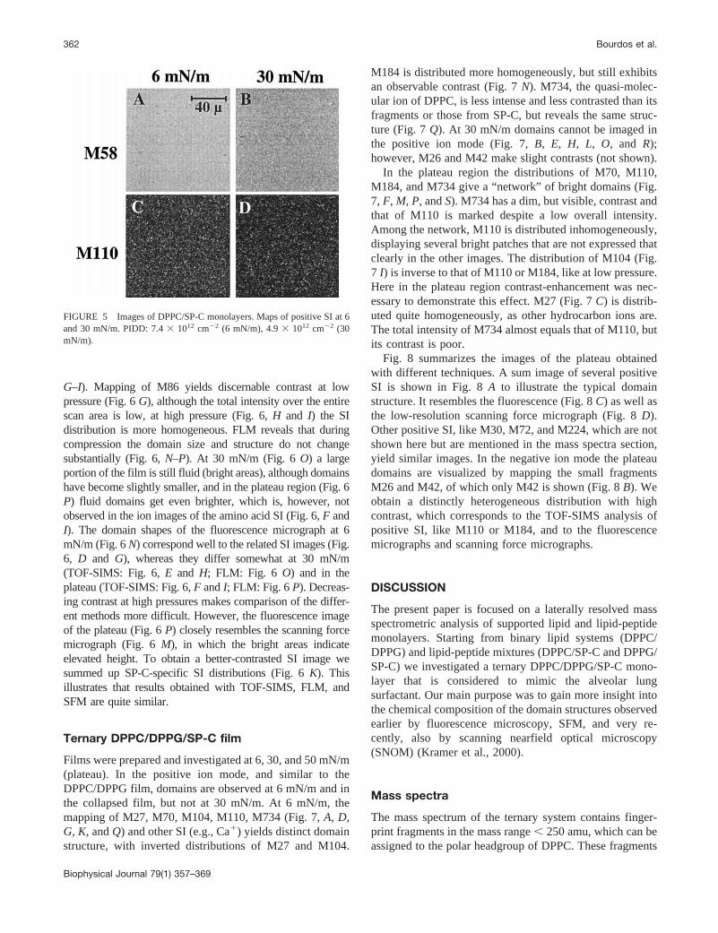
DPPC/SP-C films were prepared again at 6 and 30 mN/m(Fig. 5), DPPG/SP-C films at 6, 30, and 50 mN/m (Fig. 6);both were analyzed with TOF-SIMS and FLM. FLM-pic-tures of DPPC/SP-C films have been published before (vonNahmen, 1997). In addition, an SFM image of DPPG/SP-Cin the plateau is presented in Fig. 6M. The isotherms ofboth mixtures exhibit plateau regions at'50 mN/m, but theDPPC/SP-C film could not be transferred due to a contin-uous pressure drop. At 6 and 30 mN/m no domains areobserved in DPPC/SP-C for either a DPPC-specific (M58,Fig. 5, A andB) or SP-C-specific SI (M110, Fig. 5,C andD). The DPPG/SP-C film maintained a stable equilibriumpressure in the plateau region when the compression washalted before transferring the film. Several SI distributionsmake domains at each pressure, three of them are shown as anexample in Fig. 6: the contrast of the distributions of thehydrocarbon ion M27 (Fig. 6,A–C) and Ca1 (Fig. 6 L, onlyshown for the plateau region) is found to be inverse to that ofthe amino acid fragments M72 and M86 (Fig. 6,D–F and
FIGURE 4 Images of DPPC/DPPG monolayers. (A–H) Maps of positiveSI at 6 and 30 mN/m. PIDD: 1.03 1013 cm22 (6 mN/m), 7.03 1012 cm22
(30 mN/m); (I) 6 mN/m, Ca1; (K) 6 mN/m, sum image of several positiveSI; (L) 6 mN/m, silver-decorated; (I) and (K) are digitally enhanced.
Domain Structures in Lung Surfactant 361
Biophysical Journal 79(1) 357–369
G–I). Mapping of M86 yields discernable contrast at lowpressure (Fig. 6G), although the total intensity over the entirescan area is low, at high pressure (Fig. 6,H and I) the SIdistribution is more homogeneous. FLM reveals that duringcompression the domain size and structure do not changesubstantially (Fig. 6,N–P). At 30 mN/m (Fig. 6O) a largeportion of the film is still fluid (bright areas), although domainshave become slightly smaller, and in the plateau region (Fig. 6P) fluid domains get even brighter, which is, however, notobserved in the ion images of the amino acid SI (Fig. 6,F andI). The domain shapes of the fluorescence micrograph at 6mN/m (Fig. 6N) correspond well to the related SI images (Fig.6, D and G), whereas they differ somewhat at 30 mN/m(TOF-SIMS: Fig. 6,E and H; FLM: Fig. 6 O) and in theplateau (TOF-SIMS: Fig. 6,F andI; FLM: Fig. 6 P). Decreas-ing contrast at high pressures makes comparison of the differ-ent methods more difficult. However, the fluorescence imageof the plateau (Fig. 6P) closely resembles the scanning forcemicrograph (Fig. 6M), in which the bright areas indicateelevated height. To obtain a better-contrasted SI image wesummed up SP-C-specific SI distributions (Fig. 6K). Thisillustrates that results obtained with TOF-SIMS, FLM, andSFM are quite similar.
Ternary DPPC/DPPG/SP-C film
Films were prepared and investigated at 6, 30, and 50 mN/m(plateau). In the positive ion mode, and similar to theDPPC/DPPG film, domains are observed at 6 mN/m and inthe collapsed film, but not at 30 mN/m. At 6 mN/m, themapping of M27, M70, M104, M110, M734 (Fig. 7,A, D,G, K, andQ) and other SI (e.g., Ca1) yields distinct domainstructure, with inverted distributions of M27 and M104.
M184 is distributed more homogeneously, but still exhibitsan observable contrast (Fig. 7N). M734, the quasi-molec-ular ion of DPPC, is less intense and less contrasted than itsfragments or those from SP-C, but reveals the same struc-ture (Fig. 7Q). At 30 mN/m domains cannot be imaged inthe positive ion mode (Fig. 7,B, E, H, L, O, and R);however, M26 and M42 make slight contrasts (not shown).
In the plateau region the distributions of M70, M110,M184, and M734 give a “network” of bright domains (Fig.7, F, M, P, andS). M734 has a dim, but visible, contrast andthat of M110 is marked despite a low overall intensity.Among the network, M110 is distributed inhomogeneously,displaying several bright patches that are not expressed thatclearly in the other images. The distribution of M104 (Fig.7 I) is inverse to that of M110 or M184, like at low pressure.Here in the plateau region contrast-enhancement was nec-essary to demonstrate this effect. M27 (Fig. 7C) is distrib-uted quite homogeneously, as other hydrocarbon ions are.The total intensity of M734 almost equals that of M110, butits contrast is poor.
Fig. 8 summarizes the images of the plateau obtainedwith different techniques. A sum image of several positiveSI is shown in Fig. 8A to illustrate the typical domainstructure. It resembles the fluorescence (Fig. 8C) as well asthe low-resolution scanning force micrograph (Fig. 8D).Other positive SI, like M30, M72, and M224, which are notshown here but are mentioned in the mass spectra section,yield similar images. In the negative ion mode the plateaudomains are visualized by mapping the small fragmentsM26 and M42, of which only M42 is shown (Fig. 8B). Weobtain a distinctly heterogeneous distribution with highcontrast, which corresponds to the TOF-SIMS analysis ofpositive SI, like M110 or M184, and to the fluorescencemicrographs and scanning force micrographs.
DISCUSSION
The present paper is focused on a laterally resolved massspectrometric analysis of supported lipid and lipid-peptidemonolayers. Starting from binary lipid systems (DPPC/DPPG) and lipid-peptide mixtures (DPPC/SP-C and DPPG/SP-C) we investigated a ternary DPPC/DPPG/SP-C mono-layer that is considered to mimic the alveolar lungsurfactant. Our main purpose was to gain more insight intothe chemical composition of the domain structures observedearlier by fluorescence microscopy, SFM, and very re-cently, also by scanning nearfield optical microscopy(SNOM) (Kramer et al., 2000).
Mass spectra
The mass spectrum of the ternary system contains finger-print fragments in the mass range, 250 amu, which can beassigned to the polar headgroup of DPPC. These fragments
FIGURE 5 Images of DPPC/SP-C monolayers. Maps of positive SI at 6and 30 mN/m. PIDD: 7.43 1012 cm22 (6 mN/m), 4.93 1012 cm22 (30mN/m).
362 Bourdos et al.
Biophysical Journal 79(1) 357–369
are also obtained by related techniques, such as FAB/MS(Ayanoglu et al., 1984; Matsuhara and Hayashi, 1991). Inthe molecular ion region, peaks similar to those obtained byMALDI (Harvey, 1995) are observed. DPPC is desorbedboth as protonated and dehydrogenated quasi-molecular ion
(DPPC 6 H)1. 252Cf plasma desorption MS (Demirev,1987) exhibits similar results in the fingerprint range. DPPGis found negatively charged, which is not shown here; theprotonated phosphoglycerol residue M173 is the only pos-itive SI characteristic for DPPG (in spectra of spin-coated
FIGURE 6 Images of DPPG/SP-C monolayers.(A–I) Maps of positive SI at 6, 30, and 50 mN/m(plateau); (K) Plateau, sum image of several aminoacid-specific positive SI; (L) Plateau, Ca1; PIDD:1.9 3 1013 cm22 (6 mN/m), 8.13 1012 cm22 (30mN/m), 5.93 1013 cm22 (50 mN/m). (M) Plateau,scanning force micrograph. Bright areas are higherthan the dark (no scale bar given). (N–P) Fluores-cence micrographs at 6, 30, and 50 mN/m.
Domain Structures in Lung Surfactant 363
Biophysical Journal 79(1) 357–369
DPPG, a sodiated quasi-molecular ion (DPPG1 2Na 2H)1 could be detected in the positive ion mode). BecauseDPPC and DPPG differ in their headgroup, the massrange , 200 amu can be used for the identification ofDPPC: M104, M166, and M184, as well as other fragmentslisted in the table, are typical of DPPC but are not included
in the DPPG spectrum. Other typical phospholipid frag-ments are not discussed here because they do not yieldadditional information.
The SP-C spectrum consists of pronounced amino acidpeaks (M-45)1 (Hagenhoff, 1993), arising from a net cleav-age of the COOH2-group. Fragments of the basic residues,
FIGURE 7 Images of layers prepared from theternary mixture DPPC/DPPG/SP-C. (A–S) Maps ofpositive SI at 6, 30, and 50 mN/m; (G) is digitallyenhanced. PIDD 8.13 1012 cm22 (6 mN/m), 2.031013 cm22 (30 mN/m), 4.6 3 1013 cm22 (50mN/m).
364 Bourdos et al.
Biophysical Journal 79(1) 357–369
(Lys-45)1 and (Arg-45)1, corresponding to M101 andM129, respectively, are not found; because of their unpairedelectron they are only expected to emerge with very smallintensity according to the nitrogen rule (McLafferty andTurecek, 1993). The small fragments M30 and M44 do notonly originate from glycine and alanine, respectively, butare general constituents of peptide mass spectra (McLaf-ferty and Turecek, 1993).
Most of the nitrogen-containing SI in the mass range,100 amu interfere with non-specific hydrocarbon SI, recog-nized by asymmetrically shaped peaks. In fact, this does notmatter much, because the intense nitrogen-containing SI(with paired electrons) have even nominal masses, whereasthe intense hydrocarbon ions have odd masses or odd num-bers of hydrogen atoms, respectively. So an intense N-containing fragment interferes with a less intense hydrocar-bon, and vice versa. For instance, at nominal mass 70C4H8N
1 interferes with13CC4H91 and C5H10
1. The latterpeak is small, because it has an unpaired electron. Thecontribution of the isotopomer (13CC4H9
1) can be calcu-lated from the lighter neighbored hydrocarbon SI (C5H9
1),taking into account the isotope distribution of carbon, wherethe portion of 13C amounts to 1.1%. Thus'3% of theintensity of M70 are due to13CC4H9
1 and 7% due toC5H10
1. So as a crude estimate we may assume a value of10%, which is the “unspecific” share of a headgroup- oramino acid-based DPPC or SP-C peak. In fact, the intensityof the isotopomer increases with increasing C number.
In the negative ion spectra, which are not presented here,M42 is desorbed from SP-C more intensively than fromDPPC (nearly 10-fold). The origin of M42 (CNO2) can beattributed to the peptide bonds. A possible objection that
M26 and M42 are ubiquitous impurities because of theirsmallness is invalidated by their insignificant yield inDPPG, which does not contain such a chemical group.
The SI yields in a single compound analysis, especiallythe yields of the quasi-molecular ions (M1 H)1, (M 1alkaline)1, and (M 1 metal)1, depend on the method ofdeposition (spin coating, droplet preparation, or as LB film),on the substrate material, and on the pH. The depositiontechnique is crucial, because it is directly linked to thecoverage of the substrate surface. When one deposits thesame amount of a compound either as droplet or by spin-coating, the coverage is lower with the latter method. By LBpreparation, the molecules can be deposited in a well-ordered state, and substrate-overlayer interactions are bet-ter-specified. For example, in the case of a hydrophilictransfer of DPPC onto gold it is known that the polarheadgroup interacts directly with the substrate. For peptides,the presence of basic residues like arginine or lysine playsan important role in the desorption process (van Leyen etal., 1987). The fragmentation pattern of SP-C shown in Fig.3 is characteristic, but SI intensities vary when applyingdifferent deposition techniques.
Ion images
The TOF-SIMS sum image of DPPC/DPPG at 6 mN/mcorresponds well to the light microscopic picture of thesilver-decorated sample: both differ from the FLM results,where the condensed domains are smaller (von Nahmen etal., 1997b). The reason for this may be the fact that thesupported film is equilibrated before transfer, whereas afluorescence micrograph is taken after halting the barrier ofthe film balance a few minutes, or during compression atvery slow speed. Therefore, equilibration, which is neces-sary for an LB transfer, may cause a larger portion of thefilm being condensed as compared with a film at the air/water interface to a solid substrate and may result in ahigher degree of condensation of an LB film, compared toits analog, at the air/water interface. At 30 mN/m, where thefilm is predominantly condensed, fluorescence micrographsshow slight contrast (von Nahmen et al., 1997b), but withTOF-SIMS this contrast cannot be observed in the positiveion mode, not even by summing up and processing. In thenegative ion mode a very dim contrast was observed whensumming up some specific and unspecific SI and enhancingthe contrast of the resulting image.
Correlating the TOF-SIMS results with those obtained byFLM (von Nahmen et al., 1997b) or SD, it is likely that thefluid phase, which is represented by the bright domains ofboth the silver and fluorescence contrast, corresponds to thebright domains in the maps of M58, M184, and M734,which are dark in the maps of M27 or Ca1. The surfaceconcentration of the parental molecules is lower, but theintensity of M58, for example, is higher in the fluid than inthe condensed phase. Because we already know that DPPC
FIGURE 8 Additional images from the plateau region of DPPC/DPPG/SP-C layers: (A) Sum image of several positive SI; (B) M42 (CNO2); (C)Fluorescence micrograph; (D) Scanning force micrograph.
Domain Structures in Lung Surfactant 365
Biophysical Journal 79(1) 357–369
and DPPG mix nearly ideally at a 4:1 molar ratio (Garidelet al., 1997; Sieber, unpublished x-ray reflection data), apronounced matrix effect must account for the different SIintensities or ionization rates in the fluid and condensedphase. Although the SI spectrum of DPPG does not engen-der any specific fragments, DPPG could be mapped onlydue to the contrast generated by the DPPC-specific SI. Thusthe ion images arise from a physical contrast due to co-existing phases, like in one- or two-component LB filmsstudied earlier (Bourdos et al., 2000; Leufgen et al., 1996).
In the DPPC/DPPG/SP-C film, results are similar at 6 and30 mN/m. At 6 mN/m, the fluid domains are smaller and thedomain structure is more disrupted than in the correspond-ing binary lipid film. At 30 mN/m again there is no contrast.As in the DPPC/DPPG film, FLM and TOF-SIMS results donot correspond exactly, for the reasons given above. How-ever, in addition to the mere physical contrast, a chemicalcontrast is superposed because there is SP-C present in themixture, which can be distinguished from the lipids bycertain amino acid fragments, but superposition of twocontrasts complicates the interpretation. By looking at thelipid fragments at 6 mN/m, we could assume the existenceof co-existing phases, as for the DPPC/DPPG film, althougha possible electrostatic interaction between DPPG and SP-Cmay cause a certain degree of demixing. The choline ionM104 is worth considering because its contrast is oppositeto M184 or M734, with higher intensity in the condensedthan in the fluid phase, comparable to M27 and Ca1 (notshown for the ternary system). The distribution of the highlySP-C-specific M110 may mislead: one is tempted to see theSP-C strictly localized in the bright domains, sustained bythe fact that on the one hand SP-C makes up only 0.4% ofthe mixture, but on the other hand its fragments are de-sorbed intensely from the fluid domains; this may, however,be distorted by a matrix effect. For a fairly good interpre-tation it is therefore always necessary tocorrelatethe TOF-SIMS maps with other imaging techniques. Consideringearlier findings obtained with FLM (Nag et al., 1996; vonNahmen et al., 1997b), we can conclude that the aminoacid-specific fragments like M110 give a correct image ofthe distribution of SP-C in lipid monolayers, i.e., the peptideis associated with the fluid phase.
The lateral distribution of SP-C in a single phospholipidmatrix is quite different for DPPC and DPPG. In DPPC/SP-C there is no contrast observed at both low and highpressure in the distribution of all ions we considered and, ofwhich we show only two, representative of DPPC- andSP-C. This indicates full miscibility of SP-C and DPPC incorrespondence with microfluorescence measurements (vonNahmen, 1997). The pressure drop in the plateau uponhalting the barrier of the film balance is due to a loss ofmaterial. Interaction of DPPC with 0.5 mol % SP-C aloneobviously cannot accomplish the formation of a stable col-lapse structure.
However, domains are observed in DPPG/SP-C at each ofthe considered film pressures. As in the ternary system,SP-C induces a plateau at high pressure (.50 mN/m)which, upon halting barriers, settles to an equilibrium, in-dicating a stable collapse state that allows an LB transfer.This points at a pronounced electrostatic interaction be-tween the negatively charged DPPG and SP-C, which car-ries positive charges at its basic residues. Because the iso-therm shows no demixing, the SI images at least illustratethe formation of separated phases. At higher pressures thecontrast vanishes due to continual condensation, compara-ble to the DPPC/DPPG system; therefore, the fluorescencemicrographs are not easy to compare with the ion images,although there is a good agreement at 6 mN/m and in theplateau with respect to the sum image. In the plateau region,the fluorescence intensity gets brighter in the fluid domainsdue to higher concentration of the probe molecules (NBD-PG) per unit area. This is interpreted by the formation ofthree-dimensional multilayer stacks (Galla et al., 1998).Stack formation was assumed earlier from ellipsometricmeasurements (Post et al., 1995) and confirmed here byimaging the topography with SFM (Amrein et al., 1997).Here we observed that the (bright) areas of elevated heightin the SFM pictures correspond to the bright domains of thesum image of amino acid SI and in the fluorescence image.From our TOF-SIMS or FLM images alone we may notconclude that SP-C is enriched in the multilamellar domainsfor two reasons: 1) TOF-SIMS images are impaired by amatrix effect, and 2) in the FLM measurements presentedhere no dye-labeled SP-C is used, but labeled DPPG is.However, NBD-labeled SP-C was applied earlier with theternary mixture used here by von Nahmen et al. (1997b),yielding similar results to a film containing NBD-PC. Theuse of dye-labeled SP-C gave a clearer hint that SP-C isenriched in the stacks. It has to be stated that from theTOF-SIMS measurements alone the existence of multilayerdomains cannot be derived. The capabilities of TOF-SIMSare limited in this respect because of its extreme surfacesensitivity, depending also on the nature of the primary ionbeam (Kotter and Benninghoven, 1998). The more distant amolecule is located to the substrate, the less it will beaffected by the sputter event initiated by the collision cas-cade among the substrate molecules, so multilayers nor-mally come to reduced SI yields, as shown by Stapel et al.(1999). Moreover, quantities that describe the phenomenol-ogy of SI formation, like the disappearance cross-section,are only defined for monolayers.
With respect to the Ca1-image one should recall thatintrinsic ions are detected with TOF-SIMS; they onlyamount to'1% of the desorbed particles, the rest of whichare neutrals. The mechanisms of ion formation, fragmenta-tion, and desorption from organic molecules adsorbed tosolids are not fully understood, which applies even more tooverlayers like LB films. Ca1, in contrast, is not to the sameextent affected by matrix effects like organic fragments,
366 Bourdos et al.
Biophysical Journal 79(1) 357–369
because it does not arise from complex ionization mecha-nisms rather than simple ionization. This applies especiallyto alkaline or alkaline-earth elements with low ionizationenergies. We therefore think that the Ca1 map nearly re-flects the distribution of Ca1 at the surface.
The plateau of DPPC/DPPG/SP-C
Both DPPC-specific (M184, M734) and SP-C-specific(M70, M110, the negative M42) fragments yield the samenetwork-like domain structure, best visible in the sum im-age of some positive SI. Identical structures were alsoobtained with FLM or low-resolution SFM (von Nahmen etal., 1997a, b). The SFM images arise from a topographiccontrast due to distinct protrusions, which form when themodel surfactant film partially collapses (Amrein et al.,1997). The protrusions are supposed to be the surfactantreservoir, probably multilamellar stacks from which theexcluded material is reinserted into the monolayer when thefilm is expanded. From fluorescence data and from SFMpictures these stacks are assumed to consist of an oddnumber of layers.
However, high-resolution images exhibit no continuousformation of protrusions, rather than the existence of smallpatches (von Nahmen et al., 1997a). From this we concludethat the fluid “network” includes the adjacent intermediatemonolayer domain, and is not only a network of layeredlamellae. The similar network-like distribution of SP-C-specific SI, like M110, indicates that SP-C is presentthroughout the network, and not only in the postulatedstacks. It seems to be kept as spare material, which issupplied for the formation of more stacks upon furthercompression. This is consistent with SFM images taken atsuccessive points in the plateau region during compression,where the proportion of protruded areas increases withdecreasing molecular area. In FLM measurements on theternary system using fluorescence-labeled SP-C and PC, thefluorescence intensity increased in those areas related to thepostulated stacks. As in DPPG/SP-C, this is due to a volumeeffect where flat monolayers escape into the third dimensionin agreement with the squeeze-out hypothesis by Bangham(1979); DPPC, however, as it is demonstrated here by thedistribution of M184, seems to be located in these domainsin considerable amounts, too. This would contradict Bang-ham’s hypothesis, stating that upon film collapse only non-DPPC components are excluded from the monolayer andpure, condensed DPPC should remain at the interface, main-taining low surface tension.
Upon scrutinizing the TOF-SIMS results obtained fromthe plateau region, one has to consider the above-mentionedvolume effect, which normally brings about lower SI inten-sities from multilayers, although a higher local concentra-tion of the parental molecule is possible due to layering (aneffect being less pronounced when multi-atomic or noblegas PI sources are used, as shown by Stapel et al., 1999).
Therefore it is crucial to know to what extent both effectscompensate each other. Moreover, as discussed above, amatrix effect becomes even more complicated if differentphysical states are involved in the SI formation. All to-gether, these effects make it impossible to determinewhether SP-C is really squeezed out of the condensedmonolayer phase, but intensities of many DPPC- and SP-C-based fragments are higher in the network than in the“patches.” Because the network is supposed to contain thelayered part of the film, thus being subject to the volumeeffect, one expects lower yields in it. Thus we claim that themajor share of the SP-C content is located in the network,which corresponds very well with the FLM results. Becausethe existence of multilayers would reduce SI intensities, thediscrepancy between intensity and concentration is evenincreased. For the assessment of the distribution of DPPCthe FLM micrographs are not helpful, as they are for thedistribution of SP-C. SP-C could be replaced completely bya fluorescent analog, whereas this is not possible for DPPC,the major component of the surfactant.
A completely different behavior was observed with M27,a hydrocarbon SI, that is distributed more homogeneouslyin the plateau than other either DPPC- or SP-C-based SI.Hydrocarbon ions neither seem to be affected that stronglyby multilayer formation nor by lateral pressure, i.e., thephysical state of the film. The latter corresponds to the factthat hydrocarbon ions mainly originate from the acyl chains(Bourdos et al., 2000), of which especially the methyl andethyl endgroups are less sensitive to changes of the lateralpressure. M104 again exhibits contrast inverse to variousother SI, although not as pronounced as at 6 mN/m, whereit was shown that this SI is more intense in the fluid phase.Its intensity in the plateau may be lowered by both theexistence of multilayers and a possible higher fluidity of thenetwork.
CONCLUSION
We used TOF-SIMS to visualize chemically and physicallydifferent domain structures in mixed LB films of phospho-lipids and phospholipids with SP-C, with particular interestin the ternary system DPPC/DPPG/SP-C. For the collapsedternary film we showed in accordance with previous workthat upon compression of the monolayer a network-likeSP-C-rich structure—the surfactant reservoir—is formed,which comprises an intermediate, also SP-C-enriched,monolayer phase. Correlation of FLM and TOF-SIMS dataallows the conclusion that the major share of the SP-Cmolecules is located in the network. The squeeze-out hy-pothesis that only non-DPPC components leave the mono-layer could not be verified. Despite the unclear matrixeffect, especially in the presence of multilayers, we areallowed to assume that a considerable amount of DPPCremains in the protrusions, contradicting one aspect of thishypothesis but confirming its basic idea. Within the dis-
Domain Structures in Lung Surfactant 367
Biophysical Journal 79(1) 357–369
cussed limitations, TOF-SIMS turns out to be a valuabletool for studying biological model systems. For interpretingthe lung surfactant it is important to keep in mind that thesimple mixture of disaturated phospholipids of course doesnot exhibit the richness of biophysical activity of nativesurfactant. Our aim was to elucidate the role of the SP-C informing and stabilizing the surfactant reservoir.
We thank Detlef Knebel (Institut fu¨r Medizinische Physik und Biophysik,Universitat Munster) for the SFM image of DPPG/SP-C, and MichaelGleiche (Physikalisches Institut, Universita¨t Munster), who assisted inpreparing the silver-decorated samples and acquiring the images.
This work was supported by Grant GA 233/18-1 from the DeutscheForschungsgemeinschaft.
REFERENCES
Amrein, M., A. von Nahmen, and M. Sieber. 1997. A scanning force- andfluorescence light microscopy study of the structure and function of amodel pulmonary surfactant.Eur. Biophys. J.26:349–357.
Ayanoglu, E., A. Wegmann, O. Pilet, G. D. Marbury, J. R. Hass, and C.Djerassi. 1984. Mass spectrometry of phospholipids. Some applicationsof desorption chemical ionization and fast atom bombardment.J. Am.Chem. Soc.106:5246–5251.
Bangham, A. D. 1979. The physical properties of an effective lung sur-factant.Biochim. Biophys. Acta.573:552–556.
Benninghoven, A. 1994. Chemical analysis of inorganic and organic sur-faces and thin films by static time-of-flight secondary ion mass spec-trometry (TOF-SIMS).Angew. Chem. Int. Ed. Engl.33:1023–1044.
Benninghoven, A., B. Hagenhoff, and E. Niehues. 1993. Surface MS:probing real-world samples.Anal. Chem.65: 630A–640A.
Bertrand, P., and L. Weng. 1996. Time-of-flight secondary ion massspectrometry (ToF-SIMS).Mikrochim. Acta.13:167–182.
Bourdos, N., F. Kollmer, A. Benninghoven, M. Sieber, and H.-J. Galla.2000. Imaging of domain structures in a one-component lipid monolayerby time-of-flight secondary ion mass spectrometry.Langmuir. 16:1481–1484.
Clements, J. A. 1962. Surface phenomena in relation to pulmonary func-tion. Physiologist.5:11–28.
Cochrane, C. G., and S. D. Revak. 1991. Pulmonary surfactant protein B(SP-B): structure-function relationships.Science.254:566–568.
Demirev, P. A. 1987. 252-Californium plasma desorption mass spectrom-etry of glycerophospholipids.Biomed. Environ. Mass Spectrom.14:241–246.
Discher, B. M., K. M. Maloney, W. R. Schief, V. Vogel, and S. B. Hall.1996. Lateral phase separation in interfacial films of pulmonary surfac-tant.Biophys. J.71:2583–2590.
Galla, H.-J., N. Bourdos, A. von Nahmen, M. Amrein, and M. Sieber.1998. The role of pulmonary surfactant protein C during the breathingcycle.Thin Solid Films.329:632–635.
Garidel, P., C. Johann, L. Mennicke, and A. Blume. 1997. The mixingbehaviour of pseudobinary phosphatidylcholine-phosphatidylglycerolmixtures as a function of pH and chain length.Eur. Biophys. J.26:447–459.
Gleiche, M., L. F. Chi, and H. Fuchs. 1998. Molecular property relatedsilver decoration on fatty acid Langmuir-Blodgett monolayers.ThinSolid Films.329:268–272.
Hagenhoff, B. 1993. Sekunda¨rionenmassenspektrometrie an MolekularenOberflachenstrukturen. Ph.D. thesis. Westfa¨lische Wilhelms-Universi-tat, Munster. 178 pp.
Hagenhoff, B. 1995. Surface mass spectrometry: application to biosensorcharacterization.Biosensors and Bioelectronics.10:885–894.
Hagenhoff, B., A. Benninghoven, J. Spinke, M. Liley, and W. Knoll. 1993.Time-of-flight secondary ion mass spectrometry investigations of self-
assembled monolayers of organic thiols, sulfides, and disulfides on goldsurfaces.Langmuir.9:1622–1624.
Harvey, D. J. 1995. Matrix-assisted laser desorption/ionization mass spec-trometry of phospholipids.J. Mass Spectrom.30:1333–1346.
Harwood, J. L. 1987. Lung surfactant.Prog. Lipid. Res.26:211–256.Horowitz, A. D., B. Elledge, J. A. Whitsett, and J. E. Baatz. 1992. Effects
of lung surfactant proteolipid SP-C on the organization of model mem-brane lipids: a fluorescence study.Biochim. Biophys. Acta.1107:44–54.
Johansson, J., and T. Curstedt. 1997. Molecular structures and interactionsof pulmonary surfactant components.Eur. J. Biochem.244:675–693.
Johansson, J., T. Curstedt, and B. Robertson. 1994a. The proteins of thesurfactant system.Eur. Respir. J.7:372–391.
Johansson, J., T. Szyperski, T. Curstedt, and K. Wu¨thrich. 1994b. TheNMR structure of the pulmonary surfactant-associated polypeptide SP-Cin an apolar solvent contains a valyl-rich a-helix.Biochemistry.33:6015–6023.
King, R. J., and J. A. Clements. 1972. Surface active material from doglung. I. Method of isolation.Am. J. Physiol.223:715–726.
Kotter, F., and A. Benninghoven. 1998. Secondary ion emission frompolymer surfaces under Ar, Xe and SF5 ion bombardment.Appl. Surf.Sci.133:47–57.
Kramer, A., A. Wintergalen, M. Sieber, H. J. Galla, M. Amrein, and R.Guckenberger. 2000. Distribution of the surfactant associated protein Cwithin a lung surfactant model film investigated by near-field opticalmicroscopy.Biophys. J.78:454–465.
Kruger, P., M. Schalke, Z. Wang, R. H. Notter, R. A. Dluhy, and M.Losche. 1999. Effect of hydrophobic surfactant peptides SP-B and SP-Con binary phospholipid monolayers. I. Fluorescence and dark-field mi-croscopy.Biophys. J.77:903–914.
Leufgen, K. M., H. Rulle, A. Benninghoven, M. Sieber, and H-J. Galla.1996. Imaging time-of-flight secondary ion mass spectrometry allowsvisualization and analysis of coexisting phases in Langmuir-Blodgettfilms. Langmuir.12:1708–1711.
Liu, M. 1997. Synchronized changing of transinterface pressure, bubbleradius and surface tension: a unique feature of lung surfactant.Chem.Phys. Lipids.89:55–65.
Matsuhara, T., and A. Hayashi. 1991. FAB/Mass spectrometry of lipids.Prog. Lipid Res.30:301–322.
McLafferty, F. W., and F. Turecek. 1993. Interpretation of Mass Spectra.University Science Books, Mill Valley, CA.
Nag, K., J. Pe`rez-Gil, A. Cruz, and K. M. W. Keough. 1996. Fluorescentlylabeled pulmonary surfactant protein C in spread phospholipid mono-layers.Biophys. J. 71:246–256.
Nag, K., S. G. Taneva, J. Pe`rez-Gil, A. Cruz, and K. M. W. Keough. 1997.Combinations of fluorescently labeled pulmonary surfactant proteinsSP-B and SP-C in phospholipid films.Biophys. J.72:2638–2650.
Oosterlaken-Dijksterhuis, M. A., H. P. Haagsman, L. M. G. van Golde, andR. A. Demel. 1991a. Interaction of lipid vesicles with monomolecularlayers containing lung surfactant proteins SP-B or SP-C.Biochemistry.30:8276–8281.
Oosterlaken-Dijksterhuis, M. A., H. P. Haagsman, L. M. G. van Golde, andR. A. Demel. 1991b. Characterization of lipid insertion into monomo-lecular layers mediated by lung surfactant proteins SP-B and SP-C.Biochemistry.30:10965–10971.
Panaiotov, I., Tz. Ivanova, J. Proust, F. Boury, B. Denizot, K. Keough, andS. Taneva. 1996. Effect of hydrophobic protein SP-C on structure anddilatational properties of the model monolayers of pulmonary surfactant.Colloids Surf. B: Biointerfaces.6:243–260.
Perez-Gil, J., K. Nag, S. Taneva, and K. M. W. Keough. 1992a. Pulmonarysurfactant protein SP-C causes packing rearrangements of dipalmi-toylphosphatidylcholine in spread monolayers.Biophys. J.63:197–204.
Perez-Gil, J., J. Tucker, G. Simatos, and K. M. W. Keough. 1992b.Interfacial adsorption of simple lipid mixtures combined with hydropho-bic surfactant protein from pig lung.Biochem. Cell Biol.70:332–338.
Post, A., A. von Nahmen, M. Schmitt, J. Ruths, H. Riegler, M. Sieber, andH-J. Galla. 1995. Pulmonary surfactant protein C containing lipid filmsat the air-water interface as a model for the surface of lung alveoli.Mol.Membr. Biol.12:93–99.
368 Bourdos et al.
Biophysical Journal 79(1) 357–369
Rulle, H. 1996. Hochauflo¨sende Abbildungen Strukturiertet OrganischerOberflachen mit Flugzeit-sekunda¨rionen-Massenspektrometrie (TOF-SIMS). Ph.D. thesis. Westfa¨lische Wilhelms-Universita¨t, Munster. 88pp.
Scarpelli, E. M., and A. J. Mautone. 1994. Surface biophysics of thesurface monolayer theory is incompatible with regional lung function.Biophys. J.67:1080–1089.
Schurch, S., J. Goerke, and J. A. Clements. 1976. Direct determination ofsurface tension in the lung.Proc. Natl. Acad. Sci. U.S.A.73:4698–4702.
Schurch, S., R. Qanbar, H. Bachofen, and F. Possmeyer. 1995. Thesurface-associated surfactant reservoir in the alveolar lining.Biol.Neonate.67(Suppl. 1):61–76.
Schwieters, J., H. G. Cramer, T. Heller, U. Juergens, E. Niehuis, J.Zehnpfennig, and A. Benninghoven. 1991. High mass resolution surfaceimaging with a time-of-flight secondary ion mass spectroscopy scanningmicroprobe.J. Vac. Sci. Technol. A.9:2864–2871.
Sen, A., S.-W. Hui, M. Mosgrober-Anthony, B. A. Holm, and E. A. Egan.1988. Localization of lipid exchange sites between bulk lung surfactantsand surface monolayer: freeze fracture study.J. Colloid Interface Sci.126:355–360.
Shelly, S. A., J. E. Paciga, and J. U. Balius. 1984. Lung surfactantphospholipids in different animal species.Lipids. 19:857–862.
Stapel, D., O. Brox, and A. Benninghoven. 1999. Secondary ion emissionfrom arachidic acid LB-layers under Ar, Xe, Ga and SF5 primary ionbombardment.Appl. Surf. Sci.140:156–167.
Taneva, S. G., and K. M. W. Keough. 1994a. Dynamic surface propertiesof pulmonary surfactant proteins SP-B and SP-C and their mixtures withdipalmitoylphosphatidylcholine.Biochemistry.33:14660–14670.
Taneva, S. G., and K. M. W. Keough. 1994b. Pulmonary surfactantproteins SP-B and SP-C in spread monolayers at the air-water interface:I. Monolayers of pulmonary surfactant protein SP-B and phospholipids.Biophys. J.66:1137–1148.
Taneva, S. G., and K. M. W. Keough. 1994c. Pulmonary surfactantproteins SP-B and SP-C in spread monolayers at the air-water interface:
II. Monolayers of pulmonary surfactant protein Protein SP-C and phos-pholipids.Biophys. J.66:1149–1157.
Taneva, S. G., and K. M. W. Keough. 1994d. Pulmonary surfactantproteins SP-B and SP-C in spread monolayers at the air-water interface:III. Proteins SP-B plus SP-C with phospholipids in spread monolayers.Biophys. J.66:1158–1166.
Tchoreloff, P., A. Gulik, B. Denizot, J. E. Proust, and F. Puisieux. 1991. Astructural study of interfacial phospholipid and lung surfactant layers bytransmission electron microscopy after Blodgett sampling: influence ofsurface pressure and temperature.Chem. Phys. Lipids.59:151–165.
van Leyen, D., D. Greifendorf, and A. Benninghoven. 1987. Secondary ionformation from peptides: influence of primary structure and substrates.6th International Conference on Secondary Ion Mass Spectrometry,Versailles, Sept. 13–18, 679–682.
von Nahmen, A. 1997. Strukturelle Organisation von Surfactant Protein Chaltigen Phospholipid-Monoschichten an der Luft-Wasser-Grenzfla¨-che—Ein Modell des alveola¨ren Surfactant. Ph.D. thesis. Westfa¨lischeWilhelms-Universita¨t, Munster. 139 pp.
von Nahmen, A., A. Post, H. J. Galla, and M. Sieber. 1997b. The phasebehaviour of lipid monolayers containing pulmonary surfactant proteinC studied by fluorescence light microscopy.Eur. Biophys. J.26:359–369.
von Nahmen, A., M. Schenk, M. Sieber, and M. Amrein. 1997a. Thestructure of a model pulmonary surfactant as revealed by scanning forcemicroscopy.Biophys. J.72:463–469.
Wang, Z., O. Gurel, J. E. Baatz, and R. H. Notter. 1996b. Differentialactivity and lack of synergy of lung surfactant proteins SP-B and SP-Cin interactions with phospholipids.J. Lipid Res.37:1749–1760.
Wang, Z., S. B. Hall, and R. H. Notter. 1995. Dynamic surface activity offilms of lung surfactant phospholipids, hydrophobic proteins, and neutrallipids. J. Lipid Res.36:1283–1293.
Wang, Z., S. B. Hall, and R. H. Notter. 1996a. Roles of different hydro-phobic constituents in the adsorption of pulmonary surfactant.J. LipidRes.37:790–798.