Analysis of T cell receptor (TCR) BV-gene clonotypes in NC/Nga mice developing dermatitis resembling human atopic dermatitis Akihiro Matsuoka a , Tomohiro Kato b , Yoshinao Soma a, * , Hideto Takahama a , Masayuki Nakamura a , Hiroyuki Matsuoka c , Masako Mizoguchi a a Department of Dermatology, St. Marianna University School of Medicine, 2-16-1 Sugao, Miyamae-ku, Kawasaki 216-8511, Japan b Institute of Medical Science, St. Marianna University School of Medicine, Japan c Department of Medical Zoology, Jichi Medical School, Japan Received 10 August 2004; received in revised form 18 November 2004; accepted 25 November 2004 Journal of Dermatological Science (2005) 38, 17—24 www.intl.elsevierhealth.com/journals/jods KEYWORDS T cell receptor; BV-gene clonotypes; NC/Nga mice; Atopic dermatitis Summary Background: Our previous study showed that T cells in skin lesions of human atopic dermatitis (AD) had oligoclonal accumulation, indicating the involvement of antigen- specific immune reactions at those sites. Recently, NC/Nga mice, which develop skin lesions similar to AD, have been proposed as a model for that disease. Objective: To clarify whether NC/Nga mice are suitable as a model for human AD from the viewpoint of their antigen-specific immune responses. Methods: Reverse transcription-polymerase chain reaction (RT-PCR) and single strand conformation polymorphism (SSCP) analyses were conducted to detect TCR BV genes of clonally expanded T cells derived from NC/Nga mice at an early phase of the AD-like dermatitis, at a late phase of the dermatitis, and with no AD-like dermatitis. Results: (1) T cells with TCR BV 7, 10 and 17 reside in the skin of NC/Nga mice without the AD-like dermatitis. (2) T cells with these BV genes contain oligoclonal accumula- tions, however, expanded Tcell clonotypes are also detected in the spleen and exist constantly during the course of the AD-like dermatitis. (3) Development of the AD-like dermatitis is associated with additional oligoclonal expansion/accumulation of T cells with TCR BV 2, 4 and 6 genes. (4) Progression of the AD-like dermatitis is associated with further oligoclonal expansion/accumulation of T cells with the TCR BV 14 gene. (5) Some of the expanded TCR clonotypes are common between the individual mice and between early and late phases. * Corresponding author. Tel.: +81 44 977 8111; fax: +81 44 977 3540. E-mail address: [email protected] (Y. Soma). 0923-1811/$30.00 # 2004 Japanese Society for Investigative Dermatology. Published by Elsevier Ireland Ltd. All rights reserved. doi:10.1016/j.jdermsci.2004.11.011
Transcript
Analysis of T cell receptor (TCR) BV-geneclonotypes in NC/Nga mice developing dermatitisresembling human atopic dermatitis
Journal of Dermatological Science (2005) 38, 17—24
aDepartment of Dermatology, St. Marianna University School of Medicine, 2-16-1 Sugao,Miyamae-ku, Kawasaki 216-8511, JapanbInstitute of Medical Science, St. Marianna University School of Medicine, JapancDepartment of Medical Zoology, Jichi Medical School, Japan
Received 10 August 2004; received in revised form 18 November 2004; accepted 25 November 2004
Background: Our previous study showed that T cells in skin lesions of human atopicdermatitis (AD) had oligoclonal accumulation, indicating the involvement of antigen-specific immune reactions at those sites. Recently, NC/Nga mice, which develop skinlesions similar to AD, have been proposed as a model for that disease.Objective: To clarify whether NC/Nga mice are suitable as a model for human ADfrom the viewpoint of their antigen-specific immune responses.Methods: Reverse transcription-polymerase chain reaction (RT-PCR) and singlestrand conformation polymorphism (SSCP) analyses were conducted to detect TCRBV genes of clonally expanded Tcells derived from NC/Nga mice at an early phase ofthe AD-like dermatitis, at a late phase of the dermatitis, and with no AD-likedermatitis.Results: (1) Tcells with TCR BV 7, 10 and 17 reside in the skin of NC/Ngamice withoutthe AD-like dermatitis. (2) T cells with these BV genes contain oligoclonal accumula-tions, however, expanded T cell clonotypes are also detected in the spleen and existconstantly during the course of the AD-like dermatitis. (3) Development of the AD-likedermatitis is associated with additional oligoclonal expansion/accumulation of Tcellswith TCR BV 2, 4 and 6 genes. (4) Progression of the AD-like dermatitis is associatedwith further oligoclonal expansion/accumulation of T cells with the TCR BV 14 gene.(5) Some of the expanded TCR clonotypes are common between the individual miceand between early and late phases.
0923-1811/$30.00 # 2004 Japanese Society for Investigative Dermatology. Published by Elsevier Ireland Ltd. All rights reserved.doi:10.1016/j.jdermsci.2004.11.011
18 A. Matsuoka et al.
Conclusions: Taking these data together with the previous human AD studies, NC/Nga mice seem to be an appropriate model for human AD.# 2004 Japanese Society for Investigative Dermatology. Published by Elsevier IrelandLtd. All rights reserved.
1. Introduction
Atopic dermatitis (AD) is a chronic inflammatory skindisease with infiltrations of lymphocytes in the skinlesions. T cells as well as monocytes/macrophagesaccount for a considerable part of those infiltratinglymphocytes [1—3]. In our previous study, T cells inthe skin lesions of human AD showed oligoclonalaccumulation identified by single strand conforma-tion polymorphism (SSCP) analysis of Tcell receptor(TCR) B chain-gene products, indicating the involve-ment of antigen-specific immune reactions at thesites of AD lesions [4,5].
Recently, NC/Nga mice, which develop sponta-neous dermatitis, which resembles human AD, havebeen used as an animal model for that disease inevaluating possible therapeutic drugs. NC/Nga miceshow hypersensitivity to irradiation, anaphylaxis toovalbumin, anemia and often develop chronic glo-merulonephritis as they age. Interestingly, thisstrain of mice develops dermatitis mainly on theirfaces 7—8 weeks after birth only under conventionalenvironmental conditions and not under specificpathogen-free (SPF) conditions. This dermatitis isassociated with thickening of the epidermis, dander,incrustation and bleeding, and becomes progres-sively severe until 13—14 weeks of age. After that,the progression of the dermatitis become somehowslow but still continues until about 17 weeks of age.NC/Nga mice show elevated levels of serum IgEimmunologically, and increased numbers of mastcells and eosinophils in the dermis and impairedskin barrier function histologically. The serum IgElevels start to increase about 6 weeks of age andprogressively increase until about 12 weeks of age[6—9]. Since these features closely resemble thoseof human AD, NC/Nga mice have been proposed as amodel for human AD.
T cells in skin lesions of human AD show oligoclo-nal accumulation, indicating the involvement ofantigen-specific immune reactions at sites of der-matitis [4,5]. If T cells in the skin lesions of NC/Ngamice display a similar behavior to those of humanAD, NC/Nga mice would be a good model for humanAD from the viewpoint of cellular immunologicalmechanisms. However, whether accumulation of Tcells in the skin lesions of NC/Nga mice results fromantigen-specific immune reactions has not beeninvestigated so far. We thus, attempted in this study,
to investigate the T cell clonality in skin lesions ofNC/Nga mice.
Specifically, we used SSCP analysis of TCR B-chain-gene products as we established previously[10—12]. This enables us to distinguish nucleotidesequence differences in the diversity regions of TCRB chains. With this technique, we confirmed theadequacy of NC/Nga mice as a model for humanAD from the viewpoint of antigen-specific immuneresponses at the molecular level.
2. Materials and methods
2.1. Mice
Male NC/Nga mice were purchased from Japan SLCInc. (Shizuoka, Japan). The mice were housed andfed under conventional conditions or under SPFconditions until used.
2.2. Histological examination
Hematoxylin-eosin (H-E) staining was conducted ondorsal skin lesions from NC/Nga mice bred in con-ventional conditions and in normal skin from NC/Ngamice bred in SPF conditions.
2.3. cDNA preparation and polymerasechain reaction (PCR)
Skin and spleen samples were individually homoge-nized and immediately processed for RNA prepara-tion. Total RNAwas isolated from the samples by theacid guanidium thiocyanate-phenol-chloroformmethod [10]. The extracted RNAwas then convertedto cDNA, using reverse transcriptase and randomhexamer oligonucleotide priming at 42 8C for 1 husing a first strand cDNA synthesis Kit (Roche Diag-nostics Co., Indianapolis, IN). PCR was performedusing 20 ng of the cDNA and 50 pmol each of variableregion (VB)-specific primers and a common CB pri-mer for 40 cycles (94 8C for 1 min, 60 8C 1.5 min, and72 8C for 1.5 min) in a thermocycler using PureTaqTM Ready-To-GO PCR Beads (Amersham Bios-ciences Corp., Piscataway, NJ). The nucleotidesequences of the primers used are as follows:
2.4. Single strand conformation polymorph-ism (SSCP) analysis
SSCP analysis of TCR genes was described earlier[10—12]. In brief, nearly equal quantities of ampli-fied TCR genes were diluted in a denaturing solution(95% formamide, 10 mM EDTA, 0,1% xylene cyano1)and incubated at 90 8C for 2 min. The diluted sam-ples (3_l) were used for electrophoresis in non-denaturing 5% polyacrylamide gels, containing 10%glycero1. The gels were run at 35 W constant powerfor approximately 2 h. After electrophoresis, DNAswere transferred to nylon membranes (Immobilon-S, Millipore, Bedford, MA) and then hybridized witha biotinylated internal CB probe of 50-GAGGATCT-GAGALAATGTGACTCCAC. Finally, the bound probe
Fig. 1 Clinical features and H-E staining of dorsal skin fromconditions. (a and b) Fourteen-week-old mice bred in SPF condconditions. (e and f) Fourteen-week-old mice bred in conven
was visualized by subsequent incubation with strep-tavidin, biotinylated alkaline phosphatase, and achemiluminescent substrate system (Plex, LumineS-cence kit, Millipore).
3. Results
3.1. Histological findings
As above mentioned, the AD-like dermatitis in theNC/Nga mice starts 7—8 weeks after birth andestablished about 13—14 weeks of age and theincrease of serum IgE levels precedes the progres-sion of the dermatitis by 1—2 weeks [8]. Therefore,we here examined skin of the NC/Nga mice at 8weeks of age and 14 weeks of age as an early phaseand a late phase of the AD-like dermatitis, respec-tively.
We first investigated the skin of 8- and 14-week-old NC/Nga mice bred inthe conventional conditionand 14-week-old NC/Nga mice bred in SPF condi-tion. The skin of mice bred in SPF conditions showedno dermatitis physically (Fig. 1a) and no infiltrationof inflammatory cells histologically (Fig. 1b). How-ever, the skin of the 8-week-old NC/Ngamice bred inthe conventional condition showed mild thickening,dander, incrustation and bleeding on the back, neckand face (Fig. 1c) and histologically mild thickeningof epidermis and mild infiltration of inflammatorycells in the upper layer of dermis (Fig. 1d). Further,the skin of 14-week-old NC/Nga mice bred in con-ventional conditions showed progressed thickening,dander, incrustation and bleeding on the back, neckand face (Fig. 1e) and histological findings of hyper-keratinization and more distinctthickening of theepidermis and infiltration of inflammatory cells in
NC/Nga mice bred in SPF conditions or in conventionalitions. (c and d) Eight-week-old mice bred in conventionaltional conditions.
20 A. Matsuoka et al.
the upper layer of the dermis (Fig. 1f). Theseobservations confirm that the human AD-like der-matitis occurs only in conventional conditions.
3.2. Detection of TCR B-chain genes derivedfrom the skin
We analyzed TCR B-chain-gene products by RT-PCRand by SSCP analysis. Specifically, we extractedmRNA from the dorsal skin of 8-week-old or 14-
Fig. 2 Separation of TCR B genes derived from the dorsal skiwith no AD-like dermatitis under SPF conditions. (d—f) Eight-wunder conventional conditions. (g—i) Fourteen-week-old NCconventional conditions. Each lane indicates the outcome o17 primers and a common CB primer. Mouse numbers are sho
week-old NC/Nga mice housed under conventionalconditions, who had an early phase and a chronicphase of the dermatitis, respectively, and from NC/Nga mice kept under SPF conditions without the AD-like dermatitis. We amplified TCR BV-gene productsin 20 BV-gene families and then used SSCPanalysis todetect TCR BV genes of clonally expanded T cells ineach of the three groups. As shown in Fig. 2a—c, allthree mice without AD-like dermatitis showed TCRproducts with distinct bands in BV 7, 10 and 17 by
n of NC/Nga mice. (a—c) Fourteen-week-old NC/Nga miceeek-old NC/Nga mice with an early phase of the dermatitis/Nga mice with a late phase of the dermatitis underf electrophoresis of amplified PCR products using BV 1—wn on the upper right of each figure.
T cell receptor gene clonotype in NC/Nga mice 21
Fig. 3 Comparison of TCR clones accumulating in theskin lesions of NC/Nga mice at an early or a late phase ofthe AD-like dermatitis by RT-PCR and SSCP analysis. (a-1,b-1, c-1) Amplified PCR products with the same BV primersin each sample were electrophoresed in the same SSCPgel. Lanes 1—3: NC/Nga mice AD 1—3, with an early phaseof the dermatitis; lanes 4—6: NC/Nga mice AD 4—6, with alate phase of the dermatitis. (a-2, b-2, c-2). Bands whichmigrated to the same positions are illustrated.
SSCP analysis. In addition to these three BVs, one ofthose three mice also showed TCR products withdistinct bands in BV 15 and another showed TCRproducts with distinct bands in BV 16. Thus, at leastT cells with TCR BV 7, 10 and 17 appear to reside inthe skin independent of the AD-like dermatitis.
In mice with an early phase of the AD-like der-matitis (shown in Fig. 2d—f) TCR BV products wereobtained in the families of BV 2, 4, 6, 7, 10 and 17 inall three mice. Additionally, TCR BV 8.2 and 16 weredetected in one of those three mice and TCR BV 14and 15 were detected in another. All of the detectedBV genes showed distinct bands by SSCP analysis.This indicates that clonal expansion and accumula-tion of Tcells with TCR of BV 2, 4 and 6 occur in thesite at the early phase of the AD-like dermatitis. Incontrast, in the chronic phase, TCR products wereobtained in more BVs (2, 4, 6, 7, 10, 14, 16 and 17)with distinct bands in all three mice tested. Further,one of those three mice also showed clonal accu-mulation of T cells in BV 8.2 and 15 and anothershowed clonal accumulation of T cells in BV 15(Fig. 2g—i). Therefore, Tcells with TCR BV 14 appearto accumulate or clonally expand in the site duringthe course of the AD-like dermatitis. These dataindicate that T cells with TCR BV 2, 4, 6 and 14,and in part with BV 8.2, 15 and 16, are linked tothe occurrence and progression of the AD-likedermatitis.
3.3. Clonality of TCR B chains of T cellsaccumulating in the skin
We next investigated whether the clonallyexpanded T cells possess TCR BV of the samesequences between the individual mice andbetween the early and late phases of AD-like der-matitis by comparing the positions of the TCR BVbands on the SSCP analysis. As a result, we foundthat the TCR BV genes that formed distinct bandswere separated into different positionsmostly in theSSCP analysis. However, some of them were thesame, as shown in Fig. 3a. In the case of BV 2,one of the distinct TCR BV-gene bands was commonamong the three mice in the late phase of the AD-like dermatitis. This TCR BV clonotype was alsodetected in one of the three mice at the early phaseof the AD-like dermatitis. In the case of BV 6, one ofthe distinct TCR BV-gene bands was common amongthe three mice at the early phase of AD-like derma-titis. This TCR BV clonotype was also detected in oneof the three mice at the late phase of AD-likedermatitis.
We next asked whether Tcells of BV 7, 10 and 17,which already resided in the mice without the AD-like dermatitis, are involved in the dermatitis. When
we compared the distinct TCR BV bands between themice without the AD-like dermatitis, those with theearly phase dermatitis, and those with the latephase dermatitis, there found no drastic change
22 A. Matsuoka et al.
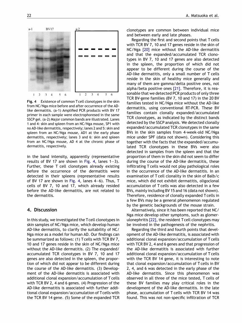
Fig. 4 Existence of common Tcell clonotypes in the skinfrom NC/Nga mice before and after occurrence of the AD-like dermatitis. (a-1) Amplified PCR products with BV 17primer in each sample were electrophoresed in the sameSSCP gel. (a-2) Major common bands are illustrated. Lanes1 and 4: skin and spleen from an NC/Nga mouse, SP1 withno AD-like dermatitis, respectively; lanes 2 and 5: skin andspleen from an NC/Nga mouse, AD1 at the early phasedermatitis, respectively; lanes 3 and 6: skin and spleenfrom an NC/Nga mouse, AD 4 at the chronic phase ofdermatitis, respectively.
in the band intensity, apparently (representativeresults of BV 17 are shown in Fig. 4, lanes 1—3).Further, these T cell clonotypes already existingbefore the occurrence of the dermatitis weredetected in their spleens (representative resultsof BV 17 are shown in Fig. 4, lanes 4—6). Thus, Tcells of BV 7, 10 and 17, which already residedbefore the AD-like dermatitis, are not related tothe dermatitis.
4. Discussion
In this study, we investigated the Tcell clonotypes inskin samples of NC/Nga mice, which develop humanAD-like dermatitis, to clarify the suitability of NC/Nga mice as a model for human AD. Our findings canbe summarized as follows: (1) Tcells with TCR BV 7,10 and 17 genes reside in the skin of NC/Nga micewithout the AD-like dermatitis. (2) The expanded/accumulated TCR clonotypes in BV 7, 10 and 17genes are also detected in the spleen, the propor-tion of which did not appear to be different duringthe course of the AD-like dermatitis. (3) Develop-ment of the AD-like dermatitis is associated withadditional clonal expansion/accumulation of T cellswith TCR BV 2, 4 and 6 genes. (4) Progression of theAD-like dermatitis is associated with further addi-tional clonal expansion/accumulation of Tcells withthe TCR BV 14 gene. (5) Some of the expanded TCR
clonotypes are common between individual miceand between early and late phases.
Regarding the first and second points that T cellswith TCR BV 7, 10 and 17 genes reside in the skin ofNC/Nga [20] mice without the AD-like dermatitisand that the expanded/accumulated TCR clono-types in BV 7, 10 and 17 genes are also detectedin the spleen, the proportion of which did notappear to be different during the course of theAD-like dermatitis, only a small number of T cellsreside in the skin of healthy mice generally andmany of them are gamma/delta positive ones, notalpha/beta positive ones [21]. Therefore, it is rea-sonable that we detected PCR products of only threeTCR BV-gene families (BV 7, 10 and 17) in the 20 BVfamilies tested in NC/Nga mice without the AD-likedermatitis, using conventional RT-PCR. These BVfamilies contain clonally expanded/accumulatedTCR clonotypes, as indicated by the distinct bandsdetected by the SSCP analysis. We detected clonallyexpanded/accumulated TCR clonotypes in the sameBVs in the skin samples from 4-week-old NC/Ngamice under SPF (data not shown). Considering thistogether with the facts that the expanded/accumu-lated TCR clonotypes in these BVs were alsodetected in samples from the spleen and that theproportion of them in the skin did not seem to differduring the course of the AD-like dermatitis, theseinfiltrating T cells would not play pathological rolesin the occurrence of the AD-like dermatitis. In anexamination of T cell clonality in the skin of Balb/cmice, which did not exhibit dermatitis, oligoclonalaccumulation of T cells was also detected in a fewBVs, mainly including BV 15 and 16 (data not shown).Therefore, residence of clonally expanded Tcells ina few BVs may be a general phenomenon regulatedby the genetic backgrounds of the mouse strain.
Alternatively, since it has been reported that NC/Nga mice develop other symptoms, such as glomer-ulonephritis [22], the resident Tcell clonotypes maybe involved in the pathogenesis of the nephritis.
Regarding the third and fourth points that devel-opment of the AD-like dermatitis, is associated withadditional clonal expansion/accumulation of T cellswith TCR BV 2, 4 and 6 genes and that progression ofthe AD-like dermatitis is associated with furtheradditional clonal expansion/accumulation of T cellswith the TCR BV 14 gene, it is interesting to notethat clonal expansion/accumulation of T cells in BV2, 4, and 6 was detected in the early phase of theAD-like dermatitis. Since this phenomenon wasobserved in all three of the mice tested, T cells ofthese BV families may play critical roles in thedevelopment of the AD-like dermatitis. In the latephase, accumulation of T cells with TCR BV 14 wasfound. This was not non-specific infiltration of TCR
T cell receptor gene clonotype in NC/Nga mice 23
BV 14-positive Tcells, since the PCR products of TCRBV 14 showed several distinct bands on the SSCPanalysis, which indicated that antigen-specific clo-nal expansion/accumulation of T cells with TCR BV14 occurs. The T cells may also contribute to theprogression of the AD-like dermatitis. Clonal expan-sion/accumulation of T cells with TCR BV 8.2 wasobserved only in some of the mice with the derma-titis (one out of the three mice each in the early andthe chronic phases), suggesting that T cells with BV8.2 may have a minimal contribution to the derma-titis. In the case of BV 15 and 16, the clonal expan-sion/accumulation was detected in one out of thethree mice in the early phase dermatitis and in twoor three out of the three mice in the late phasedermatitis, however, the accumulation of T cellswith these two BVs was also detected in one outof the three mice without dermatitis. Therefore, itremains to be resolved whether or not these T cellsplay an active role in the dermatitis.
In addition, since the TCR-B cain transcripts weredetected in more numbers of BVs in the late phasethan in the early phase, and also in the early phasethan in the dermatitis-undeveloped condition, thetotal number of the infiltrating T cells may beincreased in the dermatitis-developed condition,in particular, in the late phase. However, all thedetected BVs contained oligoclonal accumulation ofTcells, which indicated the pathological importanceof the antigen-specific T cells.
In humans, we previously reported the results ofsimilar T cell clonotype analyses [4,5]. In thosestudies, we detected oligoclonal accumulation ofTcells in inflamed skin areas of patients with chronicAD, but not in normal skin. This is comparable withthe present findings of oligoclonal accumulation of Tcells in various BV families in the late phase of theAD-like dermatitis in NC/Nga mice. It has not beenfully understood what the clonality of T cells in theskin of patients with an early phase of AD in humanis, mainly because of difficulty of sampling. There-fore, it remains to be solved whether expanded/accumulated Tcell clonotypes in the skin of patientswith early phase AD is rather limited compared tothose in the chronic phase. However, as far asobserving the chronic phase of dermatitis in NC/Nga mice and human AD, oligoclonal accumulationof Tcells is a common phenomenon between humanAD and the AD-like dermatitis in NC/Nga mice. Ofnote, the oligoclonal accumulation was observed inmost of the BVs tested in humans [4,5], while ratherlimited numbers of BVs showed oligoclonal accumu-lation in NC/Nga mice as presented here. Since thesensitivity of the SSCP analysis does not seem to beso different between human and mouse systems[10,12], involvement of rather limited numbers of
T cell clonotypes in the dermatitis can be a char-acteristic of NC/Nga mice. This may reflect differ-ent variety of environmental antigens betweenmicein cages and humans. From the aspect of T cellantigens, the NC/Nga mice can be a more simplifiedmodel of human AD. Further studies will be needed.
Regarding the fifth point that some of theexpanded TCR clonotypes are common betweenindividual mice and between early and late phases,clonal expansion/accumulation of T cells, indicatedby distinct bands on the SSCP gels, is thought tobe brought about by antigen-specific immuneresponses. In this regard, it is noteworthy that someof the newly expanded/accumulated T cells in thecourse of the AD-like dermatitis showed the sameclonotypes between individual mice. Since thegenetic backgrounds of the NC/Nga mice are iden-tical, they possess the same array of MHC antigens,which are a specific apparatus to present antigens toTCR on T cells. Therefore, detection of the same Tcell clonotypes would indicate recognition of thesame and/or very similar antigens. These antigenswould play critical roles in the immune reactions ofthe AD-like dermatitis. In humans, we reported thatidentical T cell clones with the same antigen speci-ficities are present in samples obtained from sepa-rate regions of the skin, indicating that stimulationwith antigen(s) that exist in large areas of the skinlead to the oligoclonal accumulation of Tcells. Thus,human AD and the AD-like dermatitis found in NC/Nga mice are similar from the viewpoint of T cell-stimulating antigens. In addition, we reported thatone of the antigens recognized by the clonallyexpanded T cells in skin lesions of patients withAD was house dust mite antigens purified fromDermatophagoides pteronyssinus [4]. Therefore, itis interesting and should be further investigatedwhich antigens stimulate T cells to expand in theskin lesions of NC/Nga mice.
In conclusion, the AD-like dermatitis of NC/Ngamice displays oligoclonal T cell accumulation,which is more distinct in the late phase than inthe early phase. The oligoclonal accumulation of Tcells, which indicates antigen-specific immuneresponses, is a common feature between humanAD and the AD-like dermatitis in NC/Nga mice.Thus, NC/Nga mice are a suitable model for humanAD from the viewpoint of antigen-specific T cellimmune responses.
Acknowledgements
This study was supported in part by grants from theMinistry of Education, Culture, Sports, Science and
24 A. Matsuoka et al.
Technology of Japan, and the Ministry of Health,Labor and Welfare of Japan.
[2] Menssen A, Trommler P, Vollmer S, Schendel D, Albert E,Gurtler L, et al. Evidence for an antigen-specific cellularimmune response in skin lesions of patients with psoriasisvulgaris. J Immunol 1995;155(8):4078—83.
[3] Mihm Jr MC, Soter NA, Dvorak HF, Austen KF. The structure ofnormal skin and the morphology of atopic eczema. J InvestDermatol 1976;67(3):305—12.
[4] Takahama H, Masuko-hongo K, Tanaka A, Kawa Y, Ohta N,Yamamoto K, et al. T-cell clonotypes specific forDermatophagoides pteronyssinus in the skin lesionsof patients with atopic dermatitis. Hum Immunol2002;63(7):558—66.
[5] Tanaka A, Takahama H, Kato T, Kubota Y, Kurokawa K,Nishioka K, et al. Clonotypic analysis of T cells infiltratingthe skin of patients with atopic dermatitis: evidence forantigen-driven accumulation of T cells. Hum Immunol1996;48(1—2):107—13.
[6] Matsuoka H, Maki N, Yoshida S, Arai M, Wang J, Oikawa Y, etal. A mouse model of the atopic eczema/dermatitis syn-drome by repeated application of a crude extract of house-dust mite Dermatophagoides farinae. Allergy 2003;58(2):139—45.
[7] Aioi A, Tonogaito H, Suto H, Hamada K, Ra CR, Ogawa H, etal. Impairment of skin barrier function in NC/Nga Tnd miceas a possible model for atopic dermatitis. Br J Dermatol2001;144(1):12—8.
[8] Matsuda H, Watanabe N, Geba GP, Sperl J, Tsudzuki M, HiroiJ, et al. Development of atopic dermatitis-like skin lesionwith IgE hyperproduction in NC/Nga mice. Int Immunol1997;9(3):461—6.
[9] Matsumoto M, Ra C, Kawamoto K, Sato H, Itakura A, SawadaJ, et al. IgE hyperproduction through enhanced tyrosinephosphorylation of Janus kinase 3 in NC/Nga mice, a modelfor human atopic dermatitis. J Immunol 1999;162(2):1056—63.
[10] Kato T, Ikeda Y, Zong ZP, Sasakawa H, Kurokawa M, Masuko K,et al. Characterization of T cell receptor beta chains of
accumulating T cells in skin allografts in mice. Transplanta-tion 1996;62(2):266—72.
[11] Yamamoto K, Masuko-Hongo K, Tanaka A, Kurokawa M,Hoeger T, Nishioka K, et al. Establishment and applicationof a novel T cell clonality analysis using single-strand con-formation polymorphism of T cell receptor messenger sig-nals. Hum Immunol 1996;48(1—2):23—31.
[12] Yamamoto K, Sakoda H, Nakajima T, Kato T, Okubo M, DohiM, et al. Accumulation of multiple T cell clonotypes in thesynovial lesions of patients with rheumatoid arthritisrevealed by a novel clonality analysis. Int Immunol 1992;4(11):1219—23.
[13] Barth RK, Kim BS, Lan NC, Hunkapiller T, Sobieck N, WinotoA, et al. The murine T-cell receptor uses a limited repertoireof expressed V beta gene segments. Nature 1985;316(6028):517—23.
[14] Behlke MA, Spinella DG, Chou HS, Sha W, Hartl DL, Loh DY. T-cell receptor beta-chain expression: dependence on rela-tively few variable region genes. Science 1985;229(4713):566—70.
[15] Goverman J, Minard K, Shastri N, Hunkapiller T, Hansburg D,Sercarz E, et al. Rearranged beta T cell receptor genes in ahelper T cell clone specific for lysozyme: no correlationbetween V beta and MHC restriction. Cell 1985;40(4):859—67.
[16] Chou HS, Anderson SJ, Louie MC, Godambe SA, Pozzi MR,Behlke MA, et al. Tandem linkage and unusual RNA splicing ofthe T-cell receptor beta-chain variable-region genes. ProcNatl Acad Sci USA 1987;84(7):1992—6.
[17] Saito H, Kranz DM, Takagaki Y, Hayday AC, Eisen HN, Tone-gawa S. Complete primary structure of a heterodimeric T-cell receptor deduced from cDNA sequences. Nature1984;309(5971):757—62.
[18] Behlke MA, Chou HS, Huppi K, Loh DY. Murine T-cell receptormutants with deletions of beta-chain variable region genes.Proc Natl Acad Sci USA 1986;83(3):767—71.
[19] Kappler JW, Wade T, White J, Kushnir E, Blackman M, Bill J,et al. A T cell receptor V beta segment that imparts reac-tivity to a class II major histocompatibility complex product.Cell 1987;49(2):263—71.
[20] Opelz G, Kiuchi M, Takasugi M, Terasaki PI. Autologousstimulation of human lymphocyte subpopulation. J ExpMed 1975;142(5):1327—33.