AbstractMuscular variants of the forearm are common and frequently cause neurovascular compression syndromes, especially when interfering with the compact topography of the carpal tunnel or the Canalis ulnaris. Here, we report on a male body donor with multiple muscular normal variations on both forearms. The two main findings are (1) an accessory variant muscle (AVM) on the right forearm originating from the M. brachioradialis, the distal radius, and the M. flexor pollicis longus. It spanned the wrist beneath the Fascia antebrachia and inserted at the proximal phalanx of the digitus minimus. (2) Moreover, we found a three-headed palmaris longus variant on the left arm with proximal origin tendon and a distal, trifurcated muscle belly, with separated insertions at the palmar aponeurosis, the flexor retinaculum, and, in analogy to the accessory muscle on the contralateral arm, at the base of the proximal phalanx of the digitus minimus. We found a considerable thickening of the left-hand median nerve right before entering the carpal tunnel indicative of a possible chronic compression syndrome adding clinical relevance to this anatomical case. We also discuss the notion that both, the AVM and the contralateral three-headed palmaris variant are developmental descendants of the M. palmaris longus. Additionally, we found a previously not recorded variant of the M. palmaris brevis on the left hand.
Variants of the muscular system, especially in the forearm and wrist, are common and include various deviations, such as the absence of muscles, alternative origins, insertions, or trajectories, as well as accessory muscles (Gruber 1868; Macalister 1875; Sookur et al. 2008; Spalteholz 1939). Since they often do not cause any symptoms, most of these mus-cular variants remain undetected and thus a precise quan-tification of their prevalence remains vague. Harvie et al. (2004) found that in a collective of 58 asymptomatic test persons, 47% exhibited muscular variants of the M. abduc-tor digiti minimi. Similarly, a MRI-based study detected 23 muscle variations on 42 wrists, of which in 24% there was an accessory M. abductor digiti minimi and in 16% the M. palmaris longus was missing (Zeiss and Jakab 1995). Dodds et al. (1990) detected muscular variants in 22.4% of
wrists in 58 body donor documented by anatomical dissec-tion. In contrast, in a recent report, Park (2019) found only eight anomalous muscles while conducting endoscopic car-pal tunnel surgery in 973 hands. It is noteworthy, however, that this low number of variants was caused by the limited endoscopic field of view. Yet a considerably lower number of variants found within narrow and functionally important structures, such as carpal tunnel, compared to less confined topographies of e.g. the M. palmaris longus, would well explain the low count of symptomatic cases despite a high prevalence of muscular variants on the forearm in general.
Although most variants are incidental findings, still abnor-mal muscular configurations can cause neurovascular com-pression syndromes as reported by several authors (Al-Qattan 2004; De Smet 2002; James et al. 1987; Jeffery 1971; Lisanti et al. 2001; Ruocco et al. 1998; Salgeback 1977; Spinner et al. 1996; Zeiss and Jakab 1995). In fact, approximately 50% of muscle anomaly-caused carpal tunnel neuropathies and about 20% of ulnar tunnel compression syndromes are caused by the M. palmaris longus or its variants (Zeiss and Jakab 1995). Moreover, changes in the topography by missing or accessory muscles are likely to impede with surgical interventions.
557Anatomical work-up of an individual with multiple muscular variants on both forearms
1 3
In this report, we describe a previously unknown acces-sory muscle in the hypothenar area and wrist of the right forearm, located directly below the Fascia antebrachii. Additionally, we report two additional variants on the left forearm, namely a variant of the M. palmaris brevis with an accessory origin on the fascia of the thenar musculature as well as a M. palmaris longus variant with three muscular divisions inserting on the palmar aponeurosis, the flexor reti-naculum and the proximal phalanx of the digitus minimus. Since we also found a considerably thickening of the median nerve indicative of a chronic compression syndrome, these and similar anatomical variants may have a direct clinical impact.
Material and methods
Body donor
The body donor was a 65-year-old Caucasian male, who died of sudden cardiac death. No morphological abnormali-ties such as scars or deformities were visible externally. In the latest medical record, no pre-existing conditions of the locomotor system were documented. The body donor gave his informed consent in concert with the declaration of Hel-sinki to use his cadaver for research purposes. The procedure was approved by the local ethical authorities (Project Nr. 237/2007 BO1).
Fixation
The fixation was carried out by intravasal infusion via the femoral artery using an IJT-50 injection system (Thalheimer, Ellwangen, Germany). Depending on the condition of the cadaver’s vascular system, we used perfusion pressure of 0.5–1.0 bar. The fixation solution consisted of ethanol 45.5% (v/v), glycerin 23,5% (v/v), formalin 2% (v/v), and lyso-formin 3,6% (v/v) in H2O.
Photo‑documentation
We photo-documented each preparation step under standard-ized conditions using a Nikon D300 camera with vario lens. Camera settings were standardized to 1/80 s exposure time, ISO 800 and aperture setting 16.
Results
Accessory wrist muscle on the right forearm
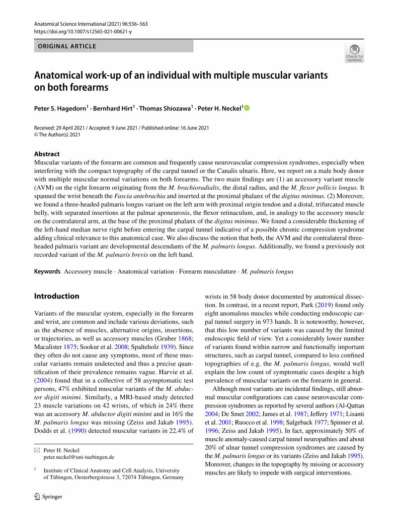
The accessory variant muscle (AVM) was located directly beneath the Fascia antebrachii on the right forearm and wrist (Figs. 1 and 2). It ran obliquely from the radial aspect
of the distal palmar forearm to the hypothenar muscles. On its course, it passed underneath the radial artery and the tendon of M. flexor carpi radialis. Thereby, it made direct contact to the Carpal tunnel, to the ulnar tunnel, and to the ulnar artery and nerve. In its trajectory, the AVM muscle fib-ers curved towards the hypothenar musculature. The muscle belly narrowed to form an approximately cylindrical tendon at the proximal third of the hypothenar, which then passed between the bellies of Mm. flexor et abductor digiti minimi into the depth of the hypothenar musculature (Figs. 1 and 2).
We severed the radial artery, M. abductor pollicis longus, M. flexor carpi radialis, and M. brachioradialis to gain a better access to the AVM’s origin (Fig. 2B–D). For a bet-ter access to the insertion, we separated the M. flexor digiti minimi at its origin (Fig. 2A, B).
We found that the AVM exhibits a thin, but wide and tripartite origin from the tendon of the M. brachioradialis, with few connective tissue fibers branching of to insert at the periosteum of the distal palmar part of the radius, as well as a third origin from the belly of the M. flexor pollicis longus. The cylindrical tendon of the AVM inserts at the base of the proximal phalanx of the digitus minimus (Figs. 1 and 2).
In comparison to previously reported accessory muscles of the hypothenar and wrist, the AVM is extraordinarily large in size with a maximum thickness of 0.4 cm, a length of 7.4 cm (muscle belly), and width of 2.3 cm as meas-ured at the belly of the muscle. Moreover, we found an even wider tripartite aponeurotic origin with a combined width of 5.5 cm. The insertion tendon is 4.5 cm long and 0.3 cm in diameter.
Due to the trajectory of the AVM and targeted pull-sim-ulations, the presumable function was a weak abduction of the digitus minimus and a flexion-ulnarduction in the wrist.
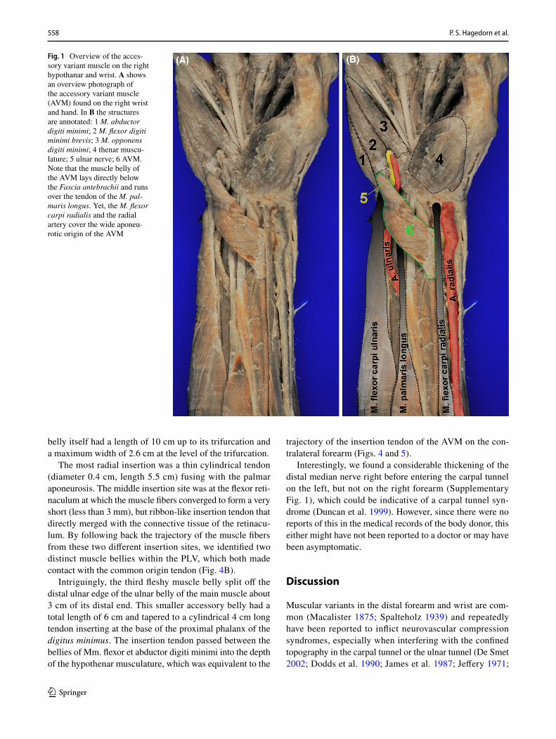
Variant of palmaris brevis on the left wrist
At the left hand, we found a variant of the M. palmaris bre-vis, consisting of larger, regularly described part with an origin at the palmar aponeurosis and an insertion just below the integument of the hypothenar region. Additionally, how-ever, there were accessory muscle fibers originating at the fascia of the thenar musculature and running underneath the palmar aponeurosis to eventually converge and join the other palmaris brevis fibers at the regular insertion site (Fig. 3).
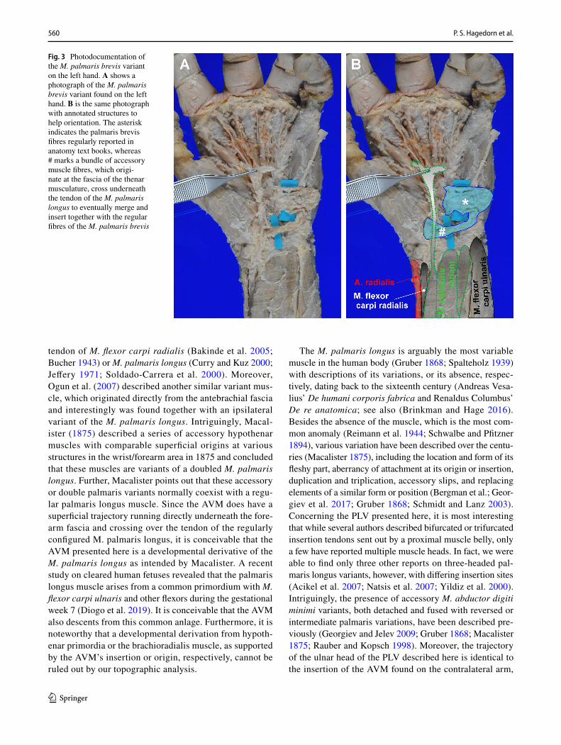
M. palmaris longus variant on the left forearm
Additionally, we found a M. palmaris longus variant (PLV) also located on the left forearm of the body donor (Fig. 4) and originating from the medial epicondyle of the humerus with a nearly cylindrical origin tendon with 15 cm of length. Interestingly, the fleshy muscle belly was trifurcated/three-headed with three different insertion tendons. The muscle
558 P. S. Hagedorn et al.
1 3
belly itself had a length of 10 cm up to its trifurcation and a maximum width of 2.6 cm at the level of the trifurcation.
The most radial insertion was a thin cylindrical tendon (diameter 0.4 cm, length 5.5 cm) fusing with the palmar aponeurosis. The middle insertion site was at the flexor reti-naculum at which the muscle fibers converged to form a very short (less than 3 mm), but ribbon-like insertion tendon that directly merged with the connective tissue of the retinacu-lum. By following back the trajectory of the muscle fibers from these two different insertion sites, we identified two distinct muscle bellies within the PLV, which both made contact with the common origin tendon (Fig. 4B).
Intriguingly, the third fleshy muscle belly split off the distal ulnar edge of the ulnar belly of the main muscle about 3 cm of its distal end. This smaller accessory belly had a total length of 6 cm and tapered to a cylindrical 4 cm long tendon inserting at the base of the proximal phalanx of the digitus minimus. The insertion tendon passed between the bellies of Mm. flexor et abductor digiti minimi into the depth of the hypothenar musculature, which was equivalent to the
trajectory of the insertion tendon of the AVM on the con-tralateral forearm (Figs. 4 and 5).
Interestingly, we found a considerable thickening of the distal median nerve right before entering the carpal tunnel on the left, but not on the right forearm (Supplementary Fig. 1), which could be indicative of a carpal tunnel syn-drome (Duncan et al. 1999). However, since there were no reports of this in the medical records of the body donor, this either might have not been reported to a doctor or may have been asymptomatic.
Discussion
Muscular variants in the distal forearm and wrist are com-mon (Macalister 1875; Spalteholz 1939) and repeatedly have been reported to inflict neurovascular compression syndromes, especially when interfering with the confined topography in the carpal tunnel or the ulnar tunnel (De Smet 2002; Dodds et al. 1990; James et al. 1987; Jeffery 1971;
Fig. 1 Overview of the acces-sory variant muscle on the right hypothanar and wrist. A shows an overview photograph of the accessory variant muscle (AVM) found on the right wrist and hand. In B the structures are annotated: 1 M. abductor digiti minimi; 2 M. flexor digiti minimi brevis; 3 M. opponens digiti minimi; 4 thenar muscu-lature; 5 ulnar nerve; 6 AVM. Note that the muscle belly of the AVM lays directly below the Fascia antebrachii and runs over the tendon of the M. pal-maris longus. Yet, the M. flexor carpi radialis and the radial artery cover the wide aponeu-rotic origin of the AVM
559Anatomical work-up of an individual with multiple muscular variants on both forearms
1 3
Lisanti et al. 2001; Ruocco et al. 1998; Spinner et al. 1996; Zeiss and Jakab 1995). In this report, we presented the ana-tomical workup of a male individual with multiple, previ-ously unknown variants on both arms. The two major find-ings were an accessory variant muscle on the right wrist and hypothenar as well as a variant of the M. palmaris longus on the left arm with one origin tendon and three separated insertion sites.
The trajectory of the insertion tendon as well as the insertion site of the AVM is similar to previously described accessory variants of the M. abductor digiti minimi or M. flexor digiti minimi (Bakinde et al. 2005; Bucher 1943; Claassen et al. 2013; Curry and Kuz 2000; Ogun et al. 2007; Ruge 1921; Saadeh and Bergman 1988; Soldado-Carrera et al. 2000; Wingerter et al. 2003; Wulle 1987); various variants have been illustrated by (Schmidt and
Lanz 2003). Yet, none of the reported variants had a tripar-tite origin from the tendon of the M. brachioradialis, the belly of the M. flexor pollicis longus, and the periosteum of the distal radius. Astonishingly similar was an acces-sory hypothenar muscle reported by (Claassen et al. 2013). However, this variant did not exhibit an isolated insertion tendon but rather fused with the muscle belly of the regu-lar M. abductor digiti minimi. Moreover, this variant origi-nated from the carpal ligament, flexor retinaculum, and the palmar side of radius, yet, unlike the AVM described here, running underneath the palmaris longus tendon (Claassen et al. 2013). Other accessory M. abductor digiti minimi variants were found to originate at the antebrachial fascia (Saadeh and Bergman 1988; Wingerter et al. 2003; Wulle 1987) and the intercompartmental septum on the medial side of the forearm (Wahba et al. 1998) as well as at the
Fig. 2 Documentation of the accessory variant muscle on the right hypothenar and wrist. A–D show photographs of the accessory vari-ant muscle (AVM) from different angles and with full access to its origin and insertion tendons. In Ai–Di the structures are annotated: 1 M. abductor digiti minimi; 2 thenar musculature; 3 ulnar nerve; 4 AVM; the asterisk in A marks fatty tissue covering large parts of
the aponeurotic insertion tendon of the AVM, which was removed in B-D. The AVM originates from the tendon of the M. brachioradia-lis, the belly of the M. flexor pollicis longus, and from the periosteum of the distal radius. The nearly cylindrical insertion tendon does not merge with other muscles of the hypothenar and inserts at the proxi-mal ulnar aspect of the proximal phalanx of the digitus minimus
560 P. S. Hagedorn et al.
1 3
tendon of M. flexor carpi radialis (Bakinde et al. 2005; Bucher 1943) or M. palmaris longus (Curry and Kuz 2000; Jeffery 1971; Soldado-Carrera et al. 2000). Moreover, Ogun et al. (2007) described another similar variant mus-cle, which originated directly from the antebrachial fascia and interestingly was found together with an ipsilateral variant of the M. palmaris longus. Intriguingly, Macal-ister (1875) described a series of accessory hypothenar muscles with comparable superficial origins at various structures in the wrist/forearm area in 1875 and concluded that these muscles are variants of a doubled M. palmaris longus. Further, Macalister points out that these accessory or double palmaris variants normally coexist with a regu-lar palmaris longus muscle. Since the AVM does have a superficial trajectory running directly underneath the fore-arm fascia and crossing over the tendon of the regularly configured M. palmaris longus, it is conceivable that the AVM presented here is a developmental derivative of the M. palmaris longus as intended by Macalister. A recent study on cleared human fetuses revealed that the palmaris longus muscle arises from a common primordium with M. flexor carpi ulnaris and other flexors during the gestational week 7 (Diogo et al. 2019). It is conceivable that the AVM also descents from this common anlage. Furthermore, it is noteworthy that a developmental derivation from hypoth-enar primordia or the brachioradialis muscle, as supported by the AVM’s insertion or origin, respectively, cannot be ruled out by our topographic analysis.
The M. palmaris longus is arguably the most variable muscle in the human body (Gruber 1868; Spalteholz 1939) with descriptions of its variations, or its absence, respec-tively, dating back to the sixteenth century (Andreas Vesa-lius’ De humani corporis fabrica and Renaldus Columbus’ De re anatomica; see also (Brinkman and Hage 2016). Besides the absence of the muscle, which is the most com-mon anomaly (Reimann et al. 1944; Schwalbe and Pfitzner 1894), various variation have been described over the centu-ries (Macalister 1875), including the location and form of its fleshy part, aberrancy of attachment at its origin or insertion, duplication and triplication, accessory slips, and replacing elements of a similar form or position (Bergman et al.; Geor-giev et al. 2017; Gruber 1868; Schmidt and Lanz 2003). Concerning the PLV presented here, it is most interesting that while several authors described bifurcated or trifurcated insertion tendons sent out by a proximal muscle belly, only a few have reported multiple muscle heads. In fact, we were able to find only three other reports on three-headed pal-maris longus variants, however, with differing insertion sites (Acikel et al. 2007; Natsis et al. 2007; Yildiz et al. 2000). Intriguingly, the presence of accessory M. abductor digiti minimi variants, both detached and fused with reversed or intermediate palmaris variations, have been described pre-viously (Georgiev and Jelev 2009; Gruber 1868; Macalister 1875; Rauber and Kopsch 1998). Moreover, the trajectory of the ulnar head of the PLV described here is identical to the insertion of the AVM found on the contralateral arm,
Fig. 3 Photodocumentation of the M. palmaris brevis variant on the left hand. A shows a photograph of the M. palmaris brevis variant found on the left hand. B is the same photograph with annotated structures to help orientation. The asterisk indicates the palmaris brevis fibres regularly reported in anatomy text books, whereas # marks a bundle of accessory muscle fibres, which origi-nate at the fascia of the thenar musculature, cross underneath the tendon of the M. palmaris longus to eventually merge and insert together with the regular fibres of the M. palmaris brevis
561Anatomical work-up of an individual with multiple muscular variants on both forearms
1 3
thereby additionally supporting the notion that the variants on both arms are variations of the palmaris longus muscle. Alternatively, the three-headed palmaris variation we found could also be the product of a bi-headed palmaris longus fused with an accessory M. abductor digiti minimi.
Numerous authors have reported neurovascular com-pression syndromes caused by muscular variations or accessory muscles (De Smet 2002; Dodds et al. 1990; James et al. 1987; Jeffery 1971; Lisanti et al. 2001; Ruocco
et al. 1998; Spinner et al. 1996; Zeiss and Jakab 1995). Interestingly, two of the previously mentioned three-headed palmaris variations were causative for median nerve compression symptoms and thus were discovered in surgery (Acikel et al. 2007; Yildiz et al. 2000). In the present case, we found a considerable thickening in the median nerve directly before entering the carpal tunnel on the left side, which arguably indicates a chronic com-pression caused by the PLV (Duncan et al. 1999). Yet, it is noteworthy that we did not find any report on median nerve compression symptoms in the medical records of the body donor suggesting either that the presence of the PLV was asymptomatic or that the body donor never sought medical advice on his symptoms. Still, it is highly conceivable that variants in close proximity to important anatomical structures can cause symptoms of nerve and artery compression and additionally hamper in-surgery orientation with potential complications.
Supplementary Information The online version contains supplemen-tary material available at https:// doi. org/ 10. 1007/ s12565- 021- 00621-y.
Acknowledgements We would like to thank Jürgen Papp, Manfred Mauz, and Tatjana Steiner for their technical support as well as Bern-hard Tillmann for his great help on literature research and his helpful
Fig. 4 Overview of the M. palmaris longus variant on the left fore-arm and wrist. A and B show an overview and detail photograph of the M. palmaris longus variant (PLV) found on the left forearm. In Ai and Bi the structures are annotated: 1 thenar musculature; 2 M. flexor digiti minimi brevis; 3 M. abductor digiti minimi; 4 PLV. The PLV has a tendon of origin and large intermediate muscle belly, which in turn trifurcates and inserts in the palmar aponeurosis, the reti-naculum flexorum, and the proximal phalanx of the digitus minimus. These insertion sites correspond to fused, but distinguishable muscle bellies (4.1–4.3). The insertion tendon of 4.1 with the attached pal-mar aponeurosis was displaced radially in B to allow a better view on the insertion site of 4.2. Note that the insertion tendon of 4.3 has the same trajectory and insertion site as the AVM on the contralateral arm
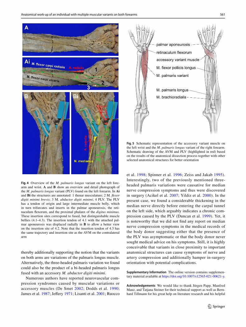
Fig. 5 Schematic representation of the accessory variant muscle on the left wrist and the M. palmaris longus variant of the right forearm. Schematic drawing of the AVM and PLV (highlighted in red) based on the results of the anatomical dissection process together with other selected anatomical structures for better orientation
comments on the work and manuscript. Moreover, we want to express our gratitude to the body donor.
Author contributions PSH: study concept and design; acquisition of data; analysis and interpretation of data; drafting of the manuscript. BH: study concept and design; critical revision of the manuscript for important intellectual content. TS: study concept and design; critical revision of the manuscript for important intellectual content. PHN: study concept and design; acquisition of data; analysis and interpreta-tion of data; drafting of the manuscript; critical revision of the manu-script for important intellectual content.
Funding Open Access funding enabled and organized by Projekt DEAL. Not applicable.
Data availability Not applicable.
Code availability Not applicable.
Declarations
Conflict of interests The authors declare no conflict of interests.
Ethical approval The body donor gave his informed consent in con-cert with the declaration of Helsinki to use his cadaver for research purposes. The procedure was approved by the local ethical authorities (Project Nr. 237/2007 BO1).
Consent to participate Not applicable.
Consent for publication Not applicable.
Open Access This article is licensed under a Creative Commons Attri-bution 4.0 International License, which permits use, sharing, adapta-tion, distribution and reproduction in any medium or format, as long as you give appropriate credit to the original author(s) and the source, provide a link to the Creative Commons licence, and indicate if changes were made. The images or other third party material in this article are included in the article’s Creative Commons licence, unless indicated otherwise in a credit line to the material. If material is not included in the article’s Creative Commons licence and your intended use is not permitted by statutory regulation or exceeds the permitted use, you will need to obtain permission directly from the copyright holder. To view a copy of this licence, visit http:// creat iveco mmons. org/ licen ses/ by/4. 0/.
References
Acikel C, Ulkur E, Karagoz H, Celikoz B (2007) Effort-related com-pression of median and ulnar nerves as a result of reversed three-headed and hypertrophied palmaris longus muscle with exten-sion of Guyon’s canal. Scand J Plast Reconstr Surg Hand Surg 41(1):45–47
Al-Qattan MM (2004) Ulnar nerve compression at the wrist by the accessory abductor digiti minimi muscle: wrist trauma as a pre-cipitating factor. Hand Surg 9(1):79–82
Bakinde N, Yotovski P, Voigt T, Rager G (2005) Accessory mus-cle in the hypothenar region: a functional approach. Ann Anat 187(2):149–152
Bergman RA, Afifi AK, Miyauchi R Anatomy Atlases: Illustrated Encyclopedia of Human Anatomic Variation. Available online:
Brinkman RJ, Hage JJ (2016) The first description of the absence of the palmaris longus muscle. J Plast Surg Hand Surg 50(1):56–58
Bucher O (1943) Über die Varietäten des M abductor digiti quinti. Anat Anz 94:317–324
Claassen H, Schmitt O, Schulze M, Wree A (2013) Variation in the hypothenar muscles and its impact on ulnar tunnel syndrome. Surg Radiol Anat 35(10):893–899
Curry B, Kuz J (2000) A new variation of abductor digiti minimi acces-sorius. J Hand Surg Am 25(3):585–587
De Smet L (2002) Median and ulnar nerve compression at the wrist caused by anomalous muscles. Acta Orthop Belg 68(5):431–438
Diogo R, Siomava N, Gitton Y (2019) Development of human limb muscles based on whole-mount immunostaining and the links between ontogeny and evolution. Development 146(20):dev180349. https:// doi. org/ 10. 1242/ dev. 180349
Dodds GA 3rd, Hale D, Jackson WT (1990) Incidence of anatomic variants in Guyon’s canal. J Hand Surg Am 15(2):352–355
Duncan I, Sullivan P, Lomas F (1999) Sonography in the diagnosis of carpal tunnel syndrome. AJR Am J Roentgenol 173(3):681–684
Georgiev GP, Jelev L (2009) Unusual coexistence of a variant abduc-tor digiti minimi and reversed palmaris longus and their possi-ble relation to median and ulnar nerves entrapment at the wrist. Rom J Morphol Embryol 50(4):725–727
Georgiev GP, Iliev AA, Dimitrova IN, Kotov GN, Malinova LG, Landzhov BV (2017) Palmaris Longus Muscle Variations: Clin-ical Significance and Proposal of New Classifications. Folia Med (plovdiv) 59(3):289–297
Gruber (1868) Über die Varietäten des Musculus palmaris longus, Mémoires de l’Academie Impériale des Sciences de St.-Péters-bourg, VII Série,Tome XI, No 14, St. Petersburg
Harvie P, Patel N, Ostlere SJ (2004) Prevalence and epidemiological variation of anomalous muscles at guyon’s canal. J Hand Surg Br 29(1):26–29
James MR, Rowley DI, Norris SH (1987) Ulnar nerve compression by an accessory abductor digiti minimi muscle presenting fol-lowing injury. Injury 18(1):66–67
Jeffery AK (1971) Compression of the deep palmar branch of the ulnar nerve by an anomalous muscle. Case report and review. J Bone Jt Surg Br 53(4):718–723
Lisanti M, Rosati M, Maltinti M (2001) Ulnar nerve entrapment in Guyon’s tunnel by an anomalous palmaris longus muscle with a persisting median artery. Acta Orthop Belg 67(4):399–402
Macalister A (1875) Additional observations on muscular anomalies in human anatomy. (Third series) with a catalogue of the prin-cipal muscular variations hitherto published. Trans R Ir Acad 25:1–134
Natsis K, Levva S, Totlis T, Anastasopoulos N, Paraskevas G (2007) Three-headed reversed palmaris longus muscle and its clinical significance. Ann Anat 189(1):97–101
Ogun TC, Karalezli N, Ogun CO (2007) The concomitant presence of two anomalous muscles in the forearm. Hand (NY) 2(3):120–122
Park SH (2019) Anomalous muscles of the wrist encountered dur-ing endoscopic carpal tunnel surgery. J Korean Neurosurg Soc 62(1):90–95
Rauber AA, Kopsch F (1998) Anatomie des Menschen, Band I Bewe-gungsapparat, 2nd edn., Tillmann B, Töndury G (eds), Georg Thime Verlag, Stuttgart
Reimann AF, Daseler EH, Anson BJ, Beaton LE (1944) The palmaris longus muscle and tendon. A study of 1600 extremities. Anat Rec 89(4):495–505
Ruge G (1921) Anleitungen zu den Präparierübungen and der men-schlichen Leiche, 5th edn. Verlag von Wilhelm Engelmann, Leipzig
563Anatomical work-up of an individual with multiple muscular variants on both forearms
1 3
Ruocco MJ, Walsh JJ, Jackson JP (1998) MR imaging of ulnar nerve entrapment secondary to an anomalous wrist muscle. Skeletal Radiol 27(4):218–221
Saadeh FA, Bergman RA (1988) An unusual accessory flexor (oppon-ens) digiti minimi muscle. Anat Anz 165(4):327–329
Salgeback S (1977) Ulnar tunnel syndrome caused by anomalous mus-cles. Case report. Scand J Plast Reconstr Surg 11(3):255–258
Schmidt H-M, Lanz U (2003) Chirurgische Anatomie der Hand, 2nd edn. Georg Thieme Verlag, Stuttgart
Schwalbe G, Pfitzner W (1894) Varietäten-statistik und anthropologie. Morphologische Arbeiten 3(3):459–490
Soldado-Carrera F, Vilar-Coromina N, Rodriguez-Baeza A (2000) An accessory belly of the abductor digiti minimi muscle: a case report and embryologic aspects. Surg Radiol Anat 22(1):51–54
Sookur PA, Naraghi AM, Bleakney RR, Jalan R, Chan O, White LM (2008) Accessory muscles: anatomy, symptoms, and radiologic evaluation. Radiographics 28(2):481–499
Spalteholz W (1939) Handatlas der Anatomie des Menschen, II, 14th edn. Verlag von S. Hirzel, Leipzig
Spinner RJ, Lins RE, Spinner M (1996) Compression of the medial half of the deep branch of the ulnar nerve by an anomalous ori-gin of the flexor digiti minimi. a case report. J Bone Jt Surg Am 78(3):427–430
Wahba MY, Singh GD, Lozanoff S (1998) An anomalous accessory flexor digiti minimi profundus muscle: a case study. Clin Anat 11(1):55–59
Wingerter S, Gupta S, Le S, Shamasunder S, Bernstein R, Rabitaille W, Kukuyeva Y, Downie S (2003) Unusual origin of the flexor digiti minimi brevis muscle. Clin Anat 16(6):531–533
Wulle C (1987) M. abductor digiti minimi longus: anatomical rarity ? Handchir Mikrochir Plast Chir 19(1):43–45
Yildiz M, Sener M, Aynaci O (2000) Three-headed reversed palmaris longus muscle: a case report and review of the literature. Surg Radiol Anat 22(3–4):217–219
Zeiss J, Jakab E (1995) MR demonstration of an anomalous muscle in a patient with coexistent carpal and ulnar tunnel syndrome. Case report and literature summary. Clin Imaging 19(2):102–105
Publisher’s Note Springer Nature remains neutral with regard to jurisdictional claims in published maps and institutional affiliations.