58
TISSUES CHAPTER 3
| Date post: | 19-Dec-2015 |
| Category: |
Documents |
| Upload: | jan-jamison-zulueta |
| View: | 23 times |
| Download: | 2 times |
TISSUESCHAPTER 3
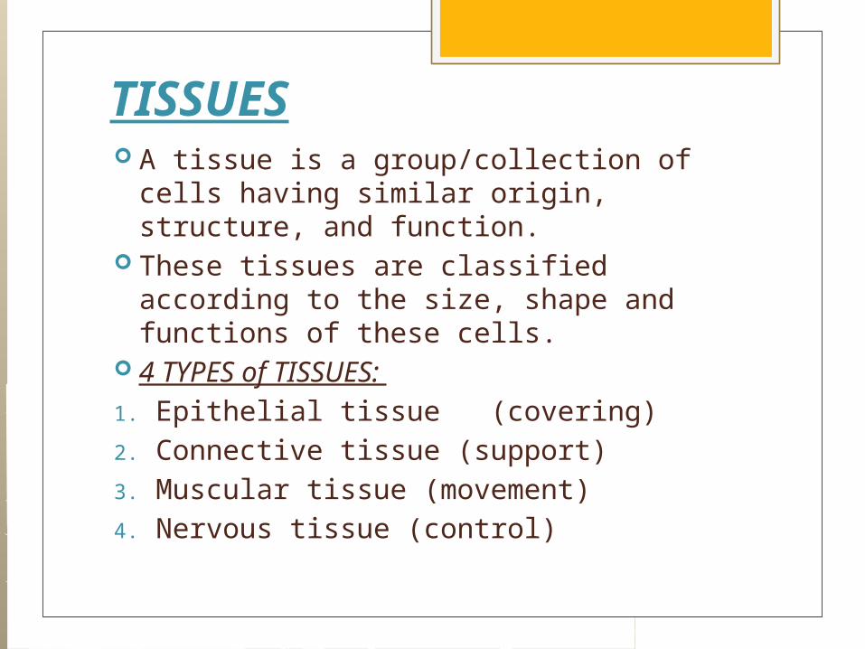
TISSUES A tissue is a group/collection of cells
having similar origin, structure, and function.
These tissues are classified according to the size, shape and functions of these cells.
4 TYPES of TISSUES: 1. Epithelial tissue (covering)2. Connective tissue (support)3. Muscular tissue (movement)4. Nervous tissue (control)

I. EPITHELIAL TISSUE: The underside of this tissue
is anchored to a connective tissue by a thin non-living layer called basement membrane or basal lamina. It (1) reproduces readily; (2) heal rapidly into new cells, replacing the damaged or lost ones. It has no blood supply, but is nourished by nutrients obtained from the blood vessels in the underlying connective tissue.
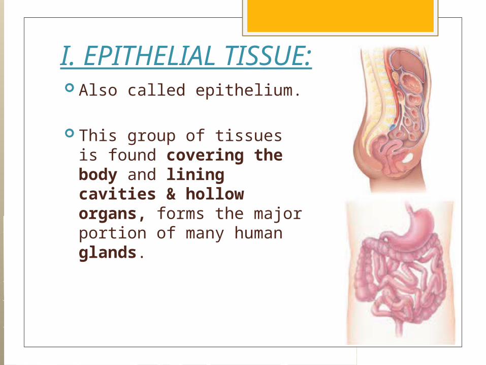
I. EPITHELIAL TISSUE: Also called epithelium.
This group of tissues is found covering the body and lining cavities & hollow organs, forms the major portion of many human glands.
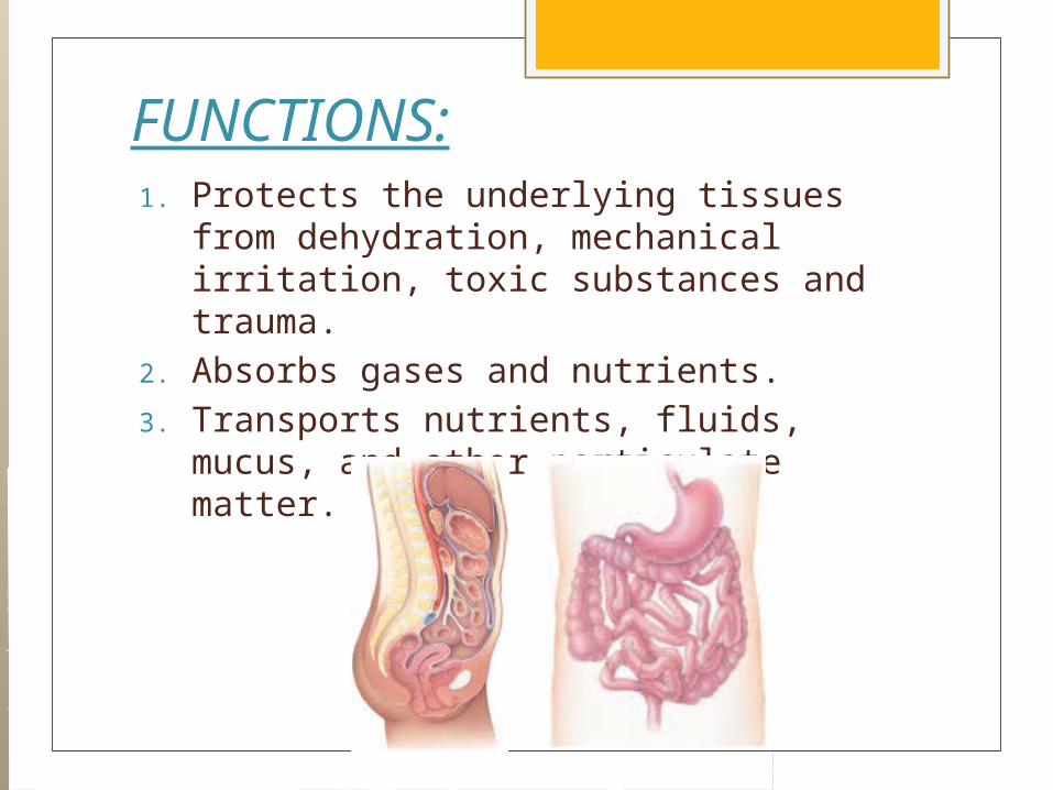
FUNCTIONS:1. Protects the underlying tissues from
dehydration, mechanical irritation, toxic substances and trauma.
2. Absorbs gases and nutrients.3. Transports nutrients, fluids, mucus, and
other particulate matter.

FUNCTIONS:4. Secreting cell products such as
enzymes, sweat, and hormones.5. In kidney epithelium, it functions in the
excretion of waste products.6. Other epithelium specialized for
sensory reception occurs in ears, nose, and taste buds.
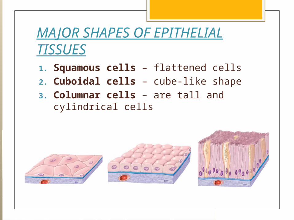
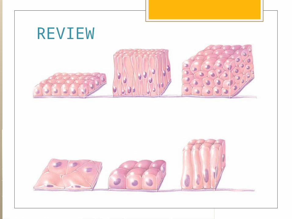
MAJOR SHAPES OF EPITHELIAL TISSUES1. Squamous cells – flattened cells2. Cuboidal cells – cube-like shape3. Columnar cells – are tall and
cylindrical cells
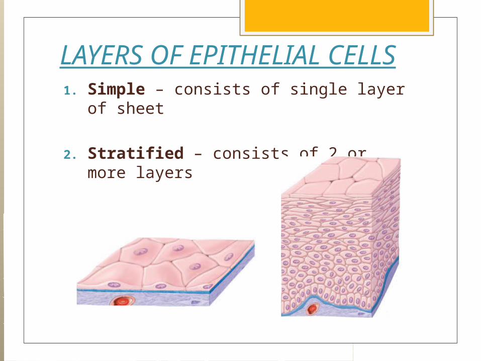
LAYERS OF EPITHELIAL CELLS1. Simple – consists of single layer of
sheet
2. Stratified – consists of 2 or more layers
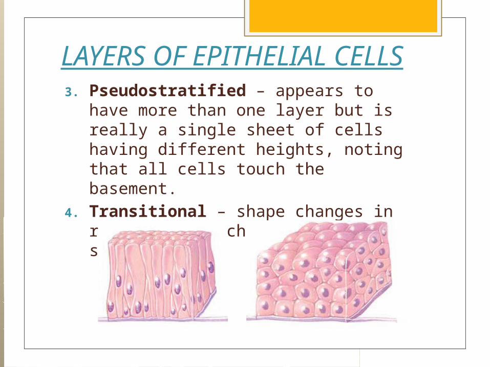
LAYERS OF EPITHELIAL CELLS3. Pseudostratified – appears to have
more than one layer but is really a single sheet of cells having different heights, noting that all cells touch the basement.
4. Transitional – shape changes in response to mechanical stretching.

GLANDS In addition to the protective and absorptive
functions, the epithelium has secretory function in the form of glands called glandular epithelium.
2 TYPES OF GLANDS1. Endocrine glands – secretes chemical
regulators called hormones that drains directly into the blood stream, thus it is called ductless glands.
2. Exocrine glands – secretes their products into the ducts, thus they are called ducted glands.

ENDOCRINE GLAND

EXOCRINE GLAND

Exocrine glands are classified according to:
I. NUMBER of CELLSA. Unicellular glands – contains only
one cell.
Ex: Goblet Cell – found in the digestive tract, secretes a glycoprotein called mucin which when added with water will produce mucus.
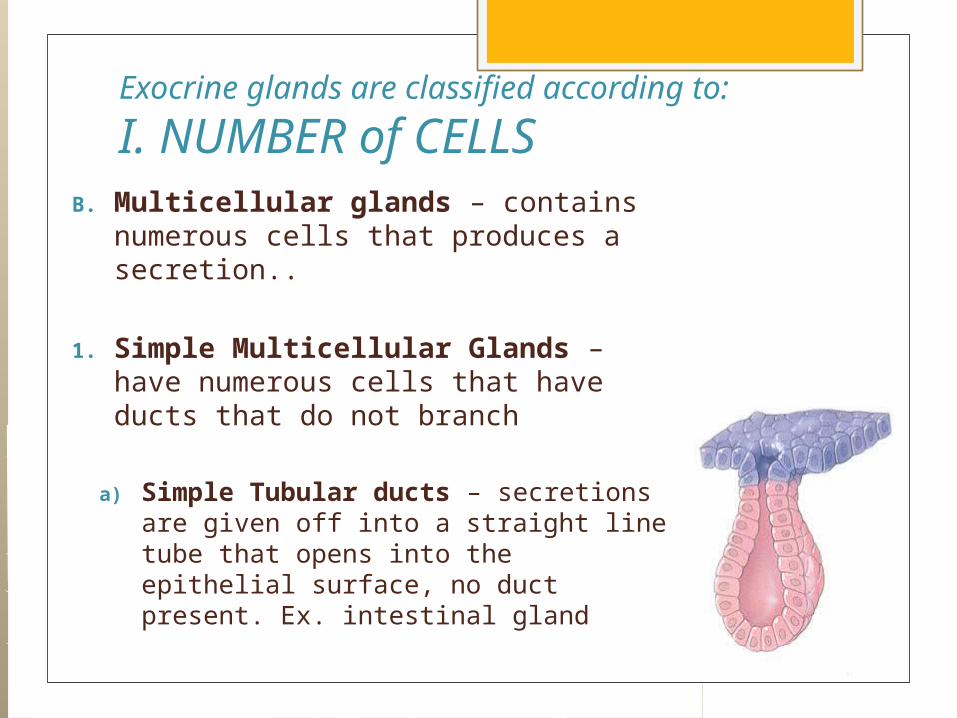
Exocrine glands are classified according to:
I. NUMBER of CELLSB. Multicellular glands – contains
numerous cells that produces a secretion..
1. Simple Multicellular Glands – have numerous cells that have ducts that do not branch
a) Simple Tubular ducts – secretions are given off into a straight line tube that opens into the epithelial surface, no duct present. Ex. intestinal gland

B. MULTICELLULAR GLANDS1. Simple Multicellular Glands (cont.)
b) Simple Coiled ducts – secretory units have coiled tubules that convey secretions into the surface, it has unbranched duct. Ex. Sweat Glands
c) Branched tubular – secretory unit is tubular and has branches, ducts maybe absent. Ex. Gastric glands, uterine glands
d) Branched acinar (alveolar) – secretory unit is shape like a sac, have several acini that are arranged along a duct. Ex. Sebaceous (oil) gland

B. MULTICELLULAR GLANDS2. Compound Multicellular Glands
a) Compound tubular gland – secretory unit is tubular with many branches. Ex. Liver, testes
b) Compound acinar glands – secretory unit is saclike with many branches. Ex. Submandibular glands, sublingual glands
c) Tubuloacinar glands – secretory units are both tubular and sac like with many branches. Ex. Pancreas, parotid glands.
REVIEW

REVIEW
II. CONNECTIVE TISSUE: Connective tissue is found everywhere in
the body. It is the most numerous tissues in the body. The origin of all connective tissues is
mesenchyme (an embryonic tissue) Serve to support the body and binds
together body parts and other tissues. Because of its matrix, connective tissue is
able to bear weight, withstand great tension, and endure abuses such as physical trauma and abrasion that no other tissue would be able to tolerate.
Functions of Connective Tissues
1. Binding and support (connective tissue proper)
2. Protection (adipose tissue, blood, bone)
3. Transport (blood)4. Insulation (Adipose tissue)

Structural elements of connective tissue:1. Extracellular matrix (ground
substance)2. Cells
General Types of CONNECTIVE TISSUES According to the CHARACTERISTICS of their Matrix and Ground Substance
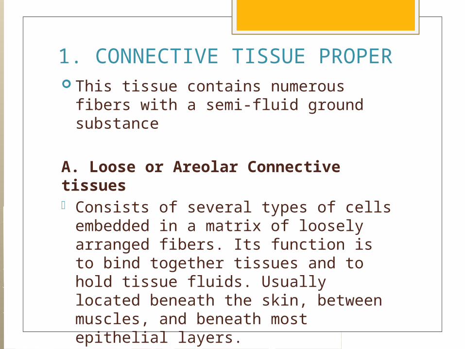
1. CONNECTIVE TISSUE PROPER This tissue contains numerous fibers
with a semi-fluid ground substance
A. Loose or Areolar Connective tissues - Consists of several types of cells
embedded in a matrix of loosely arranged fibers. Its function is to bind together tissues and to hold tissue fluids. Usually located beneath the skin, between muscles, and beneath most epithelial layers.

A. Loose or Areolar Connective tissues
1. Fibroblast – manufacture the protein fiber of the tissue and ground substance, as well as hyaluronic acid that serves as spreading factor of many cells.
A. Loose or Areolar Connective tissues
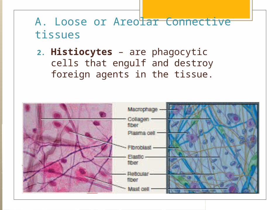
2. Histiocytes – are phagocytic cells that engulf and destroy foreign agents in the tissue.

A. Loose or Areolar Connective tissues
3. Lymphocytes – a type of WBC that helps boost up our immune system.
A. Loose or Areolar Connective tissues
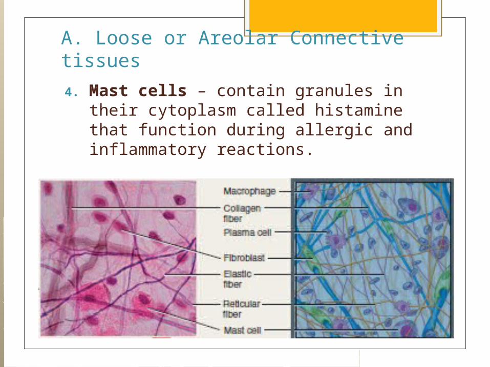
4. Mast cells – contain granules in their cytoplasm called histamine that function during allergic and inflammatory reactions.

A. Loose or Areolar Connective tissues
5. Collagen fibers – produces the protein collagen which forms fibers with high tensile strength and flexibility.
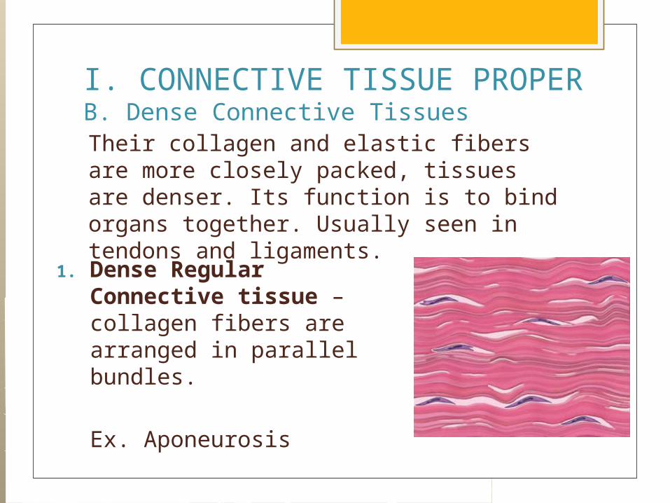
I. CONNECTIVE TISSUE PROPERB. Dense Connective TissuesTheir collagen and elastic fibers are more closely packed, tissues are denser. Its function is to bind organs together. Usually seen in tendons and ligaments.
1. Dense Regular Connective tissue – collagen fibers are arranged in parallel bundles.
Ex. Aponeurosis
I. CONNECTIVE TISSUE PROPERB. Dense Connective Tissues
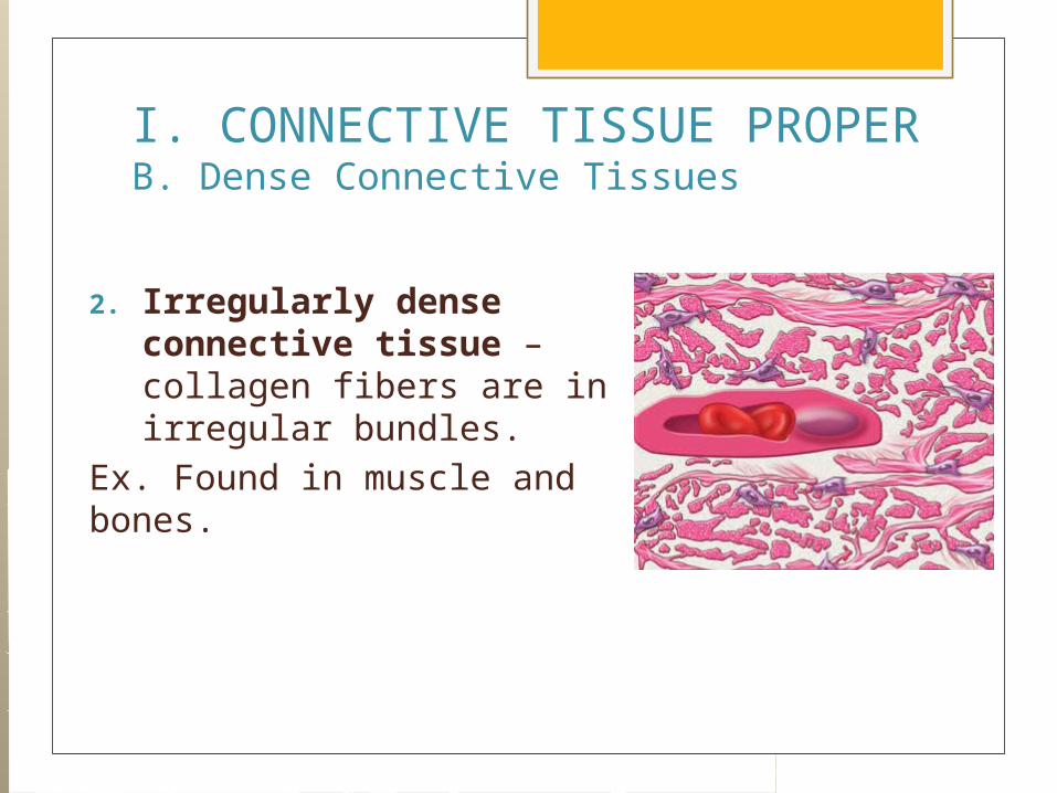
2. Irregularly dense connective tissue – collagen fibers are in irregular bundles.
Ex. Found in muscle and bones.

I. CONNECTIVE TISSUE PROPERB. Dense Connective Tissues
3. Reticular connective tissue – has delicate fibers forming a network called reticulum. This netwotk supports soft organs such as the spleen, lymph nodes, and liver. These fibers are synthesized by reticular fibers.

I. CONNECTIVE TISSUE PROPERB. Dense Connective Tissues
4. Adipose or fat tissue – these cells expand with fat droplets and push the nucleus into the rim of the cytoplasm, thus resembling a ring. Usually found beneath the skin, around the kidneys, behind the eyeballs, and on the surface of the heart. Its function is to protect, insulate the body against heat loss and serves as storage depot for fat for energy production.
I. CONNECTIVE TISSUE PROPERB. Dense Connective Tissues
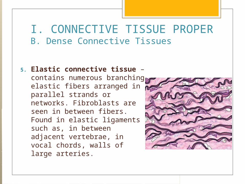
5. Elastic connective tissue – contains numerous branching elastic fibers arranged in parallel strands or networks. Fibroblasts are seen in between fibers. Found in elastic ligaments such as, in between adjacent vertebrae, in vocal chords, walls of large arteries.
II. CARTILAGE Cartilage is a type of connective tissue that
is hard but elastic in nature. These fibers are produced by cartilage cells
called chondroblast, eventually these chondroblast becomes trapped in their products and revert to mature cells called chondrocytes.
The substances of cartilage forms a rubber-like mixture of proteins and proteoglycans, associated with carbohydrate units.
No blood is seen on cartilage

TYPES OF CARTILAGEA. Hyaline Cartilage
The most abundant type of cartilage in the body, but the weakest.
Under the microscope, chondrocytes are seen in spaces called lacunae.
Seen at the end of long bones, external ears, fetal skeleton, nose, larynx, trachea, and bronchi.
Its function is for support, protection, and provides framework.

TYPES OF CARTILAGEC. Fibrous cartilage
or Fibtocartilage Strongest type of
cartilage with dense collagen fibers and limited amount of ground substance.
Seen in body areas that bear great amount of weight, like symphysis pubis, skull and vertebrae.
REVIEW
III. BONE It is the hardest connective tissue. It is made up of cells, collagen fibers,
and dense mineralized substance. Much stronger than cartilage because it
contains inorganic salts of calcium and phosphate called hydroxyappatite crystals.
This is the main component of the bone which is responsible for its hardness.
It has a rich blood supply.
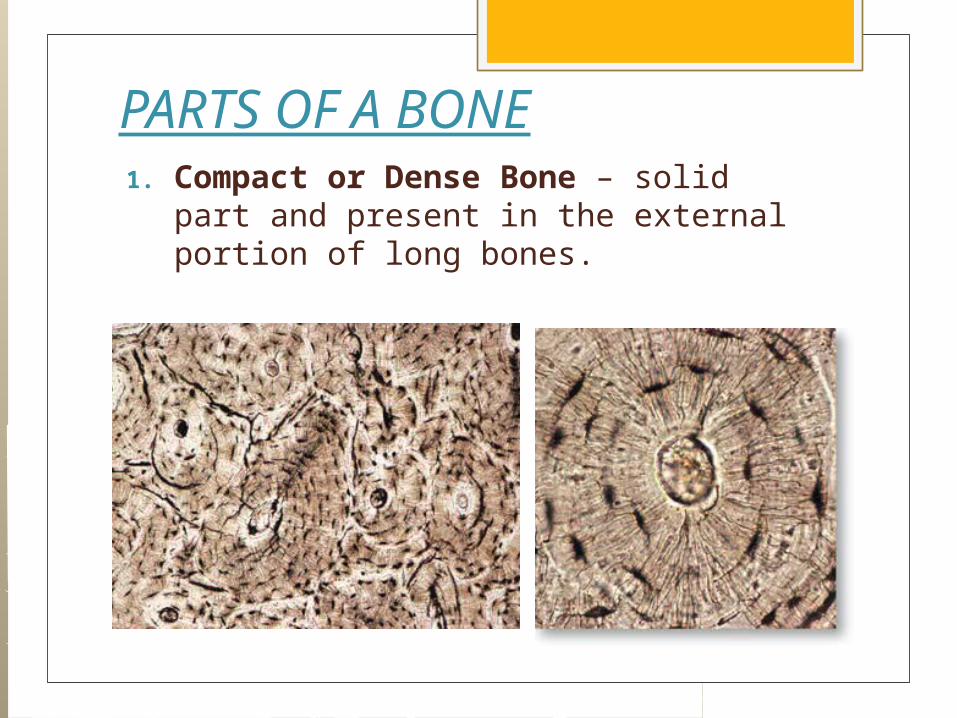
PARTS OF A BONE1. Compact or Dense Bone – solid part
and present in the external portion of long bones.
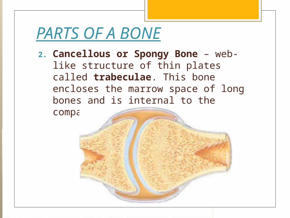
PARTS OF A BONE2. Cancellous or Spongy Bone – web-
like structure of thin plates called trabeculae. This bone encloses the marrow space of long bones and is internal to the compact bone

TYPES OF BONE CELLS1. Osteoblast – young bone cell that
synthesize the components of bone.
2. Osteocyte – mature bone cell that exist within the lacunae in the matrix.
3. Osteoclast – this destroys bone and remodels it.
IV. BLOOD Blood is fluidized connective tissue It provides communication between
different parts of the body and with the external environment. Blood involved in transportation, regulation and protection functions.
Blood is a very good suspension, blood cells are suspended in plasma.
Different types of blood cells are red blood cells (R.B.C’s), white blood cells (W.B.C’s) and platelets. Blood contains red colored pigment called hemoglobin.

IV. BLOOD
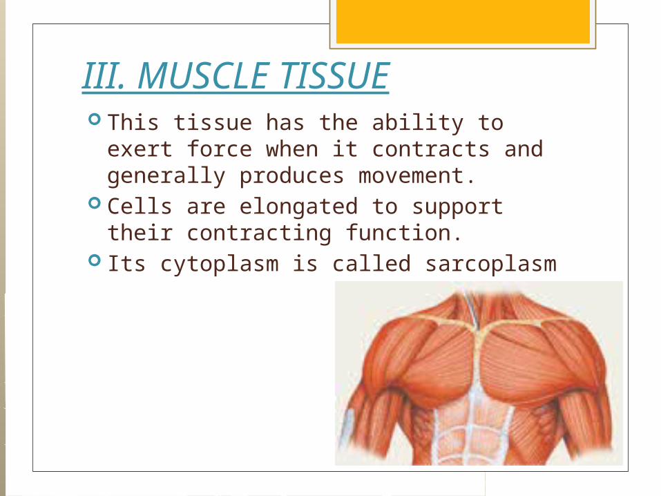
III. MUSCLE TISSUE This tissue has the ability to exert force
when it contracts and generally produces movement.
Cells are elongated to support their contracting function.
Its cytoplasm is called sarcoplasm

REVIEW

REVIEW

REVIEW

REVIEW
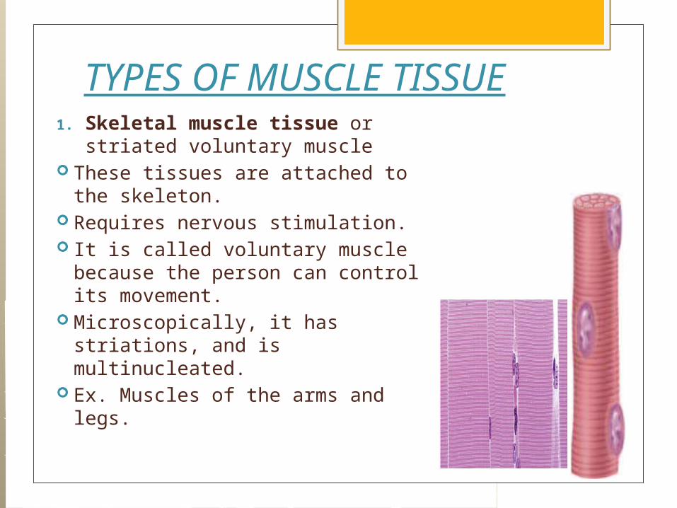
TYPES OF MUSCLE TISSUE1. Skeletal muscle tissue or
striated voluntary muscle These tissues are attached to
the skeleton. Requires nervous stimulation. It is called voluntary muscle
because the person can control its movement.
Microscopically, it has striations, and is multinucleated.
Ex. Muscles of the arms and legs.
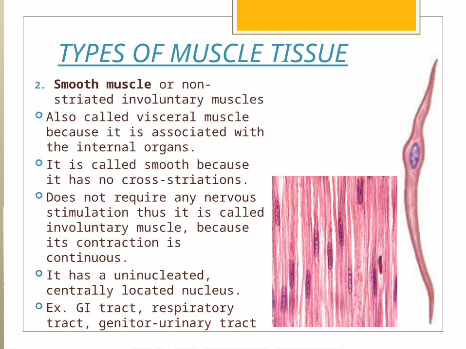
TYPES OF MUSCLE TISSUE2. Smooth muscle or non-
striated involuntary muscles Also called visceral muscle
because it is associated with the internal organs.
It is called smooth because it has no cross-striations.
Does not require any nervous stimulation thus it is called involuntary muscle, because its contraction is continuous.
It has a uninucleated, centrally located nucleus.
Ex. GI tract, respiratory tract, genitor-urinary tract

TYPES OF MUSCLE TISSUE3. Cardiac muscle or striated
involuntary muscle Found only in heart. Does not require any nervous
stimulation. It has cross striation and its
branching fibers contain cells joined to one another by a specialized cell junction called intercalated disks.
It has a uninucleated, centrally located nucleus.

IV. NERVOUS TISSUE This tissue makes up the brain,
spinal cord, and various nerves of the body.
It is the most highly organized tissue in the body because it controls and coordinates body activities.
It relays stimuli from the brain to the body.
Its function is for communication (receive, transmit, and interpret the nerve impulses)

2 TYPES OF NERVOUS TISSUE1. Neurons –
conducting cells, this is the basic unit of organization of the nervous tissue.
Functional unit of the nervous system.
2. Neuroglial cells – are supporting cells of the nervous tissue.

REVIEW

REVIEW

REVIEW

REVIEW
REVIEW


