91
Anatomy and Physiology Anatomy and Physiology Cells
| Date post: | 18-Dec-2015 |
| Category: |
Documents |
| Upload: | shana-rogers |
| View: | 227 times |
| Download: | 0 times |
Anatomy and PhysiologyAnatomy and Physiology
Cells

IntroductionIntroduction
• There are 75 trillion cells that make up the human body.
• Cells of different tissues have much in common but vary in size and shape.
• Typically, their shapes make possible their functions. Ex: nerve cells have long, threadlike extensions that transmit bioelectrical impulses.

Composite CellComposite Cell• A “typical” cell consists of three major
parts--the nucleus, the cytoplasm, and the cell membrane.
• The nucleus is usually in the center of the cell and is surrounded by a thin nuclear envelope.
• Cytoplasm surrounds the nucleus and is encircled by the thinner cell membrane (plasma membrane).
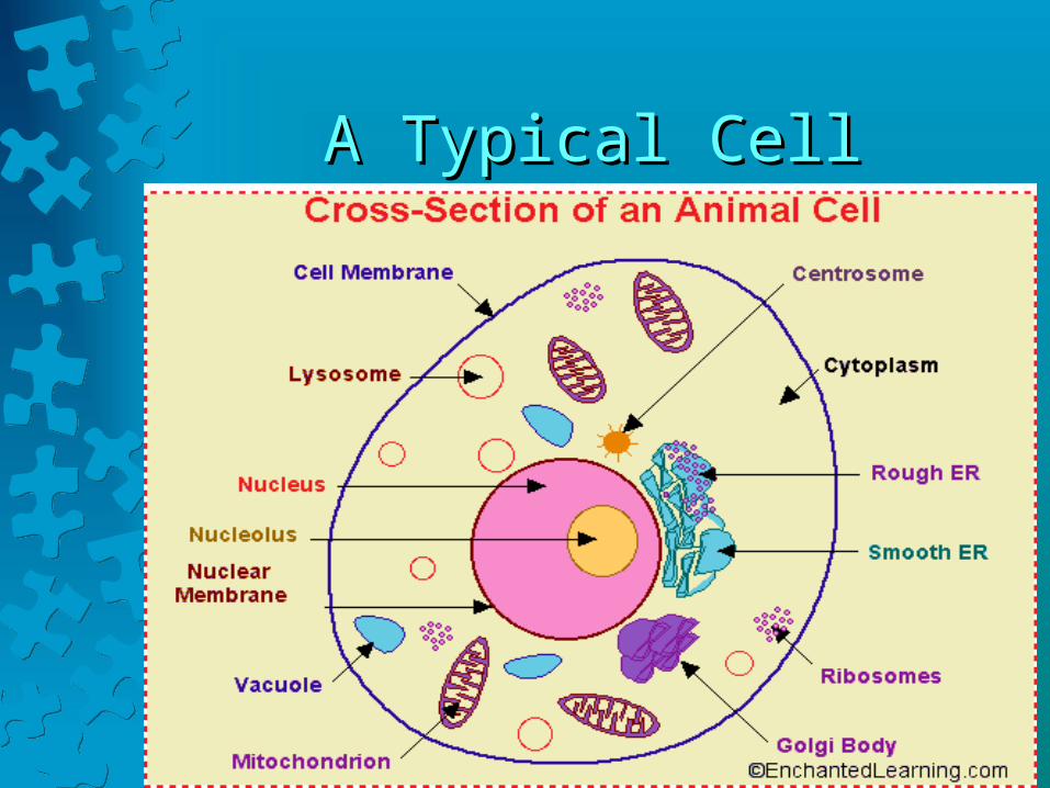
A Typical CellA Typical Cell
OrganellesOrganelles
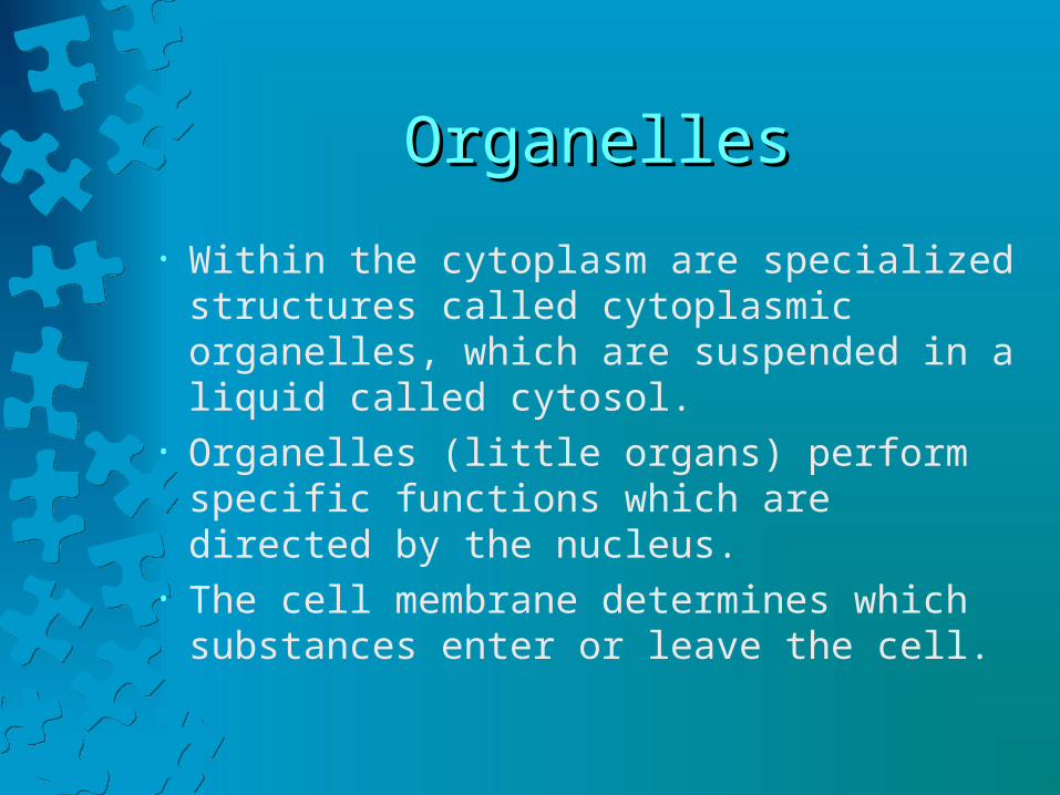
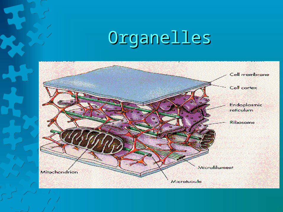
• Within the cytoplasm are specialized structures called cytoplasmic organelles, which are suspended in a liquid called cytosol.
• Organelles (little organs) perform specific functions which are directed by the nucleus.
• The cell membrane determines which substances enter or leave the cell.

Cell MembraneCell Membrane• The cell membrane regulates movement of
substances in and out of the cell and is the site of much biological activity.
• Molecules that are part of the cell membrane receive stimulation from outside the cell and transmit it into the cell, a process called signal transduction.
• The cell membrane also holds cells together.
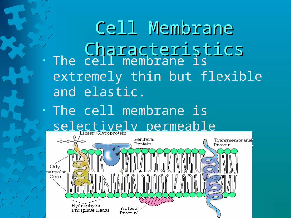
Cell Membrane CharacteristicsCell Membrane Characteristics• The cell membrane is extremely thin but
flexible and elastic.• The cell membrane is selectively permeable
(semipermeable) in that it controls which substances enter and exit.

Cell Membrane StructureCell Membrane Structure
• A cell membrane is composed mainly of lipids, proteins, and some carbohydrates.
• It is a bilayer of phospholipid molecules.• Each phospholipid molecule includes a
phosphate group and two fatty acids bound to a glycerol molecule.
• The lipid molecules can move around forming a soft and flexible fluid film.

Cell Membrane StructureCell Membrane Structure
Membrane SolubilityMembrane Solubility
• Because the membrane’s interior consists of fatty acids, it is oily. Only substances soluble in lipids can pass through this layer.
• O2 and CO2 can pass through easily but water-soluble molecules, such as amino acids, sugars, proteins, nucleic acids, and various ions, cannot pass through.
• Cholesterol molecules help to stabilize the membrane.
Membrane ProteinsMembrane Proteins• Membrane proteins are classified according to
their positions within the membrane.• Membrane-spanning (trans-membrane)
proteins extend through the lipid bilayer and may protrude from 1 or both faces.
• Peripheral membrane proteins are associated with one side of the bilayer.Membrane proteins also vary in shape--globular or elongated.

Membrane Protein FunctionsMembrane Protein Functions
• Some proteins form receptors on the cell surface that bind incoming hormones or growth factors, starting signal transduction.
• Others transport ions or molecules across the cell membrane.
• Some membrane proteins form selective channels that allow only particular ions to enter or leave.
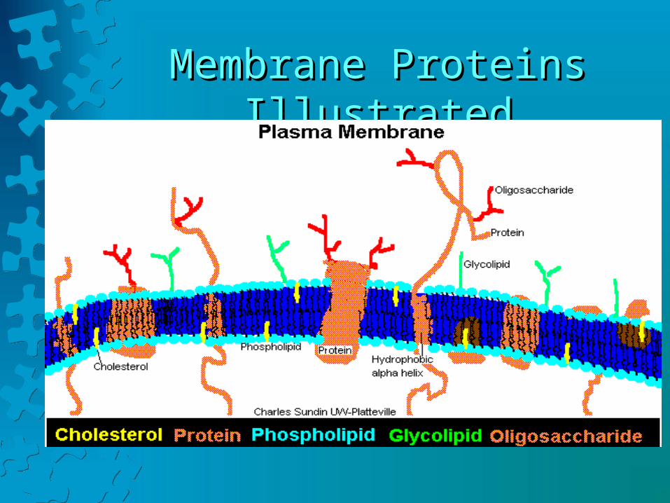
Membrane Proteins IllustratedMembrane Proteins Illustrated

More Protein FunctionsMore Protein Functions• Proteins that protrude from the inner face of the
cell anchor it to the cytoskeleton (rods and tubules that support the inner cell).
• Proteins that extend from the outer surface mark the cell as part of a particular tissue or organ--important identification for the immune system.
• CAM (cellular adhesion molecule) determines a cell’s interactions with other cells.
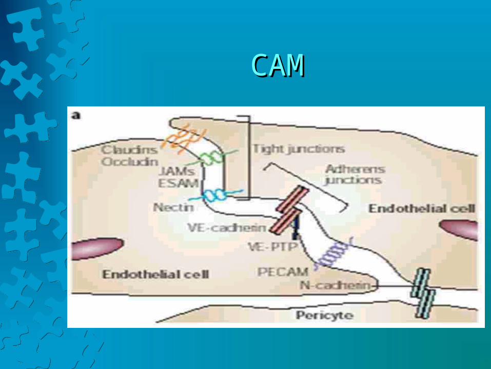
CAMCAM
CytoplasmCytoplasm
• Cytoplasm contains networks of membranes and organelles suspended in a clear liquid called cytosol.
• Cytoplasm also includes many protein rods and tubules that form a framework called a cytoskeleton.
• Cell activities occur mainly in the cytoplasm, where nutrients are received, processed, and used.

Cytoplasmic OrganellesCytoplasmic Organelles
• Endoplasmic Reticulum
• Ribosomes• Golgi Apparatus• Mitochondria• Lysosomes• Peroxisomes
• Microfilaments and microtubules
• Centrosome• Cilia and Flagella• Vesicles• Cell Nucleus
• nucleolus
• chromatin
OrganellesOrganelles

Endoplasmic ReticulumEndoplasmic Reticulum
• Endoplasmic Reticulum (ER) is composed of membrane-bound, flattened sacs, elongated canals, and fluid-filled bubble-like sacs called vesicles.
• ER provides a vast tubular network that transports molecules from one cell part to another.

Functions of ERFunctions of ER
• ER participates in synthesis of protein and lipid molecules.
• These molecules may leave the cell as secretions, or be used within the cell to produce new ER or cell membranes as the cell grows.
Two Types of ERTwo Types of ER
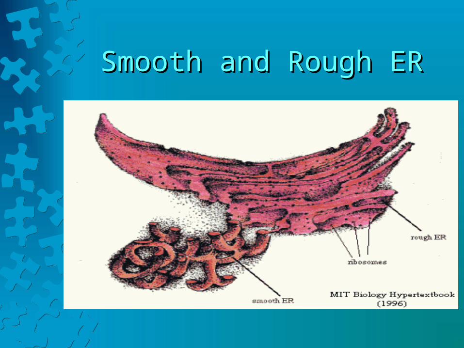
• Rough ER contains ribosomes on its surface; smooth ER is ribosome-free.
• The ribosomes of rough ER are sites or protein synthesis. The proteins may then move through ER tubules to the Golgi apparatus for further processing.
• Smooth ER contains enzymes important in lipid synthesis.
Smooth and Rough ERSmooth and Rough ER
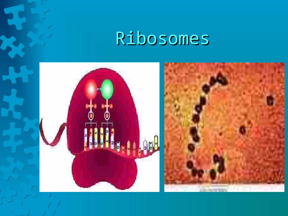
RibosomesRibosomes• All ribosomes are composed of protein and
RNA molecules.• Many ribosomes are attached to ER and
others are scattered throughout the cytoplasm.• Ribosomes provide enzymes as well as a
structural support for the RNA molecules that come together as the cell synthesizes proteins from amino acids.
RibosomesRibosomes
Golgi ApparatusGolgi Apparatus
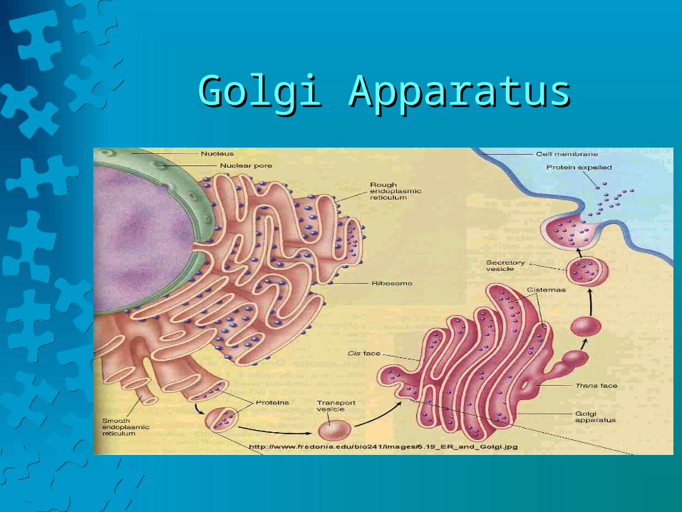
• The Golgi apparatus is composed of a stack of about 6 flattened, membranous sacs.
• This organelle refines, packages, and delivers proteins synthesized on ribosomes associated with the ER.
• Proteins arrive at the Golgi apparatus enclosed in vesicles composed of the ER membrane.
Golgi ApparatusGolgi Apparatus
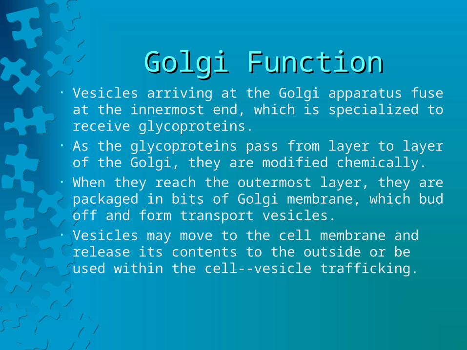
Golgi FunctionGolgi Function• Vesicles arriving at the Golgi apparatus fuse at the innermost
end, which is specialized to receive glycoproteins.• As the glycoproteins pass from layer to layer of the Golgi,
they are modified chemically.• When they reach the outermost layer, they are packaged in
bits of Golgi membrane, which bud off and form transport vesicles.
• Vesicles may move to the cell membrane and release its contents to the outside or be used within the cell--vesicle trafficking.

Vesicle FormationVesicle Formation
MitochondriaMitochondria
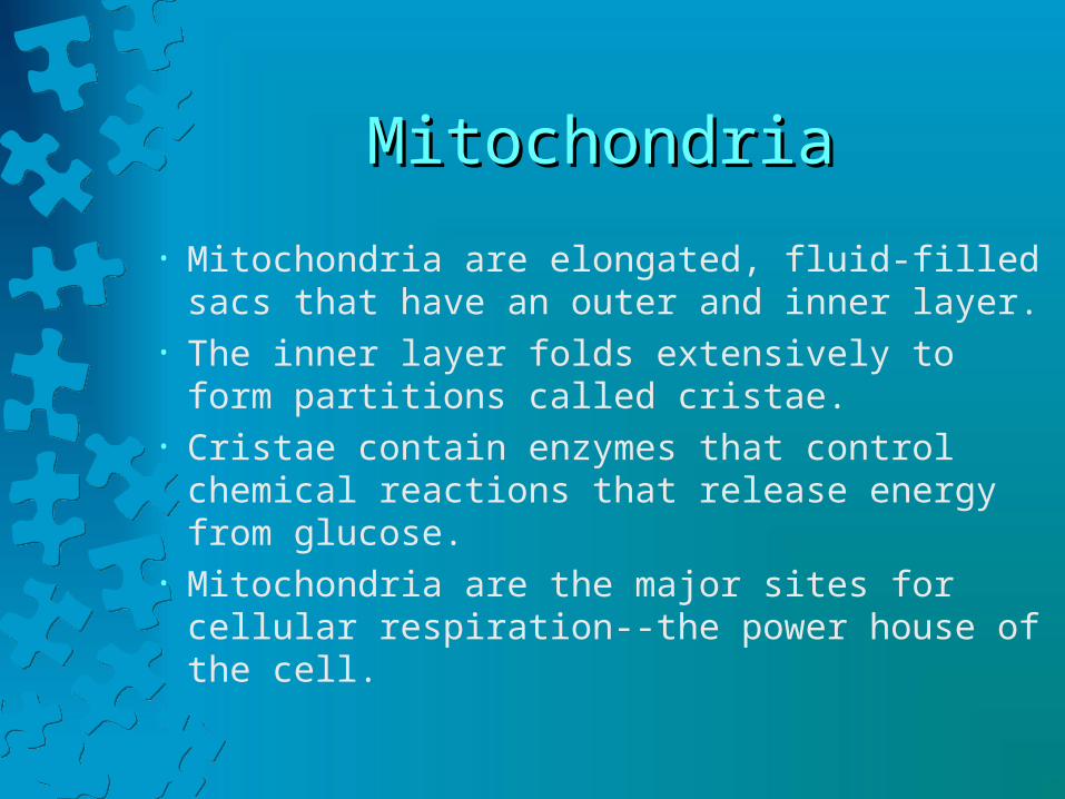
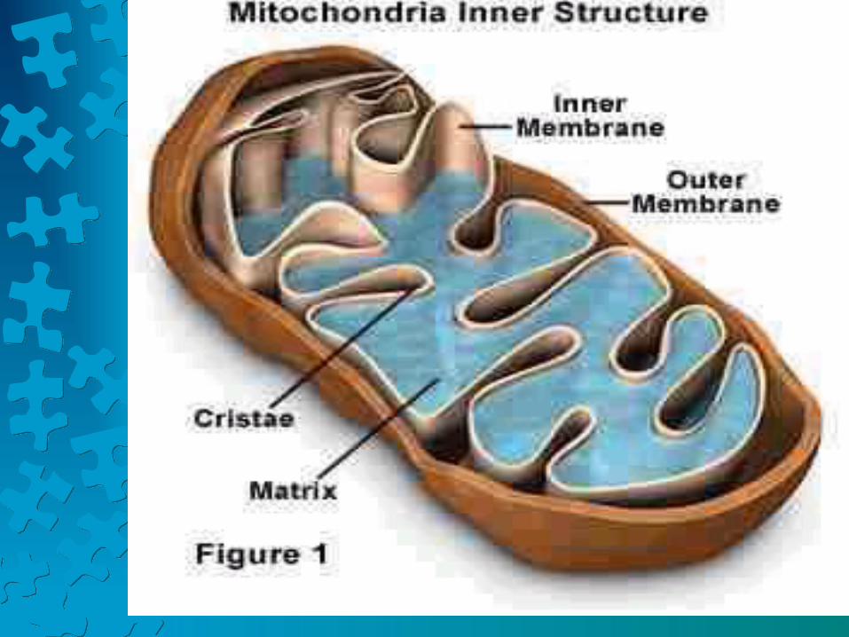
• Mitochondria are elongated, fluid-filled sacs that have an outer and inner layer.
• The inner layer folds extensively to form partitions called cristae.
• Cristae contain enzymes that control chemical reactions that release energy from glucose.
• Mitochondria are the major sites for cellular respiration--the power house of the cell.
More MitochondriaMore Mitochondria
• Mitochondria contain their own DNA--much like that of bacteria.
• According to the endosymbiont theory, mitochondria are the remnants of once free-living bacteria-like cells that were swallowed by more complex primitive cells.
• You can only inherit mitochondria from your mother.
LysosomesLysosomes
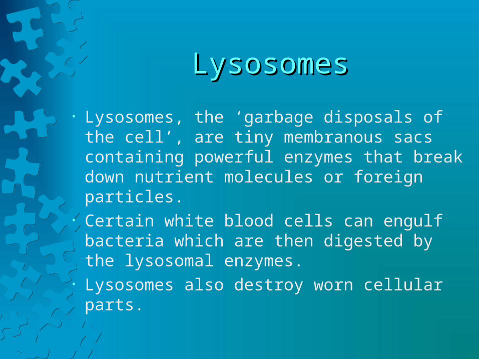
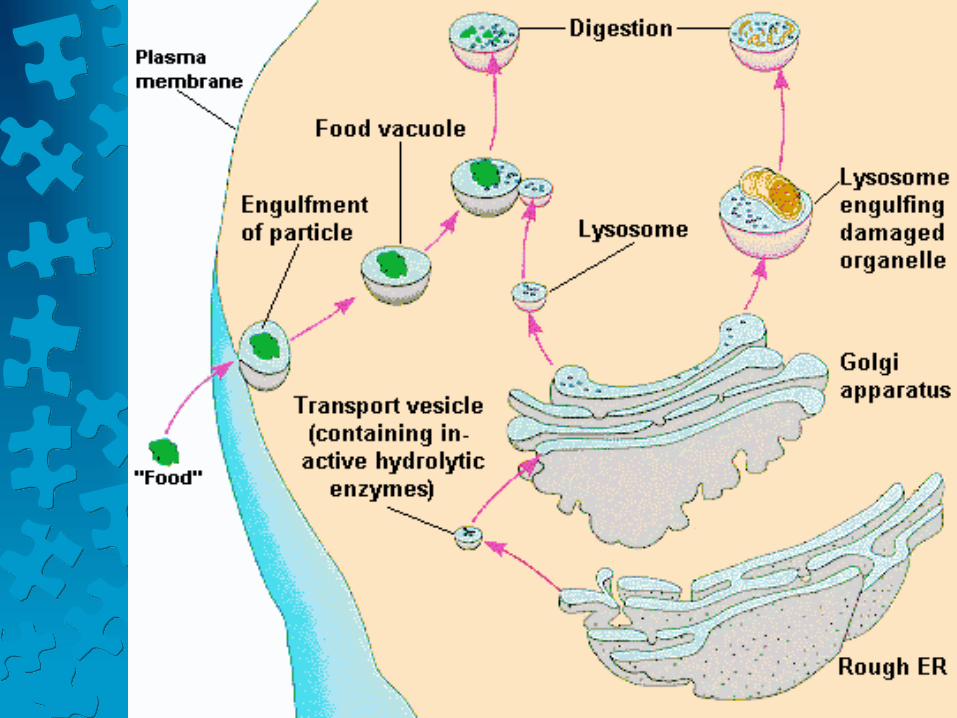
• Lysosomes, the ‘garbage disposals of the cell’, are tiny membranous sacs containing powerful enzymes that break down nutrient molecules or foreign particles.
• Certain white blood cells can engulf bacteria which are then digested by the lysosomal enzymes.
• Lysosomes also destroy worn cellular parts.
PeroxisomesPeroxisomes
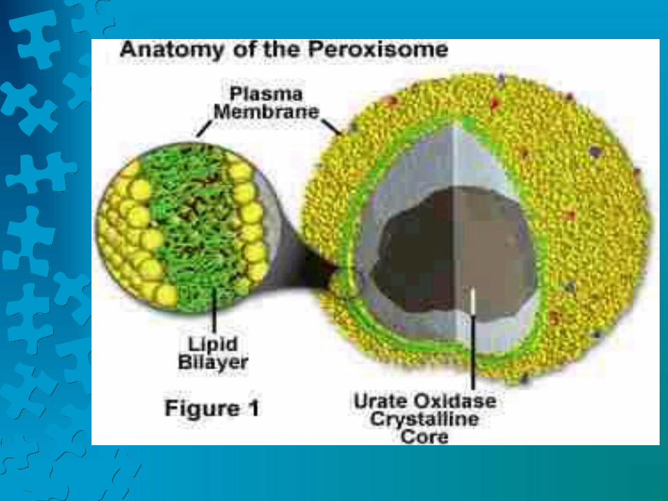
• Peroxisomes, membranous sacs abundant in liver and kidney cells, house enzymes that catalyze a variety of biochemical reactions, including synthesis of bile acids; detoxification of hydrogen peroxide; breakdown of certain lipids and rare biochemicals; and detoxification of alcohol.
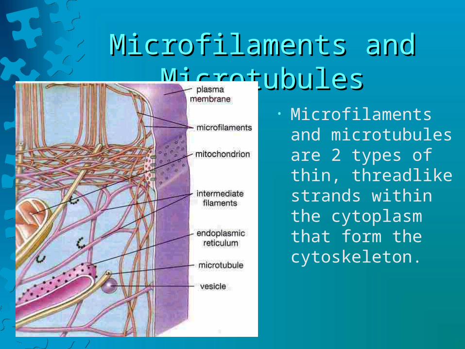
Microfilaments and MicrotubulesMicrofilaments and Microtubules
• Microfilaments and microtubules are 2 types of thin, threadlike strands within the cytoplasm that form the cytoskeleton.

CytoskeletonCytoskeleton• Microfilaments are tiny
rods of actin protein that form meshworks or bundles.
• They provide cell motility.
• In muscle cells, microfilaments form myofibrils to help muscle cells contract.
• Microtubules are long, slender tubes composed of globular tubulin proteins .
• Usually 2-3 times larger than microfilaments, microtubules are arrayed in a pattern called 9 + 2.
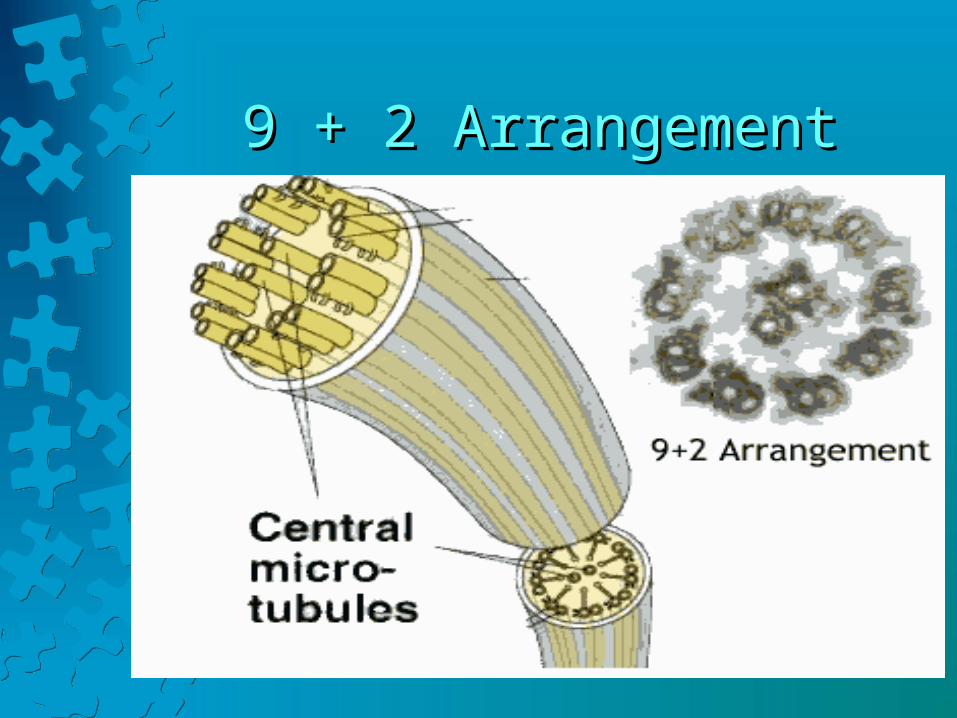
9 + 2 Arrangement9 + 2 Arrangement

CentrosomeCentrosome
• The centrosome is a nonmembranous structure near the nucleus of animal cells.
• It consists of 2 hollow cylinders called centrioles, made of microtubules.
• The centrioles lie at right angles to each other. During mitosis, they help distribute chromosomes to newly forming cells.
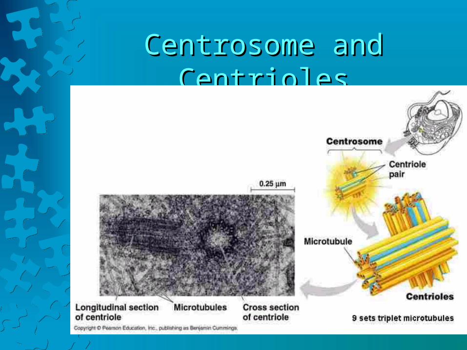
Centrosome and CentriolesCentrosome and Centrioles

Cilia and FlagellaCilia and Flagella• Cilia and flagella are motile extensions from the
surfaces of certain cells, made of microtubules in a 9 + 2 arrangement.
• Their main difference is in length.• Cilia is a tiny, hairlike structure attached beneath the
cell membrane that moves in a ‘to-and-fro’ pattern.• Flagella are longer than cilia. Usually a cell may
have only 1 cilia which moves in a whip-like motion.

Cilia and FlagellaCilia and Flagella
VesiclesVesicles
• Vesicles are membranous sacs formed by part of the cell membrane folding inward and pinching off.
• Vesicles that hold food or water are called vacuoles.
• The Golgi apparatus and ER also form vesicles that play a role in secretion.

Formation of VacuolesFormation of Vacuoles

Cell NucleusCell Nucleus
• The nucleus houses the genetic material (DNA), which directs all cell activity.
• It is a large spherical structure enclosed in a double-layered lipid nuclear envelope.
• The nuclear envelope has protein channels called nuclear pores that allow certain molecules to exit the nucleus.
• A nuclear pore is not just a hole, but a complex opening formed from 100+ proteins.
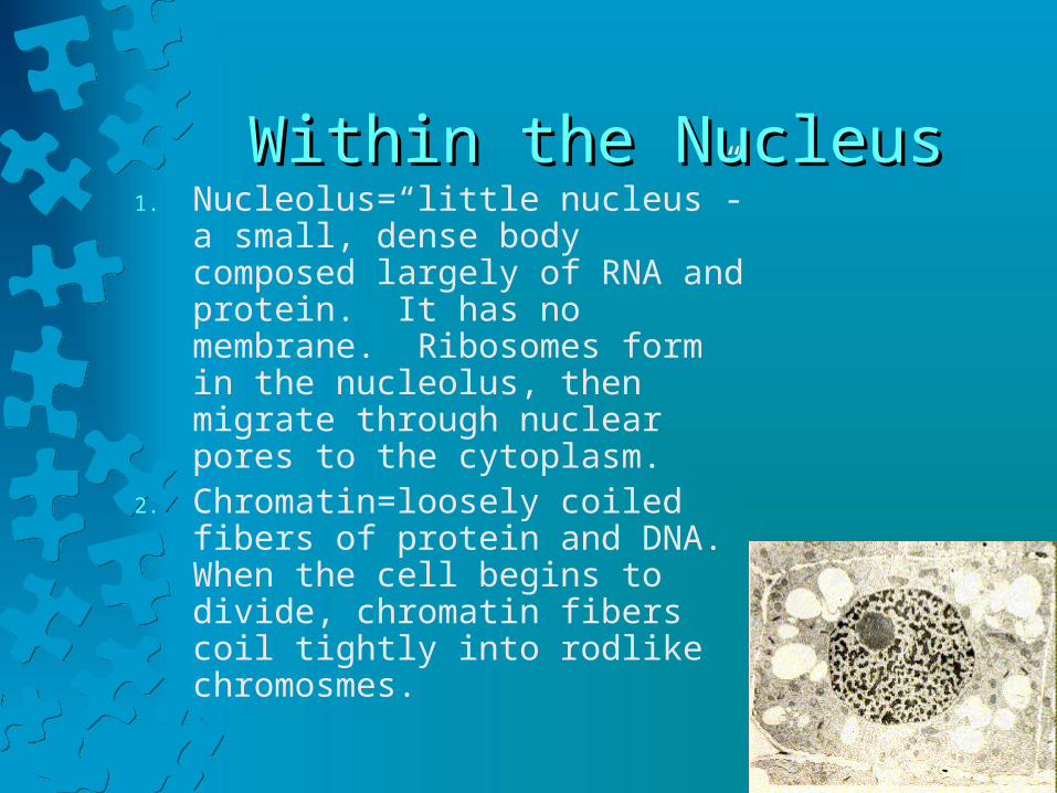
Within the NucleusWithin the Nucleus1. Nucleolus=“little nucleus”-a
small, dense body composed largely of RNA and protein. It has no membrane. Ribosomes form in the nucleolus, then migrate through nuclear pores to the cytoplasm.
2. Chromatin=loosely coiled fibers of protein and DNA. When the cell begins to divide, chromatin fibers coil tightly into rodlike chromosmes.

Movement Through Cell MembranesMovement Through Cell Membranes• The cell membrane is a selective barrier
that controls which substances enter and leave the cell.
1. Passive mechanisms do not require energy: diffusion, facilitated diffusion, osmosis, and filtration.
2. Active mechanisms use cellular energy: active transport, endocytosis, and exocytosis.

Passive Mechanisms: DiffusionPassive Mechanisms: Diffusion
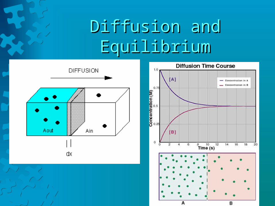
• Diffusion is the process by which molecules or ions scatter or spread spontaneously from regions of higher concentrations to regions of lower concentrations.
• Molecules move at random and mix molecules together.
• Equilibrium occurs when the molecules are equally mixed. Movement still occurs but there is no change in concentration.
Diffusion and EquilibriumDiffusion and Equilibrium
Examples of Diffusion at WorkExamples of Diffusion at Work
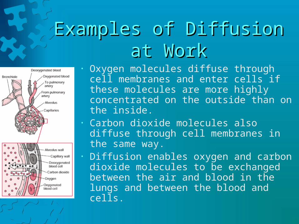
• Oxygen molecules diffuse through cell membranes and enter cells if these molecules are more highly concentrated on the outside than on the inside.
• Carbon dioxide molecules also diffuse through cell membranes in the same way.
• Diffusion enables oxygen and carbon dioxide molecules to be exchanged between the air and blood in the lungs and between the blood and cells.

DialysisDialysis
• Dialysis uses diffusion to separate smaller molecules from larger ones in a liquid.
• The artificial kidney used a process called hemodialysis to filter wastes from the blood, much as a real kidney would.
• To remove blood urea, the dialyzing fluid must have a lower urea concentration than the blood; glucose concentration in the fluid must match the blood to prevent from losing glucose.
Facilitated DiffusionFacilitated Diffusion• Facilitated diffusion occurs in most cells
when a special protein carrier molecule attached to the surface of the cell membrane helps another substance move across the membrane.
• Once inside the cell, the molecule is released. Again, the material moves from areas of high concentration to areas of low concentration.
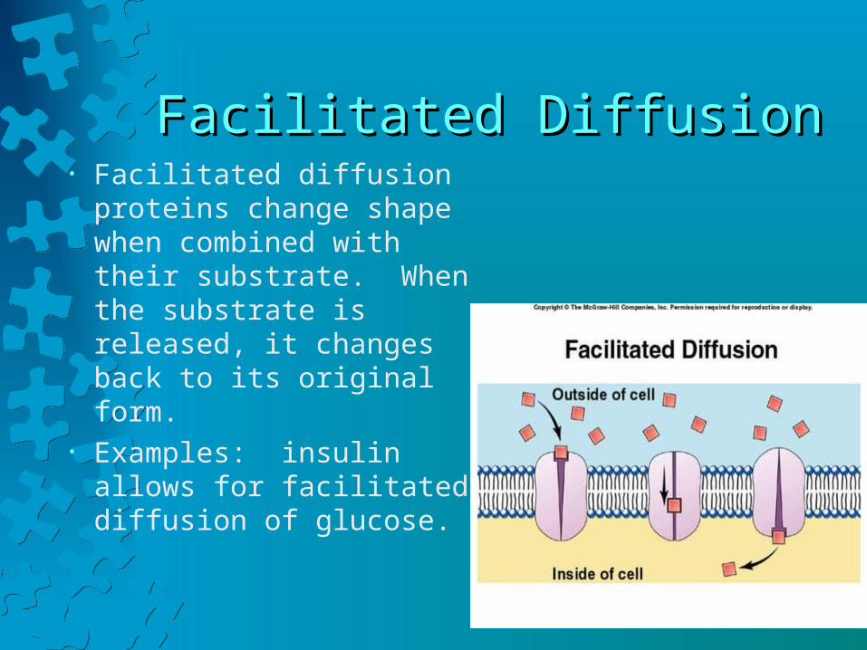
Facilitated DiffusionFacilitated Diffusion• Facilitated diffusion
proteins change shape when combined with their substrate. When the substrate is released, it changes back to its original form.
• Examples: insulin allows for facilitated diffusion of glucose.
Osmosis: A Special Case of Osmosis: A Special Case of DiffusionDiffusion
• Osmosis is the diffusion of water only, from an area of high water concentration to an area of low water concentration.
• *In solutions, a higher concentration of solute means a lower concentration of water because the solute takes up space that could be occupied by water molecules.
Osmotic PressureOsmotic Pressure
• The ability of osmosis to generate enough pressure to lift a volume of water is called osmotic pressure.
• The greater the concentration of nonpermeable solute particles in a solution, the lower the water concentration of that solution and the greater the osmotic pressure.
• Water always tends to diffuse toward solutions of greater osmotic pressure.

Three Types of SolutionThree Types of Solution
• Since cell membranes are permeable to water, water equilibrates by osmosis and the concentration of water and solutes everywhere in body fluids is the same.
• Any solution that has the same osmotic pressure as body fluids is called isotonic.
• Solutions with a higher osmotic pressure than body fluids are called hypertonic.
• A hypotonic solution has a lower osmotic pressure than body fluids.

A Cell’s Reaction to Different A Cell’s Reaction to Different SolutionsSolutions
• A cell placed in an isotonic solution will have no net gain of water.
• A cell placed in a hypertonic solution will , it will lose water.
• A cell in a hypotonic solution will gain water.
FiltrationFiltration
• Molecules pass through membranes by diffusion or osmosis because of random movements.The process of filtration forces molecules through membranes.
• Filtration is commonly used to separate solids from water. Tissue fluid forms when water and small dissolved substances are forced out through the thin, porous walls of blood capillaries.
• Larger particles, such as blood proteins, are left inside.

FiltrationFiltration• In the body, tissue fluid forms when water
and small dissolved substances are forced out through the thin, porous walls of blood capillaries, but larger particles, blood proteins, are left inside.
• The force comes from blood pressure generated mostly by heart action.

Active MechanismsActive Mechanisms
• When particles move from areas of lower concentration to one of higher concentration, energy is required.
• This energy comes from cellular metabolism and specifically from a molecule called ATP, adenosine triphosphate.
Active TransportActive Transport
• Active transport is a process that moves particles through membranes from a region of lower concentration to a region of higher concentration.
• Sodium ions can diffuse passively into cells through protein channels but active transport continually moves sodium ions through cell membranes to the outside where the concentration is higher.
• Equilibrium is never reached.

Active Transport FactsActive Transport Facts
• Active transport used specific carrier molecules in cell membranes and may use up to 40% of the cell’s energy to actively transport particles.
• The carrier molecules are proteins with binding sites that combine with the particles being transported.

Active TransportActive Transport• When the carrier protein
combines with the substrate, it changes shape which moves the particle through the membrane.
• Once inside, the particle is released and the carrier protein goes back to its original shape.
• Particles that are actively transported across cell membranes include sugars, amino acids, and
• sodium, potassium, calcium, and hydrogen ions.
Endocytosis and ExocytosisEndocytosis and Exocytosis
• Two processes use cellular energy to move substances into (endocytosis) or out of (exocytosis) a cell without crossing the cell membrane.
EndocytosisEndocytosis
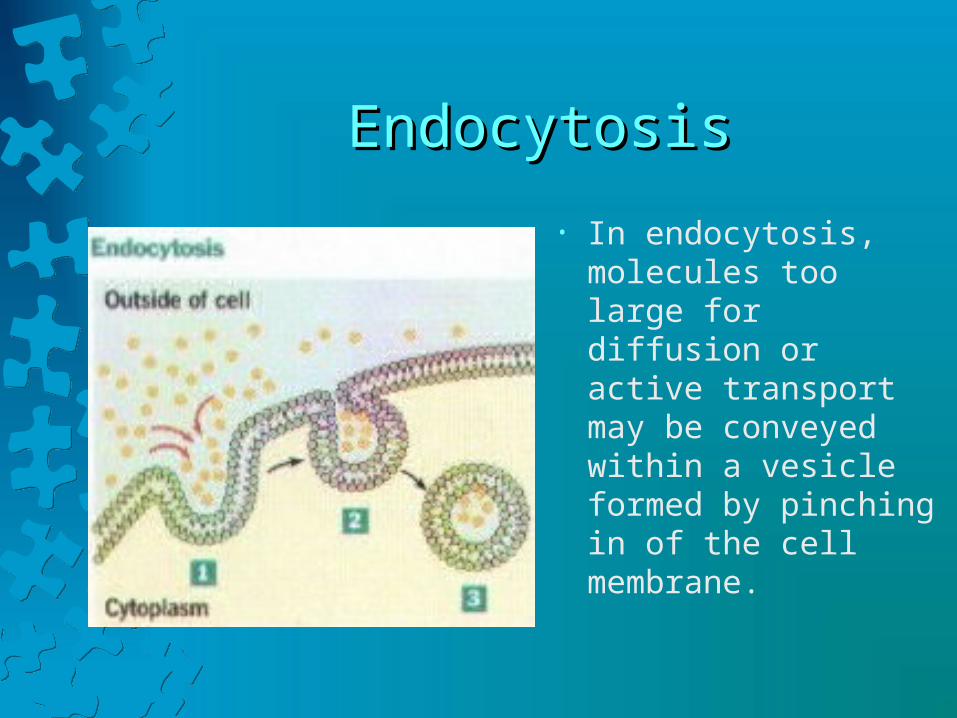
• In endocytosis, molecules too large for diffusion or active transport may be conveyed within a vesicle formed by pinching in of the cell membrane.

ExocytosisExocytosis
• In exocytosis, the reverse process secretes a substance stored in a vesicle from the cell.
• Nerve cells use exocytosis to release the neurotransmitter chemicals that signal other nerve cells, muscle cells, or glands.
Three Forms of EndocytosisThree Forms of Endocytosis
1. Pinocytosis=cell drinking
2. Phagocytosis=cell eating
3. Receptor-mediated endocytosis for specific molecules

PinocytosisPinocytosis• Pinocytosis takes in droplets of liquid from the
surroundings as a small portion of the cell membrane indents.
• A vesicle forms which detaches from the surface and moves into the cytoplasm.

PhagocytosisPhagocytosis
• Phagocytosis is when the cell takes in solids.
• Certain white blood cells are called phagocytes because they engulf bacteria.
• Once inside the cell, a lysosome then combines with the vesicle and digestive enzymes decompose the contents.

Receptor-Medicated EndocytosisReceptor-Medicated Endocytosis
• Receptor-mediated endocytosis contain proteins that extend through a portion of the cell membrane to the outer surface.
• These receptors bind only with specific molecules (ligands) which can be transmitted across.
• Ex: cholesterol
TranscytosisTranscytosis
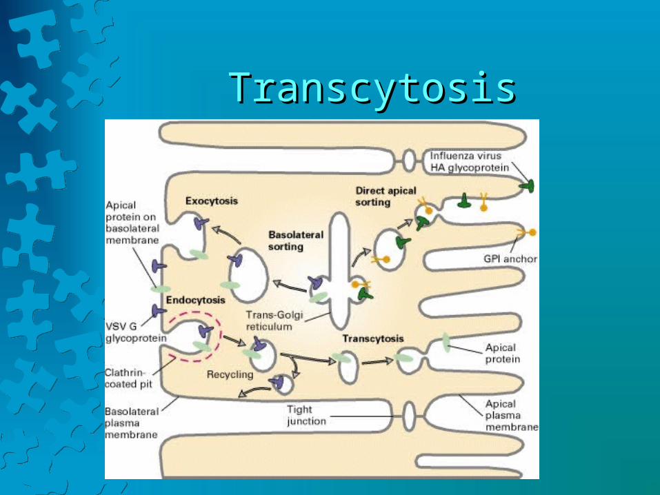
• Transcytosis combines endocytosis and exocytosis to selectively and rapidly transport a substance or particle from one end of a cell to the other.
• Transcytosis moves substances across barriers formed by tightly connected cells.
• HIV uses transcytosis to corss lining cells in the anus and female reproductive tract.
TranscytosisTranscytosis
The Cell CycleThe Cell Cycle
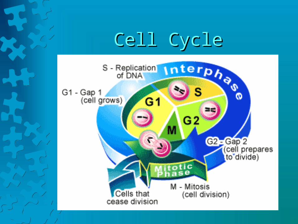
• Cell cycle—well-ordered sequence of events between the time a cell divides to form 2 daughter cells and the time those daughter cells divide.
• The cell cycle alternates between M phase, or dividing phase, and Interphase, the non-dividing phase.
Cell CycleCell Cycle
Division of the CellDivision of the Cell
• The M phase is the shortest part of the cell cycle and the phase during which the cell divides, includes:
1.Mitosis—division of the nucleus.
2.Cytokinesis—division of the cytoplasm.

MitosisMitosis
• Cells undergo mitosis as they approach the maximum cell size at which they can work efficiently.
• The 4 phases of mitosis include:• Prophase• Metaphase• Anaphase• Telophase
Prophase: The first phase of MitosisProphase: The first phase of Mitosis
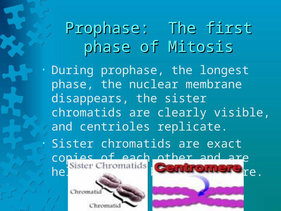
• During prophase, the longest phase, the nuclear membrane disappears, the sister chromatids are clearly visible, and centrioles replicate.
• Sister chromatids are exact copies of each other and are held together by a centromere.

ProphaseProphase
.In the nucleus:In the nucleus:Nucleoli disappear.Nucleoli disappear.The 2 identical sister The 2 identical sister chromatids are joined at the chromatids are joined at the centromere.centromere.In the cytoplasm:In the cytoplasm:Mitotic spindle forms Mitotic spindle forms between the 2 centrosomes.between the 2 centrosomes.Centrosomes move apart.Centrosomes move apart.
MetaphaseMetaphase
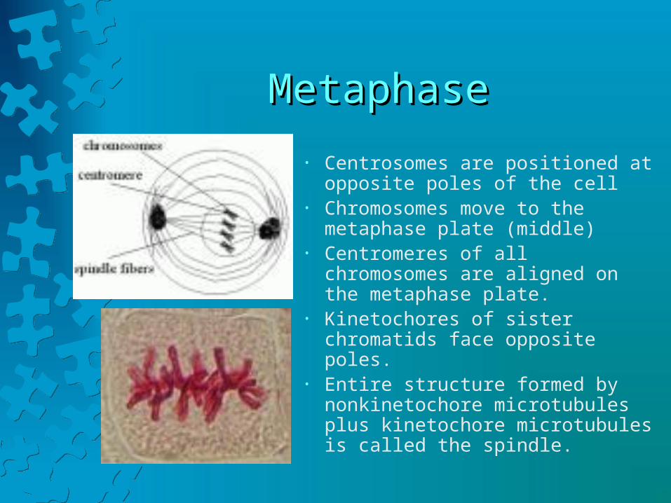
• Centrosomes are positioned at opposite poles of the cell
• Chromosomes move to the metaphase plate (middle)
• Centromeres of all chromosomes are aligned on the metaphase plate.
• Kinetochores of sister chromatids face opposite poles.
• Entire structure formed by nonkinetochore microtubules plus kinetochore microtubules is called the spindle.
AnaphaseAnaphase
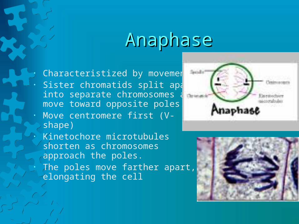
• Characteristized by movement.• Sister chromatids split apart into
separate chromosomes and move toward opposite poles.
• Move centromere first (V-shape)• Kinetochore microtubules shorten as
chromosomes approach the poles.• The poles move farther apart,
elongating the cell
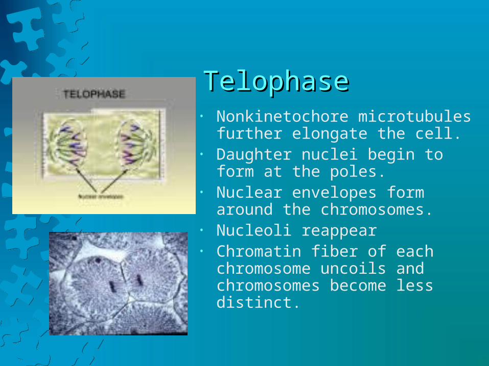
TelophaseTelophase• Nonkinetochore microtubules further
elongate the cell.• Daughter nuclei begin to form at the
poles.• Nuclear envelopes form around the
chromosomes.• Nucleoli reappear• Chromatin fiber of each chromosome
uncoils and chromosomes become less distinct.
CytokinesisCytokinesis
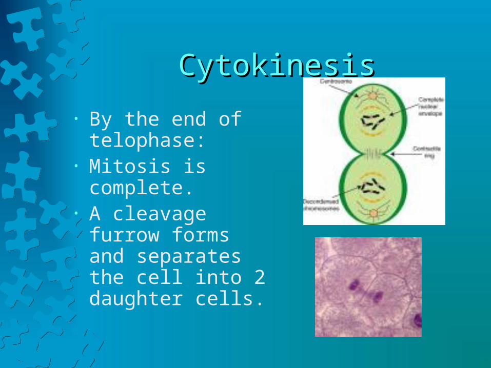
• By the end of telophase:
• Mitosis is complete.• A cleavage furrow
forms and separates the cell into 2 daughter cells.

Control of the Cell CycleControl of the Cell Cycle
• Enzymes control the cell cycle.• Certain enzymes are necessary to begin and
drive the cell cycle and other enzymes control the cycle through the phases.
CancerCancer
• Occasionally, cells lose control of the cell cycle resulting in uncontrolled dividing of the cells. This abnormal growth is called a tumor.
• If a tumor becomes malignant, the result is cancer.• This loss of control may be caused by
environmental factors or by changes in enzyme production.

Genes that Cause CanderGenes that Cause Cander
• Oncogenes activate other genes that increase cell division rate.
• Tumor suppressor genes normally hold mitosis in check.
• When tumor suppressor genes are removed or inactivated, cancer may result.

Causes of CancerCauses of Cancer
• Both genetic and environmental factors are involved.
• Environmental influences include cigarette smoke, air/water pollution, and exposure to UV radiation from the sun.
• Cancer may also be caused by viral infections that damage genes.
Stem and Progenitor CellsStem and Progenitor Cells
• In 1855, German physiologist Rudolph Virchow stated that all cells come from preexisting cells.
• Cells that retain the ability to divide repeatedly allow for continual growth and renewal.
• A stem cell divides mitotically to produce either 2 daughter cells, or one daughter cell and one stem cell.

Stem and Progenitor CellsStem and Progenitor Cells
• A cell that is partially specialized is an intermediate between a stem cell and a differentiated cell is called a progenitor cell.
• A progenitor is said to be ‘committed’ because its daughter cells can become any of a restricted number of cell types.

Totipotent vs PluripotentTotipotent vs Pluripotent
• Stem and progenitor cells are described in terms of their potential to become different types of cells.
• Totipotent mans that they can give rise to every cell type.
• Pluripotent means that can they can follow several different pathways but not all of them.

TotipotentTotipotent
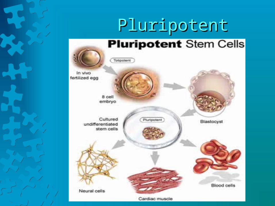
PluripotentPluripotent


