55
Anatomy and Physiology Anatomy and Physiology Chapter #3 Chapter #3
| Date post: | 18-Dec-2015 |
| Category: |
Documents |
| Upload: | augustus-briggs |
| View: | 216 times |
| Download: | 0 times |

Anatomy and Physiology Chapter Anatomy and Physiology Chapter #3#3

3.1 Introduction
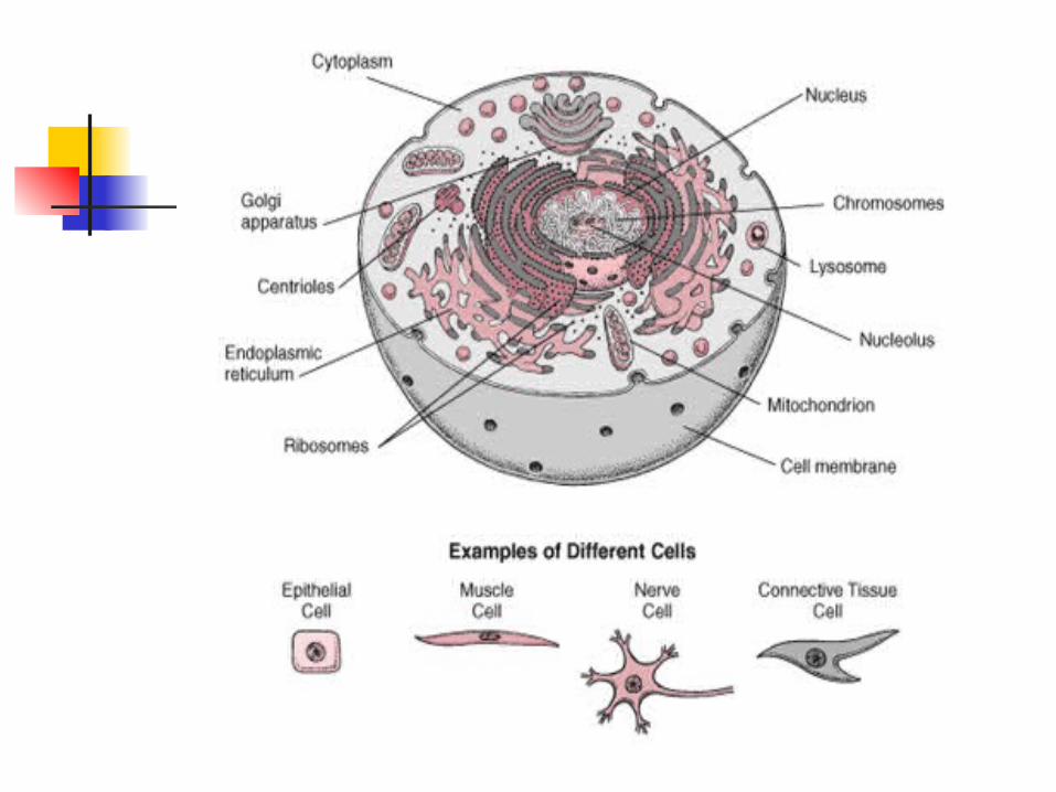
Cells vary considerable in size, shape, and function.
The shape of cells in the body vary based on their function.

3.2 Composite Cell Because cells vary so greatly in size,
shape, content, and function, describing a “typical” cell is impossible.
All of the structures described in the composite cell can NOT be found in every cell regardless of function.
The organelles are found in the cytoplasm of the cell.
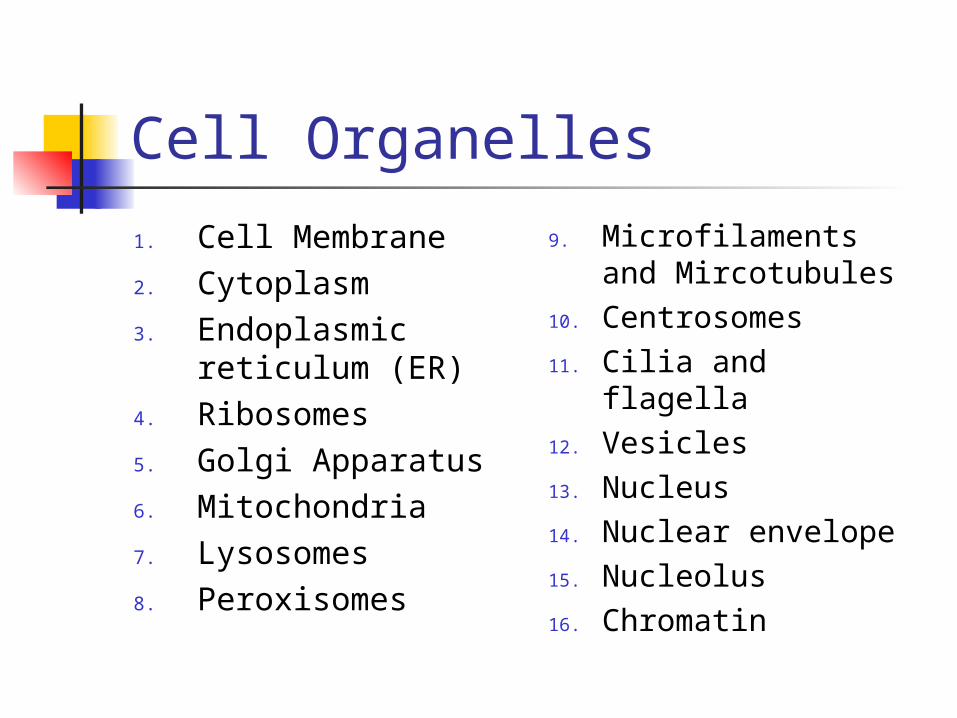
Cell Organelles
1. Cell Membrane2. Cytoplasm3. Endoplasmic
reticulum (ER)4. Ribosomes5. Golgi Apparatus6. Mitochondria7. Lysosomes8. Peroxisomes
9. Microfilaments and Mircotubules
10. Centrosomes11. Cilia and flagella12. Vesicles13. Nucleus14. Nuclear envelope15. Nucleolus16. Chromatin

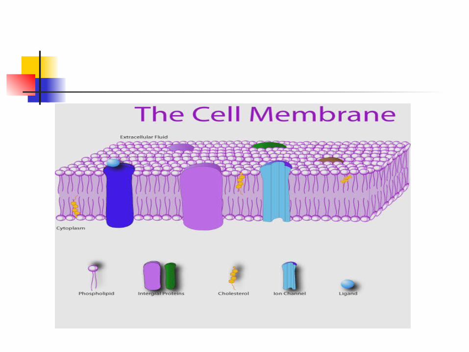
Cell Membrane Extremely thin and semipermeable. The cell membrane contains many kinds of
proteins, each with a special function. Function: The membrane allows some
substances to enter and leave the cell but not others, participates in signal transduction, and helps cells adhere to other cells. The membrane forms a boundary around the cellular contents,, and the basic structure of the cell membrane is a lipid-protein bilayer.
Structure: The basic framework of the cell membrane consists of a (bilayer) double layer of phospholipid, with fatty acid tails turned in.

Cell Memebrane The Phosphate Head is
HYDROPHILIC meaning "WATER LOVING". Because of its hydrophilic nature, the head of a Phospholipid will orient itself so that it is as close as possible to water molecules.
The Lipid Tails are HYDROPHOBIC meaning "WATER-FEARING", the Hydrophobic tails will tend to orient themselves away from water.
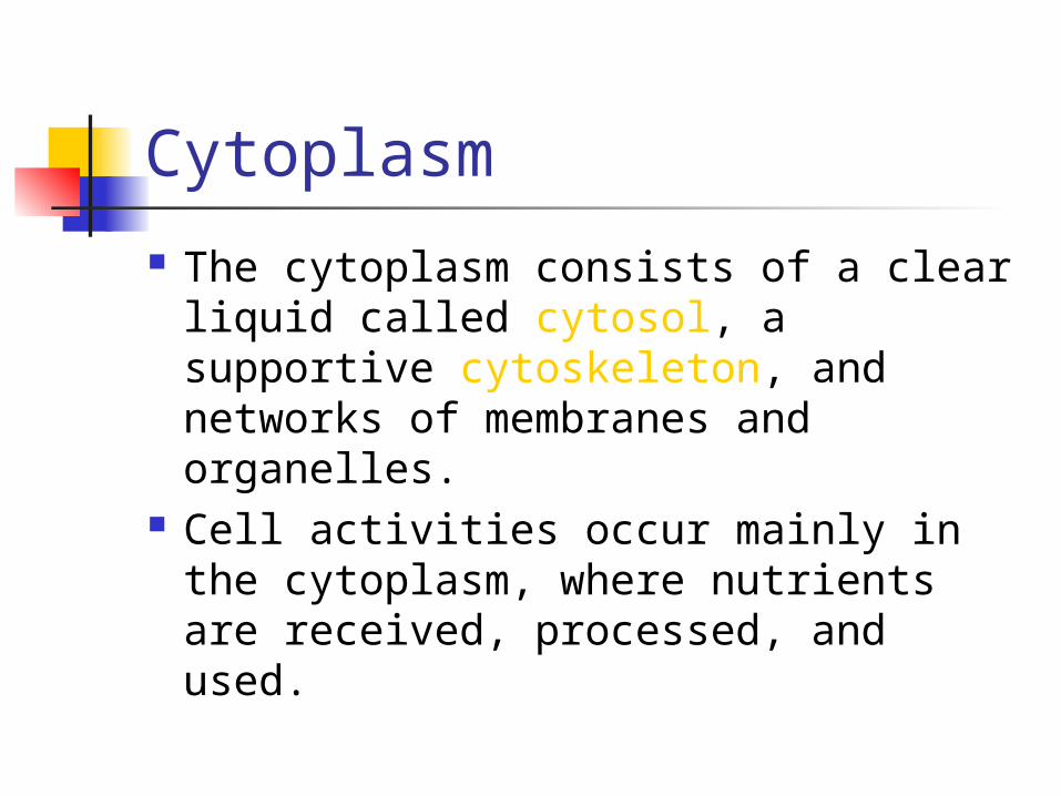
Cytoplasm
The cytoplasm consists of a clear liquid called cytosol, a supportive cytoskeleton, and networks of membranes and organelles.
Cell activities occur mainly in the cytoplasm, where nutrients are received, processed, and used.

Endoplasmic Reticulum ER is made up of membrane-bounded
flatten sacs and elongated canals. ER provides a tubular transport system inside the cell.
Rough ER: Has ribosomes on its outer layer
What does it function in the synthesis and transport of? PROTEIN
Smooth ER: No ribosomes. What does it function in the transport of? LIPIDS
Vesicles that have a role in secretion are formed by the ER.


Ribosomes
They are found in the cytoplasm and ER
They composed of Protein and RNA molecules.
Function is protein synthesis.

Popeye is active because he eats spinach.
However, for Popeye's cells to be active, his ribosomes help create proteins.


Golgi Apparatus They are composed of about 6 flattened,
membranous sacs. Function is to refine, package, and deliver
proteins synthesized on ribosomes, and it packages the cells products.
Vesicles that have a role in secretion are formed by the golgi apparatus (and ER).
Plays a central role in the transport of new molecules from inside to outside the cell.


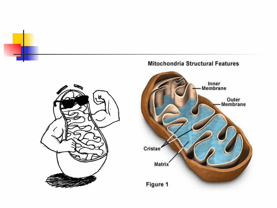
Mitochondria
Is the POWERHOUSE of the cell and contain enzymes that catalyze reactions that release energy from nutrients needed for aerobic respiration.
Enzyme is a protein that catalyzes a specific biochemical reaction.
Adenosine Triphosphate (ATP) energy.

Lysosomes
The "garbage disposals of the cell“ They contain powerful enzymes to
break up old cell components and bacteria. Example white blood cells can engulf bacteria to fight bacterial infections.


Peroxisomes
Membranous sacs are abundant in liver and kidney cells.
They contain enzymes that function in the synthesis of bile acids, breakdown of lipids, degradation of rare biochemicals, and detoxification of alcohol.

Microfilaments and microtubules Thin, threadlike processes within the
cytoplasm that function in various kinds of cell movement and that serve as the cytoskeleton of the cell.
Microfilaments, made of the protein actin, cause various cellular movements.
Mictotubules, made of the globular protein tubulin. Form “9+2” pattern.


Centrosome
is a structure made up of two hollow cylinders called centrioles.
Function in cell reproduction.


Cilia and flagella They are motile extensions from the
cell. Cilia is short and move “to-and-fro”. Cilia function is to move fluids. Flagella are longer and move in an
undulating wavelike motion. What is the only flagellated cell in
the body? SPERM


Vesicles
Or vacuoles are formed by part of the cell membrane.
They contain some liquid or solid material formerly outside the cell.
Golgi and ER also form vesicles.

Nucleus, nuclear envelope
Is bounded by a double-layered nuclear membrane (nuclear envelope) containing relatively large nuclear pores that allow the passage of certain substances.

Nucleolus
Is a small, dense body composed mainly of RNA.
Has no surrounding membrane. Ribosomes form in the nucleolus.

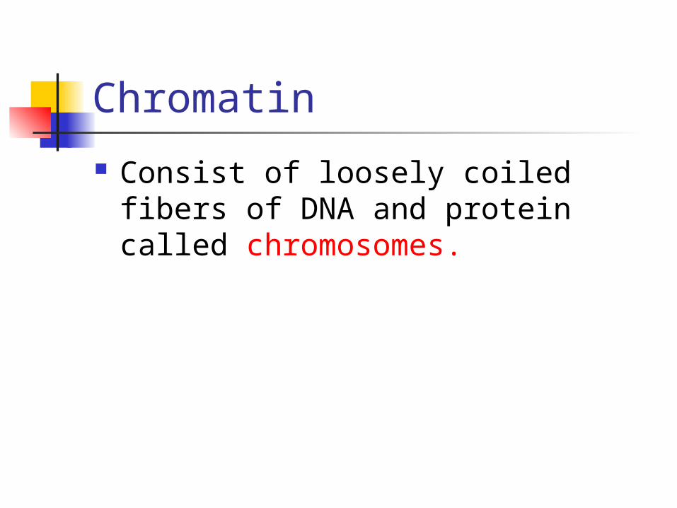
Chromatin
Consist of loosely coiled fibers of DNA and protein called chromosomes.


3.3 Movements through cell membranes Passive
mechanisms do not require energy.
1. Diffusion2. Facilitated
diffusion3. Osmosis4. Filtration
Active mechanisms require cell energy.
1. Active transport2. Endocytosis3. Exocytosis

Passive
Diffusion is the process by which molecules or ions scatter or spread spontaneously from high concentration to low concentration.
Oxygen and carbon dioxide can diffuse readily through the cell membrane.
Equilibrium is reached.

Passive
Substances that are not able to pass through the lipid bilayer need the help of membrane proteins to get across, this is called facilitated diffusion.
Carrier molecules “revolving doors” are used to carry large molecules (glucose) across the membrane.


Passive Osmosis is the diffusion of water. Solutions with a higher osmotic
pressure than body fluids are called hypertonic.
Solutions with a osmotic pressure equal to body fluids are called isotonic.
Solutions with a lower osmotic pressure than body fluids are called hypotonic.

Passive
When fluid is forced through a membrane by hydrostatic or blood pressure, the mechanism is called filtration.
Edema excess tissue fluid.

Active
Active transport is a process that moves particles through membranes from a region of low concentration to high concentration.
Equilibrium is never reached.

Active In endocytosis molecules that are too large to
be transported by other means are engulfed by portion of the cell membrane and carried into the cell surrounded by a vesicle.
1. Pinocytosis is a form in which cells engulf liquids.
2. Phagocytosis is a form in which the cell takes in larger particles, such as a white blood cell engulfing a bacterium.
3. Receptor-mediated is a form in which receptors bind specific particles, and they are drawn into the cell.

The reverse to endocytosis is exocytosis.
3.4 The Cell Cycle The series of changes a cell undergoes
from the time it is formed until it reproduces is called the cell cycle.
The cell cycle is highly regulated. Most cells do not divide continually. Cells have a maximum number of times they can divide because of built-in “clocks” called telomeres on the tips of chromosomes.
Mitosis cell division to form new cells (body cells).
Meiosis cell division that forms sex cells (gametes).

Mitosis (IPMAT)
1. Interphase2. Prophase3. Metaphase4. Anaphase5. Telophase

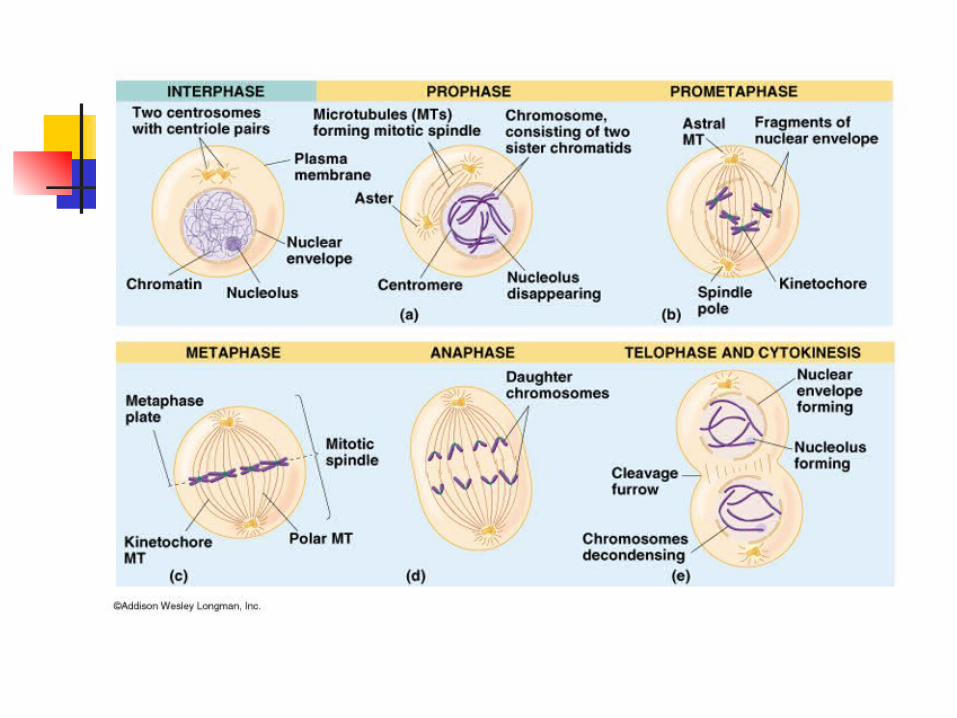
Mitosis Interphase is a period of great metabolic
activity in which the cell grows and synthesizes new molecules and organelles. During the S phase of interphase, the DNA of the cell is replicated in preparation for cell division.
Prophase chromosomes and centrioles become visible and the nuclear envelope and the nucleolus disappear.
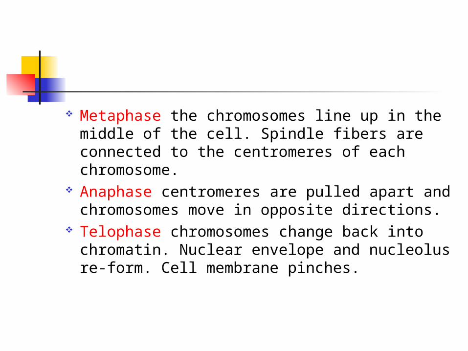
Metaphase the chromosomes line up in the middle of the cell. Spindle fibers are connected to the centromeres of each chromosome.
Anaphase centromeres are pulled apart and chromosomes move in opposite directions.
Telophase chromosomes change back into chromatin. Nuclear envelope and nucleolus re-form. Cell membrane pinches.
Cytokinesis is the division of cytoplasm.

Cell Death Apoptosis is a form of cell death that is
actually a normal part of development, sculpting organs from overgrown tissues.
In the fetus, apoptosis carves away webbing between developing fingers and toes.
Apoptosis follows a sunburn it peels away cells so damaged that they might otherwise turn cancerous.

Cancer
Cancer cells spread to distant areas (metastasize), display excessive growth (hyperplasia), and exhibit properties found in earlier stages (de-differentiation).

Work Cited Golgi apparatus image.www.sirinet.net/~jgjohnso/cell.html Mitochondria man image.
www.mitochondria.us/pages/rebep_main.html Mitochondria image.
www.mitochondria.us/pages/rebep_main.html Cell image.
www.merck.com/media/mmhe2/figures/fg001_2.gif
ER image. www.science.siu.edu/plant-biology/PLB117/JPEGs%20CD/0073.JPG
Ribosome image. //anthro.palomar.edu/biobasis/bio_5.htm

Cell membrane image. www.bioeng.auckland.ac.nz/images/
Popeye cartoon.www.//vilenski.org/science/safari/cellstructure/
ribo.html Lysosome image.
www.people.virginia.edu/~rjh9u/lysosome.html
Cytoskeleton image. www.beyondbooks.com/lif71/4f.asp
Animated sheep. www.beyondbooks.com

Centrosome image. www.faculty.tcc.cc.fl.us/scma/smithh/centrioles.jpg
Cilia and Flagella image. www.academic.brooklyn.cuny.edu/biology/bio4fv/page/flagella-movement.html
Nucleus image.www.emc.maricopa.edu/faculty/farabee/BIOBK/
BioBookCELL2.html Carrier Molecule image.
www.msad54.k12.me.us/MSAD54Pages/skow/CurrProjects/Biology/CrazyCells/Transport/transport%20proteins.htm

Edema image. www.nucleusinc.com
Mitosis image 1st pg. http://campus.queens.edu/faculty/jannr/cells/mit%20pics/mitosis%20animal.jpg
Mitosis image. www.mun.ca/.../BIOL2060/CellBiol17/CB17_19.html


