94
ANATOMY AND PHYSIOLOGY OF EAR,NOSE&THROAT. ASSESMENT OF HEARING IN CHILDREN - DR.AKSHAY.B.K.
| Date post: | 22-Jan-2018 |
| Category: |
Health & Medicine |
| Upload: | drakshay-b-k |
| View: | 291 times |
| Download: | 8 times |
ANATOMY AND PHYSIOLOGY OF EAR,NOSE&THROAT. ASSESMENT OF HEARING IN CHILDREN
-DR.AKSHAY.B.K.

ANATOMY OF EAR
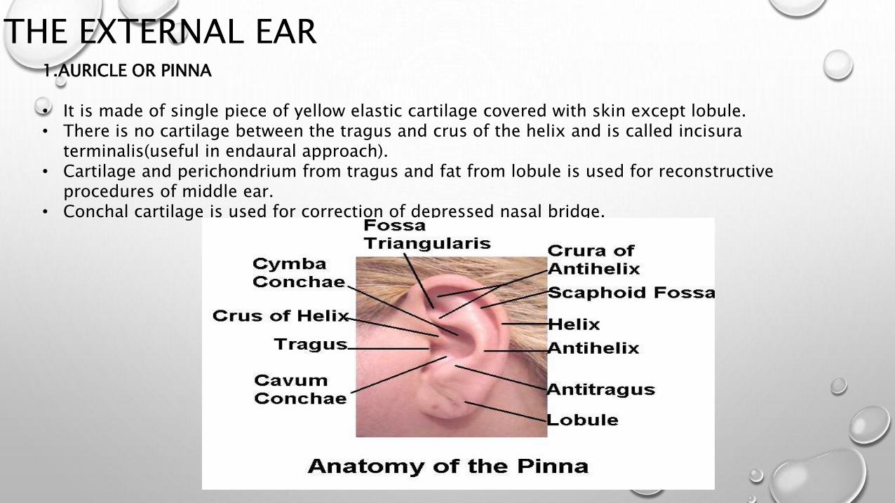
THE EXTERNAL EAR1.AURICLE OR PINNA
• It is made of single piece of yellow elastic cartilage covered with skin except lobule.• There is no cartilage between the tragus and crus of the helix and is called incisura
terminalis(useful in endaural approach).• Cartilage and perichondrium from tragus and fat from lobule is used for reconstructive
procedures of middle ear.• Conchal cartilage is used for correction of depressed nasal bridge.
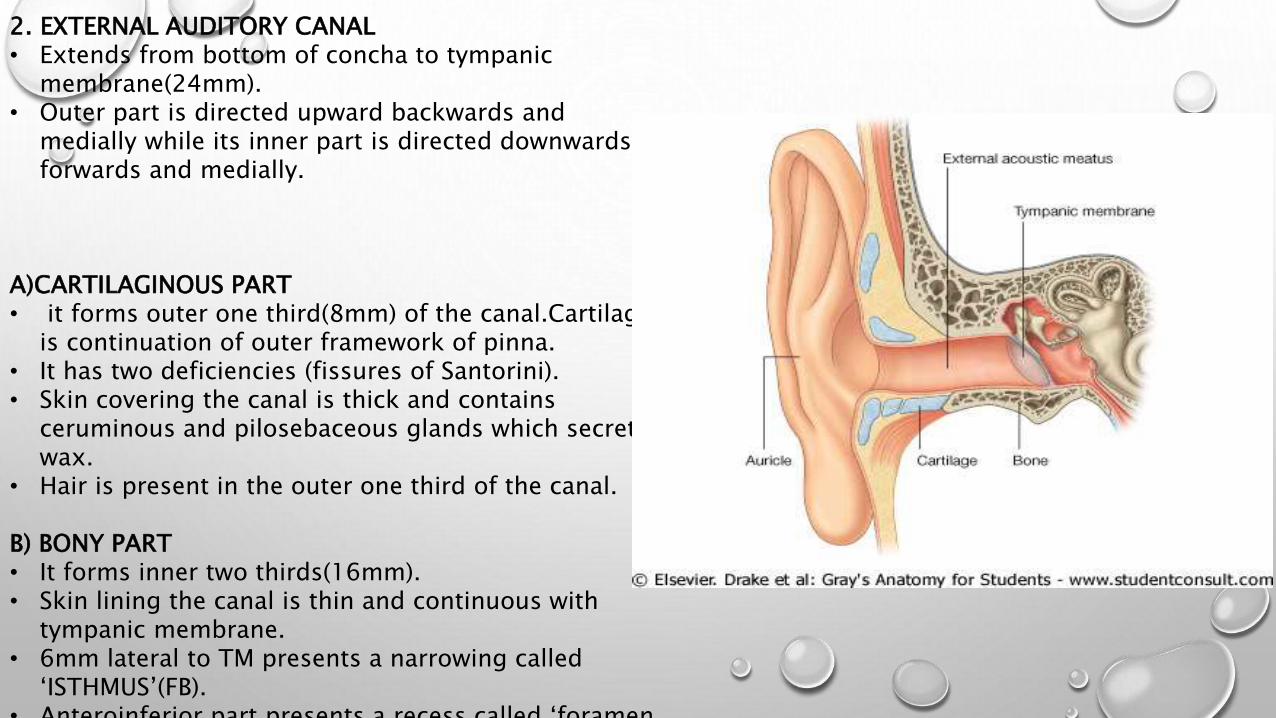
2. EXTERNAL AUDITORY CANAL• Extends from bottom of concha to tympanic
membrane(24mm).• Outer part is directed upward backwards and
medially while its inner part is directed downwards forwards and medially.
A)CARTILAGINOUS PART• it forms outer one third(8mm) of the canal.Cartilage
is continuation of outer framework of pinna. • It has two deficiencies (fissures of Santorini).• Skin covering the canal is thick and contains
ceruminous and pilosebaceous glands which secrete wax.
• Hair is present in the outer one third of the canal.
B) BONY PART• It forms inner two thirds(16mm).• Skin lining the canal is thin and continuous with
tympanic membrane.• 6mm lateral to TM presents a narrowing called
‘ISTHMUS’(FB).• Anteroinferior part presents a recess called ‘foramen
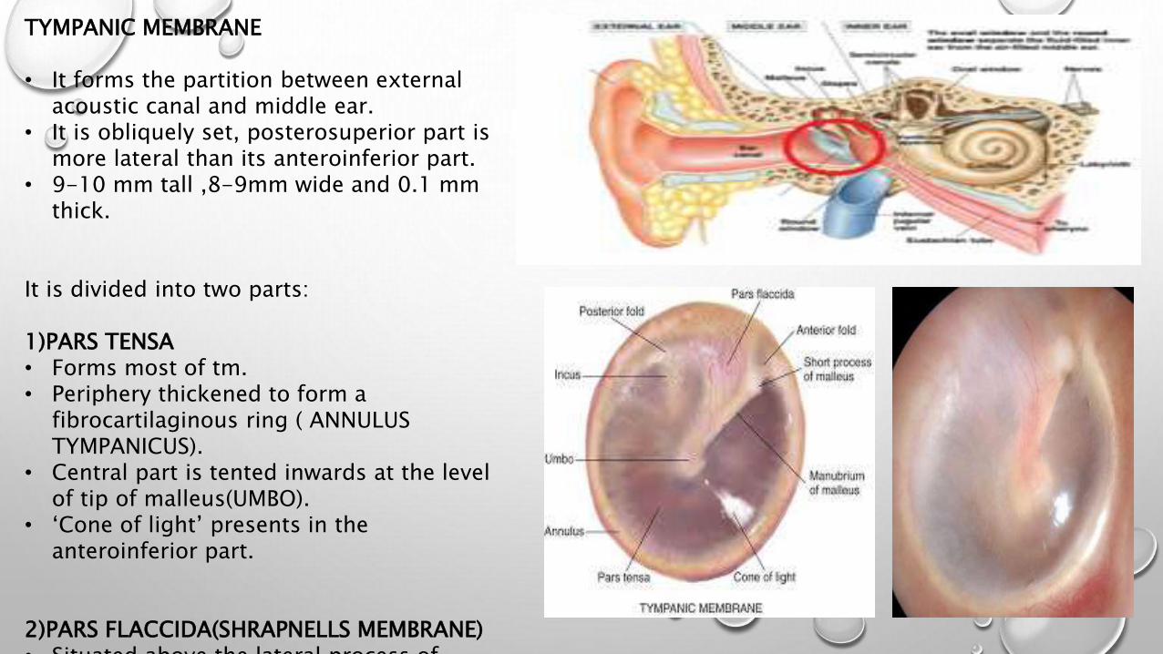
TYMPANIC MEMBRANE
• It forms the partition between external acoustic canal and middle ear.
• It is obliquely set, posterosuperior part is more lateral than its anteroinferior part.
• 9-10 mm tall ,8-9mm wide and 0.1 mm thick.
It is divided into two parts:
1)PARS TENSA • Forms most of tm.• Periphery thickened to form a
fibrocartilaginous ring ( ANNULUS TYMPANICUS).
• Central part is tented inwards at the level of tip of malleus(UMBO).
• ‘Cone of light’ presents in the anteroinferior part.
2)PARS FLACCIDA(SHRAPNELLS MEMBRANE)• Situated above the lateral process of

NERVE SUPPLY OF THE EXTERNAL EAR
PINNA• Greater auricular nerve(C2,3)• Lesser occipital nerve (C2)• Auriculotemporal nerve( V3)• Auricular branch of vagus(CNX) ARNOLDS NERVE.
EXTERNAL AUDITORY CANAL• Antr wall and roof: auriculotemporal nerve.• Postr wall and floor: auricular branch of vagus
TYMPANIC MEMBRANE:• Antr half of lateral surface : auriculotemporal
nerve• Postr half of lateral surface : auricular branch of
vagus• Medial surface : tympanic branch of CN IX
(jacobsons nerve)
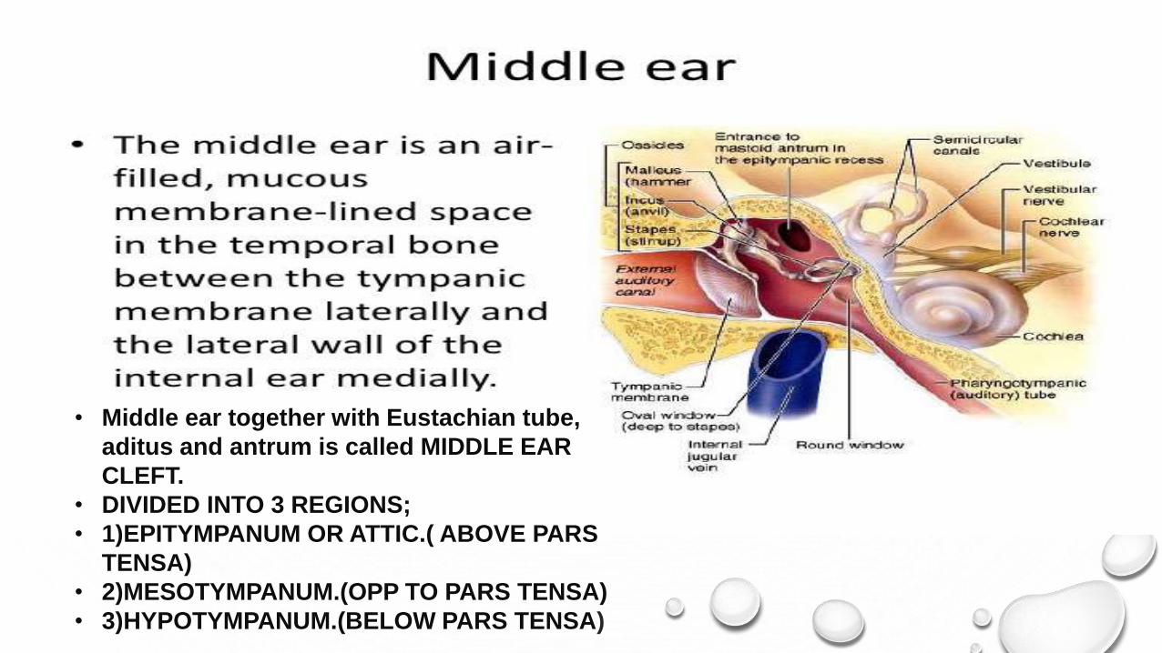
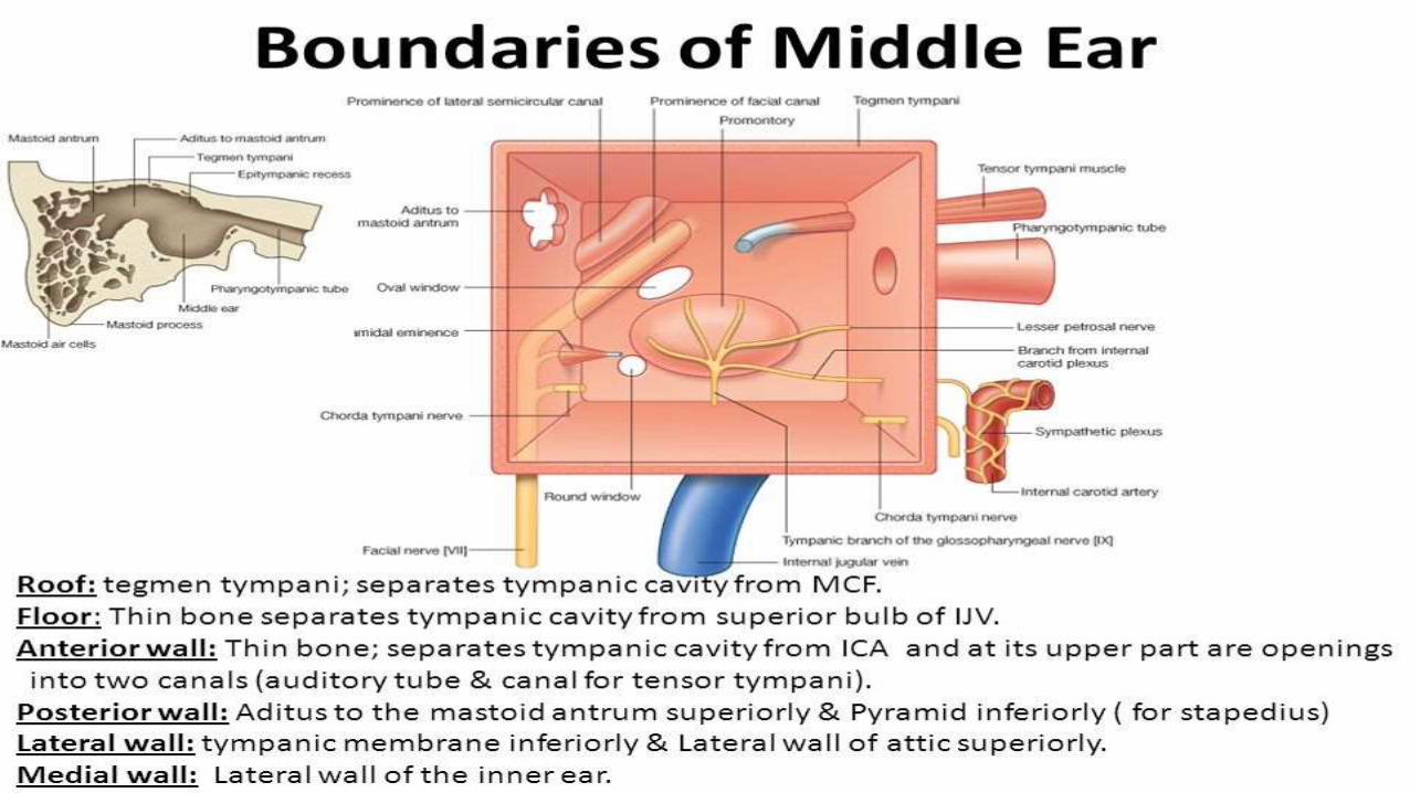
• Middle ear together with Eustachian tube,
aditus and antrum is called MIDDLE EAR
CLEFT.
• DIVIDED INTO 3 REGIONS;
• 1)EPITYMPANUM OR ATTIC.( ABOVE PARS
TENSA)
• 2)MESOTYMPANUM.(OPP TO PARS TENSA)
• 3)HYPOTYMPANUM.(BELOW PARS TENSA)
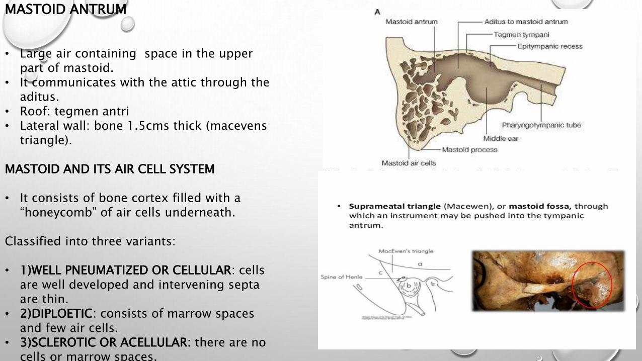
MASTOID ANTRUM
• Large air containing space in the upper part of mastoid.
• It communicates with the attic through the aditus.
• Roof: tegmen antri• Lateral wall: bone 1.5cms thick (macevens
triangle).
MASTOID AND ITS AIR CELL SYSTEM
• It consists of bone cortex filled with a “honeycomb” of air cells underneath.
Classified into three variants:
• 1)WELL PNEUMATIZED OR CELLULAR: cells are well developed and intervening septa are thin.
• 2)DIPLOETIC: consists of marrow spaces and few air cells.
• 3)SCLEROTIC OR ACELLULAR: there are no cells or marrow spaces.
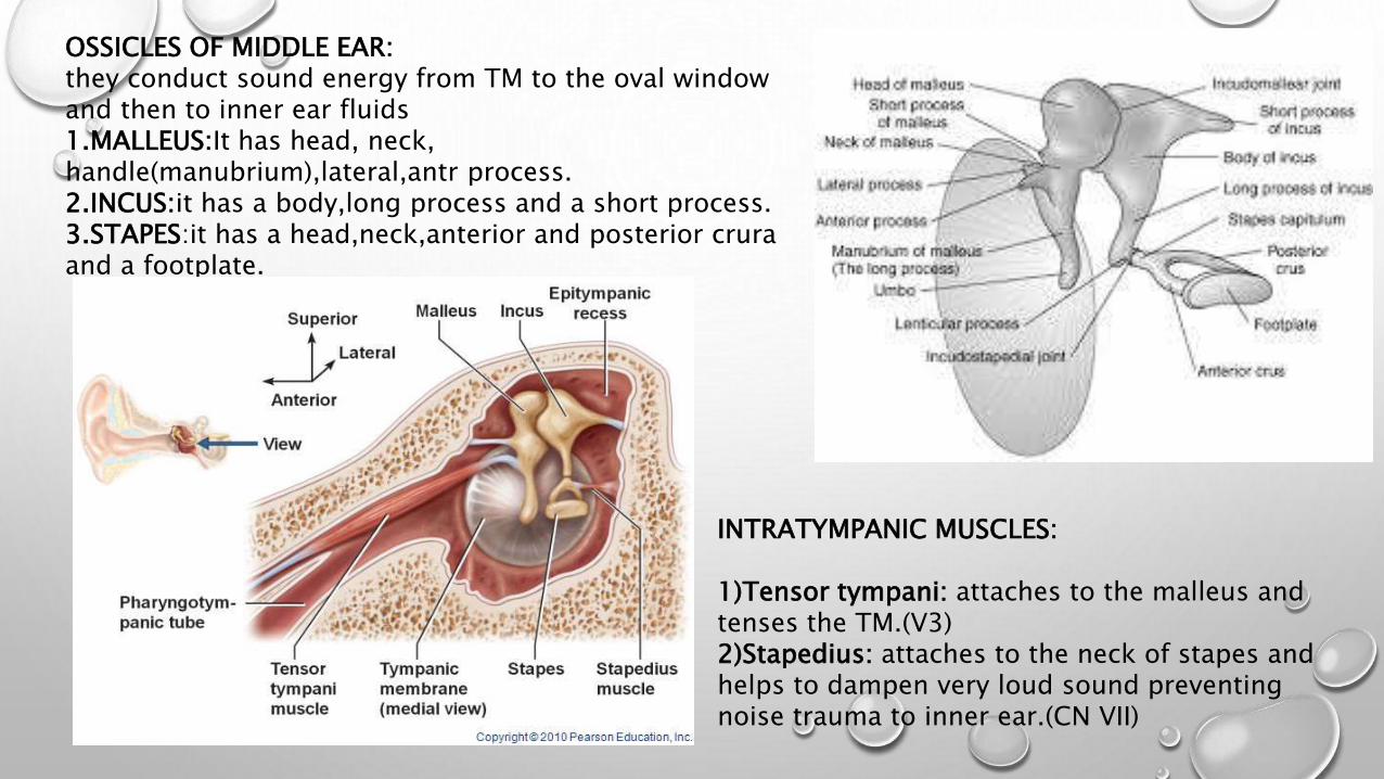
OSSICLES OF MIDDLE EAR: they conduct sound energy from TM to the oval window and then to inner ear fluids1.MALLEUS:It has head, neck, handle(manubrium),lateral,antr process.2.INCUS:it has a body,long process and a short process.3.STAPES:it has a head,neck,anterior and posterior crura and a footplate.
INTRATYMPANIC MUSCLES:
1)Tensor tympani: attaches to the malleus and tenses the TM.(V3)2)Stapedius: attaches to the neck of stapes and helps to dampen very loud sound preventing noise trauma to inner ear.(CN VII)

BLOOD SUPPLY OF MIDDLE EAR:
• Anterior tympanic branch of maxillary artery , supplies TM.• Stylomastoid branch of posterior auricular artery, supplies middle ear
&mastoid air cells.
• Four minor vessels:• Petrosal branch of middle meningeal artery.• Superior tympanic branch of middle meningeal artery.• Branch of artery of pterygoid canal.• Tympanic branch of internal carotid artery.
LYMPHATIC DRAINAGE• Pinna:pre auricular ,post auricular & parotid nodes.• Middle ear-retropharyngeal and parotid nodes
• Eustachian tube –retropharyngeal group.
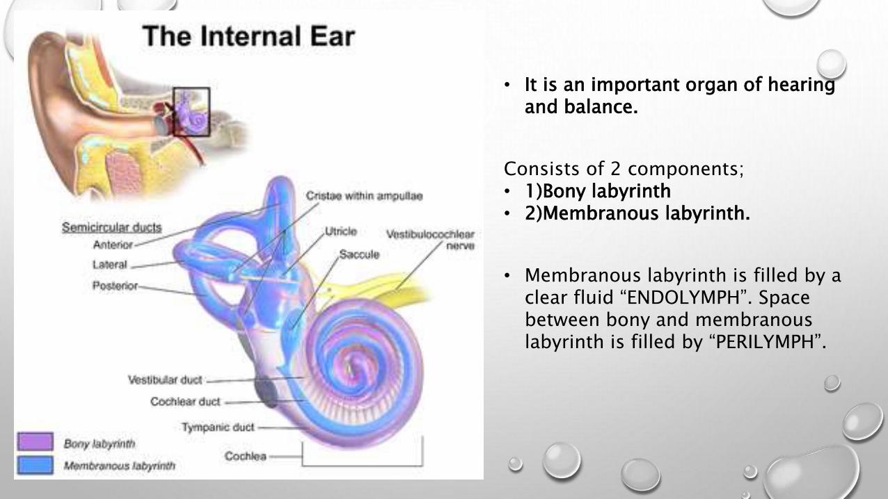
• It is an important organ of hearing and balance.
Consists of 2 components;• 1)Bony labyrinth• 2)Membranous labyrinth.
• Membranous labyrinth is filled by a clear fluid “ENDOLYMPH”. Space between bony and membranous labyrinth is filled by “PERILYMPH”.
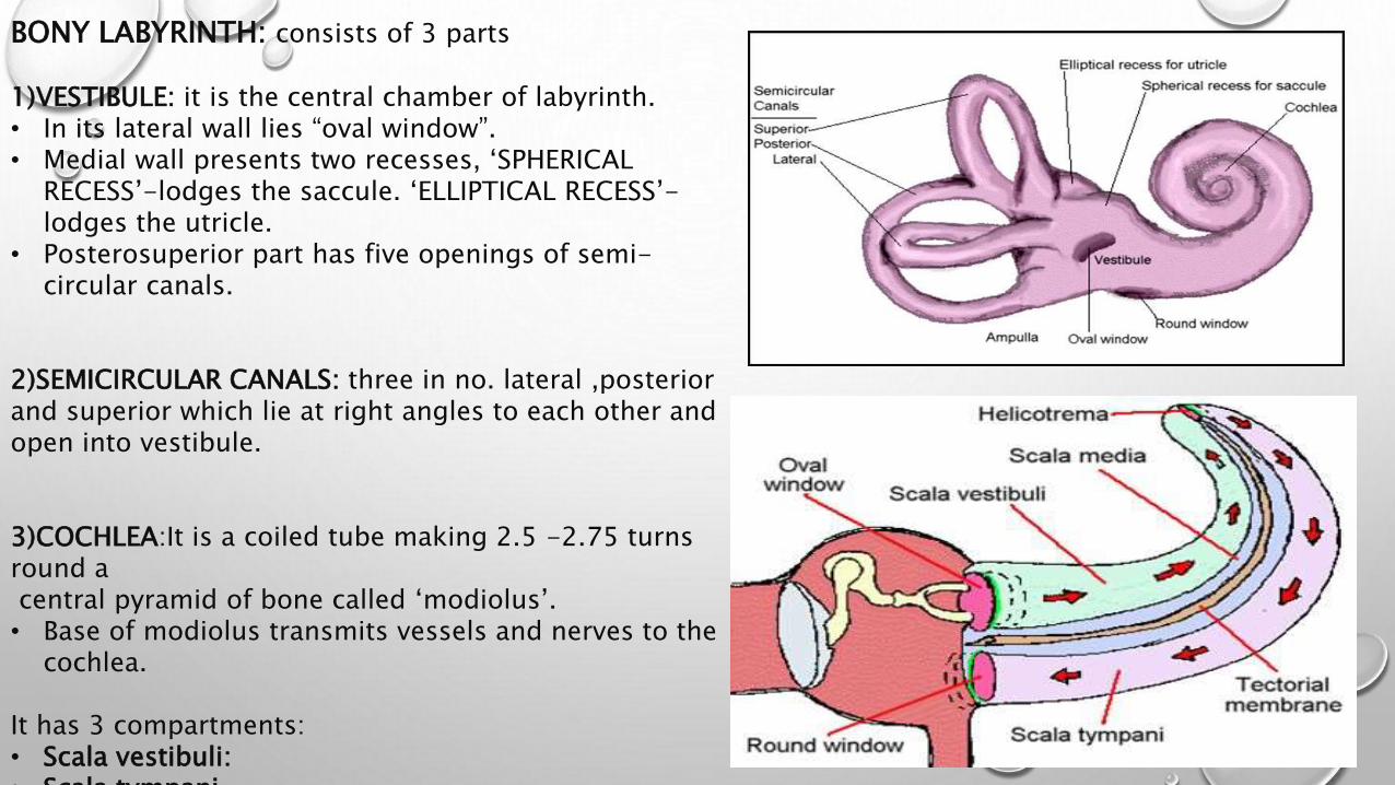
BONY LABYRINTH: consists of 3 parts
1)VESTIBULE: it is the central chamber of labyrinth.• In its lateral wall lies “oval window”.• Medial wall presents two recesses, ‘SPHERICAL
RECESS’-lodges the saccule. ‘ELLIPTICAL RECESS’-lodges the utricle.
• Posterosuperior part has five openings of semi-circular canals.
2)SEMICIRCULAR CANALS: three in no. lateral ,posterior and superior which lie at right angles to each other and open into vestibule.
3)COCHLEA:It is a coiled tube making 2.5 -2.75 turns round acentral pyramid of bone called ‘modiolus’.• Base of modiolus transmits vessels and nerves to the
cochlea.
It has 3 compartments:• Scala vestibuli:• Scala tympani.
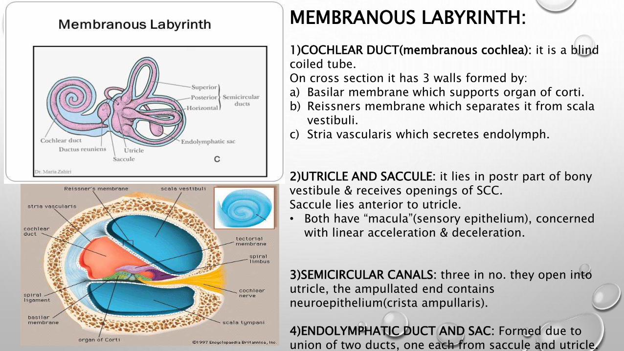
MEMBRANOUS LABYRINTH:
1)COCHLEAR DUCT(membranous cochlea): it is a blind coiled tube.On cross section it has 3 walls formed by:a) Basilar membrane which supports organ of corti.b) Reissners membrane which separates it from scala
vestibuli.c) Stria vascularis which secretes endolymph.
2)UTRICLE AND SACCULE: it lies in postr part of bony vestibule & receives openings of SCC.Saccule lies anterior to utricle.• Both have “macula”(sensory epithelium), concerned
with linear acceleration & deceleration.
3)SEMICIRCULAR CANALS: three in no. they open into utricle, the ampullated end contains neuroepithelium(crista ampullaris).
4)ENDOLYMPHATIC DUCT AND SAC: Formed due to union of two ducts, one each from saccule and utricle. its terminal part is dilated to form ‘endolymphatic sac’

Blood supply of labyrinth:It is mainly through ‘internal auditory artery or labyrinthine artery’ , a branch of anterior inferior cerebellar artery.
INNER EAR FLUIDS :
1)PERILYMPH: resembles CSF ,it fills space between bony and membranous labyrinth.it communicates through CSF via aqueduct of cochlea.Formation(2views):a)filtrate of blood serum secreted by blood capillaries of spiral ligament.b)Direct continuation of CSF.
2)ENDOLYMPH: resembles intracellular fluid, fills membranous labyrinth. Secreted by the secretory cells of stria
vascularis of cochlea.
1.2.
ORGAN OF CORTI
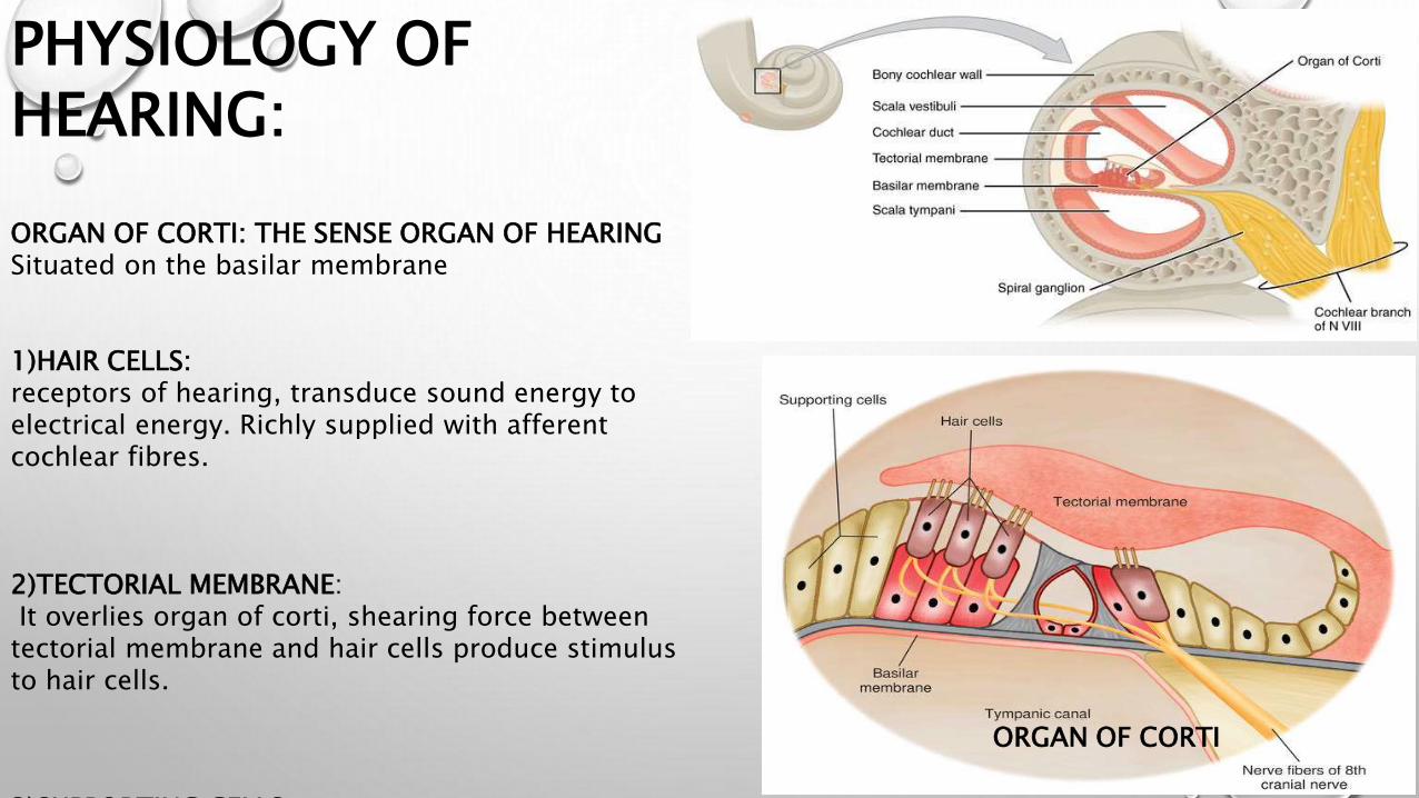
PHYSIOLOGY OF HEARING:
ORGAN OF CORTI: THE SENSE ORGAN OF HEARINGSituated on the basilar membrane
1)HAIR CELLS: receptors of hearing, transduce sound energy to electrical energy. Richly supplied with afferent cochlear fibres.
2)TECTORIAL MEMBRANE:It overlies organ of corti, shearing force between
tectorial membrane and hair cells produce stimulus to hair cells.
3)SUPPORTING CELLS.

INNER EAR
SOUND SIGNAL(environment) PINNA EXTERNAL AUDITORY CANAL TYMPANIC MEMBRANE
OSSICLES
STAPES FOOT PLATE
MECHANISM OF HEARING :
1)CONDUCTION OF SOUND
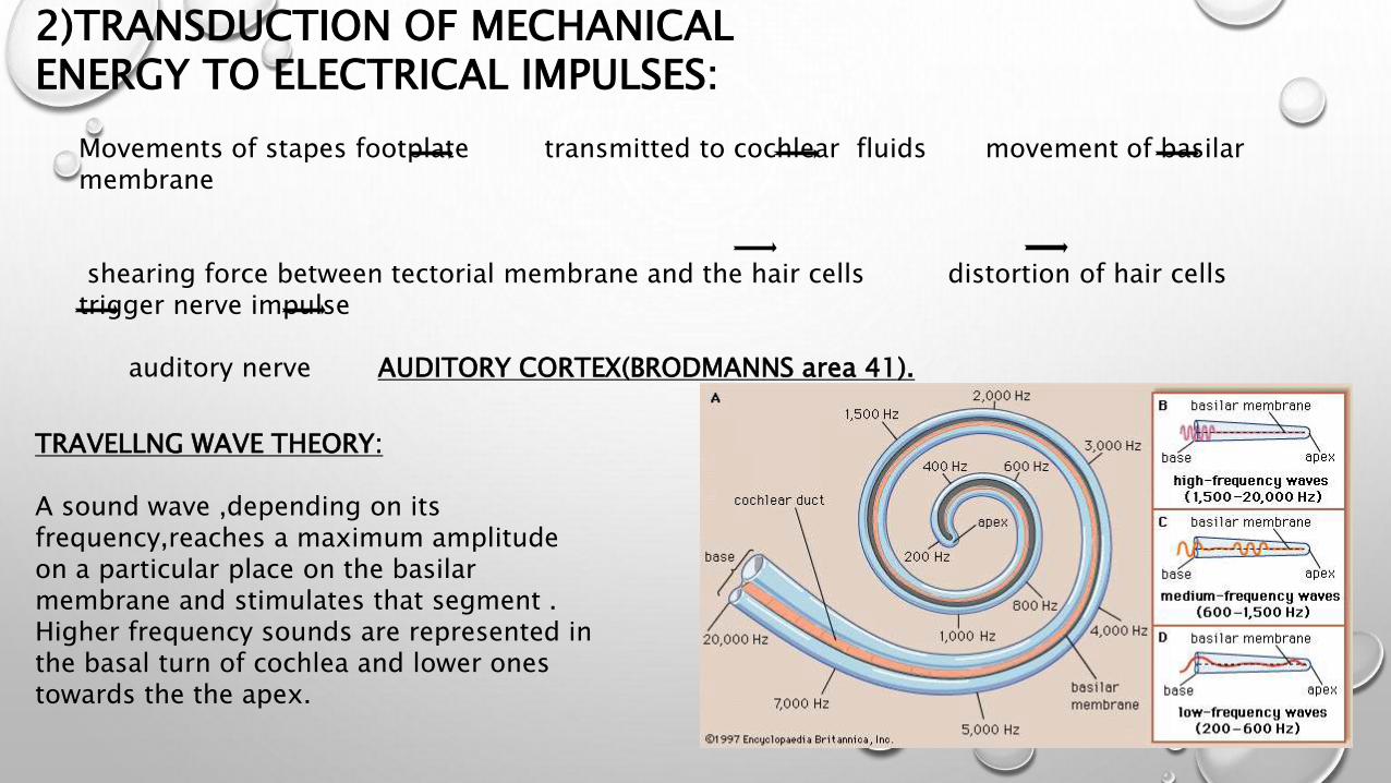
Movements of stapes footplate transmitted to cochlear fluids movement of basilar membrane
shearing force between tectorial membrane and the hair cells distortion of hair cells trigger nerve impulse
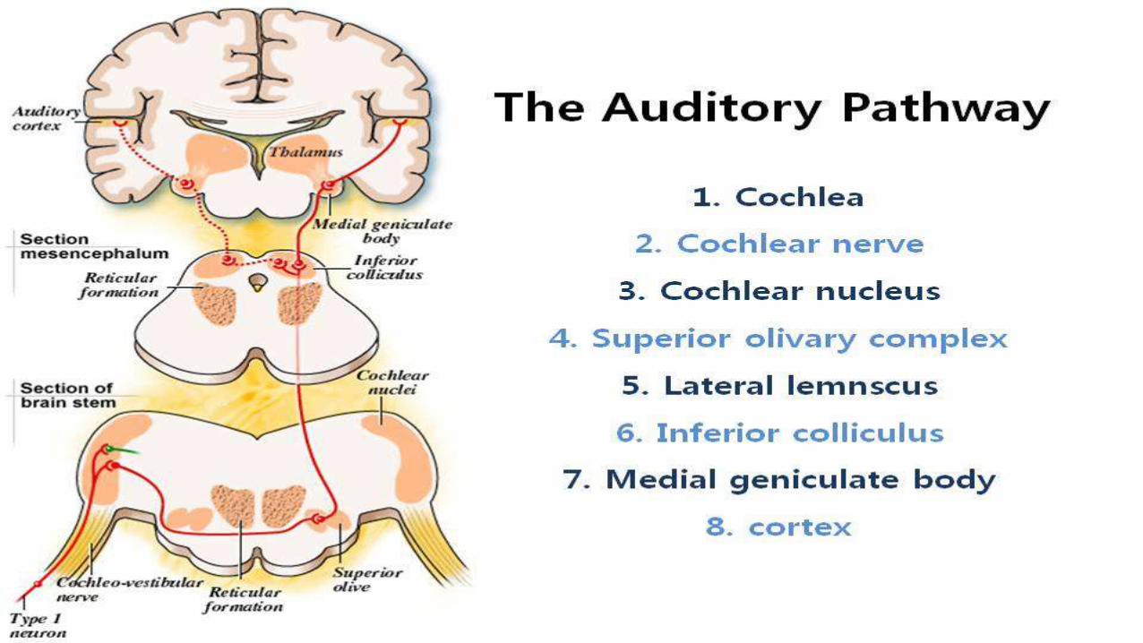
auditory nerve AUDITORY CORTEX(BRODMANNS area 41).
2)TRANSDUCTION OF MECHANICAL ENERGY TO ELECTRICAL IMPULSES:
TRAVELLNG WAVE THEORY:
A sound wave ,depending on its frequency,reaches a maximum amplitude on a particular place on the basilar membrane and stimulates that segment .Higher frequency sounds are represented in the basal turn of cochlea and lower ones towards the the apex.
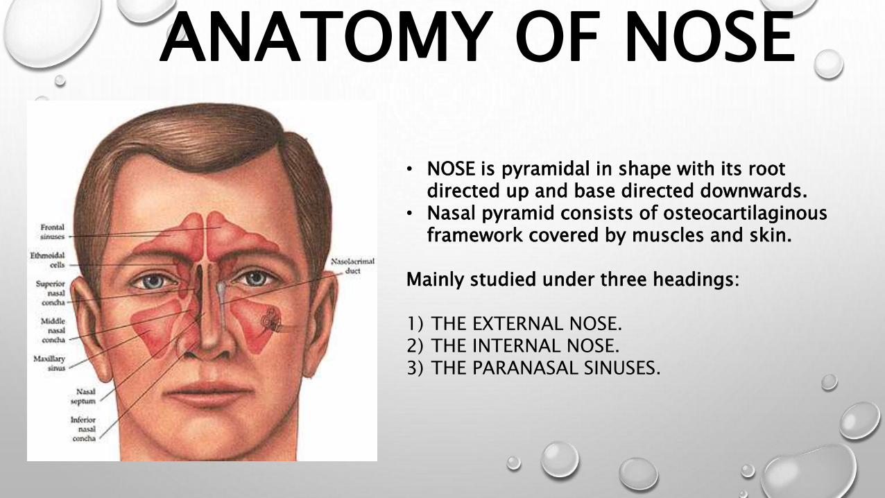
ANATOMY OF NOSE
• NOSE is pyramidal in shape with its root directed up and base directed downwards.
• Nasal pyramid consists of osteocartilaginousframework covered by muscles and skin.
Mainly studied under three headings:
1) THE EXTERNAL NOSE.2) THE INTERNAL NOSE.3) THE PARANASAL SINUSES.

1) THE EXTERNAL NOSE:
OSTEOCARTILAGINOUS FRAMEWORK: upper 1/3rd is bony while lowen2/3rds are cartilaginous,
• BONY PART: consists of two nasal bones meeting in midline,resting on nasal process of frontal bones & held between frontal processes of maxillae.
• CARTILAGINOUS PART : consists ofUpper lateral cartilages: extend from undersurface of nasal bones above to the alar cartilages below, fuse with each other & with upper border of septal cartilage in midline.• Lower free edge is seen intranasaly as ‘nasal valve’.Lower lateral cartilages(alar): • each alar cartilage is U shaped.• It has a lateral crus which forms ala and a medial crus which
runs in the columella.Lesser alar cartilages(sesamoid):• two or more in no.• Lie above and lateral to alar cartilages.• They are connected with one another & with adjoining bones
by perichondrium & periosteum.Septal cartilage:• It runs under the nasal bones to nasal tip.• Supports the dorsum of cartilaginous part of nose.

These bring about the movements of nasal tip, ala & overlying skin.• PROCERUS• NASALIS• LEVATOR LABII
SUPERIORIS ALAEQUE NASI
• DEPRESSOR SEPTI• ANTR AND POSTR
DIALATOR NARES
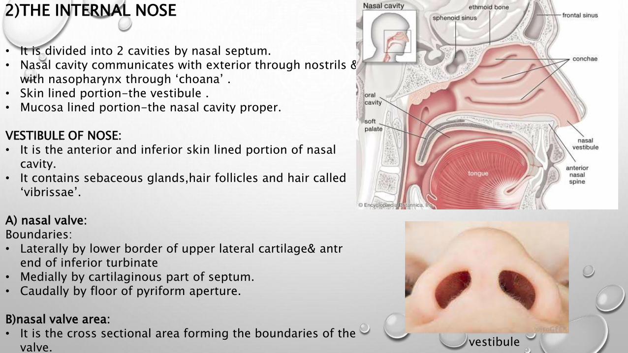
2)THE INTERNAL NOSE
• It is divided into 2 cavities by nasal septum.• Nasal cavity communicates with exterior through nostrils &
with nasopharynx through ‘choana’ .• Skin lined portion-the vestibule .• Mucosa lined portion-the nasal cavity proper.
VESTIBULE OF NOSE:• It is the anterior and inferior skin lined portion of nasal
cavity.• It contains sebaceous glands,hair follicles and hair called
‘vibrissae’.
A) nasal valve:Boundaries:• Laterally by lower border of upper lateral cartilage& antr
end of inferior turbinate• Medially by cartilaginous part of septum.• Caudally by floor of pyriform aperture.
B)nasal valve area:• It is the cross sectional area forming the boundaries of the
valve.• It is least cross sectional area of nose which regulates
vestibule

NASAL CAVITY PROPER
Each nasal cavity has a lateral wall,medial wall,roof and a floor.
ROOF:• Antr sloping part by nasal bones.• Postr part by sphenoid phone.• Middle horizontal part by cribriform
plate.
FLOOR:• Anterior 3/4th by palatine process of
maxilla.• Posterior 1/4th by horizontal part of
palatine bone.
LATERAL NASAL WALL:• Three occasionally, four turbinates
mark the lateral wall.• Turbinates are scroll like bony
projections covered by a mucous
LATERAL WALL OF NOSE
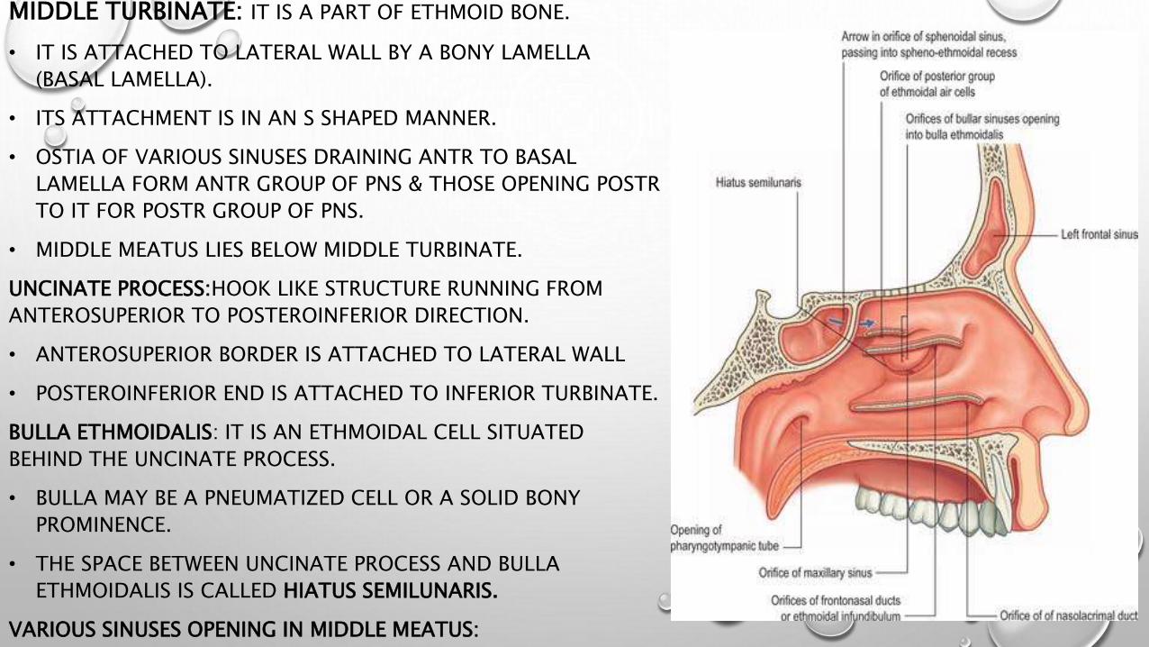
MIDDLE TURBINATE: IT IS A PART OF ETHMOID BONE.
• IT IS ATTACHED TO LATERAL WALL BY A BONY LAMELLA
(BASAL LAMELLA).
• ITS ATTACHMENT IS IN AN S SHAPED MANNER.
• OSTIA OF VARIOUS SINUSES DRAINING ANTR TO BASAL
LAMELLA FORM ANTR GROUP OF PNS & THOSE OPENING POSTR
TO IT FOR POSTR GROUP OF PNS.
• MIDDLE MEATUS LIES BELOW MIDDLE TURBINATE.
UNCINATE PROCESS:HOOK LIKE STRUCTURE RUNNING FROM
ANTEROSUPERIOR TO POSTEROINFERIOR DIRECTION.
• ANTEROSUPERIOR BORDER IS ATTACHED TO LATERAL WALL
• POSTEROINFERIOR END IS ATTACHED TO INFERIOR TURBINATE.
BULLA ETHMOIDALIS: IT IS AN ETHMOIDAL CELL SITUATED
BEHIND THE UNCINATE PROCESS.
• BULLA MAY BE A PNEUMATIZED CELL OR A SOLID BONY
PROMINENCE.
• THE SPACE BETWEEN UNCINATE PROCESS AND BULLA
ETHMOIDALIS IS CALLED HIATUS SEMILUNARIS.
VARIOUS SINUSES OPENING IN MIDDLE MEATUS:

SUPERIROR TURBINATE:• It is also a part of ethmoid bone.• Situated postr and supr to middle
turbinate.• Important landmark to identify ostium of
sphenoid sinus.
SUPERIOR MEATUS:• It is a space below supr turbinate.• Postr ethmoid cells (1-5)open into it.
SPHENOETHMOIDAL RECESS:• It is situated above the superior turbinate,
sphenoid sinus opens into it.
INFERIROR TURBINATE: a separate bone, below it lies the inferior meatus where NLD opens.
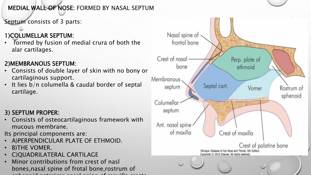
MEDIAL WALL OF NOSE: FORMED BY NASAL SEPTUM
Septum consists of 3 parts:
1)COLUMELLAR SEPTUM:• formed by fusion of medial crura of both the
alar cartilages.
2)MEMBRANOUS SEPTUM: • Consists of double layer of skin with no bony or
cartilaginous support.• It lies b/n columella & caudal border of septal
cartilage.
3) SEPTUM PROPER:• Consists of osteocartilaginous framework with
mucous membrane.Its principal components are:• A)PERPENDICULAR PLATE OF ETHMOID.• B)THE VOMER.• C)QUADRILATERAL CARTILAGE• Minor contributions from crest of nasl
bones,nasal spine of frotal bone,rostrum of sphenoid,anteriror nasal spine of maxilla,crests

PARANASAL SINUSES:These are air containing cavities in certain bones of skull.They have been divided into two groups:1)ANTERIOR GROUP: • includes maxillary, frontal & anterior ethmoidal sinuses.• Their ostia lie anterior to basal lamella.
2)POSTERIOR GROUP:• Includes posterior ethmoidal sinuses which opens into superior meatus.• Sphenoid sinus which opens in sphenoethmoidal recess.
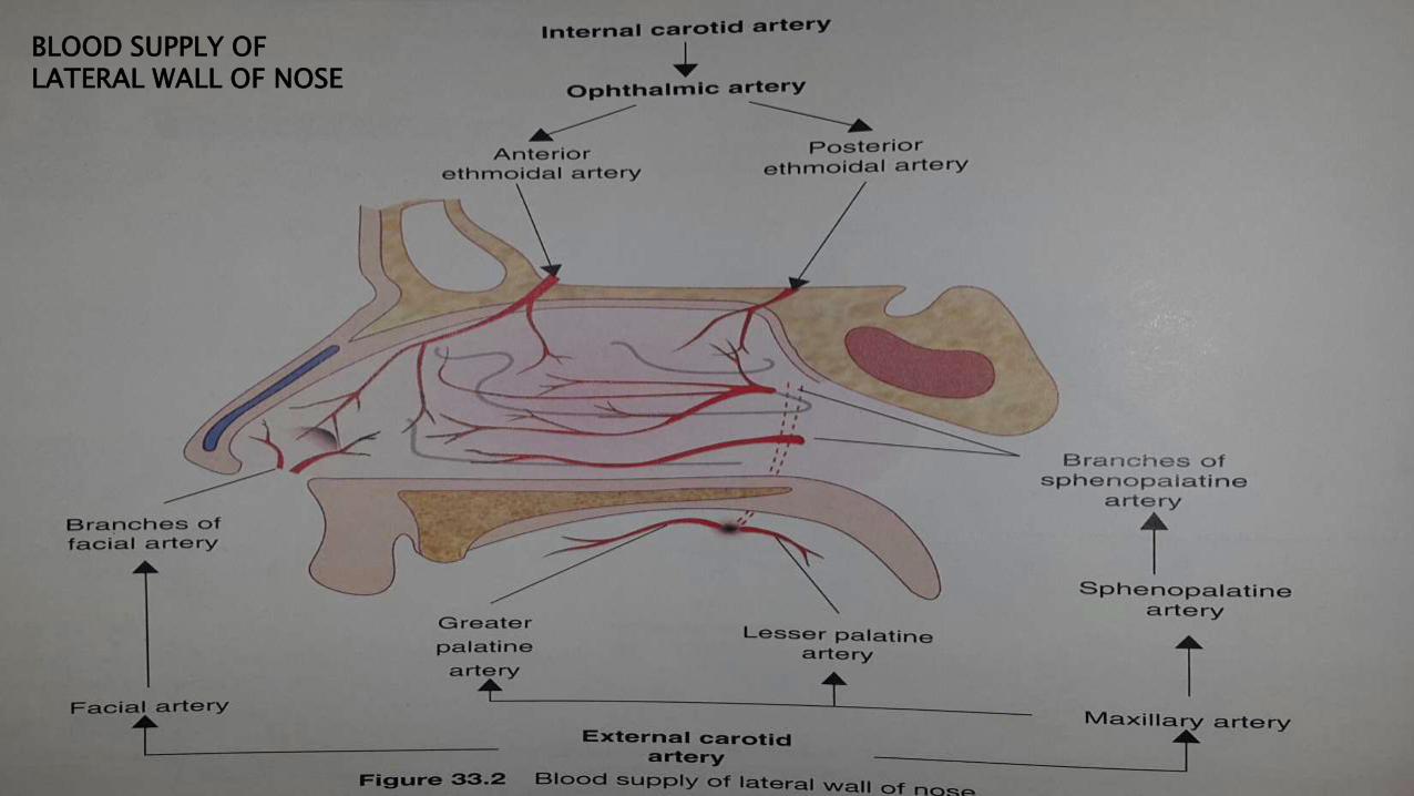
BLOOD SUPPLY OF LATERAL WALL OF NOSE

LITTLE’S AREA OR KIESSELBACH’S PLEXUS:
• It’s a vascular area in the anteroinferior part of septum.
• Anterior ethmoidal, sphenopalatine, greater palatine and septal branch of superior labial arteries & their corresponding veins form anastomosis.
• Commonest site of epistaxis.
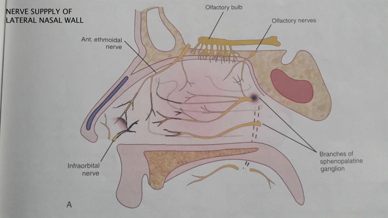
NERVE SUPPPLY OF LATERAL NASAL WALL


PHYSIOLOGY OF NOSE:
Functions of nose are classified as:1)RESPIRATION: • it is the natural pathway for breathing.• It also permits breathing and eating simultaneously.• It allows ventilation of the PNS.• Nasal cycle: nasal mucosa undergoes rhythmic cyclical congestion and decongestion, thus controlling the airflow. It varies 2-4 hrs.
2)AIRCONDITIONING OF INSPIRED AIR:
A)filtration & purification:• Vibrissae acts as filters against larger particles.• Mucus filters finer particles (0.5-3 µm) like dust ,pollen ,bacteria.B)temperature control of inspired air:• It is regulated by the large surface of nasal mucosa, mucous membrane is highly
vascular with cavernous spaces & sinusoids which controls the blood flow,thusacts a efficient ‘radiator’.
• As a result temp of inspired air is brought to that of body’s normal temperature.C)humidification:• Relative humidity of atmospheric air varies depending on climatic conditions.• Nasal mucous membrane adjusts the relative humidity of inspired air to 75% or
more.

3) PROTECTION OF LOWER AIRWAY
A) Mucociliary mechanism: nasal mucosa is rich in goblet cells,secretory glands which produce ‘mucus blanket’
It consists of superficial mucus layer & deeper serous layer floating on top of cilia which are constantly beating like a conveyer belt towards nasopharynx carrying FB.B) Enzymes & immunoglobulin:• Mucus contains “muramidase”(lysozyme) which kills bacteria
&viruses.• Also contains IgE,IgA, inteferons.C)sneezing• It is a protective reflex, FB which irritate nasal mucosa are
expelled by sneezing.
4)VOCAL RESONANCE:• Nose forms a resonating chamber for certain consonants in
speech (like M/N/NG)• When nose is blocked speech becomes denasal (i.e M/N/NG are
uttered as B/D/G.
5)NASAL REFLEXES:• Smell of food causes reflex secretion of saliva & gastric juice.• Sneezing reflex.
NASOPHARYNX
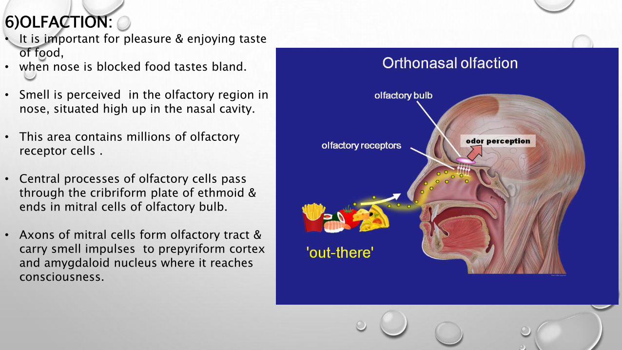
6)OLFACTION:• It is important for pleasure & enjoying taste
of food,• when nose is blocked food tastes bland.
• Smell is perceived in the olfactory region in nose, situated high up in the nasal cavity.
• This area contains millions of olfactory receptor cells .
• Central processes of olfactory cells pass through the cribriform plate of ethmoid & ends in mitral cells of olfactory bulb.
• Axons of mitral cells form olfactory tract & carry smell impulses to prepyriform cortex and amygdaloid nucleus where it reaches consciousness.

ANATOMY OF THROAT :
It is studied under three headings:
1)ANATOMY OF ORAL CAVITY.2)ANATOMY OF PHARYNX. 3)ANATOMY OF LARYNX.
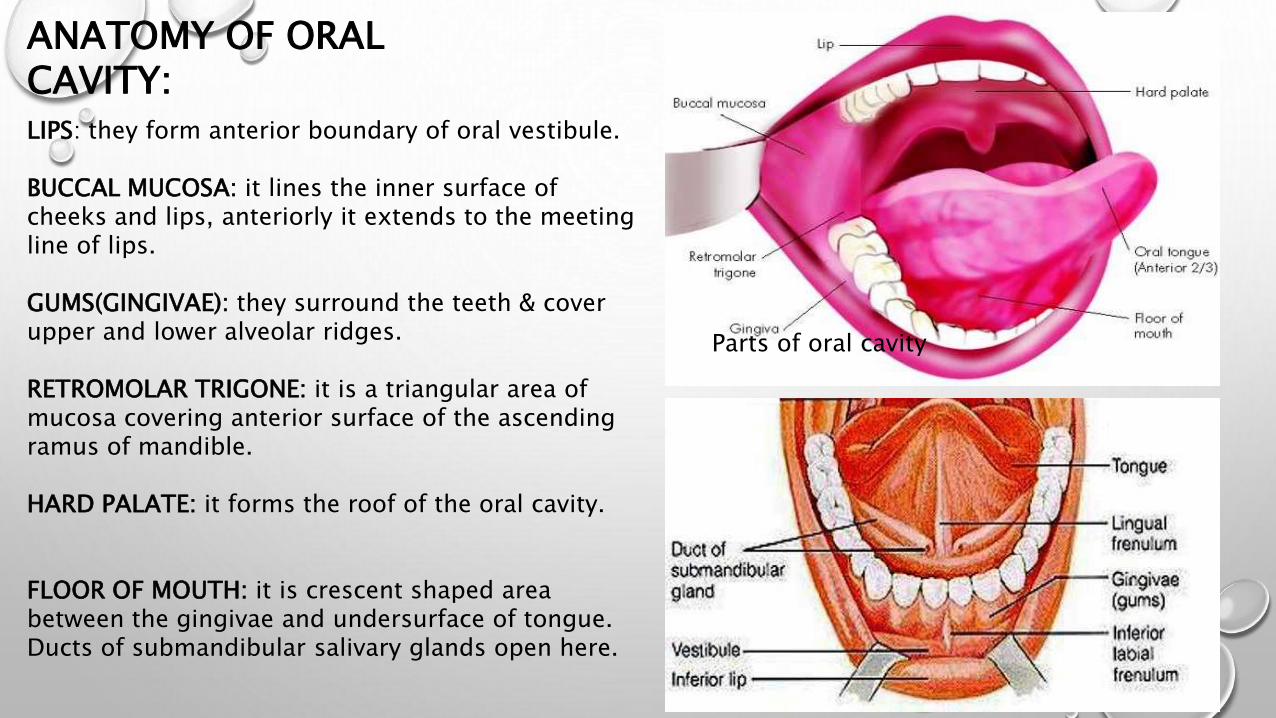
ANATOMY OF ORAL CAVITY:LIPS: they form anterior boundary of oral vestibule.
BUCCAL MUCOSA: it lines the inner surface of cheeks and lips, anteriorly it extends to the meeting line of lips.
GUMS(GINGIVAE): they surround the teeth & cover upper and lower alveolar ridges.
RETROMOLAR TRIGONE: it is a triangular area of mucosa covering anterior surface of the ascending ramus of mandible.
HARD PALATE: it forms the roof of the oral cavity.
FLOOR OF MOUTH: it is crescent shaped area between the gingivae and undersurface of tongue.Ducts of submandibular salivary glands open here.
Parts of oral cavity
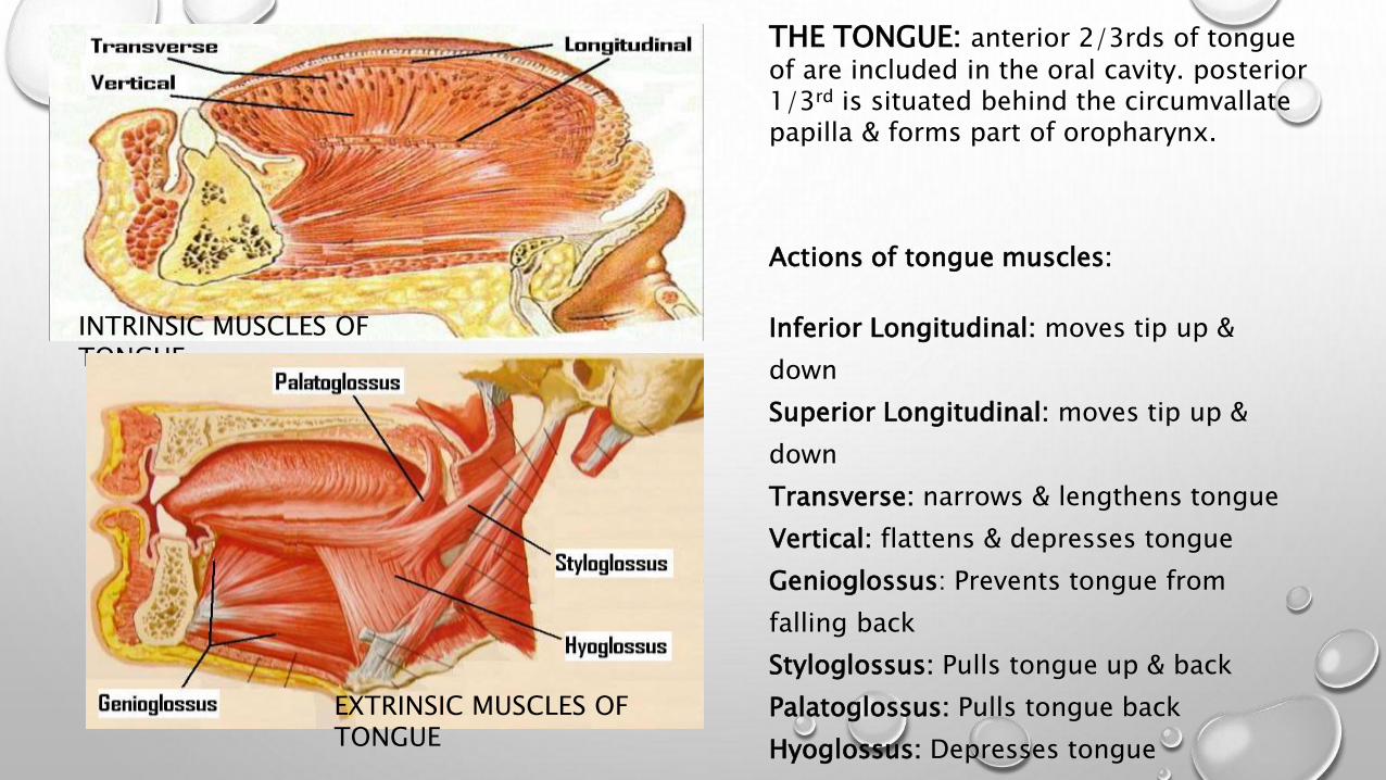
THE TONGUE: anterior 2/3rds of tongue of are included in the oral cavity. posterior 1/3rd is situated behind the circumvallate papilla & forms part of oropharynx.
Actions of tongue muscles:
Inferior Longitudinal: moves tip up &
down
Superior Longitudinal: moves tip up &
down
Transverse: narrows & lengthens tongue
Vertical: flattens & depresses tongue
Genioglossus: Prevents tongue from
falling back
Styloglossus: Pulls tongue up & back
Palatoglossus: Pulls tongue back
Hyoglossus: Depresses tongue
INTRINSIC MUSCLES OF TONGUE
EXTRINSIC MUSCLES OF TONGUE
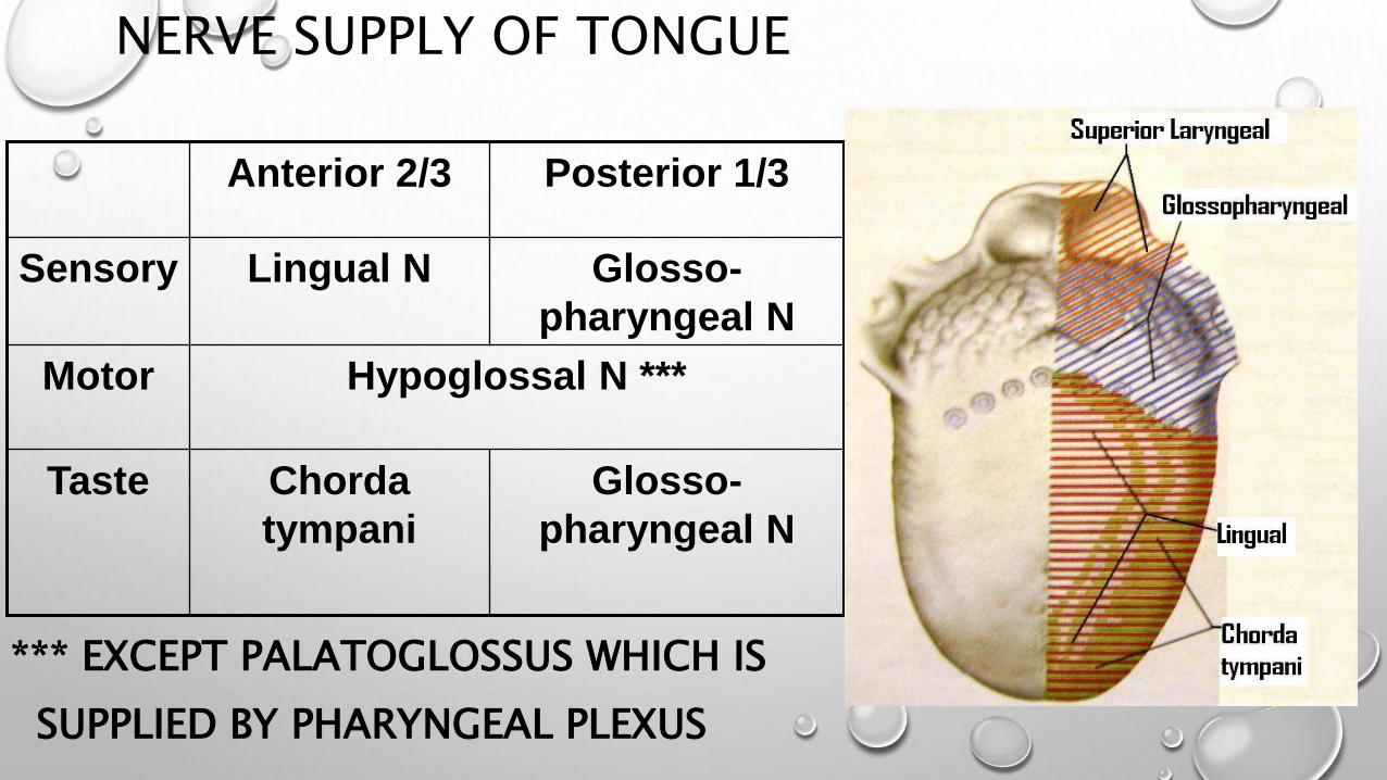
NERVE SUPPLY OF TONGUE
*** EXCEPT PALATOGLOSSUS WHICH IS
SUPPLIED BY PHARYNGEAL PLEXUS
Anterior 2/3 Posterior 1/3
Sensory Lingual N Glosso-
pharyngeal N
Motor Hypoglossal N ***
Taste Chorda
tympani
Glosso-
pharyngeal N

Papillae in tongue
THESE ARE ELEVATED PROJECTIONS
ON THE SURFACE OF TONGUE.
• LINGUAL TASTE BUDS SIT ON
LATERAL BORDERS OF RAISED
PAPILLAE.
Papillae in tongue
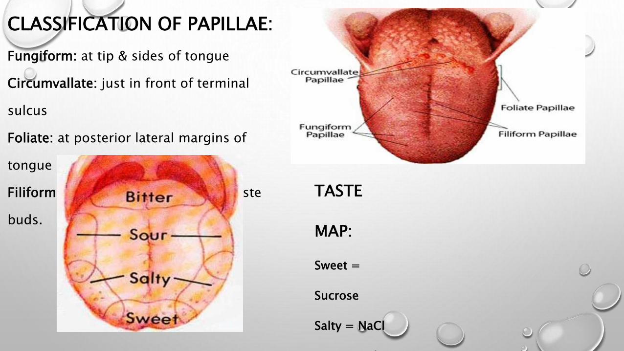
CLASSIFICATION OF PAPILLAE:
Fungiform: at tip & sides of tongue
Circumvallate: just in front of terminal
sulcus
Foliate: at posterior lateral margins of
tongue
Filiform: center of tongue, have no taste
buds.
TASTE
MAP:
Sweet =
Sucrose
Salty = NaCl
Sour = HCl

LYMPHATIC DRAINAGE OF ORAL CAVITY & NECK

ANATOMY OF PHARYNX
• Pharynx is a conical fibromuscular tube forming upper part of the air and food passages.
• It is 12-14cm long extending from base of skull to lower border of cricoid cartilage.
• Width of pharynx 3.5cm at its base but narrows to 1.5cm at pharyngo-esophageal junction.
• STRUCTURE OF PHARYNGEAL WALL:(From within outwards it has 4 layers)
1)MUCOUS MEMBRANE: it is formed by ciliated columnar in the nasopharynx and squamous epithelium elsewhere.2)PHARYNGOBASILAR FASCIA: it is a fibrous layer which lines the muscular coat.3)MUSCULAR COAT: it consists of 2 layers of muscles with 3 muscles in each• A)External layer: containing superior, middle &
inferior constrictors.• B)Internal layer: containing
stylopharyngeus,salpingopharyngeus,palatopharyngeus muscles.
4)BUCCOPHARYNGEAL FASCIA: it covers the outer surface of constrictor muscles.
LAYERS OF PHARYNX

MUSCLES OF PHARYNX

KILLIANS DEHISCENCE: • infr constrictor has two parts :thyropharyngeus &
cricopharyngeus. • The gap between these 2 layers is called killians
dehiscence.• Common site for herniation of pharyngeal mucosa
in cases of pharyngeal pouch.
WALDEYERS RING:• Scattered throughout the sub epithelial layer of
pharynx is the lymphoid tissue which is aggregated at places to form masses collectively called waldeyers ring;
• A)the adenoids.• B)palatine tonsils.• C)lingual tonsil.• D)tubal tonsils.• E)lateral pharyngeal bands• F)nodules in posterior pharyngeal wall.
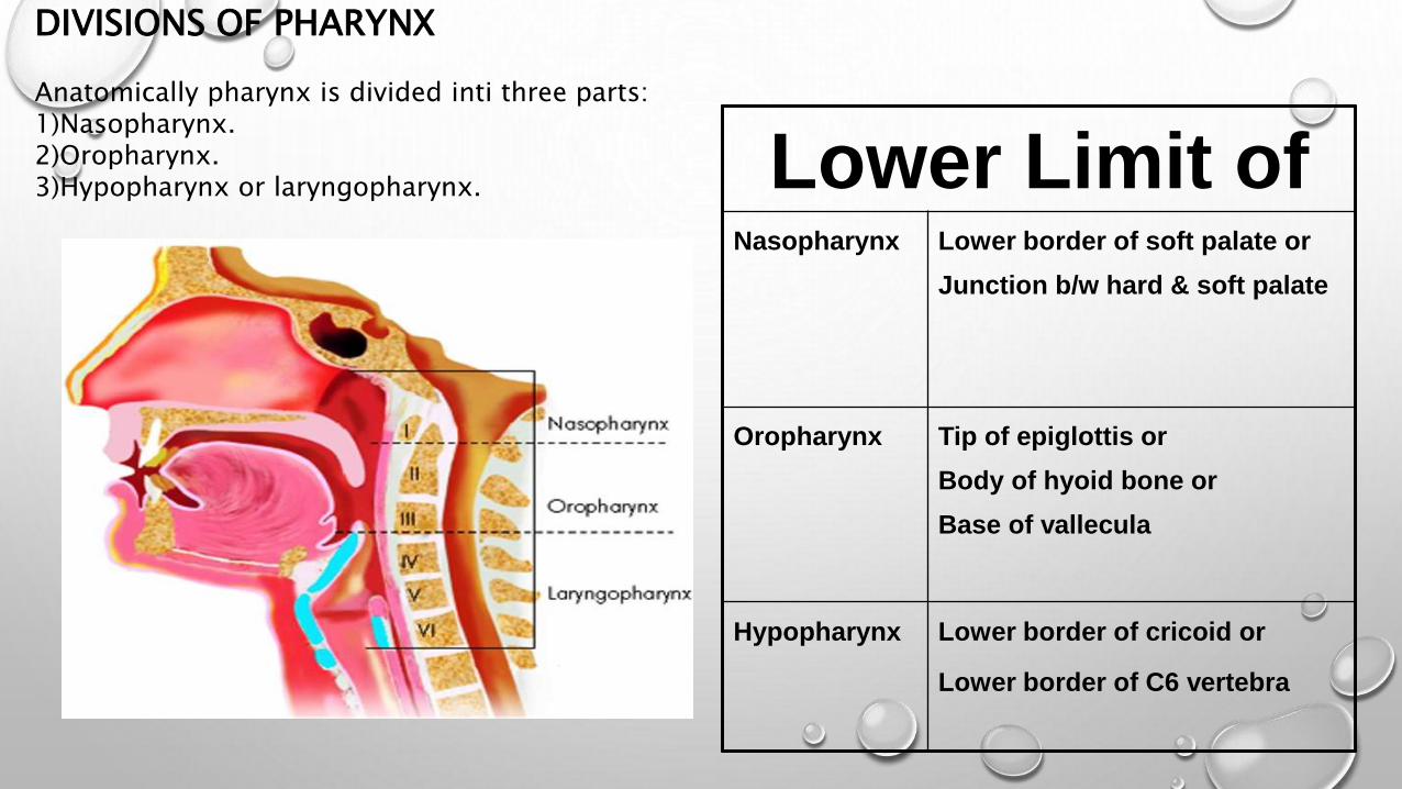
DIVISIONS OF PHARYNX
Anatomically pharynx is divided inti three parts:1)Nasopharynx.2)Oropharynx.3)Hypopharynx or laryngopharynx. Lower Limit of
Nasopharynx Lower border of soft palate or
Junction b/w hard & soft palate
Oropharynx Tip of epiglottis or
Body of hyoid bone or
Base of vallecula
Hypopharynx Lower border of cricoid or
Lower border of C6 vertebra

NASOPHARYNX:• Its the uppermost part of pharynx, extending from the
base of skull to the level of the soft palate.
• ROOF: formed by basisphenoid & basiocciput.
• POSTERIOR WALL: formed by arch of the atlas vertebra covered by prevertebral muscles & fascia.
• FLOOR: formed by soft palate anteriorly & deficient posteriorly to form nasopharyngeal isthmus.
• ANTERIOR WALL: formed by choana, separated from each other by nasal septum.
• LATERAL WALL: each lateral wall presents the pharyngeal opening of Eustachian tube.
• Above and behind the elevation formed by Eustachian tubal opening, is a recess called FOSSA OF ROSSENMULLER ,which is the commonest site for origin of carcinoma.

NASOPHARYNGEAL ISTHMUS: It separates nasopharynx from oropharynx.• Bounded anteriorly by soft palate
,posteriorly by mucosal ridge on nasopharyngeal wall called PASSAVANT’S ridge (palatopharyngeus).
• Closure of this isthmus prevents nasal regurgitation & nasal intonation.
NASOPHARYNGEAL TONSIL(ADENOIDS): • It is a sub epithelial collection of lymphoid
tissue at the junction of roof and postrwall of nasopharynx.
• It increases in size up to age of 6 yearsand then gradually atrophies.
EPITHELIAL LINING:• Lined by pseudostratified ciliated
columnar epithelium.

FUNCTIONS OF NASOPHARYNX Acts as passage for air which has been warmed & humidified in the nose into
larynx & trachea.
Through Eustachian tube it ventilates the middle ear and equalizes air pressure on both side of TM. This function is important for hearing.
Elevation of soft palate against posterior pharyngeal wall helps to cut off nasopharynx from oropharynx. This function is important during swallowing,vomiting,gagging&speech.
Acts as a resonating chamber during voice production.
Acts as a drainage channel for the mucus secreted by nasal & nasopharyngeal glands.
LYMPHATICS: Drain into upper deep cervical nodes either directly or indirectly through
retropharyngeal and parapharyngeal lymph nodes.

OROPHARYNX
It extends from the plane of hard palate above to the plane of hyoid bone below.
BOUNDARIES:1)Posterior wall: • it lies opposite to 2nd & upper part of 3rd cervical
vertebrae.2)Anterior wall:• formed by base of tongue,postr to circumvallate
papillae.• Lingual tonsils one on either side, situated in the
base of tongue.• Valleculae: they are cup shaped depressions lying
between the base of tongue & anterior surface of epiglottis.
3)Lateral wall:• Formed by palatine tonsil.• Anterior pillar(palatoglossal arch).• Posterior fold(palatopharyngeal arch).
LYMPHATICS:• They drain into upper jugular chain particularly
the jugulodigastric node.

OROPHARYNGEAL ISTHMUS
SEPARATES ORAL CAVITY FROM OROPHARYNX
BOUNDARIES ARE:
SUPERIOR: JUNCTION BETWEEN HARD & SOFT PALATE
INFERIOR: CIRCUMVALLATE PAPILLAE
LATERAL: ANTERIOR TONSILLAR PILLARS
(PALATOGLOSSUS)

FUNCTIONS OF OROPHARYNX:
1) As a chamber for passage of food and air.
2) Helps in pharyngeal phase of deglutition.
3) Forms part of vocal tract for certain speech sounds.
4) Helps in appreciation of taste.
5) Provides local defence & immunity against harmful intruders into air and food passages. (waldeyers ring: they are strategically placed at the portals of air and food entry, pathogens which happen to enter these lymphoid masses are dealt by IgM & IgG antibodies).
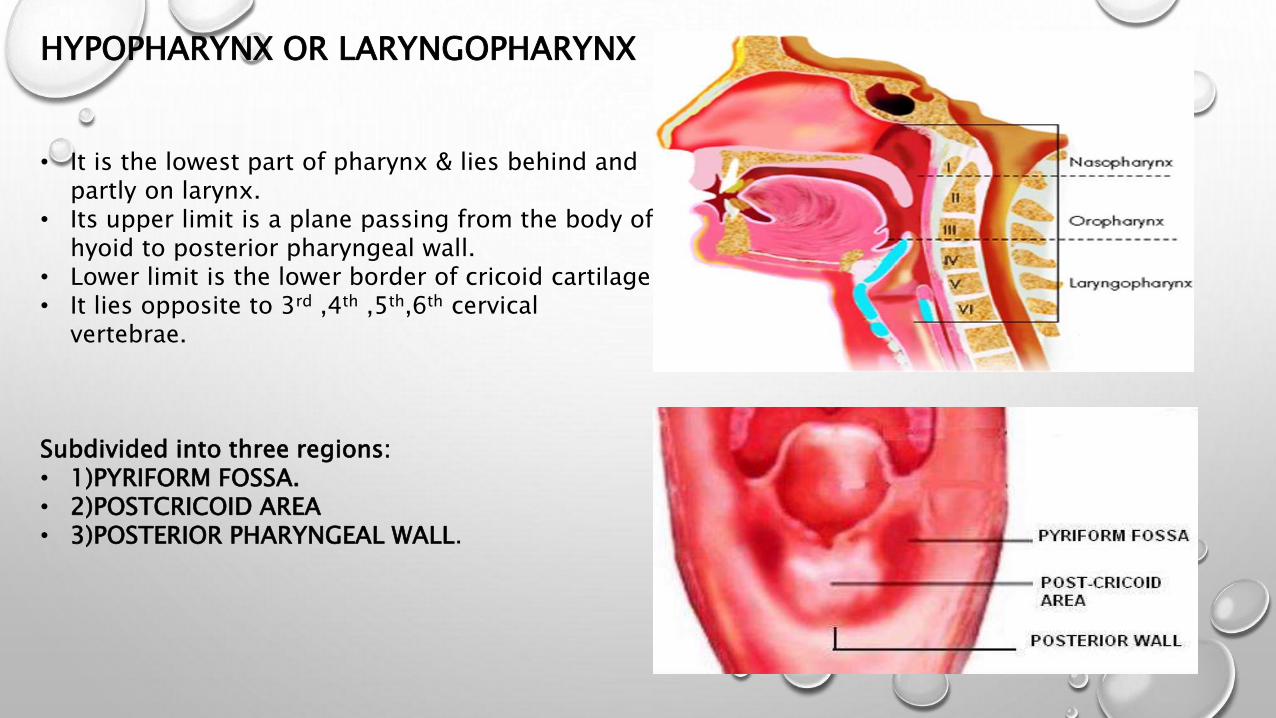
• It is the lowest part of pharynx & lies behind and partly on larynx.
• Its upper limit is a plane passing from the body of hyoid to posterior pharyngeal wall.
• Lower limit is the lower border of cricoid cartilage.• It lies opposite to 3rd ,4th ,5th,6th cervical
vertebrae.
Subdivided into three regions:• 1)PYRIFORM FOSSA.• 2)POSTCRICOID AREA• 3)POSTERIOR PHARYNGEAL WALL.
HYPOPHARYNX OR LARYNGOPHARYNX

HYPOPHARYNX OR LARYNGOPHARYNX:
1)PYRIFORM FOSSA:• it lies on either side of larynx & extends from
pharyngoepiglottic fold to upper end of oesophagus.
• It forms the lateral channel of food.
2)POSTCRICOID REGION:• It is the part of the anterior wall of
laryngopharynx b/n upper & lower borders of cricoid lamina.
3)POSTERIOR PHARYNGEAL WALL:• Extends from the level of hyoid bone to the
level of cricoarytenoid joint.
LYMPHATIC DRAINAGE: Pyriform sinus drains into upper jugular chain. Lymphatic from posterior pharyngeal wall & post
cricoid region drain into lateral pharyngeal or parapharyngeal nodes and then to deep cervical lymph nodes.
FUNCTIONS OF LARYNGOPHARYNX:
It is a common pathway for food and air. Provides a vocal tract for resonance of certain speech
sounds helps in deglutition,i.e cricopharyngeal sphincter must
relax when pharyngeal muscles are contracting.

ANATOMY OF LARYNX
The larynx lies in front of hypopharynx opposite the 3rd to 6th cervical vertebrae.
THE LARYNX IS DIVIDED ANATOMICALLY IN TO :
SUPRA GLOTTIS .
GLOTTIS
SUB GLOTTIS .
BY THE FALLS AND
TRUE VOCAL CORDS.

THE SUPRAGLOTTIS CONSISTS OF
SUPERIORLY THE
EPIGLOTTIS AND ARYEPIGLOTTIC FOLDS AS
THEY SWEEP DOWN TO THE ARYTENOIDS.
ITS LOWER BORDER IS THE VENTRICULAR
BANDS (FALSE CORDS) WHICH FORM THE
UPPER BORDER OF THE GLOTTIS .
THE GLOTTIS INCLUDES THE VOCAL CORDS
AND ANTERIOR COMMISSURE AND
POSTERIOR COMMISSURE.
THE SUB GLOTTIS BECOMES THE TRACHEA
AT THE LOWER BORDER OF THE CRICOID .(
BETWEEN TRUE V.C . AND LOWER BORDER
OF THE CRICOID ) .

THE FRAMEWORK OF THE LARYNXCONSISTS OF :
• HYOID BONE
• NUMBER OF CARTILAGES
(THYROID,CRICOID,ARYTENOID,CORNICULATE,CUNEIFORM)
• CONNECTED BY LIGAMENTS, MEMBRANES AND INTRINSIC
AND EXTRINSIC MUSCLES TO GIVE IT STABILITY.
• LINED WITH A MUCOUS MEMBRANE
THAT IS CONTINUOUS ABOVE WITH THE PHARYNX
AND BELOW WITH THAT OF THE TRACHEA .

CARTILAGINOUS SKELETON OF LARYNX
9 DIFFERENT CARTILAGES ARE PRESENT IN THE LARYNX .
• UNPAIRED CARTILAGES:
(THYROID, CRICOID , EPIGLOTTIS)
• PAIRED CARTILAGES:
(ARYTENOID , CORNICULATE ,CUNEIFORM)

1)THYROID CARTILAGE: Shield like. Largest of the laryngeal cartilages. Has two laminae meet in the midline inferiorly. The angle of fusion between the laminae is about 90 degree in men and 120 degrees in women. The fused anterior borders in men form a
projection, which can be easily palpated known as Adams apple.
The laminae diverge posteriorly. The posterior border of the two laminae are prolonged as two slender processes known as the superior and inferior cornua.
2)CRICOID CARTILAGE: The only cartilage forming a complete ring. Shaped like a signet ring. Composed of a deep broad quadrilateral
lamina posteriorly and a narrow arch anteriorly.
The lamina of the cricoid cartilage has articular facets for arytenoid cartilage .
Anterior view
Posterior view

3)EPIGLOTTIS:• Leaf shaped yellow elastic cartilage .• Projects upwards behind the tongue and the body of
the hyoid bone.• Superior margin is free. • The sides of the epiglottis is attached to the arytenoid
cartilages by aryepiglottic folds.• Attached to body of hyoid bone by hyoepiglottic
ligament. • Stalk like process called petiole
petiole
In neonates and infants the epiglottis is omega shaped.
This long, deeply grooved, floppy epiglottis protects the
nasotracheal air passage during sucking.
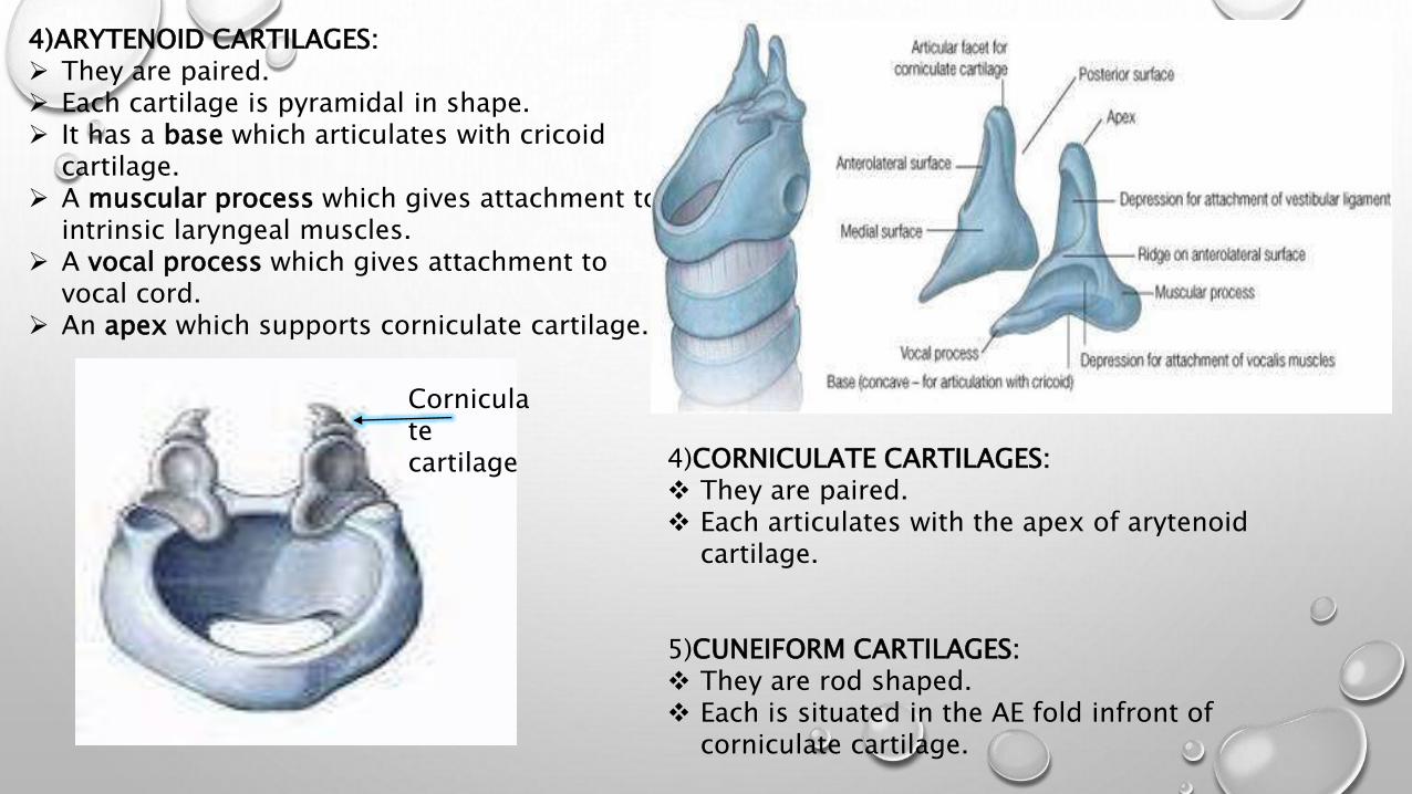
4)ARYTENOID CARTILAGES: They are paired. Each cartilage is pyramidal in shape. It has a base which articulates with cricoid
cartilage. A muscular process which gives attachment to
intrinsic laryngeal muscles. A vocal process which gives attachment to
vocal cord. An apex which supports corniculate cartilage.
4)CORNICULATE CARTILAGES: They are paired. Each articulates with the apex of arytenoid
cartilage.
5)CUNEIFORM CARTILAGES: They are rod shaped. Each is situated in the AE fold infront of
corniculate cartilage.
Corniculate cartilage
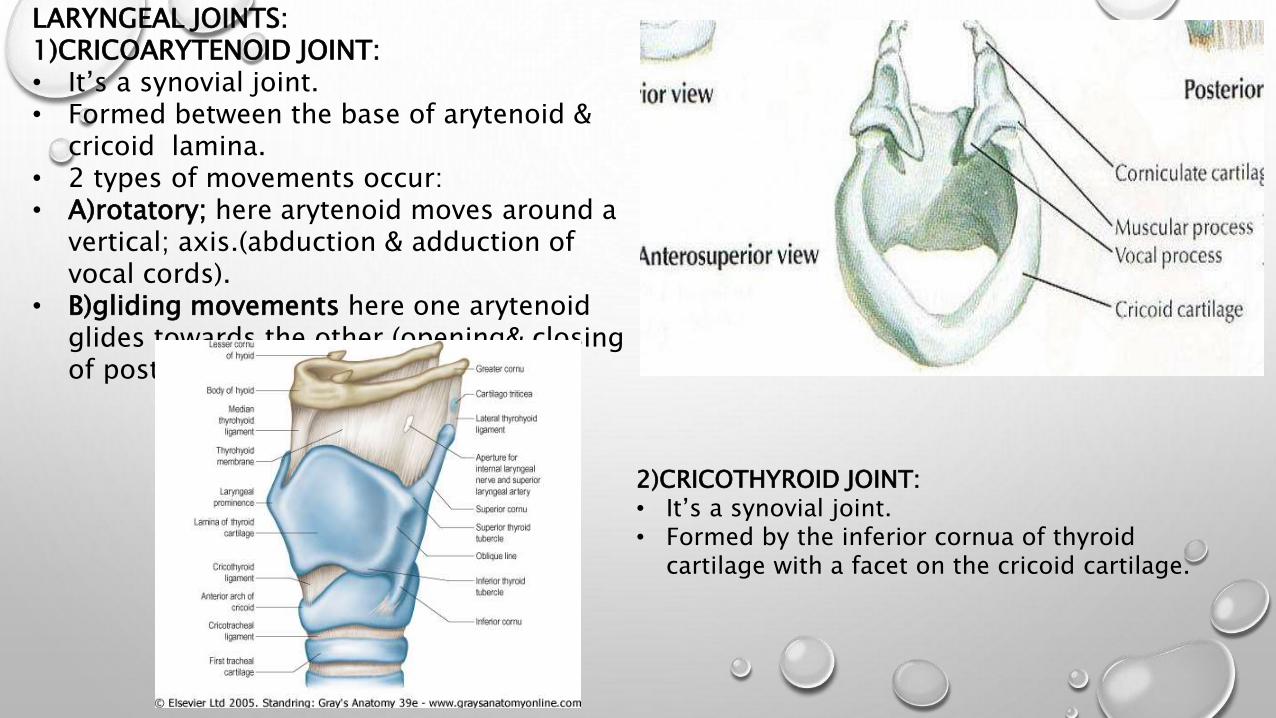
LARYNGEAL JOINTS:1)CRICOARYTENOID JOINT:• It’s a synovial joint.• Formed between the base of arytenoid &
cricoid lamina.• 2 types of movements occur:• A)rotatory; here arytenoid moves around a
vertical; axis.(abduction & adduction of vocal cords).
• B)gliding movements here one arytenoid glides towards the other.(opening& closing of postr part of glottis)
2)CRICOTHYROID JOINT:• It’s a synovial joint.• Formed by the inferior cornua of thyroid
cartilage with a facet on the cricoid cartilage.
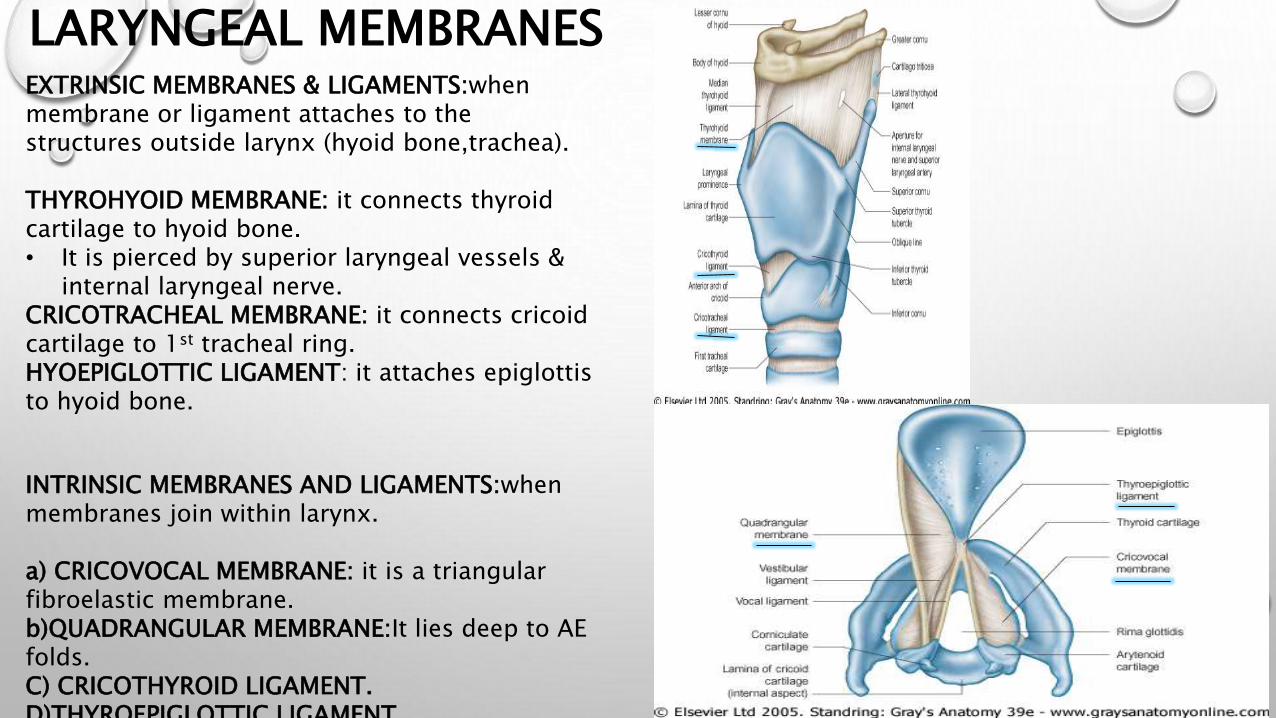
LARYNGEAL MEMBRANESEXTRINSIC MEMBRANES & LIGAMENTS:whenmembrane or ligament attaches to the structures outside larynx (hyoid bone,trachea).
THYROHYOID MEMBRANE: it connects thyroid cartilage to hyoid bone.• It is pierced by superior laryngeal vessels &
internal laryngeal nerve.CRICOTRACHEAL MEMBRANE: it connects cricoid cartilage to 1st tracheal ring.HYOEPIGLOTTIC LIGAMENT: it attaches epiglottis to hyoid bone.
INTRINSIC MEMBRANES AND LIGAMENTS:whenmembranes join within larynx.
a) CRICOVOCAL MEMBRANE: it is a triangular fibroelastic membrane.b)QUADRANGULAR MEMBRANE:It lies deep to AE folds.C) CRICOTHYROID LIGAMENT.D)THYROEPIGLOTTIC LIGAMENT
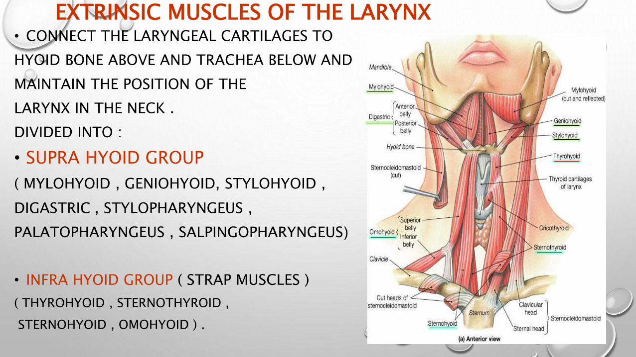
EXTRINSIC MUSCLES OF THE LARYNX• CONNECT THE LARYNGEAL CARTILAGES TO
HYOID BONE ABOVE AND TRACHEA BELOW AND
MAINTAIN THE POSITION OF THE
LARYNX IN THE NECK .
DIVIDED INTO :
• SUPRA HYOID GROUP
( MYLOHYOID , GENIOHYOID, STYLOHYOID ,
DIGASTRIC , STYLOPHARYNGEUS ,
PALATOPHARYNGEUS , SALPINGOPHARYNGEUS(
• INFRA HYOID GROUP ( STRAP MUSCLES )
( THYROHYOID , STERNOTHYROID ,
STERNOHYOID , OMOHYOID ) .
INTRINSIC MUSCLES OF LARYNX• THE INTRINSIC MUSCLES ARE ALL PAIRED AND MOVE THE
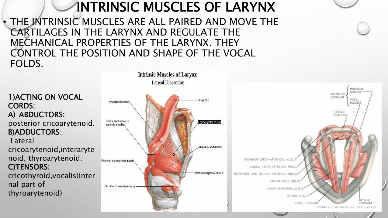
CARTILAGES IN THE LARYNX AND REGULATE THE MECHANICAL PROPERTIES OF THE LARYNX. THEY CONTROL THE POSITION AND SHAPE OF THE VOCAL FOLDS.
1)ACTING ON VOCAL CORDS:A) ABDUCTORS: posterior cricoarytenoid.B)ADDUCTORS:Lateral
cricoarytenoid,interarytenoid, thyroarytenoid.C)TENSORS:cricothyroid,vocalis(internal part of thyroarytenoid)
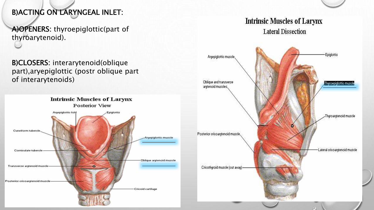
B)ACTING ON LARYNGEAL INLET:
A)OPENERS: thyroepiglottic(part of thyroarytenoid).
B)CLOSERS: interarytenoid(oblique part),aryepiglottic (postr oblique part of interarytenoids)

• INTERIOR OF LARYNX :
THE LARYNGEAL CAVITY EXTENDS FROM THE LEVEL OF 3RD CERVICAL
VERTEBRA TO THE LOWER BORDER OF THE CRICOID CARTILAGE (C6) LEVEL.
AT THE LEVEL OF CRICOID CARTILAGE IT BECOMES CONTINUOUS WITH
THAT OF THE TRACHEA.
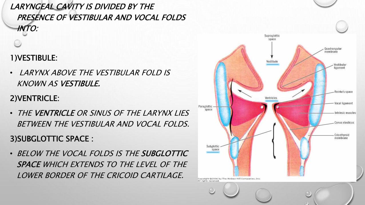
LARYNGEAL CAVITY IS DIVIDED BY THE
PRESENCE OF VESTIBULAR AND VOCAL FOLDS
INTO:
1)VESTIBULE:
• LARYNX ABOVE THE VESTIBULAR FOLD IS
KNOWN AS VESTIBULE.
2)VENTRICLE:
• THE VENTRICLE OR SINUS OF THE LARYNX LIES
BETWEEN THE VESTIBULAR AND VOCAL FOLDS.
3)SUBGLOTTIC SPACE :
• BELOW THE VOCAL FOLDS IS THE SUBGLOTTIC
SPACE WHICH EXTENDS TO THE LEVEL OF THE
LOWER BORDER OF THE CRICOID CARTILAGE.

• FALSE VOCAL CORD ( THE VENTRICULAR BANDS): WHICH ARE FORMED BY THE
MUCOUS MEMBRANE COVERING THE VENTRICULAR LIGAMENT AND THE UPPER PART OF
THE EXTERNAL PORTION OF THE THYROARYTENOID MUSCLE.
• TRUE VOCAL CORDS :2 PEARLY WHITE BANDS EXTENDING FROM THE MIDDLE OF
THYROID ANGLE TO THE VOCAL PROCESSES OF ARYTENOIDS. EACH VOCAL LIGAMENT IS
THE UPPER EDGE OF CRICOVOCAL MEMBRANE COVERED BY CLOSELY BOUND MUCOUS
MEMBRANE.THE BLOOD SUPPLY IS POOR, HENCE THE PEARLY WHITE APPEARANCE OF THE
VOCAL CORDS.
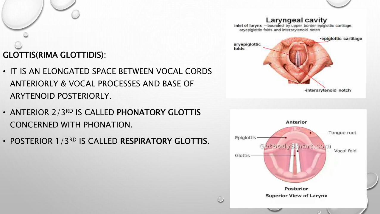
GLOTTIS(RIMA GLOTTIDIS):
• IT IS AN ELONGATED SPACE BETWEEN VOCAL CORDS
ANTERIORLY & VOCAL PROCESSES AND BASE OF
ARYTENOID POSTERIORLY.
• ANTERIOR 2/3RD IS CALLED PHONATORY GLOTTIS
CONCERNED WITH PHONATION.
• POSTERIOR 1/3RD IS CALLED RESPIRATORY GLOTTIS.
MUCOUS MEMBRANES OF THE LARYNX:• Lines the larynx except over the posterior surface of epiglottis
,true vocal cords & corniculate cartilage.• Epithelium is ciliated columnar type except over the vocal
cords & upper part of vestibule where it is stratified squamous type.
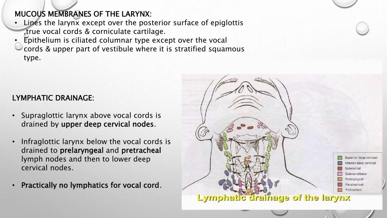
LYMPHATIC DRAINAGE:
• Supraglottic larynx above vocal cords is drained by upper deep cervical nodes.
• Infraglottic larynx below the vocal cords is drained to prelaryngeal and pretracheallymph nodes and then to lower deep cervical nodes.
• Practically no lymphatics for vocal cord.

BLOOD SUPPLY OF THE LARYNX : IS DERIVED FROM THE
• LARYNGEAL BRANCHES OF THE SUPERIOR THYROID ARTERY . .
• LARYNGEAL BRANCHES OF THE INFERIOR THYROID ARTERY .
• THE CRICOTHYROID BRANCH OF THE SUPERIOR THYROID
ARTERY.
THE VEINS LEAVING THE LARYNX ACCOMPANY THE ARTERIES;
• THE SUPERIOR VESSELS DRAIN TO THE INTERNAL JUGULAR VEIN
BY
WAY OF THE SUPERIOR THYROID OR FACIAL VEINS ,
• THE INFERIOR VESSELS DRAIN BY WAY OF INFERIOR THYROID
VEIN
INTO THE BRACHIOCEPHALIC VEINS.
• SOME VENOUS DRAINAGE ALSO OCCUR
THROUGH THE MIDDLE THYROID VEIN INTO THE INTERNAL
JUGULAR VEIN.

NERVE SUPPLY OF THE LARYNX :
THE LARYNX IS SUPPLIED BY BRANCHES OF VAGUS NERVE .
• SUPERIOR LARYNGEAL NERVE HAS TWO LARYNGEAL BRANCHES :
INTERNAL BRANCH . ENTIRELY SENSORY . IT PIERCES THE THYROHYOID MEMBRANE WITH THE
SUPERIOR LARYNGEAL ARTERY AND VEIN . IT SUPPLIES THE CAVITY OF THE LARYNX AS FAR DOWN
THE LEVEL OF THE VOCAL CORDS .
EXTERNAL BRANCH . TRAVELS DOWN ON THE INFERIOR CONSTRICTOR MUSCLE OF THE PHARYNX .
IT SUPPLIES THE CRICOTHYROID MUSCLE AND PART OF THE ANT. SUBGLOTTIS .

RECURRENT LARYNGEAL BRANCH OF
THE VAGUS NERVE (CN X) :
IT HAS MUCH LONGER COURSE ON THE LEFT SIDE THAN
ON THE RIGHT SIDE .
ON THE LT. SIDE IT TURNS ROUND THE ARCH OF
AORTA .
ON THE RT. SIDE IT TURNS ROUND THE SUBCLAVIAN
ARTERY.
IN THE NECK IT LIES BETWEEN THE TRACHEA AND
ESOPHAGUS AS IT APPROACHES THE LARYNX .
IT IS DIVIDED IN TO :
• AN ANTERO LATERAL ( MOTOR BRANCH ) :
WHICH SUPPLY ALL THE INTRINSIC MUSCLES OF THE
LARYNX EXCEPT THE CRICOTHYROID M.
• POSTERO MEDIAL ( SENSORY BRANCH) : WHICH
SUPPLIES THE CAVITY OF THE LARYNX BELOW THE
LEVEL OF VOCAL CORDS .

PHYSIOLOGY OF LARYNX:
1)PROTECTION OF LOWER AIRWAYS:it protects the lower air passage in three different ways;a)Sphincteric closure of laryngeal opening.(closure of laryngeal inlet,falsecords,true cords on swallowing of food).b)Temporary cessation of respiration.(reflex generated by afferent fibres of IX thnerve when food touches posterior pharyngeal wall).c)Cough reflex(important mechanism to dislodge & expel a foreign particle when it comes in contact with respiratory mucosa).
2)PHONATION: voice is produced by the following mechanism(AERODYNAMIC MYOELASTIC THEORY OF VOICE PRODUCTION) Vocal cords are kept adducted. Infraglottic air pressure is generated by the exhaled air from the lungs. The air force opens the cord & is released as small puffs which vibrate the
vocal cords to produce sound. The sound is converted into speech by the modulatory action of
lips,tongue,palate,pharynx & teeth, Intensity depends on the air pressure produced by lungs. Pitch depends on the frequency with which vocal cords vibrate.

3)RESPIRATION:• Larynx regulates the flow of air into the lungs.• Vocal cords abduct during inspiration and
adduct during expiration.
• 4)FIXATION OF CHEST:• when larynx is closed,chest wall gets fixed &
various thoracic & abdominal muscles can act best.
• This function is important in digging,pulling,climbing,coughing,vomiting.

ASSESMENT OF HEARING IN CHIDREN:
SCREENING FOR HEARING: Hearing impairment can have a major impact on a child’s development, early identification
improves prognosis.
Data from the Colorado newborn screening program suggest that if hearing-impaired infants are identified and treated by age 6 months, these children should develop the same level of language as their age-matched peers who are not hearing impaired.
The recommended hearing screening techniques are either otoacoustic emissions (OAE) testing or auditory brainstem evoked responses (ABRs).
IDENTIFICATION OF HEARING IMPAIRMENT:
• The impact of hearing impairment is greatest on an infant who has yet to develop language;
• consequently, identification, diagnosisand treatment should begin as soon as possible.
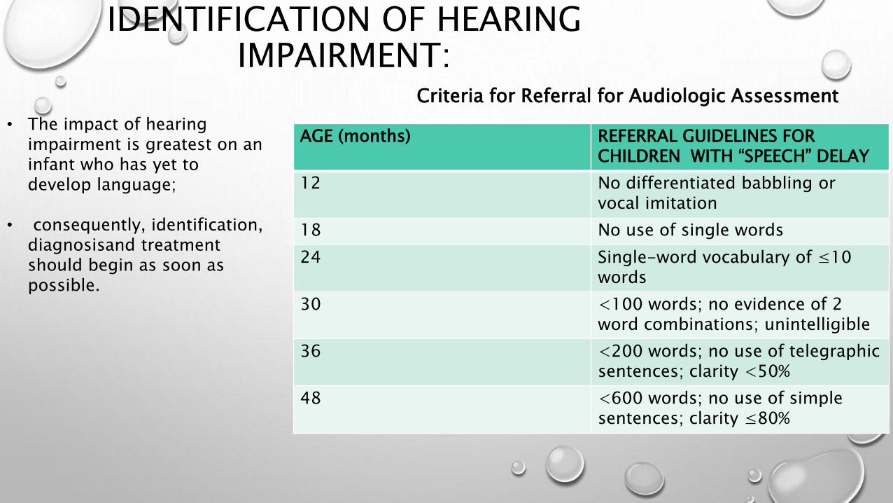
Criteria for Referral for Audiologic Assessment
AGE (months) REFERRAL GUIDELINES FOR CHILDREN WITH “SPEECH” DELAY
12 No differentiated babbling or vocal imitation
18 No use of single words
24 Single-word vocabulary of ≤10 words
30 <100 words; no evidence of 2 word combinations; unintelligible
36 <200 words; no use of telegraphic sentences; clarity <50%
48 <600 words; no use of simple sentences; clarity ≤80%

GUIDELINES FOR REFERRAL OF CHILDREN WITH SUSPECTED HEARING LOSS
AGE (mo) NORMAL DEVELOPMENT
0-4 Should startle to loud sounds, quiet to mother’s voice, momentarily cease activity when sound is presented at a conversational level
5-6 Should correctly localize to sound presented in a horizontal plane, begin to imitate sounds in own speech repertoire or at least reciprocally vocalize with an adult
7-12 Should correctly localize to sound presented in any plane Should respond to name, even when spoken quietly
13-15 Should point toward an unexpected sound or to familiar objects or persons when asked
16-18 Should follow simple directions without gestural or other visual cues; can be trained to reach toward an interesting toy at midline when a sound is presented
19-24 Should point to body parts when asked; by 21-24 mo, can be trained to perform play audiometry

0-6 months of age Behavioural observation audiometry (BOA) unconditioned, reflexive responses to
complex (not frequency-specific) test sounds such as noise, speech, or music presented from a loudspeaker or uncalibrated noisemakers.
Does not give indication of threshold. Can rule out severe and profound losses
only. Reflexive response can be inhibited,
particularly with repeated presentation of stimulus.
Tests of preference to assess hearing for this age group are therefore objective tests.
Responses to sound in neonates and young infants
Eye blink (cochleopalpebral reflex)Gross motor responseStilling Startle Eye movement/widening Crying Grimacing (Heart rate changes/breathing changes)
ASSESMENT OF HEARING
Objective Test Methods
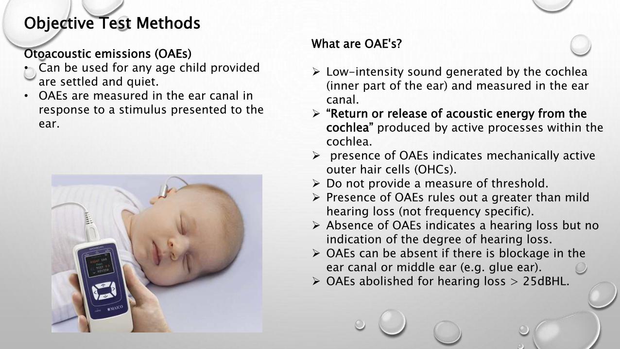
Otoacoustic emissions (OAEs) • Can be used for any age child provided
are settled and quiet. • OAEs are measured in the ear canal in
response to a stimulus presented to the ear.
What are OAE's?
Low-intensity sound generated by the cochlea (inner part of the ear) and measured in the ear canal.
“Return or release of acoustic energy from the cochlea” produced by active processes within the cochlea.
presence of OAEs indicates mechanically active outer hair cells (OHCs).
Do not provide a measure of threshold. Presence of OAEs rules out a greater than mild
hearing loss (not frequency specific). Absence of OAEs indicates a hearing loss but no
indication of the degree of hearing loss. OAEs can be absent if there is blockage in the
ear canal or middle ear (e.g. glue ear). OAEs abolished for hearing loss > 25dBHL.
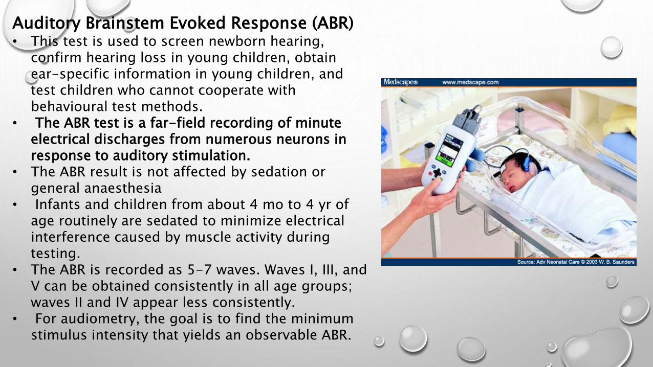
Auditory Brainstem Evoked Response (ABR)• This test is used to screen newborn hearing,
confirm hearing loss in young children, obtain ear-specific information in young children, and test children who cannot cooperate with behavioural test methods.
• The ABR test is a far-field recording of minute electrical discharges from numerous neurons in response to auditory stimulation.
• The ABR result is not affected by sedation or general anaesthesia
• Infants and children from about 4 mo to 4 yr of age routinely are sedated to minimize electrical interference caused by muscle activity during testing.
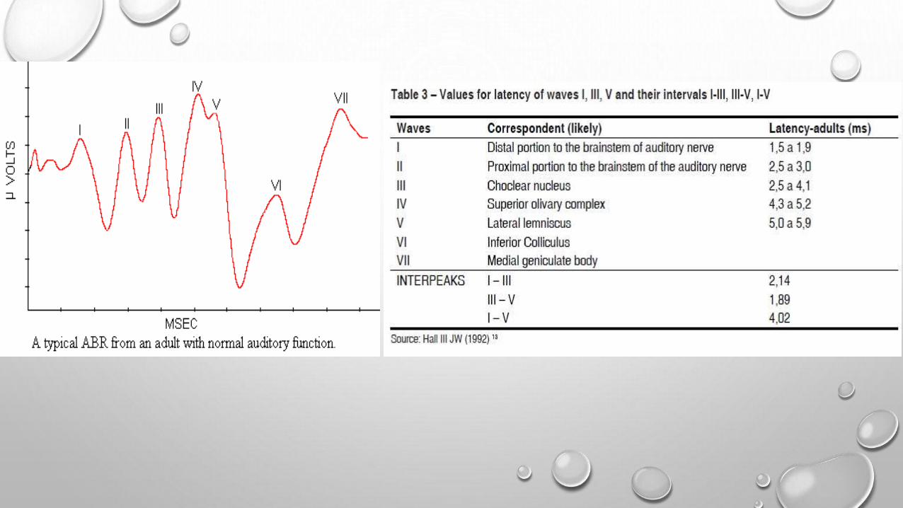
• The ABR is recorded as 5-7 waves. Waves I, III, and V can be obtained consistently in all age groups; waves II and IV appear less consistently.
• For audiometry, the goal is to find the minimum stimulus intensity that yields an observable ABR.
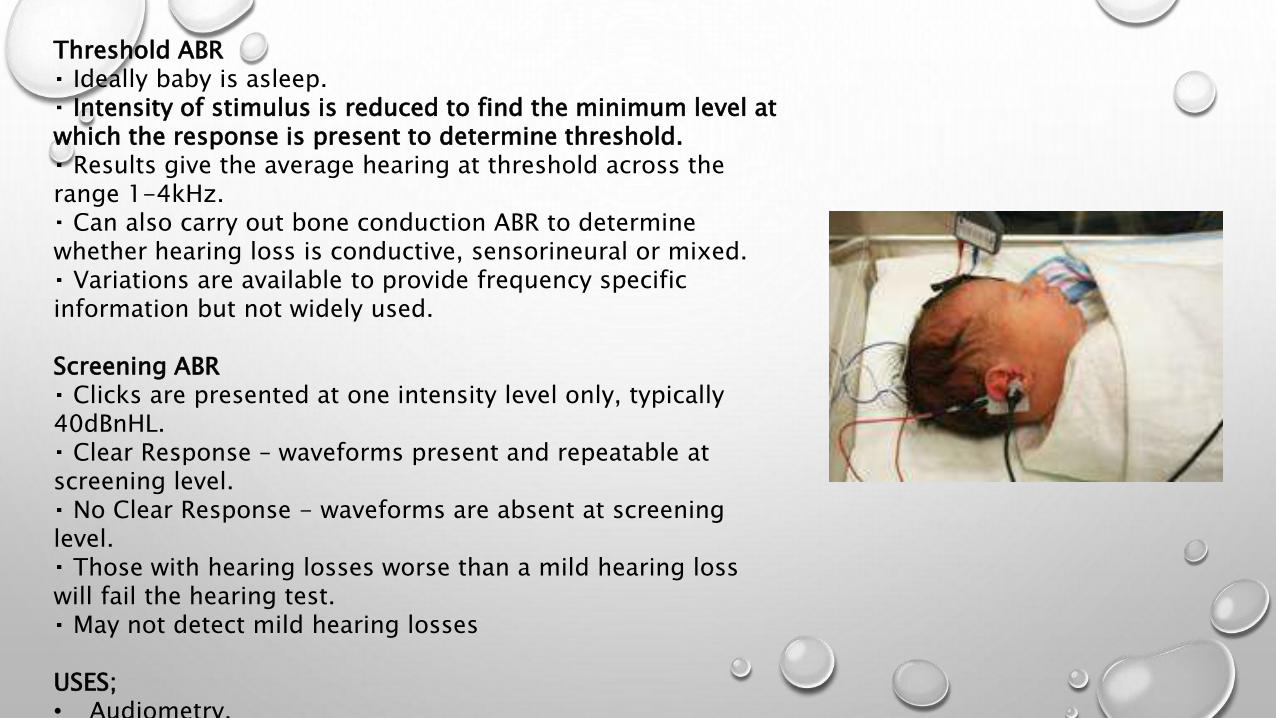
Threshold ABR Ideally baby is asleep. Intensity of stimulus is reduced to find the minimum level at
which the response is present to determine threshold. Results give the average hearing at threshold across the
range 1-4kHz. Can also carry out bone conduction ABR to determine
whether hearing loss is conductive, sensorineural or mixed. Variations are available to provide frequency specific
information but not widely used.
Screening ABR Clicks are presented at one intensity level only, typically
40dBnHL. Clear Response – waveforms present and repeatable at
screening level. No Clear Response - waveforms are absent at screening
level. Those with hearing losses worse than a mild hearing loss
will fail the hearing test. May not detect mild hearing losses
USES;• Audiometry.
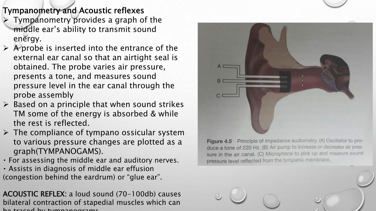
Tympanometry and Acoustic reflexes Tympanometry provides a graph of the
middle ear’s ability to transmit sound energy.
A probe is inserted into the entrance of the external ear canal so that an airtight seal is obtained. The probe varies air pressure, presents a tone, and measures sound pressure level in the ear canal through the probe assembly
Based on a principle that when sound strikes TM some of the energy is absorbed & while the rest is reflected.
The compliance of tympano ossicular system to various pressure changes are plotted as a graph(TYMPANOGAMS).
For assessing the middle ear and auditory nerves. Assists in diagnosis of middle ear effusion
(congestion behind the eardrum) or “glue ear”.
ACOUSTIC REFLEX: a loud sound (70-100db) causes bilateral contraction of stapedial muscles which can be traced by tympanograms.

6-18 months of age
Distraction test Traditional test of choice. Based on the ability of this age group to localise
sounds at and close to ear level. Works on the fact that younger children need social
reinforcement for responding to sounds. In practice difficult above 12m. Requires 2 testers. A ‘distractor’ in front who controls the attention of
the child, the test and assesses whether there has been a response
A ‘presenter’ behind the child presenting the sounds Tests hearing to a range of sounds – frequency
specific warble tones, high frequency rattle, voice. Lots of potential flaws and possibilities for missing
child with hearing loss.

6months –2years of age
Visual Reinforcement Audiometry (VRA)
A conditioned response to sound (usually head turn) ,which is reinforced by a visual reward.
Conditioned by simultaneous presentation of sound and reinforcer (reward).
Sounds typically presented from a loud speaker. Once child is reliably conditioned for the test, only
then can go on and start testing the child’s hearing. Assesses hearing across frequency range, typically
0.5kHz to 4kHz which are important for speech and language access.
Present sound, child turns head, present re-inforceronce child has turned.
Can also be presented using insert earphones or headphones to obtain ear specific information.
Also can do bone conduction VRA (tests the underlying hearing - the cochlea, inner part of the ear).
Tests the hearing in the better hearing ear
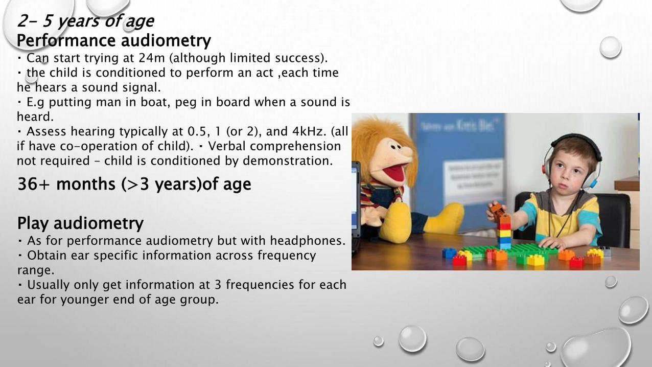
2- 5 years of age Performance audiometry
Can start trying at 24m (although limited success). the child is conditioned to perform an act ,each time
he hears a sound signal.E.g putting man in boat, peg in board when a sound is
heard. Assess hearing typically at 0.5, 1 (or 2), and 4kHz. (all
if have co-operation of child). Verbal comprehension not required – child is conditioned by demonstration.
36+ months (>3 years)of age
Play audiometry As for performance audiometry but with headphones. Obtain ear specific information across frequency
range. Usually only get information at 3 frequencies for each
ear for younger end of age group.
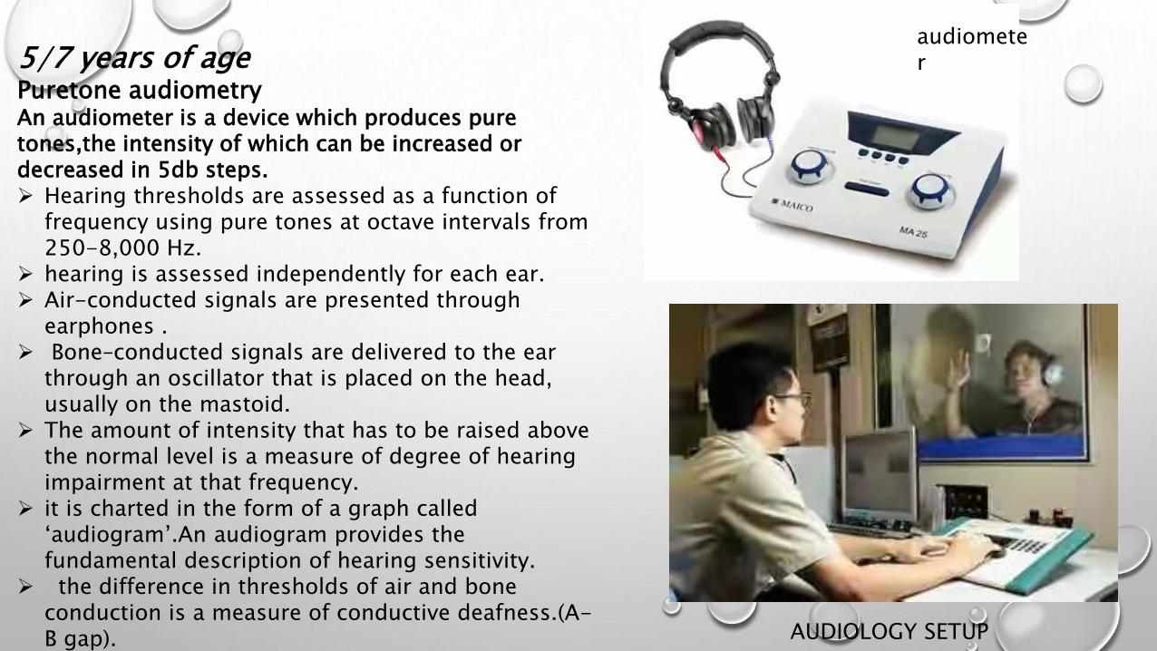
5/7 years of age Puretone audiometry An audiometer is a device which produces pure tones,the intensity of which can be increased or decreased in 5db steps. Hearing thresholds are assessed as a function of
frequency using pure tones at octave intervals from 250-8,000 Hz.
hearing is assessed independently for each ear. Air-conducted signals are presented through
earphones . Bone–conducted signals are delivered to the ear
through an oscillator that is placed on the head, usually on the mastoid.
The amount of intensity that has to be raised above the normal level is a measure of degree of hearing impairment at that frequency.
it is charted in the form of a graph called ‘audiogram’.An audiogram provides the fundamental description of hearing sensitivity.
the difference in thresholds of air and bone conduction is a measure of conductive deafness.(A-B gap).
audiometer
AUDIOLOGY SETUP

AUDIOGRAM

Speech-Recognition Threshold
• it is the minimum intensity level at which approximately 50% of the words(spondee) are repeated correctly.
• Spondee words are 2 syllable words or phrases that have equal stress on each syllable, such as baseball, hotdog.
• The SRT should correspond to the average of pure-tone thresholds at 500, 1,000, and 2,000 Hz.
• The SRT is relevant as an indicator of a child’s potential for development and use of speech and language.
• it also serves as a check of the validity of a test because children who malingerer might show a discrepancy between the pure-tone average and SRT.
SPEECH DISCRIMINATION SCORE:• A list of phonetically balanced words( pin,sin) delivered to each ear
separately above his SRT & % of words correctly heard is recorded.• Measure of patients ability to understand speech.

• REFERENCES
• DISEASES OF EAR,NOSE AND THROAT BY PL DHINGRA.
• SCOTT BROWNS OTOLARYNGOLOGY.
• NELSON TEXT BOOK OF PAEDIATRICS.
• ASSESMENT OF HEARING –BEKSHIRE UNIVERSITY

THANK YOU….


