Page 1
1
Henry A. Gremillion, DDS, MAGDLSU School of Dentistry
Anatomy of the Head and Neck with
Clinical Application
Goals of Comprehensive Dentistry
• Optimum oral health
• Anatomic harmony
• Functional harmony- TM joints- musculature- occlusion
• Orthopedic stability
Chief concern-bitemporal headache -pain with jaw function-sore teeth upon waking -neck pain
Should I treat this patient?
What is/are the diagnosis(es)?
How should I treat this patient?
What factors are important in this case?
The Puzzle
Pain PathwaysWhat We See
What We Don’t See/Know!!!
The Many Faces of Pain
Page 2
2
Differential Diagnosis
The systematic consideration of the patient’s signs and symptoms in order to distinguish one disease
from another.
Differential Diagnosis
• Teeth
• Paranasal sinuses
• Otologic
• Joint
• Muscle
• Vascular
• Neurogenous
DIAGNOSIS IS THE KEY!
Must Consisider:- anatomy- physiology- neurology- psychology
Must Consisider:- anatomy- physiology- neurology- psychology
OsteologyAnatomy of the Skull
Supraorbital foramen- supraorbital nerve and vessels
Optic canal- optic nerve, ophthalmic artery
Superior orbital fissure- nasociliary, frontal, and lacrimal branches of V1, occulomotor nerve, trochlear nerve, abducens nerve, superior and inferior ophthalmic veins
Inferior orbital fissure- V2, zygomatic nerve, infraorbital vessels
Page 3
3
Left Blowout Fx
Mandible
Maxilla
Battle's sign, also called mastoid ecchymosis : consists of bruising over the mastoid process (just behind the auricle), as a result of extravasation of
blood along the path of the posterior auricular artery.
It is an indication of fracture of the base of the posterior portion of the skull, and may suggest underlying brain trauma
Ethmoid
Vomer
Sphenoid
Palatine
Maxilla
Frontal
Occipital
Temporal
Parietal
Nasal
Superior nuchal line
Inferior nuchal line
Page 4
4
Cone Beam Computed Tomography(CBCT)
LeFort I,II,III Fractures LeFort III Facial Fracture
Page 5
5
CORONOID HYPERTROPHY
• Limited range of motion (gradually developing)
• May be painless
• Most common in adolescent males
EAGLE’S SYNDROMEELONGATED STYLOID PROCESS
EAGLE’S SYNDROME
• Pain on swallowing
• Pain upon palpation of lateral pharyngeal wall
• Pain on turning head (associated dizziness?)
Page 6
6
Surgical Removal Of Styloid Process
Page 7
7
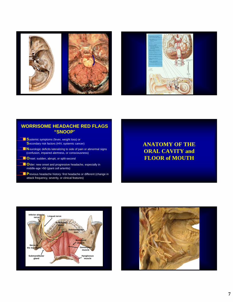
WORRISOME HEADACHE RED FLAGS“SNOOP”
Older: new onset and progressive headache, especially in middle-age >50 (giant cell arteritis)
Systemic symptoms (fever, weight loss) or
Secondary risk factors (HIV, systemic cancer)
Neurologic deficits lateralizing to side of pain or abnormal signs (confusion, impaired alertness, or consciousness)
Onset: sudden, abrupt, or split-second
Previous headache history: first headache or different (change in attack frequency, severity, or clinical features)
ANATOMY OF THE ORAL CAVITY andFLOOR of MOUTH
Lingual nerveInferior alveolar
nerve
Submandibulargland
Sublingualgland
Submandibularduct
Mylohyoidmuscle
Hyoglossusmuscle
Geniohyoidmuscle
Nerve to the myolhyoid
Page 8
8
Sublingual salivary gland
Submandibular duct
Lingual nerve
Mylohyoid muscle
Lingual vein
Hypoglossal nerve
Hyoglossus muscle
Lingual artery
Sublingual artery & vein
Geniohyoidmuscle
Lingual nerve
Submandibular ganglion
Superior pharyngeal constrictorStyloglossus muscle Palatoglossus msucleStylohyoid ligamentStylopharyngeus muscleHyoglossus muscle (cut)Lingual artery
Hypoglossal nerve
External carotid artery
Internal jugular vein
Deep lingual arteryVenae comitantes
Submandibularduct
Sublingual Gland and Submandibular Duct
Lingual Nerve
Page 9
9
Tongue positionand its relationship
to sleep-relatedbreathing disorders
such as sleep apnea…genioglossus activity
Tongue
Oropharynx
Tongue
ObstructedOropharynx
SLEEP-RELATED BREATHING DISTURBANCES
Enlarged & Inflamed Tonsils
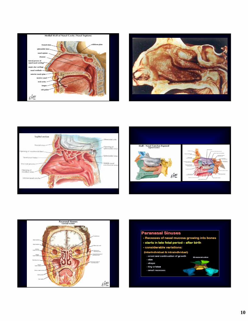
Nasal Cavity & Paranasal Sinuses
Page 10
10
cribiform platefrontal sinus
anterior nasal spine
incisive canal
oral cavity
tongue
soft palate
choanae
nasal septum
major alar cartilage
nasal vestibule
sphenoidal sinus
lateral process ofseptal nasal cartilage
Page 11
11
Mucous Retention Cyst
1. Under flap2. Sinus3. Infratemporal Fossa
DISPLACED ROOT / TOOTHDISPLACED ROOT / TOOTH Root Tip in Maxillary Sinus
Page 12
12
Third Molar Displaced into Maxillary Sinus
Third Molar Displaced into Infratemporaral Foss
Fractured Tuberosity with Maxillary Sinus Exposure
Sin
Sinus Lift with Iliac Bone Graft
PARANASALORIGINSOF PAIN
Page 13
13
Paranasal SinusesParanasal Sinuses
Headache and facial pain are commonly related to infection, inflammation, and/or obstruction of the outflow of the tracts of
the paranasal sinuses.
Acute / Chronic Sinusitis:PAINFUL COMPLICATIONS
Mucosal inflammation and thickening in cases of acute sinusitis
Partial or complete obstruction of sinus ostia
Pressure sensation
Maxillary mucoceles
Osteomyelitis
• Sphenoid sinus
• Frontal sinus
• Ethmoid sinus
• Maxillary sinus
• Pansinusitis
• Vertex, other parts of the cranium
• Frontal region
• Between the eyes
• Maxilla, dental structures
• Pain may be coalescent, less localized, associated with frontal headaches, constant pressure
Acute / Chronic Sinusitis:Acute / Chronic Sinusitis:
Sinus involved Site(s) of referral
Pansinusitis
Page 14
14
MUCOSALCONTACT
HEADACHE
Mucosal Contact HeadacheMucosal Contact Headache
• Dull and aching
• Diffuse peri-/retro-ocular, supraorbital pain
• History of chronic maxillary sinusitis
• Allergy prone
• Associated with upper respiratory tract infection
• Impedance of normal mucosal activity
Page 15
15
Pharyngeal Region
Plate 58Plate 58A Plate 58B
Page 16
16
EAREAR
Eustachian tube dysfunctionEustachian tube dysfunction
• Normal function– Dilatation
– Primarily involves the tensor veli palatini
– Swallowing causes momentary eustachian tube dilitation which equalizes pressure
– Secondarily involves • Levator veli palatini
• Salpingopharyngeus
• Superior constrictorPlate 89Plate 89
Ear Pain ( Otalgia )Ear Pain ( Otalgia )
• Acute Otitis Externa• Acute Otits Media
– Severe ear pain often– Fluid/pressure behind
the TM– Most common in
children– Treatment
• Antibiotics• Myringotomy ( ear tubes )
Page 17
17
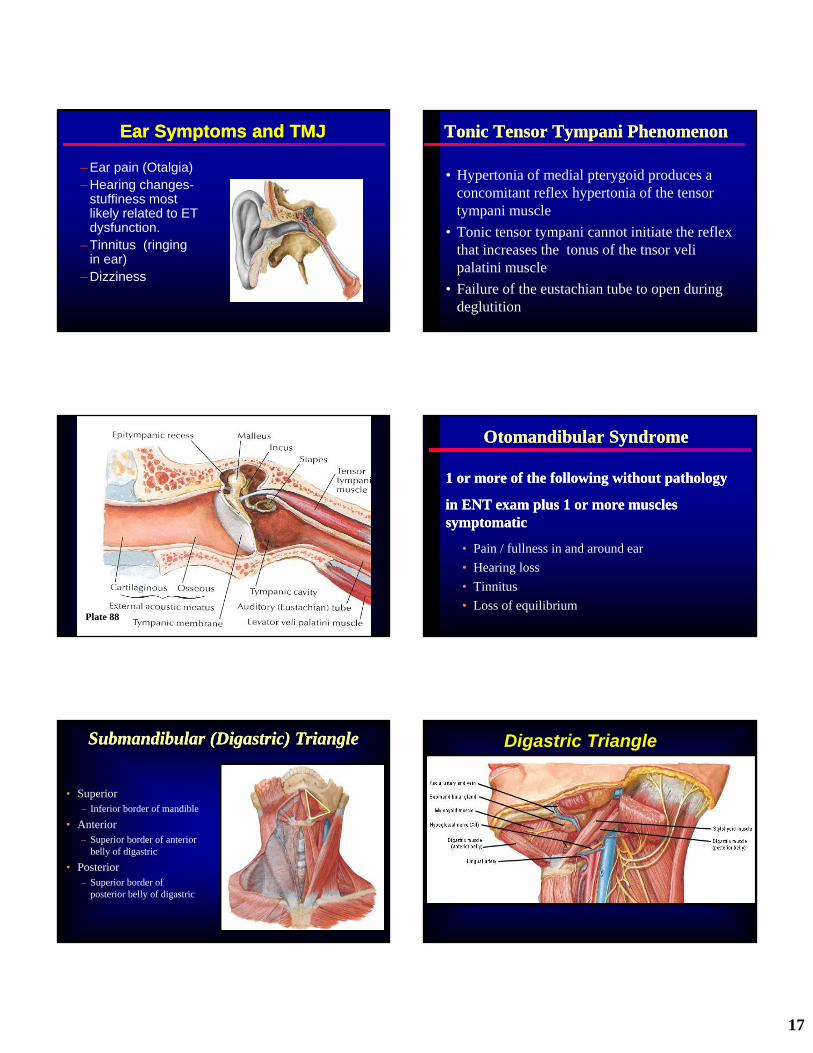
Ear Symptoms and TMJEar Symptoms and TMJ
– Ear pain (Otalgia)– Hearing changes-
stuffiness most likely related to ET dysfunction.
– Tinnitus (ringing in ear)
– Dizziness
Tonic Tensor Tympani PhenomenonTonic Tensor Tympani Phenomenon
• Hypertonia of medial pterygoid produces a concomitant reflex hypertonia of the tensor tympani muscle
• Tonic tensor tympani cannot initiate the reflex that increases the tonus of the tnsor veli palatini muscle
• Failure of the eustachian tube to open during deglutition
Plate 88
Otomandibular SyndromeOtomandibular Syndrome
• Pain / fullness in and around ear
• Hearing loss
• Tinnitus
• Loss of equilibrium
1 or more of the following without pathology
in ENT exam plus 1 or more muscles symptomatic
1 or more of the following without pathology
in ENT exam plus 1 or more muscles symptomatic
Submandibular (Digastric) TriangleSubmandibular (Digastric) Triangle
• Superior– Inferior border of mandible
• Anterior– Superior border of anterior
belly of digastric
• Posterior– Superior border of
posterior belly of digastric
Digastric Triangle
Page 18
18
Brachial plexus
Masseter muscle
Anterior digastric muscle
Sternohyoid muscle
Omohyoid muscle(superior belly)
Thyrohyoid muscleMiddle pharyngeal constrictor muscle
Scalene musclesposterior
middleanterior
Posterior digastric muscle
Stylohyoid muscle
Sternocleidomastoid muscleSternal headClavicular headOmohyoid muscle
(inferior belly)
Inferior pharyngeal constrictor muscle
Hyoglossus muscle
Mylohyoid muscle
Styloglossus muscle
Trapezius muscle
Lesser’s triangleLesser’s triangle
Major Salivary GlandsMajor Salivary Glands
Parotid gland-pure serous
Submandibular gland-primarily serous
Sublingual gland-primarily mucous
Parotid gland-pure serous
Submandibular gland-primarily serous
Sublingual gland-primarily mucous
Patient: Betty
• 51 year old Caucasian female
• Medical history significant for:– left temporomandibular surgery X2
– hypothyroidism
Patient: Betty
• Chief pain concern:– “I have pain in my jaw and throat when I eat. The
pain radiates to my ear. It feels like a toothache.”
Page 19
19
Patient: Betty
• Aggravating factors:– chewing and drinking
– certain aromas
• Alleviating/relieving factors:– none identified
SialolithiasisSialolithiasis
Diagnosis
• History– pain with salivation
• Inspection• Palpation
SialolithiasisSialolithiasis
Diagnosis
• Imaging– occlusal– lateral jaw– panoramic– sialogram
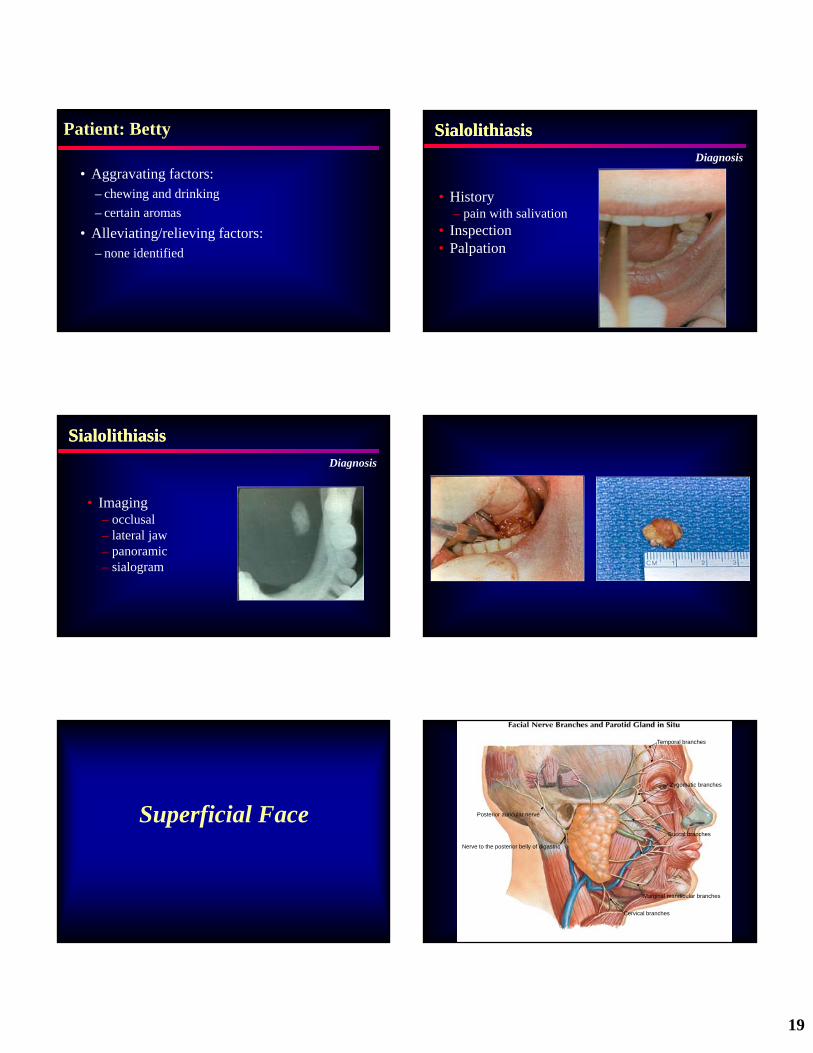
Superficial Face
Temporal branches
Buccal branches
Cervical branches
Marginal mandibular branches
Zygomatic branches
Posterior auricular nerve
Nerve to the posterior belly of digastric
Page 20
20
BELL’S PALSY
• Cranial nerve VII paralysis
• May occur post-dental procedure
• Usually unilateral
• Gradual or sudden onset
• Viral relationship???
Patient: Juan
• 28 year old Hispanic male
• Medical history:– unexplained intermittent facial
swelling and lymphadenopathy• previously treated with Pen VK 500
mg
Patient: Juan
• Chief pain concern(s):– “pain on the right side of my face; headaches in the
temples; clicking in my right jaw; face feels numb and tingles on the right side; throbbing when I eat”
Patient: Juan
• Aggravating factors:– eating
– opening wide
– yawning
• Alleviating/relieving factors:– antibiotics (Pen VK 500)
– analgesics (Ibuprofen)-- “takes the edge off”
Page 21
21
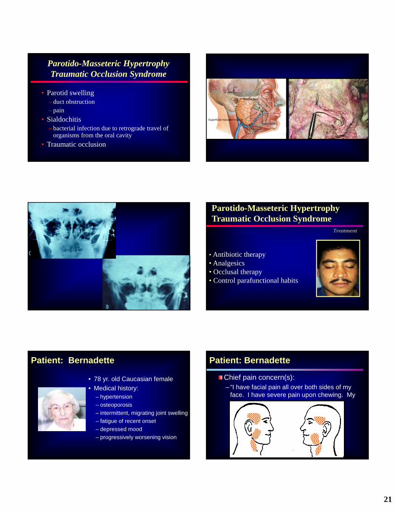
Parotido-Masseteric HypertrophyTraumatic Occlusion Syndrome
• Parotid swelling– duct obstruction– pain
• Sialdochitis– bacterial infection due to retrograde travel of
organisms from the oral cavity
• Traumatic occlusion
Parotid duct
Superficial masseter muscle
Buccinator
Parotido-Masseteric HypertrophyTraumatic Occlusion Syndrome
Treatment
• Antibiotic therapy• Analgesics• Occlusal therapy• Control parafunctional habits
Patient: Bernadette
• 78 yr. old Caucasian female
• Medical history:– hypertension
– osteoporosis
– intermittent, migrating joint swelling
– fatigue of recent onset
– depressed mood
– progressively worsening vision
Patient: Bernadette
Chief pain concern(s):– “I have facial pain all over both sides of my
face. I have severe pain upon chewing. My neck hurts.”
Page 22
22
Patient: Bernadette
Aggravating factors:– eating
– talking
– clenching
Alleviating/relieving factors:– jaw rest
– “eating in stages”
Temporal ArteritisTemporal ArteritisCharacteristics
• Jaw claudication• Craniofacial pain
– dental pain– TM joint pain– otalgia– headache
Temporal ArteritisTemporal ArteritisCharacteristics
• Visual symptoms• Anorexia• Anemia• Low grade fever/malaise• Neurologic deficits• Systemic involvement
– polymyalgia rheumatica
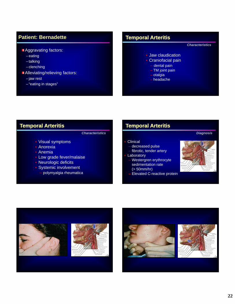
Temporal ArteritisTemporal ArteritisDiagnosis
• Clinical– decreased pulse– fibrotic, tender artery
• Laboratory– Westergren erythrocyte
sedimentation rate (> 50mm/hr)
– Elevated C-reactive protein
Page 23
23
Temporal ArteritisTemporal ArteritisDiagnosis
• Biopsy– usually the superficial temporal
artery– 1.5 cm segment due to “skip”
lesions
Temporal ArteritisTemporal ArteritisTreatment
• Glucocorticoid therapy– parenteral (in patients with visual
symptoms)– oral
> Prednisone 40-60 mg / day initially with gradual taper over 6-12 months
Deep masseter
Styloid process
Superficial masseter
TM joint capsule
Lateral TM joint ligament
Deep masseter
Lateral Pterygoid
Page 24
24
*SLP
ILP
1. Muscles active on jaw opening-lateral pterygoid (inferior belly), suprahyoid and digastric muscles
2. Muscles active on jaw closure-temporalis, masseter, medial pterygoid muscles, lateral pterygoid (superior belly)
3. Excursive movements-lateral pterygoid
IMPORTANT ASSOCIATED STRUCTURES
Muscles involved in joint function
Functional Anatomy/Biomechanics of the Masticatory System
Temporomandibular JointTemporomandibular Joint
Masticatory System: Unique Features
• Right and left function as one unit• Articulating surfaces are
fibrocartilaginous• Articular disc separates the joint into
two compartments• Ginglymoarthrodial joint (hinge-
gliding)
Page 25
25
Masticatory System: Unique Features
• Right and left function as one unit• Articulating surfaces are
fibrocartilaginous• Articular disc separates the joint into
two compartments• Ginglymoarthrodial joint (hinge-
gliding)• Articulation has a rigid end point on
closure of the teeth
1. Part of temporal bone2. Glenoid fossa is concave
structure covered with thin layer of fibrocartilage
3. Articular eminence is convex, posterior slope has an average angle of 60o
OSSEOUS STRUCTURES Glenoid fossa and
articular eminence
Articular tubercle
Zygomatic archposterior root
Articular eminence
1. Adult condyle is elliptical
2. Mediolateral dimension is about 20 mm and is twice the size of its antero-posterior width
3. Articular surface is covered by a layer of fibrocartilage
OSSEOUS STRUCTURES Condyle
1. Bioconcave structure, divided the joint space into superior and inferior spaces
2. Attachmentsa. Anterior-capsule and superior
belly lateral pterygoid
b. Posterior-bilaminar zone (retrodiskal tissues)
c. Medial/lateral condyle
SOFT TISSUES Articular Disk
(Meniscus)
3. Made up of three zonesa. Posterior band – 3 mm thickb. Intermediate zone – 1 mm
thickc. Anterior band – 2 mm thick
4. Consists of avascular connective tissue with some cartilaginous elements
SOFT TISSUES Articular Disk
(Meniscus)
1
2
3
RDT
Page 26
26
1
2
3
RDT
M
L
5. Functionsa. Load adapterb. Fluid distributionc. Divides joint space into two
compartments allowing complex movements consisting of rotation and translation
SOFT TISSUES Articular Disk
(Meniscus)
1. Lines all non-loaded surfaces
2. Made up of intimal layer of cells 1-4 deep
a. Type A – phagocyticb. Type B - secretory
3. Functions of synovial fluidsa. Lubricationb. Nutritionc. Maintains and protects
articular cartilage
JOINT SPACES Synovial Membrane
TM Joint Surfaces
Without lubrication
• relatively smooth
• have high surface energy
• may shear and rupture
TM Joint Biomechanics
The role of lubricant
• Reduces area of contact
• Reduces surface energy
• Reduces shearing
TM Joint Biomechanics
Lubrication• Boundary
• Surface (weeping)
Page 27
27
Synovial Organ
Functions• Semi-permeable membrane which allows for
adjustment of pressures within the TM joint.
Bauer W, et al. Physiological Rev 1940; 20:272-312
1. Resting (-4 mm Hg)
2. Opening (-54 mm Hg)
3. Closing (+64 mm Hg)
JOINT SPACES Intra-articular Joint
Pressures
Synovial Fluid
As the intra articular pressure increases, the viscosity of the synovial fluid decreases.
This may impair the lubricating ability of the fluid… thus increasing the frictional resistance.
TM Joint Mechanical Stress
• impaired diffusion
• local ischemic changes– may lead to cell death
– free radical formation
• decreased lubrication– increased frictional resistance
Increased sustained TM joint pressures result in:
1. Branches of the 3rd division of the trigeminal nerve
a. Auriculotemporal
b. Masseteric
c. Deep temporal
2. Fibers for pain and proprioception are mainly located in the bilaminar zone and capsule
IMPORTANT ASSOCIATED STRUCTURES
Sensory Innervation of the TMJ
TM Joint: Normal BiomechanicsTM Joint: Normal Biomechanics
Page 28
28
Articular Disc Displacement
Retrodiscal tissue
Articulardisc
Articular Disc DisplacementWith Reduction
Degenerative temporomandibular jointdisease is the result of maladaptation
to increased joint loading.
Westesson, Rohlin 1984Axelson, et al. 1992, 1993
Stegenga, et al. 1992deBont, Stegenga 1993
Page 29
29
[email protected]
Henry A. Gremillion, DDS1100 Florida Avenue
New Orleans, LA 70119