43
www.clutchprep.com ANATOMY & PHYSIOLOGY - CLUTCH CH. 19 - THE URINARY SYSTEM

! www.clutchprep.com
!
ANATOMY & PHYSIOLOGY - CLUTCH
CH. 19 - THE URINARY SYSTEM

CONCEPT: ANATOMY OF THE URINARY SYSTEM
Urinary System Anatomy:
● Kidneys are a pair of organs near the lower back.
□ Function is filter blood and collect the filtrate, called urine.
● From kidneys, urine flows through the ureters and collects in the bladder.
□ From the bladder, urine leaves the body through the urethra.
EXAMPLE: Overview of urinary system anatomy.
ANATOMY & PHYSIOLOGY - CLUTCH
CH. 19 - THE URINARY SYSTEM
Page 2

Kidney Anatomy:
● Kidneys are bean-shaped organs with two layers.
□ Renal Cortex= outer layer. □ Renal Medulla= inner layer.
□ Renal Pyramids are striations (stripe patterns) in renal medulla.
□ Renal Pelvis collects the urine and delivers it to the ureter.
● The kidney has its own dedicated blood supply.
□ Renal Artery brings blood to the kidneys to be filtered. □ Renal Vein takes filtered blood back to the heart.
EXAMPLE: Anatomy of a kidney.
ANATOMY & PHYSIOLOGY - CLUTCH
CH. 19 - THE URINARY SYSTEM
Page 3

Introduction to the Nephron:
● Nephron=functional unit of the kidney. (Small, individual structure that does all the filtering.) Two parts:
□ Tubular Elements hold the filtrate/urine.
-(Filtrate=Fluid from blood being modified into urine.)
□ Vascular Elements are the blood vessels associated with the nephron.
Tubular Elements of the Nephron:
● Bowman’s Capsule collects the filtrate as it flows out of the blood supply.
● From Bowman’s Capsule, filtrate flows through the:
□ Proximal Tubule
□ Loop of Henle
-Descending Thin Limb
-Ascending Thin Limb
-Ascending Thick Limb
□ Distal Tubule
□ Collecting Duct
Vascular Elements of the Nephron:
● Glomerulus is the ball of capillaries that provides blood to be filtered.
□ Filtrate flows glomerulus→Bowman’s Capsule.
□ Afferent Arteriole brings blood to the glomerulus.
□ Efferent Arteriole takes blood away from the glomerulus.
● The rest of the tubular elements also have associated capillaries.
□ Peritubular Capillaries surround the proximal and distal tubules.
□ Vasa Recta follow the Loop of Henle.
Affe
rent
Arte
riole
Effe
rent
Arte
riole
Glomerulus
Bowman'sCapsule
Proximal Tubule
DescendingThin Limb
Loop of Henle
AscendingThin Limb
AscendingThick Limb
Distal Tubule
CollectingDuct
ToBladder
ANATOMY & PHYSIOLOGY - CLUTCH
CH. 19 - THE URINARY SYSTEM
Page 4

PRACTICE 1: A cystoscopy is a medical procedure wherein a physician inserts a camera and scope through the external genitals into the bladder to examine the internal bladder wall. Through which of the following structures does the camera travel to get from the outside to the bladder?
a) Nephron. b) Ureter. c) Urethra. d) Renal artery. e) Renal vein.
PRACTICE 2: Using a micropipette and some outstanding surgical precision, a cannula is inserted into an individual nephron at the tip of the loop of Henle. Fluorescent dye in infused into the filtrate inside the nephron through the cannula and flows with the filtrate (but not “backwards”). In which of the following parts of the nephron will the fluorescent dye be found? (Choose all that apply.)
a) Collecting duct. b) Distal tubule. c) Ascending thick limb. d) Ascending thin limb. e) Descending limb. f) Proximal tubule. g) Bowman’s capsule.
PRACTICE 3: Caffeine causes an increase in urinary output because it causes a change in the radius of the blood vessels that directly supply blood to, and take blood away from, each individual nephron. Which of the following blood vessels are affected by caffeine? (Choose all that apply.)
a) Vasa recta. b) Renal artery. c) Renal vein. d) Afferent arteriole. e) Efferent arteriole.
ANATOMY & PHYSIOLOGY - CLUTCH
CH. 19 - THE URINARY SYSTEM
Page 5
CONCEPT: KIDNEY ANATOMY
● The kidneys are located lateral to the vertebral column on the posterior abdominal wall; Near the 3rd lumbar vertebrae □ The right kidney is slightly ______________________ to the left due to the liver
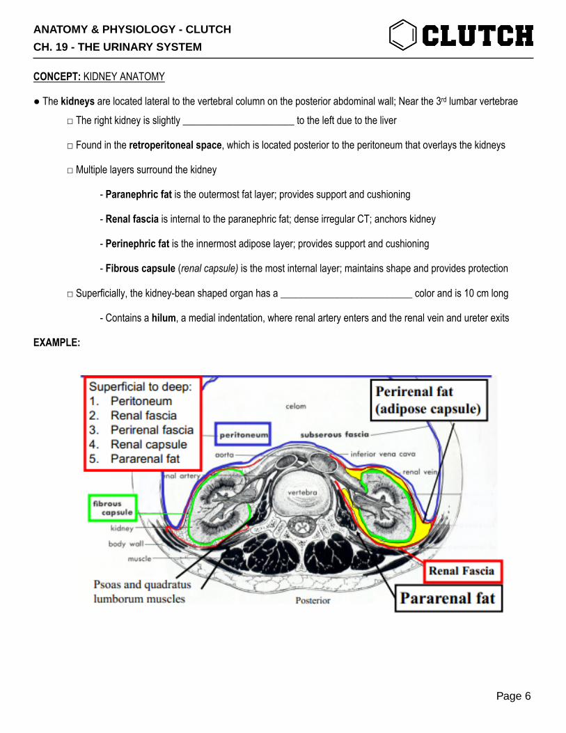
□ Found in the retroperitoneal space, which is located posterior to the peritoneum that overlays the kidneys
□ Multiple layers surround the kidney
- Paranephric fat is the outermost fat layer; provides support and cushioning
- Renal fascia is internal to the paranephric fat; dense irregular CT; anchors kidney
- Perinephric fat is the innermost adipose layer; provides support and cushioning
- Fibrous capsule (renal capsule) is the most internal layer; maintains shape and provides protection
□ Superficially, the kidney-bean shaped organ has a __________________________ color and is 10 cm long
- Contains a hilum, a medial indentation, where renal artery enters and the renal vein and ureter exits
EXAMPLE:
ANATOMY & PHYSIOLOGY - CLUTCH
CH. 19 - THE URINARY SYSTEM
Page 6
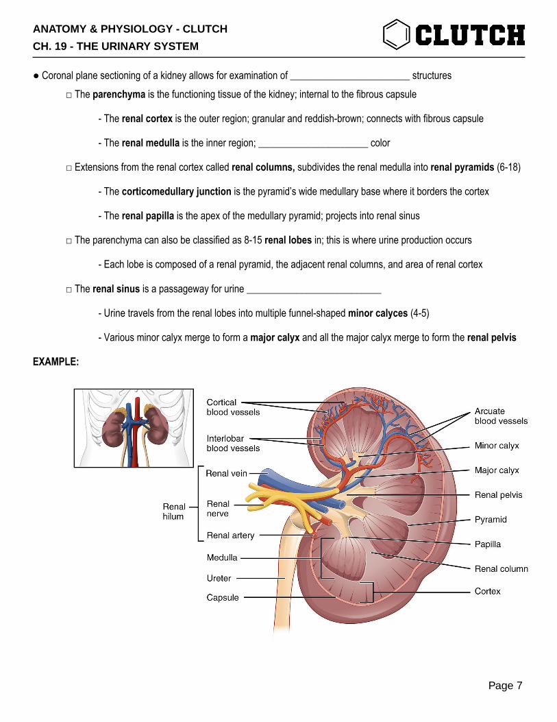
● Coronal plane sectioning of a kidney allows for examination of ________________________ structures □ The parenchyma is the functioning tissue of the kidney; internal to the fibrous capsule
- The renal cortex is the outer region; granular and reddish-brown; connects with fibrous capsule
- The renal medulla is the inner region; ______________________ color
□ Extensions from the renal cortex called renal columns, subdivides the renal medulla into renal pyramids (6-18)
- The corticomedullary junction is the pyramid’s wide medullary base where it borders the cortex
- The renal papilla is the apex of the medullary pyramid; projects into renal sinus
□ The parenchyma can also be classified as 8-15 renal lobes in; this is where urine production occurs
- Each lobe is composed of a renal pyramid, the adjacent renal columns, and area of renal cortex
□ The renal sinus is a passageway for urine ___________________________
- Urine travels from the renal lobes into multiple funnel-shaped minor calyces (4-5)
- Various minor calyx merge to form a major calyx and all the major calyx merge to form the renal pelvis
EXAMPLE:
ANATOMY & PHYSIOLOGY - CLUTCH
CH. 19 - THE URINARY SYSTEM
Page 7

Nephron Structure ● The nephron is the functional filtration unit of the kidney; made up of two main structures: 1. Renal corpuscle is the bulbous portion of the nephron mostly found in the renal cortex,
- Glomerulus which consists of convoluted capillaries called glomerular capillaries
- Blood enters through the ____________________ afferent arteriole
- Blood exits through the smaller efferent arteriole
- Bowman’s capsule (Glomerular capsule) has an internal and external layer; where filtrate travels
- Visceral layer is the innermost, surrounds the glomerular capillaries; permeable
- Capsular space surrounds the visceral layer; receives filtrate
- Parietal layer the outermost, has simple squamous epithelial layer; ____________________
□ Vascular pole region where glomerular capillaries connect to the bloodstream (arterioles)
□ Tubular pole where rental tubule originates
EXAMPLE:
ANATOMY & PHYSIOLOGY - CLUTCH
CH. 19 - THE URINARY SYSTEM
Page 8

2. Renal tubule is the rest of the nephron; a simple epithelial layer on basement membrane
- Proximal convoluted tubule (PCT) increases reabsorption of the nephron through its apical microvilli
- Originates from the urinary (tubular) pole; located in the ________________
- Reabsorbs organic materials, potassium ions, sodium ions, chloride ions, and water
- Loop of Henle (Nephron loop) is important for filtrate flow
- Originates from proximal convoluted tubule; extends from cortex (PCT & DCT) into the medulla
- Descending limb: from PCT à hairpin loop; permeable to water but low permeability to ions
- Ascending limb: hairpin loop à to DCT; impermeable to water but permeable to ions
- Distal convoluted tubule (DCT) is involved in secretion; located in the cortex
- Connects the nephron loop to a collecting tubule
- When compared with the PCT it has a ___________________ diameter and lack microvilli
EXAMPLE:
ANATOMY & PHYSIOLOGY - CLUTCH
CH. 19 - THE URINARY SYSTEM
Page 9

● There are two types of nephrons that are classified by the positioning of the renal cortex and length of the Loop of Henley □ Cortical nephrons (85%)
- Their renal corpuscles reside mainly in the cortex
- They have a ____________________ nephron loop that barely penetrates the medulla
□ Juxtamedullary nephrons (15%)
- Renal corpuscles are adjacent to the corticomedullary junction
- They have long nephron loops that penetrate deep into the ____________________
- Important in creating a salt concentration gradient in the interstitial space of the loop; salty medulla
- Helps ADH in regulating urine concentration
EXAMPLE:
ANATOMY & PHYSIOLOGY - CLUTCH
CH. 19 - THE URINARY SYSTEM
Page 10
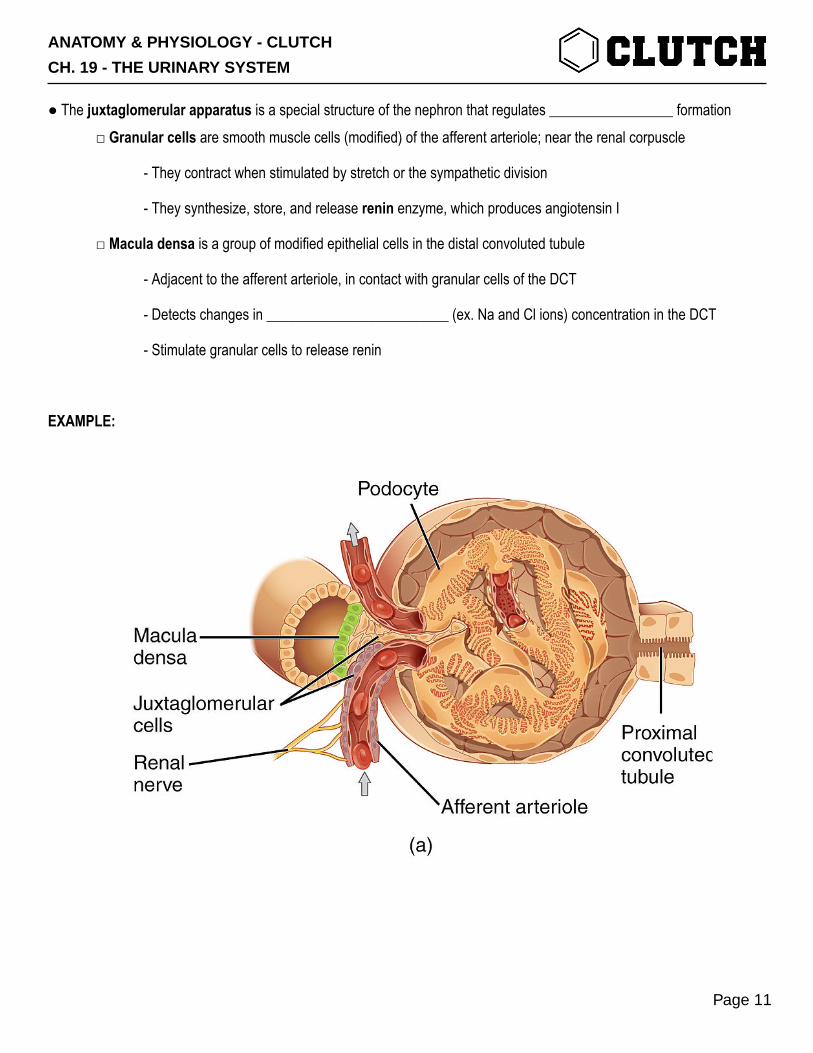
● The juxtaglomerular apparatus is a special structure of the nephron that regulates _________________ formation □ Granular cells are smooth muscle cells (modified) of the afferent arteriole; near the renal corpuscle
- They contract when stimulated by stretch or the sympathetic division
- They synthesize, store, and release renin enzyme, which produces angiotensin I
□ Macula densa is a group of modified epithelial cells in the distal convoluted tubule
- Adjacent to the afferent arteriole, in contact with granular cells of the DCT
- Detects changes in _________________________ (ex. Na and Cl ions) concentration in the DCT
- Stimulate granular cells to release renin
EXAMPLE:
ANATOMY & PHYSIOLOGY - CLUTCH
CH. 19 - THE URINARY SYSTEM
Page 11
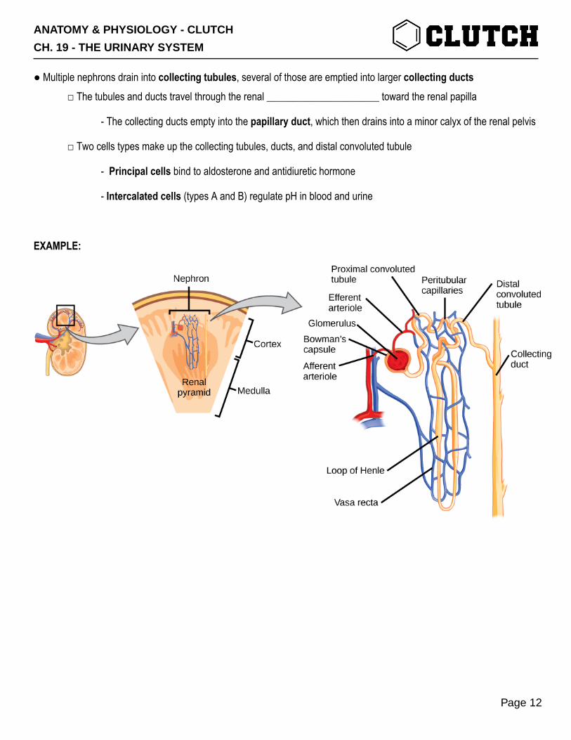
● Multiple nephrons drain into collecting tubules, several of those are emptied into larger collecting ducts □ The tubules and ducts travel through the renal ______________________ toward the renal papilla
- The collecting ducts empty into the papillary duct, which then drains into a minor calyx of the renal pelvis
□ Two cells types make up the collecting tubules, ducts, and distal convoluted tubule
- Principal cells bind to aldosterone and antidiuretic hormone
- Intercalated cells (types A and B) regulate pH in blood and urine
EXAMPLE:
ANATOMY & PHYSIOLOGY - CLUTCH
CH. 19 - THE URINARY SYSTEM
Page 12

CONCEPT: FLOW INTO AND OUT OF THE KIDNEY
● Blood and filtrate flow in and out of the nephrons in the kidney in order to remove __________ substances from the body □ Blood enters the kidney through:
- Abdominal aorta à renal artery à segmental arteries à interlobar arteries
à (at the corticomedullary junction) arcuate arteries à interlobular arteries à numerous arterioles
- Blood is then delivered to a glomerulus by an afferent arteriole and exits by the efferent arteriole
- Peritubular capillaries: intertwined around PCT and DCT
- Vasa Recta: intertwined around the loop of Henley
□ Blood leaves the kidney through:
- Blood is drained from the peritubular and vasa recta capillary beds à Interlobular veins à merge to
form arcuate veins (base of medullary pyramids) à merge to form interlobar veins (renal columns) à
merging into renal vein
EXAMPLE:
ANATOMY & PHYSIOLOGY - CLUTCH
CH. 19 - THE URINARY SYSTEM
Page 13

● As blood is filtered through the glomerulus, water and solutes move to the capsular space, forming the filtrate
□ Filtrate flows into the nephron:
- Filtrate is the filtered substance from the glomerular capillaries à proximal convoluted tubule (filtrate
becomes tubular fluid) à nephron loop à distal convoluted tubule à collecting tubules à collecting
ductsà papillary duct (at this point the tubular fluid becomes urine) à renal sinus spaces (1. minor calyx,
2. major calyx, 3. renal pelvis) à ureter à urinary bladder à urethra
EXAMPLE:
ANATOMY & PHYSIOLOGY - CLUTCH
CH. 19 - THE URINARY SYSTEM
Page 14

CONCEPT: BASIC RENAL PROCESSES
● Overall, a nephron does four processes:
1) Filtration= movement of fluid from glomerular capillaries into the tubular elements of the nephron.
-Filtrate= Filtered fluid now inside of nephron.
2) Reabsorption= movement of specific substances (e.g. glucose) from filtrate back into blood.
-Happens across epithelial cells that line tubular parts of nephron.
3) Secretion= movement of specific substances from blood into filtrate.
-Opposite process from reabsorption.
-Used to clear specific molecules (e.g. penicillin) from the body.
4) Excretion= permanent removal of substance from body (via urine).
● These processes can be summarized in an equation for excretion:
□ Excretion=Filtration+Secretion-Reabsorption
ANATOMY & PHYSIOLOGY - CLUTCH
CH. 19 - THE URINARY SYSTEM
Page 15

PRACTICE 1: Which of the following processes add substances and/or fluid to the inside of the nephron (i.e. the filtrate)? (Choose all that apply.)
a) Filtration. b) Reabsorption. c) Secretion. d) Excretion.
ANATOMY & PHYSIOLOGY - CLUTCH
CH. 19 - THE URINARY SYSTEM
Page 16

CONCEPT: THE GLOMERULUS
Filtration from Glomerulus to Bowman’s Capsule:
● Glomerulus is the big ball of capillaries that supplies filtrate to the nephron.
□ Filtration happens from glomerular capillaries→Bowman’s Capsule.
□ Afferent Arteriole brings blood to glomerulus. □ Efferent Arteriole takes blood away from glomerulus.
● Filtration out of glomerulus is a bulk flow process, so subject to Starling’s Equation of Ultrafiltration.
□ Glomerular capillaries are located between two arterioles (!)—the afferent and efferent arterioles.
-Downstream arteriole→↑R→↑Pc→even more filtration out of glomerulus than in normal capillary beds.
● Arterioles have lots of smooth muscle and can do lots of vasoconstriction or vasodilation.
□ Gives extra control over Pc.
-Vasodilation upstream and/or vasoconstriction downstream→↑Pc→↑Glomerular Filtration.
-Vasoconstriction upstream and/or vasodilation downstream→↓Pc→↓Glomerular Filtration.
EXAMPLE: The glomerulus and its associated arterioles.
ANATOMY & PHYSIOLOGY - CLUTCH
CH. 19 - THE URINARY SYSTEM
Page 17

PRACTICE 1: Caffeine causes diuresis (an increase in urine output) by acting on both the afferent and efferent arterioles. Specifically, caffeine causes an increase in the blood pressure in the glomerulus by causing a change in the radii (either vasoconstriction or vasodilation) of the afferent and efferent arterioles. Which of the following matches the blood vessel with the radius change caused by caffeine? Hint: Both changes contribute to an increase in pressure in the glomerulus. (Choose all that apply.)
a) Afferent arteriole: Vasoconstriction. b) Afferent arteriole: Vasodilation. c) Efferent arteriole: Vasoconstriction. d) Efferent arteriole: Vasodilation.
ANATOMY & PHYSIOLOGY - CLUTCH
CH. 19 - THE URINARY SYSTEM
Page 18
CONCEPT: REABSORPTION I: GENERAL CONCEPTS OF REABSORPTION IN THE KIDNEY
Introduction to Reabsorption:
● About 180 L of fluid per day is filtered from the glomerulus into Bowman’s Capsule.
□ But, only about 1.5 L gets excreted (peed out).
● Reabsorption is the movement of H2O, salts, amino acids, glucose, etc. from tubular elements back into blood.
□ Not random—tubular elements lined with epithelial cells that have specific transporters to control reabsorption.
-Some reabsorption does happen through tight junctions between cells.
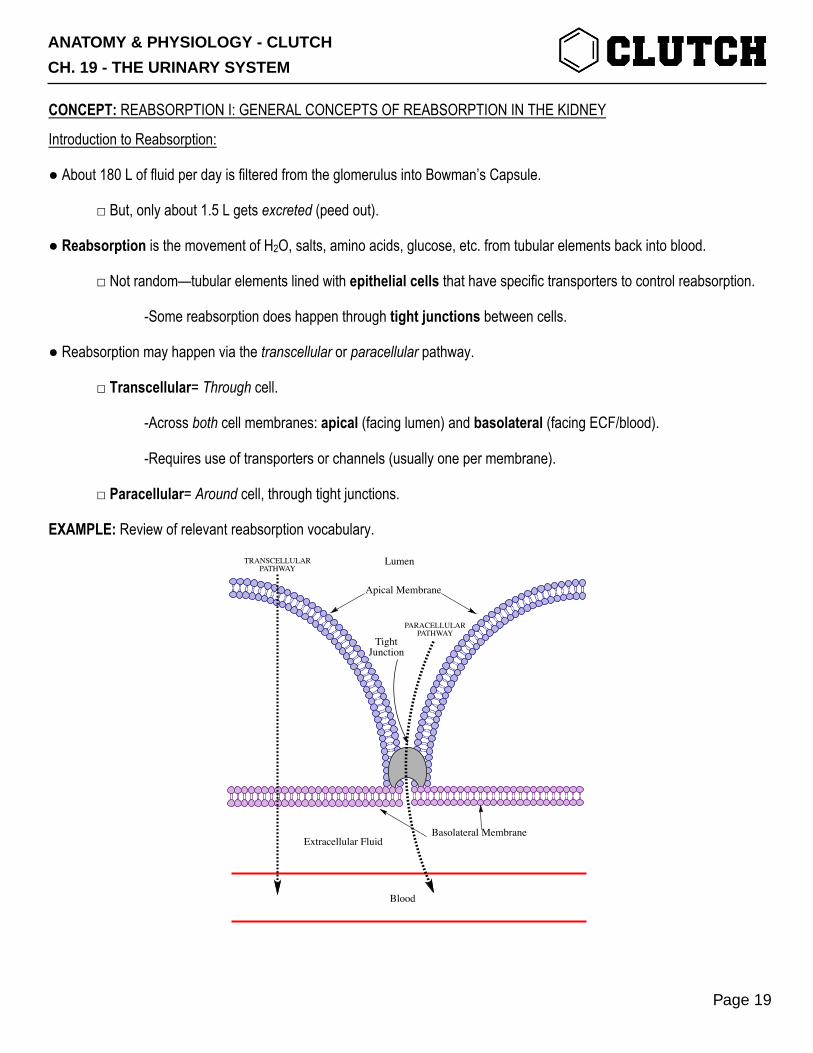
● Reabsorption may happen via the transcellular or paracellular pathway.
□ Transcellular= Through cell.
-Across both cell membranes: apical (facing lumen) and basolateral (facing ECF/blood).
-Requires use of transporters or channels (usually one per membrane).
□ Paracellular= Around cell, through tight junctions.
EXAMPLE: Review of relevant reabsorption vocabulary.
Lumen
Blood
Extracellular FluidBasolateral Membrane
TightJunction
Apical Membrane
TRANSCELLULARPATHWAY
PARACELLULARPATHWAY
ANATOMY & PHYSIOLOGY - CLUTCH
CH. 19 - THE URINARY SYSTEM
Page 19

Mechanisms of Transcellular Reabsorption:
● Lots of reabsorption depends on movement of Na+ and the normal Na+ concentration gradient across cell membranes.
□ Na+/K+ ATPases on the basolateral membrane maintain ↑[Na+]out and ↓[Na+]in.
-Driving force pulls Na+ into the cell from the filtrate.
● Na+ itself can be reabsorbed through an apical Epithelial Na+ Channel (ENaC) and basolateral Na+/K+ ATPase.
● Sodium-Linked Transport allows important organic molecules—e.g. glucose and amino acids—to be reabsorbed.
□ Apical transporter moves both the molecule and some Na+.
-Potential energy in Na+ gradient powers movement of other molecule. (This is secondary active transport.)
□ Some basolateral channel or transporter allows molecule to exit cell and enter blood.
● H2O follows the movement of Na+ and other molecules (by osmosis).
□ Aquaporins are channels that let H2O cross cell membranes.
□ (H2O may also cross through tight junctions.)
EXAMPLE: Glucose is reabsorbed by sodium-linked transport—an apical Sodium-Glucose Linked Transporter (SGLT) and basolateral Glucose Transporter (GLUT).
Lumen
Extracellular Fluid
Blood
SGLT
Na+/K+ ATPase
Na+
K+
Na+
Glu
Glu
GLUT
ATP
Nephron Epithelial CellBlood
Filtrate
Na+Na+
K+
ATP
ENaC
ADH
ANATOMY & PHYSIOLOGY - CLUTCH
CH. 19 - THE URINARY SYSTEM
Page 20

The Transport Maximum (Tm):
● Reabsorption relies on proteins, and proteins are saturable—they can be maxed out.
● Transport Maximum (Tm) is the transport rate when the transporters are working at the maximum.
□ If the concentration of solute in the filtrate exceeds Tm, that solute won’t be reabsorbed and will be excreted.
EXAMPLE: Increasing concentration of a substance in the filtrate increases transport rate up to the Tm.
EXAMPLE: In healthy patients, SGLTs are easily capable of reabsorbing all of the glucose from the filtrate→no glucose in urine. Diabetic patients with very high [Glucose]blood surpass the Tm and have “glycosuria”—glucose in their pee.
Transport Rate(Amount Reabsorbed)
Concentration in Filtrate
Tm
ANATOMY & PHYSIOLOGY - CLUTCH
CH. 19 - THE URINARY SYSTEM
Page 21
PRACTICE 1: Sodium-Glucose Linked Transporters (SGLTs) can be inhibited by a class of drugs called, logically, SGLT Inhibitors. Which of the following is the expected effect of an SGLT Inhibitor on the transport maximum Tm for glucose in the kidney?
a) Increased Tm. b) Decreased Tm. c) No change in Tm.
ANATOMY & PHYSIOLOGY - CLUTCH
CH. 19 - THE URINARY SYSTEM
Page 22

CONCEPT: REABSORPTION II: REABSORPTION (AND SECRETION) IN THE PROXIMAL TUBULE
Reabsorption in the Proximal Tubule:
● Proximal Tubule is the first part of the tubular elements of the nephron, after Bowman’s Capsule.
□ Does lots of reabsorption of water and physiologically-important/useful solutes.
Can track concentration in filtrate of various important/relevant solutes:
● Glucose and amino acids are ~100% reabsorbed into body.
□ Sodium-Linked Transport into epithelial cells.
● Water moves by osmosis when anything else is reabsorbed.
□ ↓↓Volume of water in filtrate by end of proximal tubule
● Na+ gets reabsorbed through ENaCs and as part of secondary active transport.
□ But, because of big ↓Volume, concentration of Na+ doesn’t change much.
● Urea is an important nitrogenous (has N in it) waste product.
□ Some gets reabsorbed, but not as much as other stuff. With ↓Volume→↑[Urea].
● Osmolarity, overall, doesn’t change much either.
□ Same reason as for Na+ -- lots of solute is reabsorbed, but also big ↓volume.
● Bicarbonate is partially reabsorbed.
□ Very complicated mechanism (more soon).
Concentration
in Filtrate
Proximal Tubule Length
Urea
Na+Osmolarity
GlucoseAmino Acids
HCO3-
WATER
ANATOMY & PHYSIOLOGY - CLUTCH
CH. 19 - THE URINARY SYSTEM
Page 23

Reabsorption of Bicarbonate (HCO3-) in the Proximal Tubule:
● Bicarbonate (HCO3-) is an important buffer (helps regulate pH) in the blood.
● Bicarbonate is reabsorbed in the proximal tubule by a complicated mechanism that is coupled to H+ secretion.
1. Apical Na+/H+ Exchanger uses Na+ to pump H+ out of PT cell into filtrate.
2. In filtrate, H++HCO3- ⇋H2CO3 (carbonic acid).
3. Carbonic Anhydrase (CA) catalyzes H2CO3→H2O+CO2.
4. CO2 diffuses across cell membrane into PT cell. (CO2 is a nonpolar gas.)
5. In cytosol, CA reverses reaction, remaking H2CO3.
6. H2CO3 deprotonates.
-HCO3- is pumped across basolateral membrane into blood.
-H+ secreted again, and cycle continues.
Proximal Tubule Cell
Blood
Proximal Tubule Filtrate
Na+
H+ HCO3- + H+
H2CO3
H2O + CO2
Na+
K+
HCO3-
Na+
H2O + CO2
H2CO3
NHE ATP
CACA
1
2
3
4
5
6
ANATOMY & PHYSIOLOGY - CLUTCH
CH. 19 - THE URINARY SYSTEM
Page 24

Secretion in the Proximal Tubule:
● Secretion=movement of substance from blood into the tubular filtrate.
□ i.e. the opposite of reabsorption.
● Proximal tubule secretes two important substances: H+ and the antibiotic penicillin.
EXAMPLE: H+ and penicillin are secreted out of the blood into the filtrate (and therefore urine) by the proximal tubule.
Na+
H+
NHE
Proximal Tubule CellProximal Tubule Filtrate Blood
ANATOMY & PHYSIOLOGY - CLUTCH
CH. 19 - THE URINARY SYSTEM
Page 25

PRACTICE 1: Sodium-Glucose Linked Transporter (SGLT) Inhibitors are a class of drugs that, by blocking the activity of SGLTs, decrease the reabsorption of glucose and sodium in the proximal tubule. Which of the following is the expected effect of SGLT inhibitors on the volume of fluid present at the end of the proximal tubule (relative to the volume present in an untreated nephron)?
a) Increased volume. b) Decreased volume. c) No change in volume.
PRACTICE 2: Hydrochlorothiazide (HCTZ) is a carbonic anhydrase inhibitor. On which membrane of proximal tubule cells does HCTZ act?
a) Apical membrane. b) Basolateral membrane. c) Apolateral membrane.
ANATOMY & PHYSIOLOGY - CLUTCH
CH. 19 - THE URINARY SYSTEM
Page 26
CONCEPT: REABSORPTION III: THE LOOP OF HENLE
The Loop of Henle and Concentration Gradients in the Kidney:
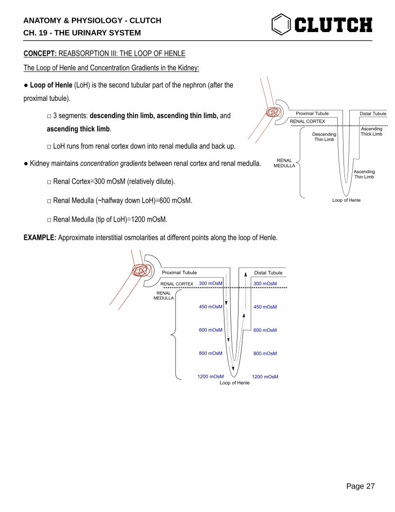
● Loop of Henle (LoH) is the second tubular part of the nephron (after the proximal tubule).
□ 3 segments: descending thin limb, ascending thin limb, and ascending thick limb.
□ LoH runs from renal cortex down into renal medulla and back up.
● Kidney maintains concentration gradients between renal cortex and renal medulla.
□ Renal Cortex≅300 mOsM (relatively dilute).
□ Renal Medulla (~halfway down LoH)≅600 mOsM.
□ Renal Medulla (tip of LoH)≅1200 mOsM.
EXAMPLE: Approximate interstitial osmolarities at different points along the loop of Henle.
Proximal Tubule
Loop of Henle
Distal Tubule
RENAL CORTEX
RENALMEDULLA
300 mOsM
450 mOsM
600 mOsM
800 mOsM
1200 mOsM
300 mOsM
450 mOsM
600 mOsM
800 mOsM
1200 mOsM
Proximal Tubule
DescendingThin Limb
Loop of Henle
AscendingThin Limb
AscendingThick Limb
Distal Tubule
RENAL CORTEX
RENALMEDULLA
ANATOMY & PHYSIOLOGY - CLUTCH
CH. 19 - THE URINARY SYSTEM
Page 27

The Descending Thin Limb and Reabsorption of H2O:
● Descending Thin Limb is very permeable to _______________, but not really to other solutes.
□ H2O gets reabsorbed all along length of descending thin limb.
-↓Volume so that [Filtrate]=[Interstitial Fluid] (remember the concentration gradient).
□ At beginning: [Filtrate]= 300 mOsM □ At end: [Filtrate]= 1200 mOsM
● Net Effect of descending thin limb on filtrate=↓Volume→↑OsM.
EXAMPLE: The descending thin limb’s high permeability to H2O allows for lots of reabsorption of H2O.
ANATOMY & PHYSIOLOGY - CLUTCH
CH. 19 - THE URINARY SYSTEM
Page 28
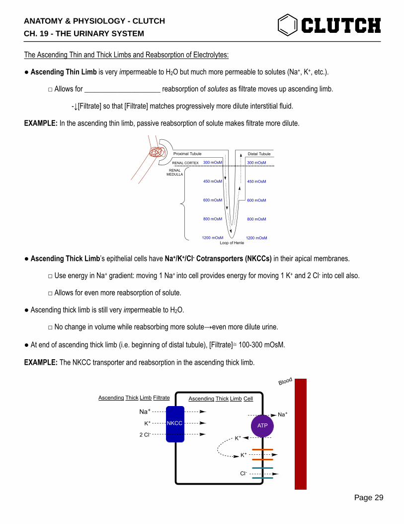
The Ascending Thin and Thick Limbs and Reabsorption of Electrolytes:
● Ascending Thin Limb is very impermeable to H2O but much more permeable to solutes (Na+, K+, etc.).
□ Allows for _____________________ reabsorption of solutes as filtrate moves up ascending limb.
-↓[Filtrate] so that [Filtrate] matches progressively more dilute interstitial fluid.
EXAMPLE: In the ascending thin limb, passive reabsorption of solute makes filtrate more dilute.
● Ascending Thick Limb’s epithelial cells have Na+/K+/Cl- Cotransporters (NKCCs) in their apical membranes.
□ Use energy in Na+ gradient: moving 1 Na+ into cell provides energy for moving 1 K+ and 2 Cl- into cell also.
□ Allows for even more reabsorption of solute.
● Ascending thick limb is still very impermeable to H2O.
□ No change in volume while reabsorbing more solute→even more dilute urine.
● At end of ascending thick limb (i.e. beginning of distal tubule), [Filtrate]≅ 100-300 mOsM.
EXAMPLE: The NKCC transporter and reabsorption in the ascending thick limb.
Proximal Tubule
Loop of Henle
Distal Tubule
RENAL CORTEX
RENALMEDULLA
300 mOsM
450 mOsM
600 mOsM
800 mOsM
1200 mOsM
300 mOsM
450 mOsM
600 mOsM
800 mOsM
1200 mOsM
Ascending Thick Limb Cell
Blood
Ascending Thick Limb Filtrate
Na+
K+
Na+
K+
ATPNKCC
2 Cl-
K+
Cl-
ANATOMY & PHYSIOLOGY - CLUTCH
CH. 19 - THE URINARY SYSTEM
Page 29

PRACTICE 1: Reabsorption in which of the following part(s) of the loop of Henle adds water volume to the renal medulla? (Choose all that apply.)
a) Descending thin limb. b) Ascending thin limb. c) Ascending thick limb.
PRACTICE 2: Reabsorption in which of the following parts of the loop of Henle adds solute content (salts, etc.) to the renal medulla? (Choose all that apply.)
a) Descending thin limb. b) Ascending thin limb. c) Ascending thick limb.
PRACTICE 3: Furosemide (brand name Lasix®) is a drug that inhibits the Na+/K+/Cl- Cotransporter (NKCC). Which of the following is the expected effect of furosemide treatment on the osmolarity of filtrate at the beginning of the distal tubule (relative to the osmolarity at that point in an untreated nephron)?
a) Increased osmolarity. b) Decreased osmolarity. c) No change in osmolarity.
ANATOMY & PHYSIOLOGY - CLUTCH
CH. 19 - THE URINARY SYSTEM
Page 30

CONCEPT: REABSORPTION IV: THE DISTAL TUBULE
The Distal Tubule and the Na+/Cl- Cotransporter
● Distal Tubule is the tubular element in the nephron after the ascending thick limb of the loop of Henle.
□ Similar properties to the ascending thick limb—active transport reabsorption of solutes, low permeability to H2O.
● Na+/Cl- Cotransporter (NCC) is the major active transporter on distal tubule epithelial cells.
□ NCCs are on the apical membrane—use energy in Na+ gradient to pull Cl- into cell.
-Na+ crosses basolateral membrane on Na+/K+ ATPase.
-Cl- diffuses across basolateral membrane through chloride channels.
● Net Effect: Solute reabsorption without water reabsorption→↓[Filtrate] (more dilute; often ~100 mOsM by end).
EXAMPLE: Reabsorption in the distal tubule happens through the NCC.
Distal Tubule Cell
Blood
DIstal Tubule Filtrate
Na+Na+
ATPNCC
Cl-
Cl-
K+
ANATOMY & PHYSIOLOGY - CLUTCH
CH. 19 - THE URINARY SYSTEM
Page 31

CONCEPT: REABSORPTION V: THE COLLECTING DUCT AND EFFECTS OF ADH AND ALDOSTERONE
The Collecting Duct and Final Determination of Urine Osmolarity:
● Collecting Duct (CD) is the final tubular element in the nephron, after the distal tubule.
□ Permeability of CD to water and solutes varies with hormonal (anti-diuretic hormone and aldosterone) levels.
● CD runs downward from renal cortex through renal medulla (just like descending loop of Henle).
□ So, filtrate in CD runs through the renal cortex→renal medulla concentration gradient.
□ If permeability to H2O is high, H2O will be reabsorbed so ↑[Filtrate] until [Filtrate]=[Renal ISF].
EXAMPLE: Water is reabsorbed out of the collecting duct as a result of concentration gradients in the renal medulla.
Loop of Henle
AscendingThin Limb
AscendingThick Limb
Distal Tubule Collecting Duct
ToBladder
300 mOsM
450 mOsM
600 mOsM
800 mOsM
1200 mOsM
300 mOsM
450 mOsM
600 mOsM
800 mOsM
1200 mOsM
H2O
H2O
H2O
H2O
ANATOMY & PHYSIOLOGY - CLUTCH
CH. 19 - THE URINARY SYSTEM
Page 32

Prinicipal Cells in the Collecting Duct and their Transporters:
● Principal Cells are the main epithelial cell type in the CD.
● Principal Cells express two important membrane channels: Aquaporins and Epithelial Na+ Channels.
□ Aquaporins (Aqs) are channels on both membranes that allow H2O to cross through the cell.
-↑Aqs→↑H2O Reabsorption.
□ Epithelial Na+ Channels (ENaCs) are apical channels that allow Na+ to enter the cell from the filtrate.
-↑ENaC→↑Na+ reabsorption. (↑Na+ reabsorption→↑H2O Reabsorption by osmosis).
EXAMPLE: A sample principal cell in the collecting duct with Aqs and ENaCs.
Collecting Duct Principal Cell
Blood
Collecting Duct Filtrate
Na+
K+
Na+
K+
ATP
ENaC
K+
AqAqH2O
ANATOMY & PHYSIOLOGY - CLUTCH
CH. 19 - THE URINARY SYSTEM
Page 33

Effects of Anti-Diuretic Hormone in the Collecting Duct:
● Anti-Diuretic Hormone (ADH) (aka vasopressin) is a peptide hormone secreted from the posterior pituitary.
□ Secreted in response to ↓Blood Pressure and ↑Blood Osmolarity.
● ADH binds to V2 Receptors on CD principal cells.
□ Effect: ↑Number of Aq channels inserted in apical membrane.
□ ↑ADH→↑Apical Aqs in CD Principal Cells→↑H2O Reabsorption→↑Blood Volume, ↓Blood OsM, and ↑[Urine]
EXAMPLE: ADH on principal cells causes more Aqs to be inserted into the apical membrane→↑H2O Reabsorption
EXAMPLE: Feedback loop for secretion and effects of ADH.
Collecting Duct Principal CellBlood
Collecting Duct Filtrate
Na+
K+
Na+
K+
ATP
ENaC
K+
AqAqH2O
ADH(+)
(More)
Blood Pressure+
Blood Osmolarity[ADH]blood
(from post. pituitary)
Vasoconstriction
Aquaporins in CD Epithelial Cells
H2O Reabsorption (filtrate→blood)
Blood Volume
UrineOsmolarity
UrineVolume
+
+
++
- +
+
--
ANATOMY & PHYSIOLOGY - CLUTCH
CH. 19 - THE URINARY SYSTEM
Page 34
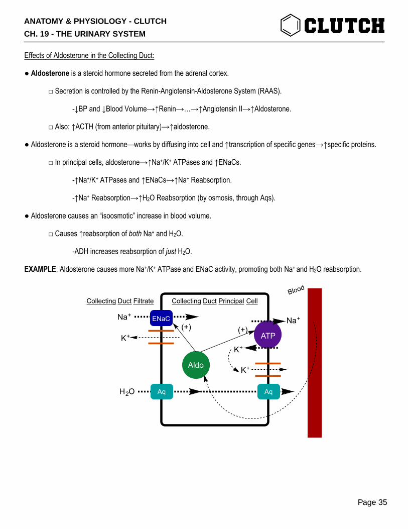
Effects of Aldosterone in the Collecting Duct:
● Aldosterone is a steroid hormone secreted from the adrenal cortex.
□ Secretion is controlled by the Renin-Angiotensin-Aldosterone System (RAAS).
-↓BP and ↓Blood Volume→↑Renin→…→↑Angiotensin II→↑Aldosterone.
□ Also: ↑ACTH (from anterior pituitary)→↑aldosterone.
● Aldosterone is a steroid hormone—works by diffusing into cell and ↑transcription of specific genes→↑specific proteins.
□ In principal cells, aldosterone→↑Na+/K+ ATPases and ↑ENaCs.
-↑Na+/K+ ATPases and ↑ENaCs→↑Na+ Reabsorption.
-↑Na+ Reabsorption→↑H2O Reabsorption (by osmosis, through Aqs).
● Aldosterone causes an “isoosmotic” increase in blood volume.
□ Causes ↑reabsorption of both Na+ and H2O.
-ADH increases reabsorption of just H2O.
EXAMPLE: Aldosterone causes more Na+/K+ ATPase and ENaC activity, promoting both Na+ and H2O reabsorption.
Collecting Duct Principal CellBlood
Collecting Duct Filtrate
Na+
K+
Na+
K+
ATP
ENaC
K+
AqAqH2O
ADH
Aldo
(+) (+)
ANATOMY & PHYSIOLOGY - CLUTCH
CH. 19 - THE URINARY SYSTEM
Page 35

PRACTICE 1: A 25-year-old man goes for a 10K run along the beach in San Diego on a hot, humid summer day. He sweats heavily and, as a result, becomes dehydrated. Which of the following describes the effect of this dehydration on his blood levels of anti-diuretic hormone (ADH)?
a) Increased ADH. b) Decreased ADH. c) No change in ADH.
PRACTICE 2: A 25-year-old man goes for a 10K run along the beach in San Diego on a hot, humid summer day. He sweats heavily and, as a result, becomes dehydrated. Which of the following describes the number of aquaporin channels inserted in the apical membrane of his collecting duct principal cells, relative to a normally hydrated person?
a) More aquaporins. b) Less aquaporins. c) No difference in aquaporins.
PRACTICE 3: A 25-year-old man goes for a 10K run along the beach in San Diego on a hot, humid summer day. He sweats heavily and, as a result, becomes dehydrated. After his run he stops at the bathroom to urinate. Which of the following describes the osmolarity of his urine relative to a normally hydrated person?
a) Higher osmolarity. b) Lower osmolarity. c) No difference in osmolarity.
ANATOMY & PHYSIOLOGY - CLUTCH
CH. 19 - THE URINARY SYSTEM
Page 36
CONCEPT: CLEARANCE—MEASURING GLOMERULAR FILTRATION RATE AND RENAL PLASMA FLOW
Excretion and Clearance:
● Excretion is what actually gets peed out after filtrate contents is modified by the nephron.
□ Excretion=Filtration+Secretion-Reabsorption.
□ Rate of excretion measured by calculating clearance.
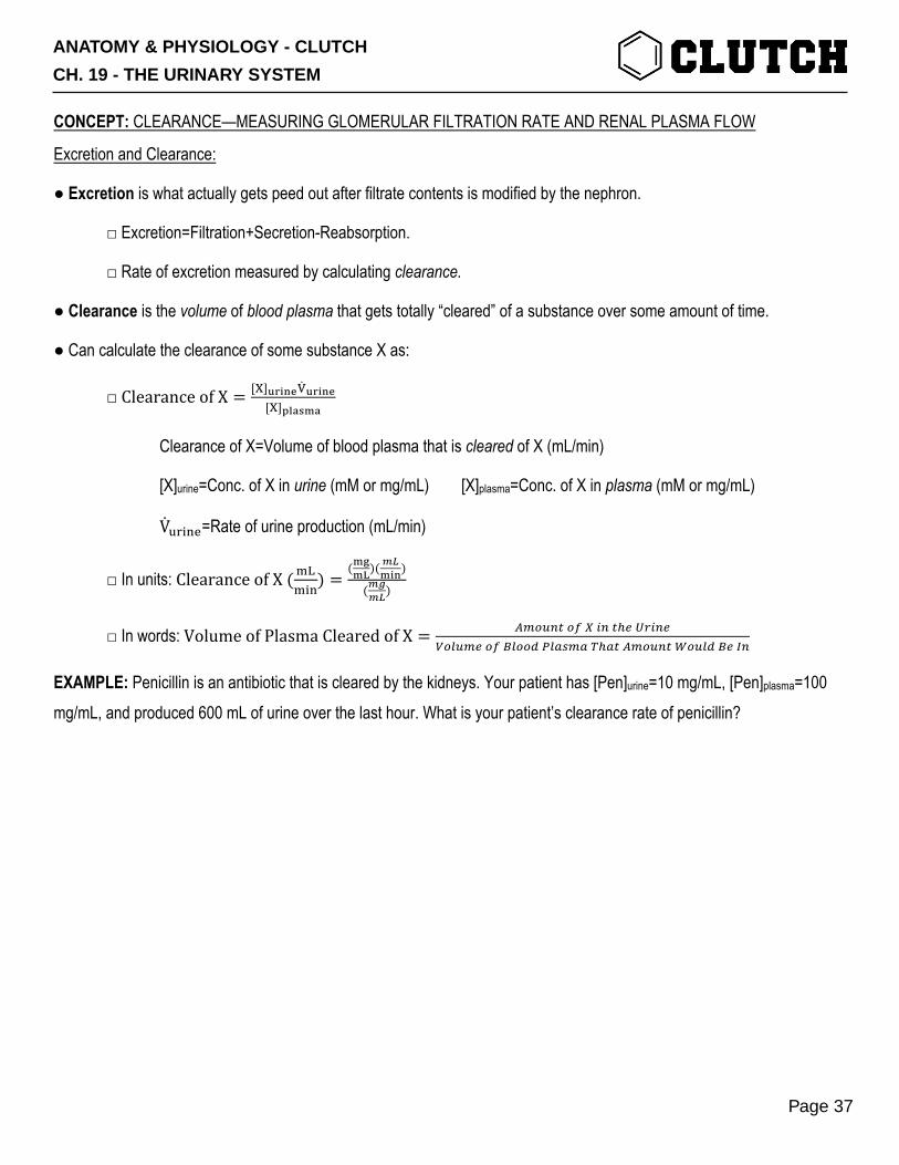
● Clearance is the volume of blood plasma that gets totally “cleared” of a substance over some amount of time.
● Can calculate the clearance of some substance X as:
□ ClearanceofX = [.]01234501234[.]6789:8
Clearance of X=Volume of blood plasma that is cleared of X (mL/min)
[X]urine=Conc. of X in urine (mM or mg/mL) [X]plasma=Conc. of X in plasma (mM or mg/mL)
V<=>?@=Rate of urine production (mL/min)
□ In units: ClearanceofX(BCB>?
) =(:E:F)(
GH:23)
(GIGH)
□ In words:VolumeofPlasmaClearedofX = OPQRSTQUVWSTXYZ[WSY\Q]RPYQU^]QQ_`]abPacXaTOPQRSTdQR]_^YeS
EXAMPLE: Penicillin is an antibiotic that is cleared by the kidneys. Your patient has [Pen]urine=10 mg/mL, [Pen]plasma=100 mg/mL, and produced 600 mL of urine over the last hour. What is your patient’s clearance rate of penicillin?
ANATOMY & PHYSIOLOGY - CLUTCH
CH. 19 - THE URINARY SYSTEM
Page 37

Glomerular Filtration Rate—Definition and Measurement:
● Glomerular Filtration Rate (GFR) is how much fluid moves from the glomerulus into Bowman’s capsule per minute.
□ Normally ≅125 mL.
● Inulin is a substance injected into the blood that is used to measure ___________.
□ Inulin filters easily from the glomerulus into Bowman’s capsule.
□ After being filtered, inulin is never touched by the nephron—no reabsorption or secretion.
-All inulin that is filtered gets ______________________.
-All fluid that is filtered loses all of its inulin (while lots of the H2O, Na+, etc. from that fluid gets reabsorbed).
● To measure GFR, you can inject inulin, wait some time, and calculate clearance.
□ 𝐺𝐹𝑅 = ClearanceofInulin = [k?<l>?]01234501234[k?<l>?]6789:8
● Creatinine is a waste product from the body that is handled very similarly to inulin by the kidneys.
□ Can also be used to calculate GFR.
EXAMPLE: You have a patient with [Inulin]plasma=6 mg/mL, [Inulin]urine=30 mg/mL, Vurine=120 mL in 1 hour. Calculate this patient’s
GFR. Is this patient healthy?
AfferentArteriole
EfferentArteriole
Bowman'sCapsule
Nephron
To BeExcreted
Back toCirculation
PeritubularCapillaries
ANATOMY & PHYSIOLOGY - CLUTCH
CH. 19 - THE URINARY SYSTEM
Page 38
Filtered Loads—Comparing to GFR to Determine Renal Handling:
● Filtered Load is the amount of a substance that gets filtered into Bowman’s capsule from the glomerulus.
□ Filtered Load of X (mg/min)= [X]plasma (mg/mL)×GFR (mL/min)
□ The amount of a substance that gets into nephron before reabsorption/secretion starts happening.
● Comparing filtered load and clearance tells you how a substance is handled by the kidneys.
□ If filtered load>clearance→substance is reabsorbed.
□ If filtered load<clearance→substance is secreted.
● Can do something similar by comparing clearance of some substance with clearance of inulin.
□ Works since inulin is neither reabsorped nor secreted→filtered load in inulin=clearance of inulin.
□ If clearance<clearanceinulin→substance is reabsorbed.
□ If clearance>clearanceinulin→substance is secreted.
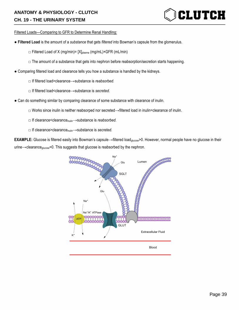
EXAMPLE: Glucose is filtered easily into Bowman’s capsule→filtered loadglucose>0. However, normal people have no glucose in their
urine→clearanceglucose=0. This suggests that glucose is reabsorbed by the nephron.
Lumen
Extracellular Fluid
Blood
SGLT
Na+/K+ ATPase
Na+
K+
Na+
Glu
Glu
GLUT
ATP
ANATOMY & PHYSIOLOGY - CLUTCH
CH. 19 - THE URINARY SYSTEM
Page 39

Renal Plasma Flow—Definition and Measurement:
● Renal Plasma Flow (RPF) is the volume of blood plasma going to the kidneys per minute.
□ Normally ≅500-600 mL.
● Para-amino hippuric acid (PAH) is a substance injected into the blood that is used to measure RPF.
□ PAH filters easily from the glomerulus into Bowman’s capsule and is secreted into the proximal tubule.
-PAH is not reabsorbed.
□ All the PAH in the plasma coming in to the kidney is either filtered or secreted.
-Plasma leaving the kidney has no PAH.
-All the plasma that enters the kidney is fully cleared of PAH.
-So, ClearancePAH=RPF.
● To measure RPF, you can inject PAH, wait some time, and calculate clearance.
□ 𝑅𝑃𝐹 = ClearanceofPAH = [pqr]01234501234[pqr]6789:8
EXAMPLE: You have a patient with [PAH]plasma=10 mg/mL, [PAH]urine=600 mg/mL, Vurine=600 mL in 1 hour. Calculate this patient’s
RPF.
AfferentArteriole
EfferentArteriole
Bowman'sCapsule
Nephron
To BeExcreted
Back toCirculation
PeritubularCapillaries
ANATOMY & PHYSIOLOGY - CLUTCH
CH. 19 - THE URINARY SYSTEM
Page 40

CONCEPT: URINARY TRACT
● The urinary tract includes the ureters, urinary bladder, and urethra; stores, transports, and eliminates urine from the body □The ureters are paired, long, muscular tubes that transport urine from the kidneys to the urinary bladder
- Originate at the renal pelvis, descends towards the urinary bladder, running anterior to the psoas major muscles
- Are ____________________________ and has three tunics
1. Mucosa has transitional epithelium that is impermeable to urine
2. Muscularis has an inner and outer smooth muscle layers which contract to propel urine through ureters
3. Adventitia is made of collagen and elastic tissue to anchor ureter to posterior abdominal wall
- They penetrate the posterior wall of the urinary bladder through the ureteral opening at an __________ angle
EXAMPLE:
ANATOMY & PHYSIOLOGY - CLUTCH
CH. 19 - THE URINARY SYSTEM
Page 41
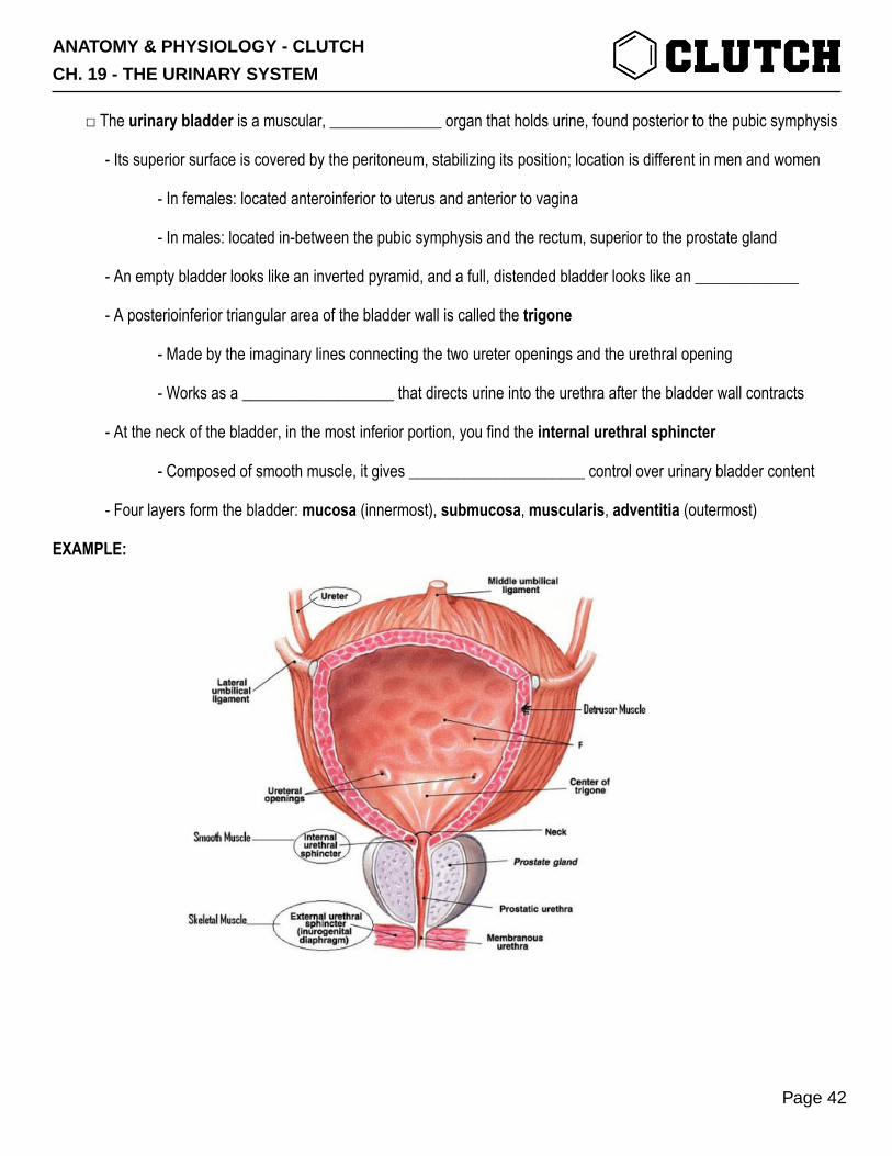
□ The urinary bladder is a muscular, ______________ organ that holds urine, found posterior to the pubic symphysis
- Its superior surface is covered by the peritoneum, stabilizing its position; location is different in men and women
- In females: located anteroinferior to uterus and anterior to vagina
- In males: located in-between the pubic symphysis and the rectum, superior to the prostate gland
- An empty bladder looks like an inverted pyramid, and a full, distended bladder looks like an _____________
- A posterioinferior triangular area of the bladder wall is called the trigone
- Made by the imaginary lines connecting the two ureter openings and the urethral opening
- Works as a ___________________ that directs urine into the urethra after the bladder wall contracts
- At the neck of the bladder, in the most inferior portion, you find the internal urethral sphincter
- Composed of smooth muscle, it gives ______________________ control over urinary bladder content
- Four layers form the bladder: mucosa (innermost), submucosa, muscularis, adventitia (outermost)
EXAMPLE:
ANATOMY & PHYSIOLOGY - CLUTCH
CH. 19 - THE URINARY SYSTEM
Page 42

□ The urethra is a muscular tube that runs from the neck of the bladder to the external urethral opening
- In both sexes the urethra ___________________ in both length and function
- In females: only functions to transports urine out of the body; is about 4cm long
- In males: functions to transports urine and semen out of the body; 19cm long; has 3 segments
1. Prostatic urethra: most dilated portion, extends through the prostate gland inferior to bladder
2. Membraneous urethra: least dilated & shortest portion, extends through urogenital diaphragm
- Has the skeletal muscle that forms the external urethral sphincter
3. Spongy urethra (penile urethra): longest portion, extends through penis;
- Has the has corpus spongiosum
- Extends to external urethral orifice
- Has an external urethral sphincter composed of skeletal muscle, giving __________ control of urine elimination
EXAMPLE:
ANATOMY & PHYSIOLOGY - CLUTCH
CH. 19 - THE URINARY SYSTEM
Page 43


