121
Last Updated: August 8, 2013 Anatomy & Physiology Model Guide Book

Last Updated: August 8, 2013
Anatomy & Physiology Model Guide Book

ii

iii
Table of Contents
Tissues ........................................................................................................................................................... 7
The Bone (Somso QS 61) ........................................................................................................................... 7
Section of Skin (Somso KS 3 & KS4) .......................................................................................................... 8
Model of the Lymphatic System in the Human Body ............................................................................. 11
Bone Structure ........................................................................................................................................ 12
Skeletal System ........................................................................................................................................... 13
The Skull .................................................................................................................................................. 13
Artificial Exploded Human Skull (Somso QS 9)........................................................................................ 14
Skull ......................................................................................................................................................... 15
Auditory Ossicles ..................................................................................................................................... 16
Thorax ..................................................................................................................................................... 17
Vertebral Column .................................................................................................................................... 17
Vertebrae ................................................................................................................................................ 18
Scapula & Humerus ................................................................................................................................. 20
Radius, Ulna, & Hand .............................................................................................................................. 21
Clavicle & Pelvis ...................................................................................................................................... 22
Femur, Tibia, Fibula, Patella .................................................................................................................... 23
Foot ......................................................................................................................................................... 24
Human Skeleton ...................................................................................................................................... 25
Muscular System ......................................................................................................................................... 27
Muscular Arm with Hand ........................................................................................................................ 27
Leg Muscle .............................................................................................................................................. 28
Cartilages of the Larynx (Somso GS 6) .................................................................................................... 30
Functional Model of the Knee Joint ........................................................................................................ 31
Functional Model of the Shoulder Joint.................................................................................................. 32
Functional Model of the Elbow Joint ...................................................................................................... 33
Functional Model of the Hip Joint........................................................................................................... 34
Muscles of the Arm with Shoulder Blade (Somo NS 15) ......................................................................... 35
Muscles of the Leg with Base of Pelvis (Somso NS 10) ........................................................................... 38
iv
Male Muscle Figure (Somso AS 3) ........................................................................................................... 40
Muscles of the Human Arm (Altay 6000.31) ........................................................................................... 41
Head & Neck Musculature (Altay 6030.10) ............................................................................................ 42
Cardiovascular/Respiratory System ............................................................................................................ 43
Two Section Heart (HE-1010) .................................................................................................................. 43
Human Heart & Mediastinum ................................................................................................................. 44
The Heart (Somso HS 26) ........................................................................................................................ 45
Model of Heart (ALS 300) ........................................................................................................................ 46
The Heart (HS 4) ...................................................................................................................................... 48
Vascular Arm ........................................................................................................................................... 49
Fine Structure of the Artery & Veins (Somso HS 25) .............................................................................. 50
Respiratory Organs & Heart .................................................................................................................... 51
Lungs, Larynx & Heart (C 243) ................................................................................................................. 52
Lungs with Heart, Diaphragm, & Larynx (Somso HS 8) ........................................................................... 53
Lung with Alveoli (Somso HS 23) ............................................................................................................ 54
Lung Apparatus ....................................................................................................................................... 55
Lung (Basic Model) .................................................................................................................................. 57
Nervous System .......................................................................................................................................... 59
Neuron (Somso BS 35) ............................................................................................................................ 59
Model of a Synapse (BS 35/3) ................................................................................................................. 60
Striated Muscle Fiber with Motor End-Plate (Somso BS 36) .................................................................. 61
Transparent Human Brain (BS 25) .......................................................................................................... 62
Transparent Model of the Skull (Somso QS 65/7) .................................................................................. 64
The Human Brain (Somso BS 23) ............................................................................................................ 65
Brain Stem (Somso BS 23/2) ................................................................................................................... 66
The Human Brain (C 15) .......................................................................................................................... 67
Brain with Arteries .................................................................................................................................. 68
Brain (Basic Model) ................................................................................................................................. 69
Fifth Cervical Vertebra with Spinal Cord (Somso BS 30) ......................................................................... 70
Model of Spinal Cord with Spinal Canal (Somso BS 31) .......................................................................... 71
Deluxe Spinal Cord .................................................................................................................................. 72

v
Spinal Cord with Nerve Branches (Somso BS 33) .................................................................................... 73
Neuron (Altay 6160.27) ........................................................................................................................... 74
Special Senses ............................................................................................................................................. 75
The Human Eye (Somso CS 20) ............................................................................................................... 75
Eye in Orbit.............................................................................................................................................. 76
Giant Eye ................................................................................................................................................. 77
The Ear (J 515) ......................................................................................................................................... 78
Ear (P-2134)............................................................................................................................................. 79
The Organ of Hearing (Somso DS 3) ........................................................................................................ 80
The Labyrinth (Somso DS 6) .................................................................................................................... 81
Spiral Organ of Corti (Somso DS 10) ....................................................................................................... 82
Nose & Nasal Cavity (Somso FS 6) .......................................................................................................... 83
Urinary System ............................................................................................................................................ 85
Kidney (K 10) ........................................................................................................................................... 85
Kidney & Nephron ................................................................................................................................... 86
Kidney (Somso LS 5) ................................................................................................................................ 87
Kidney (Somso K 12) ............................................................................................................................... 88
Glomerulus .............................................................................................................................................. 89
Renal Lobule ............................................................................................................................................ 90
Urinary System ........................................................................................................................................ 91
Kidney (Altay 6140.14) ............................................................................................................................ 92
Reproductive System .................................................................................................................................. 93
Male Pelvis (Somso MS 2) ....................................................................................................................... 93
Male Pelvis .............................................................................................................................................. 94
Male Pelvis (H 11) ................................................................................................................................... 95
Female Pelvis .......................................................................................................................................... 98
Female Pelvis (Somso MS 3) ................................................................................................................... 99
Human Female Breast ........................................................................................................................... 100
Female Genital Organs (Somso MS 5)................................................................................................... 101
Uterus (Basic Model) ............................................................................................................................. 102
Female Pelvis (H 10) .............................................................................................................................. 103

vi
Relief Model of the Ovary (Somso MS 51) ............................................................................................ 106
Digestive System ....................................................................................................................................... 107
Molar Tooth (Somso ES 11/5) ............................................................................................................... 107
Models of Teeth .................................................................................................................................... 108
Stomach (Basic Model) ......................................................................................................................... 109
The Stomach (Somso JS 4) .................................................................................................................... 110
Pancreas with Spleen & Part of the Duodenum (Somso JS 11) ............................................................ 111
Intestinal Villi ........................................................................................................................................ 112
Model of a Liver Cell (Somso JS 15) ...................................................................................................... 113
Internal Organs ..................................................................................................................................... 114
Liver & Gallbladder ............................................................................................................................... 115
Liver ....................................................................................................................................................... 116
Human Digestive System ...................................................................................................................... 117
Liver/Gallbladder (Basic Model) ........................................................................................................... 119
Colon (Basic Model) .............................................................................................................................. 120
Rectum (Basic Model) ........................................................................................................................... 121

7 | T i s s u e s
Tissues
The Bone (Somso QS 61)
1. Periosteum
2. Outer general lamellae
3. Perforating fibers of Sharpey
4. Osteon with Haversian vessel
Presentation of the spiral run of
the collagenous fibrils within
the single lamellae
5. Osteon w/presentation of the
flattened bone cells
6. Bone cell
7. Branch of the bone cell
8. Intermediate lamellae
9. Volkmann’s canals and vessels
10. Endosteum
11. Spongy substance
12. Cavities for the bone cells in the
macerated bone
Somso Model QS 61 Wedge-shaped segment from the compact part of a long bone

8 | T i s s u e s
Section of Skin (Somso KS 3 & KS4)
I. Epidermis
II. Corium (Dermis)
III. Subcutis (Hypodermis)
1. External Horny Layer (Stratum corneum)
1a. Clear Layer (Stratum lucidum) – (KS 3 only)
2. Internal Hornless Germinative Zone (Stratum germinativum)
2a. Granular Layer (Stratum granulosum)
2b. Prickle-cell Layer (Stratum spinosum)
2c. Cylindrical Layer (Stratum basale)
3. Papillae
4. Touch-corpuscles (Meissner’s corpuscles)
5. Adipose Tissue (Panniculus adiposus)
6. Lamellated Corpuscles (Pacinian corpuscles)
7. Sweat Glands (Eccrine glands)
8. Hairs (Pili)
8a. Medullary Substance (Substantia medullaris)
8b. Cortical Substance (Substantia corticalis)
8c. Cuticle of the Hair (Cuticula pili)
8d. Inner Root-Sheath
8e. Outer Root-Sheath
8f. Hyaloid Membrane
8g. Fibrous Layer
9. Hair (Pilus)
Somso Model KS 3
Block model with representation in three sections:
A.The hairy skin; B.The skin of the arm pit; C.The
hairless skin of the sole of the foot.
Somso Model KS 4
Block model showing the skin with hair
in different planes of section.

9 | T i s s u e s
Section of Skin Continued… 10. Hair Shaft (Scapus pili)
11. Hair Root (Radix pili)
11a. Hair Bulb (Bulbus pili)
12. Hair Papilla (Papilla pili)
13. Sebaceous Glands (Glandulae sebaceae)
14. Arrector Pili Muscles (Arrectores pilorum)
15. Sweat Gland of the Arm-Pit (Apocrine gland)
15a. Smooth Muscle Cells
15b. Hyaline Top
15c. Part of the pushed out cell body
Model KS 4 shows additionally…
16. Krause’s end bulbs – with nerve coils in connective tissue cover
17. Ruffini’s corpuscles – plexus of bare nerve fiber in connective tissue cover in the corium (dermis)
and subcutis (hypodermis)
18. Merkel’s touch disks in the epidermis
19. Mechanoreceptors as dendriform fibers in the hair follicle

10 | T i s s u e s
Cutaneous Sensory Organs…
a) Free nerve endings - For pain & heat sensitivity
b) Merkel’s touch cells in the epithelium
c) Meissner’s tactile corpuscles - For pressure & touch sensitivity
d) Nerves encircling the root sheath of the hair - For touch sensitivity
e) Vater-Pacini corpuscles - Mechanoreceptors
f) Krause’s end corpuscle - Receptors of frost
Hair Organs…
1-8. Layers of skin
9. Hair shaft
10. Sebaceous gland
11. Hair follicle
12. External root sheath
13. Arrector pili muscle
14. Internal root sheath
15. Hair shaft inside its root sheathes
16. Hair bulb
17. Papilla with blood vessel
Vessels and glands of the skin…
I. Capillary loops in papilla
II. Principal venous network (blue) with supply arteries (red)
III. Sweat gland outlets
IV. Sweat gland

11 | T i s s u e s
Model of the Lymphatic System in the Human Body
1. Occipital lymph nodes
2. Superficial cervical lymph nodes
3. Cubital lymph nodes
4. Deep cervical lymph nodes
5. Axillary lymph nodes
6. Right lymphatic duct
7. Tracheobronchial lymph nodes
8. Hepatic lymph nodes
9. Coeliac lymph nodes
10. Common iliac lymph nodes
11. Inguinal lymph nodes
12. Parotid gland lymph nodes
13. Submandibular lymph nodes
14. Submental lymph nodes
15. Jugular trunk
16. Thoracic duct
17. Subclavian trunk
18. Bronchial lymph nodes
19. Cisterna chili
20. Gastric lymph nodes
21. Mesenteric lymph nodes
a. Right venous angle
b. Right lung
c. Liver
d. Gall bladder
e. Portal vein
f. Caecum with appendix vermiformis
g. Bifurcation of trachea
h. Stomach
i. Transverse colon

12 | T i s s u e s
Bone Structure
1. Artery
2. Vein
3. Lymph Vessel
4. Nerve
5. Haversian lamellae
6. Interstitial lamellae
7. Osteocytes
8. Lacunae
9. Canaliculi
These structures all lie in the Haversian canal

13 | S k e l e t a l S y s t e m
Skeletal System
The Skull
A. Coronal suture
B. Coronoid process
C. Ethmoid
D. External acoustic meatus
E. Frontal
F. Sphenoid
G. Lacrimal
H. Mandible
I. Mandibular condyle
J. Mandibular fossa
K. Maxilla
L. Mastoid process
M. Nasal
N. Parietal
O. Squamosal suture
P. Styloid process
Q. Superior orbital fissure
R. Temporal
S. Zygomatic
T. Zygomatic process of temporal
14 | S k e l e t a l S y s t e m
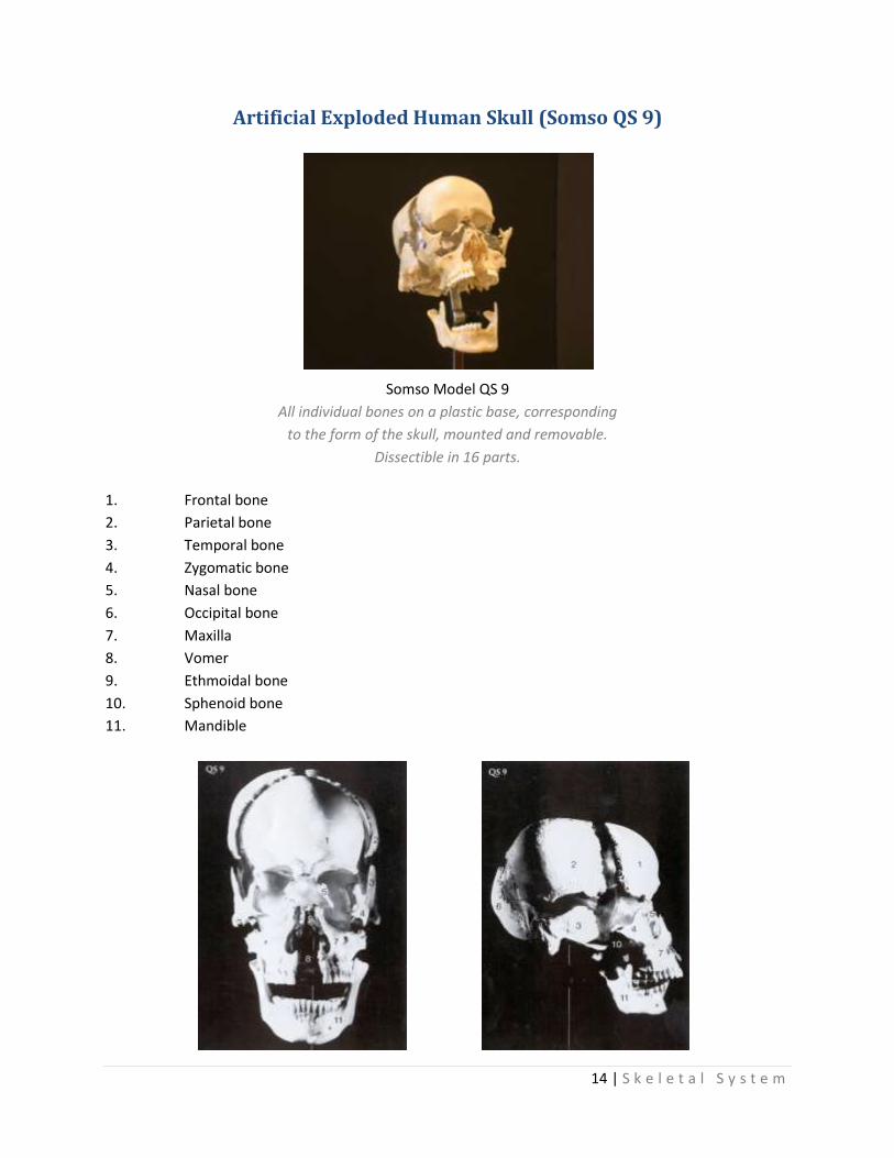
Artificial Exploded Human Skull (Somso QS 9)
1. Frontal bone
2. Parietal bone
3. Temporal bone
4. Zygomatic bone
5. Nasal bone
6. Occipital bone
7. Maxilla
8. Vomer
9. Ethmoidal bone
10. Sphenoid bone
11. Mandible
Somso Model QS 9
All individual bones on a plastic base, corresponding
to the form of the skull, mounted and removable.
Dissectible in 16 parts.

15 | S k e l e t a l S y s t e m
Skull (Lateral View)
A. Frontal
B. Parietal
C. Occipital
D. Temporal
E. Sphenoidal
F. Ethmoid
G. Lacrimal
H. Nasal
I. Zygomatic
J. Maxilla
K. Mandible
L. Styloid Process
a. Coronal Suture
b. Squamosal Suture
c. Lambdoidal Suture

16 | S k e l e t a l S y s t e m
Auditory Ossicles

17 | S k e l e t a l S y s t e m
Thorax
A. Manubrium
B. Body
C. Xiphoid Process
D. Costal Cartilage
E. False Ribs
F. Vertebrosternal or True Ribs
G. Vertebrocostal Ribs
Vertebral Column (Lateral View)

18 | S k e l e t a l S y s t e m
Vertebrae
Atlas (First Cervical Vertebra)
A. Transverse Process
B. Anterior Tubercle
C. Anterior Arch
D. Superior Articular Surface
E. Foramen Transversarium
F. Posterior Arch
G. Posterior Tubercle
Axis (Second Cervical Vertebra)
A. Odontoid Process
B. Body
C. Superior Articular Surface
D. Foramen Transversarium
E. Spinous Process
Cervical Vertebra
A. Body
B. Transverse Process
C. Pedicle
D. Superior Articular Process
E. Inferior Articular Process
F. Lamina
G. Spinous Process
H. Foramen Transversarium
Thoracic Vertebra
A. Transverse Process
B. Superior Articular Process
C. Pedicle
D. Demi-facet for head of rib
E. Body
F. Demi-facet for head of rib
G. Inferior Articular Process
H. Spinous Process
I. Facet for articular part of tubercle of rib

19 | S k e l e t a l S y s t e m
Vertebrae Cont…
Lumbar Vertebra (lateral view)
A. Superior Articular Process
B. Transverse Process
C. Pedicle
D. Body
E. Inferior Articular Process
F. Spinous Process
Lumbar Vertebra (from above and behind)
A. Transverse Process
B. Inferior Articular Process
C. Mammilary Process
D. Accessory Process
E. Superior Articular Process

20 | S k e l e t a l S y s t e m
Scapula & Humerus
Left Scapula (Dorsal View)
A. Acromion
B. Coracoid Process
C. Scapular Notch
D. Superior Border
E. Medial Angle
F. Supraspinatous Fossa
G. Spine
H. Infraspinatous Fossa
I. Vertebral Border
J. Inferior Angle
K. Axillary Border
L. Neck of Scapula
M. Lateral Angle
Left Scapula (Lateral View)
A. Coracoid
B. Supraglenoid Tubercle
C. Acromion
D. Spine
E. Glenoid Cavity
F. Axillary Border
G. Inferior Angle
Left Humerus (Anterior View)
A. Head
B. Greater Tubercle
C. Crest of Greater Tubercle
D. Intertubercular Groove
E. Radial Fossa
F. Lateral Epicondyle
G. Capitulum
H. Coronoid Fossa
I. Trochlea
J. Medial Epicondyle
K. Surgical Neck

21 | S k e l e t a l S y s t e m
Radius, Ulna, & Hand
Left Radius (Anterior Aspect)
A. Head
B. Neck
C. Styloid Process
D. Radial Tuberosity
Left Ulna (Anterior Aspect)
A. Olecranon
B. Semilunar Notch
C. Coronoid Process
D. Radial Notch
E. Articulates with Radius
F. Styloid Process
Left Hand (Dorsal Surface)
A. Navicular
B. Lunate
C. Triangular
D. Pisiform
E. Hamate
F. Capitate
G. Lesser Multangular
H. Greater Multangular
a. Base
b. Body
c. Head
22 | S k e l e t a l S y s t e m
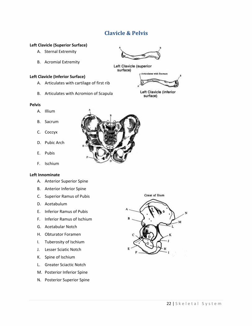
Clavicle & Pelvis
Left Clavicle (Superior Surface)
A. Sternal Extremity
B. Acromial Extremity
Left Clavicle (Inferior Surface)
A. Articulates with cartilage of first rib
B. Articulates with Acromion of Scapula
Pelvis
A. Illium
B. Sacrum
C. Coccyx
D. Pubic Arch
E. Pubis
F. Ischium
Left Innominate
A. Anterior Superior Spine
B. Anterior Inferior Spine
C. Superior Ramus of Pubis
D. Acetabulum
E. Inferior Ramus of Pubis
F. Inferior Ramus of Ischium
G. Acetabular Notch
H. Obturator Foramen
I. Tuberosity of Ischium
J. Lesser Sciatic Notch
K. Spine of Ischium
L. Greater Sciactic Notch
M. Posterior Inferior Spine
N. Posterior Superior Spine

23 | S k e l e t a l S y s t e m
Femur, Tibia, Fibula, Patella
Left Femur (Dorsal View)
A. Head
B. Neck
C. Tubercle
D. Body
E. Adductor Tubercle
F. Medial Epicondyle
G. Medial Condyle
H. Lateral Condyle
I. Lateral Epicondyle
(A) Tibia (Anterior View)
A. Intercondyloid
B. Lateral Condyle
C. Medial Condyle
D. Tuberosity
E. Medial Malleolus
(B) Fibula (Anterior View)
a. Lateral Malleolus
b. Styloid Process
Patella (Posterior View)
A. Facet for Medial Condyle
B. Facet for Articulation with lateral condyle of femur

24 | S k e l e t a l S y s t e m
Foot
Left Foot (Dorsal Surface)
A. Calcaneus
B. Talus
C. Navicular
D. First Cuneiform
E. Second Cuneiform
F. Third Cuneiform
G. Cuboid
Left Foot (Lateral Aspect)
A. Second Cuneiform
B. Third Cuneiform
C. Navicular
D. Talus
E. Calcaneus
F. Cuboid

25 | S k e l e t a l S y s t e m
Human Skeleton
1. Frontal
2. Parietal
3. Ethmoid
4. Temporal
5. Zygomatic
6. Occipital
7. Nasal
8. Maxilla
9. Mandible
10. Hyoid
11. Cervical Vertebra (7)
12. Manubrium
13. Clavicle
14. Coracoid
15. Scapula
16. Sternum
17. Xiphoid Process
18. Ribs
19. Thoracic Vertebra (12)
20. Humerus
21. Radius
22. Ulna
23. Carpals
24. Metacarpals
25. Phalanges
26. Lumbar Vertebra (5)
27. Illium
28. Sacrum (5)
29. Coccyx (3-5)
30. Pubis
31. Ischium
32. Femur
33. Patella
34. Tibia
35. Fibula
36. Talus
37. Calcaneus
38. Tarsals
39. Metatarsals

26 | S k e l e t a l S y s t e m

27 | M u s c u l a r S y s t e m
Muscular System
Muscular Arm with Hand
1. Clavicle
2. Acromion
3. Spine of Scapula
4. Scapula
5. Humerus
6. Ulna
7. Radius
8. Metacarpal Bones
9. Deltoid Muscle
10. Supraspinatus Muscle
11. Infraspinatus Muscle
12. Subclavius Muscle
13. Teres Minor Muscle
14. Teres Major Muscle
15. Latissimus Dorsal Muscle
16. Triceps Muscle
17. Biceps (brachii) Muscle
18. Coraco-brachialis Muscle
19. Brachialis Muscle
20. Anconeus Muscle
21. Brachio-radialis Muscle
22. Extensor Carpi Radialis Longus
23. Extensor Carpi Radialis Brevis
24. Pronator Teres Muscle
25. Flexor Carpi Ulnaris Muscle
26. Palmaris Longus Muscle
27. Flexor Carpi Ulnaris Muscle
28. Flexor Digitorum Sublimis
29. Flexor Digitorum Profundus
30. Abductor Pollicis Longus
31. Flexor Pollicis Longus
32. Extensor Pollicis Brevis
33. Extensor Digitorum Communis
34. Extensor Digiti Minimi
35. Extensor Carpi Ulnaris
36. Dorsal Interosseous
37. Extensor Pollicis Longus
38. Abductor Pollicis Brevis
39. Flexor Pollicis Brevis
40. Adductor Pollicis
41. Lumbricales Muscles
42. Flexor Digiti Minimi
43. Abductor Digiti Minimi
44. Extensor Retinaculum
45. Flexor Retinaculum
46. Brachial Artery
47. Radial Artery
48. Brachial Vein
49. Median Nerve
50. Annular Ligaments of Tendon-Sheath
51. Cruciate Ligaments of Tendon-Sheath

28 | M u s c u l a r S y s t e m
Leg Muscle
1. Tibia, Shinbone
2. Fibula, Calf bone, Peroneal bone
3. Calcaneum, Heel bone
4. Metatarsal bones
5. Patella
6. Popliteal Space
7. Rectus femoris Muscle
8. Vastus Lateralis Muscle
9. Vastus Medialis Muscle
10. Sartorius Muscle
11. Ilio-psoas Muscle
12. Pectineus Muscle
13. Adductor Longus Muscle
14. Gracilis Muscle
15. Semimembranosus Muscle
16. Semitendinosus Muscle
17. Adductor Magnus Muscle
18. Biceps Femoris Muscle (Long head)
19. Biceps Femoris Muscle (Short head)
20. Gluteus Maximus Muscle
21. Plantaris Muscle
22. Popliteus Muscle
23. Tibialis Anterior Muscle
24. Extensor Digitorum Longus
25. Extensor Hallucis Longus
26. Peroneus Longus
27. Peroneus Brevis
28. Peroneus Tertius
29. Gastrocnemius Muscle
30. Calcaneal (Achilles) Tendon
31. Soleus Muscle
32. Flexor Digitorum Longus
33. Flexor Hallucis Longus

29 | M u s c u l a r S y s t e m
Leg Muscle cont…
34. Tibialis Posterior Muscle
35. Abductor Hallucis Muscle
36. Abductor Digiti Minimi Muscle
37. Extensor Hallucis Brevis
38. Extensor Digitorum Brevis
39. Dorsal Interosseous Muscle
40. Superior Extensor Retinaculum
41. Patellae Ligament
42. Femoral Artery
43. Deep Femoral Artery
44. Popliteal Artery
45. Posterior Tibial Artery
46. Peroneal Artery
47. Dorsal Artery
48. Femoral Nerve
49. Tibial Nerve
50. Sural Nerve

30 | M u s c u l a r S y s t e m
Cartilages of the Larynx (Somso GS 6)
1. Thyroid cartilage
a. Incisura thyroidea superior
b. Incisura thyroidea inferior
c. Upper long horn (Cornu superior)
d. Lower short horn (Cornu inferius)
2. Cricoid cartilage
3. Cartilage of the epiglottis
4. Arytenoid cartilages
e. Processus vocalis
f. Processus muscularis
g. Cartilage of Santorini
5. Tracheal cartilage
6. Hyoid bone
h. Corpus
i. Greater horn (Cornu majus)
j. Lesser horn (Cornu minus)
Somso Model GS 6 – Functional Model

31 | M u s c u l a r S y s t e m
Functional Model of the Knee Joint

32 | M u s c u l a r S y s t e m
Functional Model of the Shoulder Joint
1. Articular capsule (Capsula articularis)
2. Supplemental ligaments of the articular capsule (Lig. Glenohumeralia)
3. Tendon of the biceps muscle
4. Coracoacromial ligament (Lig. Coracoacromiale)
5. External clavicular ligaments (Lig. Coracoclaviculare)
a. Conoid ligament (Lig. Conoideum)
b. Trapezoid ligament (Lig. Trapezoideum)
6. Transverse ligament of the scapula (Lig. Transversum scapulae)
7. Lig. Acromioclaviculare
I. Scapula
II. Clavicle
III. Humerus
Somso Model NS 53 – Functional Model
Shoulder-joint model from
the front (ventral)

33 | M u s c u l a r S y s t e m
Functional Model of the Elbow Joint
1. Internal or ulnar lateral ligament (Ligamentum collaterale mediale)
2. Radial ligament (Ligamentum radiale)
3. Orbicular ligament (Ligamentum anulare)
4. Bicipital tendon
I. Humerus
II. Ulna
III. Radius
Somso Model NS 52 – Functional Model
Elbow-joint model from
the front (ventral)

34 | M u s c u l a r S y s t e m
Functional Model of the Hip Joint
1. Ilio-femoral ligament (Ligamentum iliofemorale)
2. Pubo-femoral ligament (Ligamentum pubofemorale)
3. Ishio-capsular ligament (Ligamentum ishiofemorale)
4. Orbicular zone (Zona orbicularis)
5. Teres femoris ligament (Ligamentum capitis femoris)
I. Hip bone (Os coxae)
II. Femur
III. Head of the femur (Caput femoris)
Somso Model NS 51 – Functional Model
Hip-joint model from
the front (ventral)
Hip-joint model from
the back (dorsal)
35 | M u s c u l a r S y s t e m
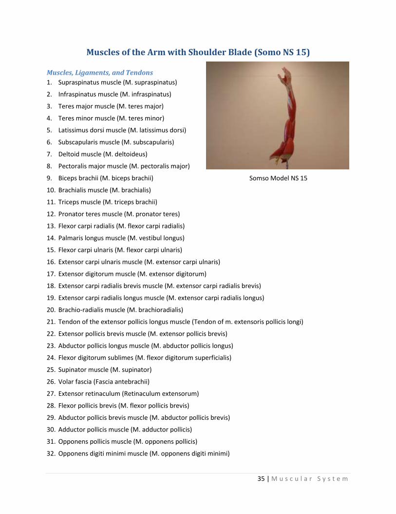
Muscles of the Arm with Shoulder Blade (Somo NS 15)
Muscles, Ligaments, and Tendons
1. Supraspinatus muscle (M. supraspinatus)
2. Infraspinatus muscle (M. infraspinatus)
3. Teres major muscle (M. teres major)
4. Teres minor muscle (M. teres minor)
5. Latissimus dorsi muscle (M. latissimus dorsi)
6. Subscapularis muscle (M. subscapularis)
7. Deltoid muscle (M. deltoideus)
8. Pectoralis major muscle (M. pectoralis major)
9. Biceps brachii (M. biceps brachii)
10. Brachialis muscle (M. brachialis)
11. Triceps muscle (M. triceps brachii)
12. Pronator teres muscle (M. pronator teres)
13. Flexor carpi radialis (M. flexor carpi radialis)
14. Palmaris longus muscle (M. vestibul longus)
15. Flexor carpi ulnaris (M. flexor carpi ulnaris)
16. Extensor carpi ulnaris muscle (M. extensor carpi ulnaris)
17. Extensor digitorum muscle (M. extensor digitorum)
18. Extensor carpi radialis brevis muscle (M. extensor carpi radialis brevis)
19. Extensor carpi radialis longus muscle (M. extensor carpi radialis longus)
20. Brachio-radialis muscle (M. brachioradialis)
21. Tendon of the extensor pollicis longus muscle (Tendon of m. extensoris pollicis longi)
22. Extensor pollicis brevis muscle (M. extensor pollicis brevis)
23. Abductor pollicis longus muscle (M. abductor pollicis longus)
24. Flexor digitorum sublimes (M. flexor digitorum superficialis)
25. Supinator muscle (M. supinator)
26. Volar fascia (Fascia antebrachii)
27. Extensor retinaculum (Retinaculum extensorum)
28. Flexor pollicis brevis (M. flexor pollicis brevis)
29. Abductor pollicis brevis muscle (M. abductor pollicis brevis)
30. Adductor pollicis muscle (M. adductor pollicis)
31. Opponens pollicis muscle (M. opponens pollicis)
32. Opponens digiti minimi muscle (M. opponens digiti minimi)
Somso Model NS 15

36 | M u s c u l a r S y s t e m
Muscles of the Arm with Shoulder Blade cont…
Muscles, Ligaments, and Tendons cont…
33. Flexor digiti minimi (M. flexor digiti minimi brevis)
34. Abductor digiti minimi muscle (M. abductor digiti minimi)
35. Lumbrical muscles (Mm. lumbricales)
36. Dorsal interosseous muscles of the hand (Mm. interossei dorsales)
37. Flexor retinaculum of upper limb (Lig. Carpi transversum)
38. Tendon sheath (Vagina fibrosa digitorum manus)
39. Crucial strings of the tendon sheath (Pars cruciformis vaginae fibrosae)
40. Annular strings of the tendon sheath (Pars anularis vaginae fibrosae)
41. Tendon of the flexor pollicis longus (Tendo m. flexoris pollicis longi)
42. Tendons of the flexor digitorum sublimis (Tendines m. flexoris digitoris superficialis)
43. Tendons of the flexor digitorum profundus (Tendines m. flexoris digitoris profundi)
44. Chiasma tendinum
Arteries
45. Axillary arteries (A. axillaries)
46. Subscapular artery (A. subscapularis)
47. Circumflex scapular artery (A. circumflexa scapulae)
48. Posterior circumflex humeral artery (A. circumflexa humeri posterior)
49. Brachial artery (A. brachialis)
50. Profunda brachii artery (A. profunda brachii)
51. Ramus deltoideus
52. Medial collateral artery (A. collateralis media)
53. Radial collateral artery (A. collateralis radialis)
54. Superior ulnar collateral artery (A. collateralis ulnaris superior)
55. Radial artery (A. radialis)
56. Ramus vestibul superficialis
57. Ramus carpeus dorsalis
58. A. metacarpea dorsalis
59. Ulnar artery (A. ulnaris)
60. Common interosseous artery (A. interossea communis)
61. Posterior interosseous artery (A. interossea posterior)
62. Median artery (A. mediana)
63. Arcus vestibul superficialis
64. Palmar digital arteries (A. Digitales palmares communes)
65. Palmar digital arteries (A. Digitales palmares propriae)
66. Dorsal digital arteries (A. Digitales dorsales)

37 | M u s c u l a r S y s t e m
Muscles of the Arm with Shoulder Blade cont…
Nerves
67. Plexus brachialis
68. Axillary nerve (N. axillaris)
69. Medial cutaneous nerve of the forearm (N. cutaneous antebrachii medialis)
70. Musculo-cutaneous nerve (N. musculocutaneous)
71. Median nerve (N. medianus)
72. Anterior interosseous nerve (N. interosseous anterior)
73. Ramus vestibul (N. ulnaris)
74. Ulnar nerve (N. ulnaris)
75. Ramus vestibul (N. ulnaris)
76. Ramus dorsalis (N. ulnaris)
77. Ramus profundus
78. Ramus superficialis
79. N. digitalis vestibul communis
80. Radial nerve (N. radialis)
81. Ramus superficialis (N. radialis)
Bones
82. Scapula
83. Spine of the scapula (Spina scapulae)
84. Coracoid process (Processus coracoideus)
85. Clavicle (Clavicula)
86. Humerus
87. Medial epicondyle of the humerus (Epicondylus medialis)
88. Lateral epicondyle of the humerus (Epicondylus lateralis)
89. Olecranon (tip of the elbow)
90. Ulna
91. Radius
92. Pisiform bone (Os pisiforme)
93. Carpus
94. Metacarpal bone (Ossa metacarpalia)
95. Phalanx I – II – III (Ossa digitorum manus, phalanx proximalis media and distalis)

38 | M u s c u l a r S y s t e m
Muscles of the Leg with Base of Pelvis (Somso NS 10)
Muscles
1. Psoas major muscle
2. Iliacus muscle
3. Gluteus maximus muscle
4. Gluteus medius muscle
5. Piriformis muscle
6. Obturator internus muscle
7. Gemellus muscle
a. Superior gemellus muscle
b. Inferior gemellus muscle
8. Quadratus femoris muscle
9. Tensor fasciae latae muscle
10. Sartorius muscle
11. Quadriceps femoris muscle
a. Rectus femoris muscle
b. Vastus medialis muscle
c. Vastus lateralis muscle
d. Vastus intermedius muscle
e. Common tendon of the muscles
12. Pectineus muscle
13. Adductor longus muscle
14. Adductor magnus muscle
15. Gracilis muscle
16. Semitendinosus muscle
17. Semimembranosus muscle
18. Biceps femoris
a. Caput longum
b. Caput breve
19. Tibialis anterior muscle
20. Extensor hallucis longus muscle
21. Extensor digitorum longus muscle
22. Fibularis longus muscle (peroneus)
23. Fibularis brevis muscle (peroneus)
24. Triceps surae muscle
a-b. Gastrocnemius muscle
a. Caput mediale
b. Caput laterale
c. Soleus muscle
d. Achilles tendon
25. Plantaris muscle
26. Popliteus muscle
27. Flexor digitorum longus
28. Tibialis posterior muscle
29. Flexor hallucis longus
30. Extensor hallucis brevis muscle
31. Extensor digitorum brevis muscle
32. Abductor hallucis muscle
33. Flexor hallucis brevis
34. Abductor digiti minimi muscle
35. Flexor digiti minimi brevis
36. Flexor digitorum brevis
37. Lumbrical muscles
38. Dorsal interosseous muscles of the foot
Somso Model NS 10
39 | M u s c u l a r S y s t e m
Muscles of the Leg with Base of Pelvis cont.
Vessels
39. External iliac artery
a. External iliac vein
40. Internal iliac artery
41. Obturator artery
42. Superior gluteal artery
43. Inferior gluteal artery
44. Internal pudendal artery
45. Femoral artery
a. Femoral vein
46. Profunda femoris artery
47. Descending branch of the lateral circumflex
artery
48. Ascending branch of the lateral circumflex
artery
49. A. circumflexa femoris medialis ramus
profundus
50. Perforating arteries
51. Popliteal artery
52. Lateral superior genicular artery
53. Medial superior genicular artery
54. Lateral inferior genicular artery
55. Medial inferior genicular artery
56. Posterior tibial artery
57. Anterior tibial artery
58. Peroneal artery
Nerves
59. Femoral nerve
60. Saphenous nerve
61. Obturator nerve
62. Plenus sacralis
63. Sciatic nerve
64. Medial popliteal nerve & posterior tibial
nerve
65. Common peroneal nerve
66. Deep peroneal nerve
67. Superficial peroneal nerve
Bones
68. Fifth lumbar vertebra
69. Sacrum
70. Coccyx
71. Iliac crest
72. Anterior superior iliac spine
73. Pubic bone
74. Pubic symphysis
75. Ischial spine
76. Ischial tuberosity
77. Greater trochanter
78. Lesser trochanter
79. Femur
80. Medial epicondyle of the femur
81. Lateral epicondyle of the femur
82. Kneecap (Patella)
83. Shin-bone (Tibia)
84. Medial malleolus
85. Head of fibula
86. Fibula
87. Lateral malleolus
88. Heel bone (Calcaneus)
89. Navicular bone of the foot
90. I. Metatarsal bone
91. V. Metatarsal bone

40 | M u s c u l a r S y s t e m
Male Muscle Figure (Somso AS 3)
Muscles
1. Frontal ventor of occipito-frontal muscle
2. Orbicularis oculi muscle
3. Nasal muscle
4. Levator muscle of superior lip
5. Greater cygomatic muscle
6. Orbicularis oris muscle
7. Depressor muscle of the mouth
8. Superior auricular muscle
9. Posterior auricular muscle
10. Occipital ventor of occipito-frontal muscle
11. Sternocleidomastoid muscle
12. Scaleni muscles
13. Levator muscle of scapula
14. Trapecial muscle
15. Infraspinous muscle
16. Latissimus dorsi muscle
17. Greatest gluteal muscle
18. Deltoid muscle
19. Serratus anterior muscle
20. Pectoralis major muscle
21. External oblique muscle of abdomen
22. Rectus abdominis muscle
23. Sheath of straight muscle of abdomen
24. Triceps muscle
25. Biceps muscle
26. Extensor carpi radialis longus muscle
27. Extensor digitorum muscle
28. Retinaculum of the extensors
29. Extensor pollicis brevis muscle
30. Brachioradial muscle
31. Sartorius muscle
32. Rectus femoris muscle
33. Pectineal muscle
34. Gracilis muscle
35. Adductor longus muscle
36. Biceps muscle of femur
37. Semitendinous muscle
38. Semimembranous muscle
39. Gastrocnemial muscle
40. Soleus muscle
41. Calcanean tendon
42. Extensor digitorum longus muscle
43. Tibialis anterior muscle
44. Extensor digitorum brevis muscle
45. Long peroneal muscle
46. Inferior retinaculum of extensor muscle
Bones
a. Skull
b. Clavicle
c. Anterior superior iliac spine
d. Popliteal
e. Patella
f. Tibia
g. Medial malleolus
h. Lateral malleolus
i. Calcaneus (heel bone)
Somso Model AS 3

41 | C i r c u l a t o r y / R e s p i r a t o r y S y s t e m
Muscles of the Human Arm (Altay 6000.31) 7 Parts
1. Clavicle 2. Scapula 3. Spine of scapula 4. Acromion of scapula 5. Coracoid process 6. Subscapularis muscle 7. Subclavius muscle 8. Latissimus dorsi muscle 9. Pectoralis minor muscle 10. Supraspinatus muscle 11. Infraspinatus muscle 12. Teres minor muscle 13. Teres major 14. Deltoid 15. Humerus 16. Biceps brachii muscle
a. Long head b. Short head
17. Coracobrachialis muscle 18. Brachialis muscle 19. Brachial artery 20. Median nerve 21. Ulnar nerve 22. Triceps brachii muscle
a. Medial head b. Long head c. Lateral head
23. Olecranon
24. Radial nerve 25. Profunda brachii artery 26. Medial intermuscular septum 27. Brachioradialis muscle 28. Pronator teres muscle 29. Flexor carpi radialis muscle 30. Palmaris longus muscle 31. Flexor carpi ulnaris muscle 32. Flexor digitorum superficialis
m. 33. Flexor pollicis longus muscle 34. Flexor digitorum profundus m. 35. Pronator quadrates muscle 36. Radial artery 37. Ulnar artery 38. Common interosseous artery 39. Anterior interosseous artery 40. Anterior interosseous nerve 41. Superficial palmar branch 42. Extensor carpi radialis longus
m. 43. Extensor carpi radialis brevis
m. 44. Extensor digitorum muscle 45. Extensor digiti minimi muscle 46. Extensor carpi ulnaris muscle 47. Anconeus muscle 48. Supinator muscle 49. Abductor pollicis longus
muscle 50. Extensor pollicis brevis muscle 51. Extensor pollicis longus muscle
52. Extensor indicis muscle 53. Ulna 54. Radius 55. Recurrent interosseous artery 56. Posterior interosseous artery 57. Posterior interosseous nerve 58. Abductor pollicis brevis muscle 59. Flexor pollicis brevis muscle 60. Flexor digitorum superficialis
tendon 61. Lumbrical muscles 62. Flexor digiti minimi brevis muscle 63. Abductor digiti minimi muscle 64. Superficial palmar arch 65. Deep palmar branch 66. Deep palmar arch 67. Common palmar digital artery 68. Proper palmar digital artery 69. Common palmar digital nerve 70. Proper palmar digital nerve 71. Adductor pollicis muscle 72. Palmar interosseous muscles 73. Palmar metacarpal artery 74. Metacarpal bone (II) 75. Tendon of extensor digitorum 76. Dorsal aponeurosis (middle finger) 77. Dorsal interosseous muscles 78. Dorsal metacarpal artery 79. Dorsal digital artery

42 | C i r c u l a t o r y / R e s p i r a t o r y S y s t e m
Head & Neck Musculature (Altay 6030.10)
1. Frontal belly of occipitofrontalis m.
2. Epicranial aponeurosis 3. Occipital belly of
occipitofrontalis muscle 4. Orbicularis oculi muscle 5. Depressor supercilii muscle 6. Procerus muscle 7. Nasalis muscle 8. Orbicularis muscle 9. Levator labii superior
alaeque nasi muscle 10. Levator labii superior muscle 11. Zygomaticus minor muscle 12. Zygomaticus major muscle 13. Risorius muscle 14. Depressor anguli oris muscle 15. Depressor labii inferior
muscle 16. Mentalis muscle 17. Buccinator muscle 18. Auricularis superior muscle 19. Auricularis anterior muscle 20. Auricularis posterior muscle 21. Masseter muscle 22. Temporalis muscle 23. Medial pterygoid muscle 24. Lateral pterygoid muscle
25. Sternocleidomastoid muscle 26. Anterior belly of digastric
muscle 27. Posterior belly of digastric
muscle 28. Stylopharyngeus muscle 29. Mylohyoid muscle 30. Hyoglossus muscle 31. Sternohyoid muscle 32. Superior belly of omohyoid
muscle 33. Inferior belly of omohyoid
muscle 34. Geniohyoid muscle 35. Sternothyroid muscle 36. Thyrohyoid muscle 37. Scalenus anterior muscle 38. Scalenus medius muscle 39. Posterior scalene muscle 40. Levator scapulae muscle 41. Splenius capitis muscle 42. Semispinalis capitis muscle 43. Rhomboid minor muscle 44. Rhomboid major muscle 45. Trapezius muscle 46. Pectoralis major muscle 47. Deltoid muscle 48. Pectoralis minor muscle 49. Supraspinatus muscle 50. Serratus anterior muscle
51. External intercostales muscles
52. Intercostales interni muscles
53. Hyoid bone 54. Thyroid cartilage 55. Thyroid gland 56. Common carotid artery 57. Internal carotid artery 58. External carotid artery 59. Superior thyroid artery 60. Lingual artery 61. Facial artery 62. Occipital artery 63. Posterior auricular artery 64. Superficial temporal artery 65. Maxillary artery 66. Subclavian artery 67. Vertebral artery 68. Thyrocervical trunk 69. Inferior thyroid artery 70. Transverse cervical artery 71. Suprascapular artery 72. Internal thoracic artery 73. Axillary artery 74. Superior thoracic artery 75. Thoracoacromial artery 76. Lateral thoracic artery

43 | C i r c u l a t o r y / R e s p i r a t o r y S y s t e m
Cardiovascular/Respiratory System
Two Section Heart (HE-1010)
External Anterior Aspect 1. Aorta
2. Brachiocephalic trunk (innominate artery)
3. Left common carotid artery
4. Left subclavian artery
5. Pulmonary trunk
6. Ligamentum arteriosum
7. Left pulmonary artery
8. Right auricle
10. Left auricle
11/12. Left/Right Ventricles (respectively)
13. Right coronary artery
14. Left coronary artery
15. Circumflex branch, left coronary artery
16. Great cardiac vein
17. Small cardiac vein
18. Anterior cardiac veins
19. Anterior interventricular descending branch,
left coronary artery
20. Oblique vein of left atrium
External Posterior Aspect 9. Superior vena cava 21. Marginal branch, right coronary artery 22. Coronary sinus 23. Middle cardiac vein 24. Posterior interventricular descending branch,
right coronary artery
25. Posterior vein of left ventricle
26. Inferior vena cava
27. Left pulmonary veins
28. Right pulmonary veins
29. Right pulmonary artery
30. Left atrium
31. Right atrium
Internal Anterior Aspect 1. Mitral valve 2. Aortic valve 3. Orifice left coronary artery 4. Orifice right coronary artery 5. Pulmonary valve 6. Left & right anterior papillary muscle 7. Medial (conal) papillary muscle 8. Moderator band 9. Supraventricular crest 10. Trabeculae carneae
Internal Posterior Aspect 1. Tricuspid valve 2. Muscular interventricular septum 3. Left & right posterior papillary muscle 4. Chordae tendineae 5. Membranous septum 6. Right anterior papillary muscle 7. Pectinate muscle
Model HE-1010

44 | C i r c u l a t o r y / R e s p i r a t o r y S y s t e m
Human Heart & Mediastinum
1. Aorta 2. Brachiocephalic (innominate) artery 3. Left common carotid artery 4. Left subclavian artery 5. Superficial cardiac plexus 6. Left vagus nerve 7. Recurrent laryngeal nerve 8. Ligamentum arteriosus 9. Right pulmonary artery 10. Left bronchus 11. Left bronchial artery 12. Pulmonary nerve plexus 13. Intercostal arteries 14. Thoracic duct 15. Azygos vein 16. Esophagus 17. Right vagus nerve 18. Right bronchial artery 19. Trachea 20. Right main bronchus 21. Right pulmonary artery 22. Tracheobronchial lymph nodes 23. Right ventricle 24. Left ventricle 25. Left atrium 26. Right atrium
27. Right auricle 28. Conus arteriosus 29. Left auricle 30. Apex 31. Pulmonary artery 32. Aorta 33. Pericardium 34. Superior vena cava 35. Right coronary artery 36. Anterior cardiac veins 37. Left coronary artery 38. Anterior interventricular artery 39. Circumflex artery 40. Great cardiac vein 41. Marginal branch of left coronary artery 42. Oblique vein of left atrium 43. Coronary sinus 44. Dorsal interventricular vein 45. Left dorsal ventricular vein 46. Inferior vena cava 47. Azygos vein 48. Left pulmonary veins 49. Right pulmonary veins 50. Marginal branch of right coronary artery 51. Coronary sinus 52. Anterior interventricular sulcus
Model 56-6818 (5X Magnification)

45 | C i r c u l a t o r y / R e s p i r a t o r y S y s t e m
The Heart (Somso HS 26)
A. Apex
B. Interventricular septum
I. Right auricle II. Left auricle
a. Right auricular appendage b. Left auricular appendage
III. Right ventricle IV. Left ventricle
c. Superior vena cava d. Inferior vena cava e. Tricuspid valve f. Pulmonary artery g. Pulmonary veins h. Bicuspid valve i. Aorta k. Semilunar valve
A. Tendinous cord 2. Papillary muscles 3. Ascending branch of aorta 4. Right coronary artery 5. Left coronary artery 6. Great heart-vein 7. Arch of aorta
Somso Model HS 26 (4 parts)

46 | C i r c u l a t o r y / R e s p i r a t o r y S y s t e m
Model of Heart (ALS 300)

47 | C i r c u l a t o r y / R e s p i r a t o r y S y s t e m
Model of Heart Cont... ALS 300
1. Left subclavian artery 2. Left common carotid artery 3. Right brachiocephalic artery 4. Right & left brachiocephalic vein 5. Superior vena cava 6. Ascending branch of the aorta 7. Right atrium 8. Right coronary artery 9. Pulmonary valve 10. Right tricuspid valve 11. Superior infundibulum 12. Superior right papillary muscles 13. Right ventricle 14. Ventricular septum 15. Left branch of the pulmonary artery 16. Left pulmonary veins 17. Pulmonary artery 18. Left atrium 19. Bicuspid valve or mitral valve 20. Aortic valve 21. Superior left papillary muscles 22. Left ventricle 23. Right auricle of the heart 24. Left auricle of the heart 25. Right coronary artery 26. Superior branch of the left coronary artery 27. Aortic arch 28. Coronary sinus 29. Posterior branch of the left coronary artery 30. Posterior coronary vein 31. Azygous vein 32. Right pulmonary artery 33. Right pulmonary veins 34. Inferior vena cava 35. Posterior branch of the right coronary artery

48 | C i r c u l a t o r y / R e s p i r a t o r y S y s t e m
The Heart (HS 4)
A. Apex B. Interventricular septum I. Right auricle II. Left auricle
a) Right auricular appendage b) Left auricular appendage
III. Right ventricle IV. Left ventricle
c) Superior vena cava d) Inferior vena cava e) Tricuspid valve f) Pulmonary artery g) Pulmonary vein h) Bicuspid valve i) Aorta k) Semilunar valves
1. Tendenous cord 2. Papillary muscles 3. Ascending branch of aorta 4. Right coronary artery 5. Left coronary artery 6. Coronary Sinus 7. Arch of aorta
Somso Model HS 4 (2 parts)

49 | C i r c u l a t o r y / R e s p i r a t o r y S y s t e m
Vascular Arm
Arteries
1. axillary 2. humeral circumflex 3. brachial 4. brachial profunda 5. superior ulnar collateral 6. inferior ulnar collateral 7. anterior ulnar recurrent 8. radial 9. ulnar 10. anterior interosseous 11. posterior interosseous 12. deep palmar arch 13. superficial palmar arch 14. middle collateral 15. radial collateral
Veins
16. median 17. vestibul 18. axillary 19. median cubital 20. cephalic 21. accessory cephalic
Model ZKJ-688-E

50 | C i r c u l a t o r y / R e s p i r a t o r y S y s t e m
Fine Structure of the Artery & Veins (Somso HS 25)
1. Artery
2. Artery Exit
3. Vein
4. Vein Exit
5. Intima
a. Endothelium
b. Fenestrated Layer
6. Membrana Elastica Interna
7. Media
a. Smooth Muscle Cells
b. Collagenous Fibers
c. Elastic Fibers
8. Membrana Elastica Externa
9. Adventitia
a. Collagenous Fibers
b. Elastic Fibers
c. Figure-of-eight turns of the
Collagenous Fibers
10. Common Connective Tissue Sheath for
Artery & Vein
11. Valve of the vein, closed
12. Valvel of the vein, open
13. Muscular cells in the adventitia
14. Direction of blood flow in the artery
15. Direction of blood flow in vein
Somso HS 25

51 | C i r c u l a t o r y / R e s p i r a t o r y S y s t e m
Respiratory Organs & Heart
1. Hyoid bone 2. Greater horn of hyoid bone 3. Lesser horn of hyoid bone 4. Median thyrohyoid ligament 5. Lateral thyrohyoid ligament 6. Thyrohyoid membrane 7. Thyrohyoid cartilage 8. Superior horn of thyroid cartilage 9. Inferior horn of thyroid cartilage 10. Epiglottis 11. Arytenoid cartilage 12. Cricoid cartilage 13. Cricothyroid ligament 14. Cricotracheal ligament 15. Cricothyroid muscle 16. Thyroid gland 17. Tracheal cartilages 18. Vocal cords 19. Esophagus 20. Trachea 21. Bifurcation of trachea 22. Right bronchus 23. Left bronchus 24. Right lung 25. Left lung 26. Heart 27. Right atrium 28. Left atrium 29. Right ventricle 30. Left ventricle 31. Bicuspid valve 32. Tricuspid valve 33. Semilunar valve of pulmonary artery 34. Semilunar valve of aorta 35. Aorta 36. Superior vena cava 37. Inferior vena cava 38. Pulmonary artery 39. Pulmonary vein 40. Right coronary artery 41. Left coronary artery
42. Anterior coronary veins 43. Posterior coronary veins 44. Coronary sinus 45. Right innominate vein 46. Left innominare vein 47. Right subclavian vein 48. Right jugular vein 49. Left subclavian vein 50. Left jugular vein 51. Right subclavian artery 52. Right carotid artery 53. Left subclavian artery 54. Left carotid artery 55. Aortic arch 56. Thoracic/Descending aorta 57. Thoracic vertebra 58. Intervertebral disc
Model #192, A42

52 | C i r c u l a t o r y / R e s p i r a t o r y S y s t e m
Lungs, Larynx & Heart (C 243)
1. Hyoid bone 2. Thyroid cartilage 3. Cricoid cartilage 4. Trachea 5. Thyroid gland 6. Median thyroid ligament
I. Lung a. Upper lobe b. Right middle lobe c. Lower lobe
I. Lung d. Upper lobe e. Right middle lobe f. Lower lobe
III. Heart with arteries 1. Right atrium 2. Right ventricle 3. Left atrium 4. Left ventricle
a. Right auricle of the heart b. Left auricle of the heart c. Bicuspid valve d. Tricuspid valve e. Valve of pulmonary artery f. Papillary muscle g. Aortic valve
5. Aorta 6. Common carotid artery 7. Common carotid artery 8. Subclavian artery 9. Thoracic aorta 10. Superior vena cava 11. Internal jugular vein 12. Subclavian vein 13. Pulmonary veins 14. Pulmonary artery 15. Inferior vena cava 16. Great cardiac vein 17. Left coronary artery 18. Esophagus 19. Thoracic vertebrae
Model C 243
53 | C i r c u l a t o r y / R e s p i r a t o r y S y s t e m
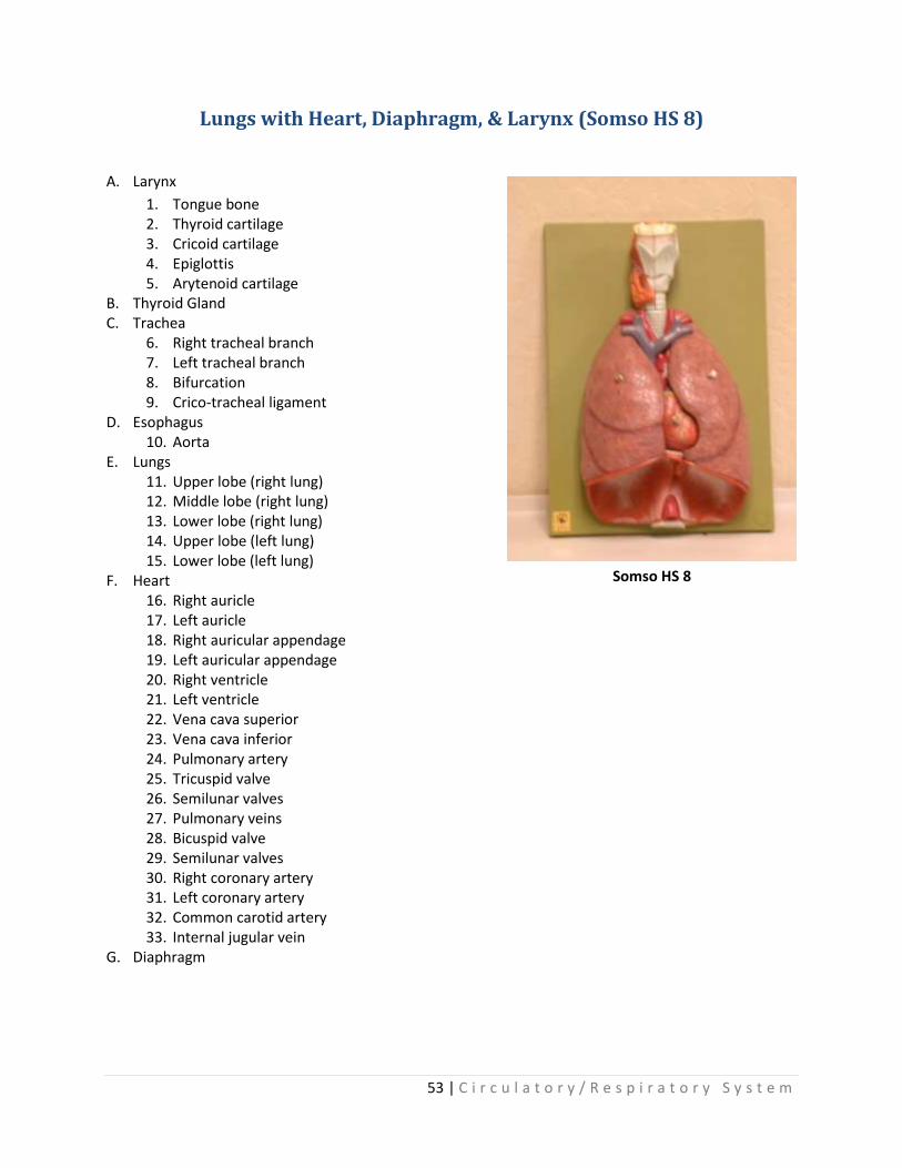
Lungs with Heart, Diaphragm, & Larynx (Somso HS 8)
A. Larynx
1. Tongue bone 2. Thyroid cartilage 3. Cricoid cartilage 4. Epiglottis 5. Arytenoid cartilage
B. Thyroid Gland C. Trachea
6. Right tracheal branch 7. Left tracheal branch 8. Bifurcation 9. Crico-tracheal ligament
D. Esophagus 10. Aorta
E. Lungs 11. Upper lobe (right lung) 12. Middle lobe (right lung) 13. Lower lobe (right lung) 14. Upper lobe (left lung) 15. Lower lobe (left lung)
F. Heart 16. Right auricle 17. Left auricle 18. Right auricular appendage 19. Left auricular appendage 20. Right ventricle 21. Left ventricle 22. Vena cava superior 23. Vena cava inferior 24. Pulmonary artery 25. Tricuspid valve 26. Semilunar valves 27. Pulmonary veins 28. Bicuspid valve 29. Semilunar valves 30. Right coronary artery 31. Left coronary artery 32. Common carotid artery 33. Internal jugular vein
G. Diaphragm
Somso HS 8

54 | C i r c u l a t o r y / R e s p i r a t o r y S y s t e m
Lung with Alveoli (Somso HS 23)
Somso Model HS 23
1. Intra-pulmonary bronchi 2. Non-cartilaginous bronchioles 3. Respiratory bronchioles 4. Alveoli 5. Mucous membrane 6. Elastic fibers 7. Smooth muscle 8. Pulmonary plexus 9. Mucous glands of the bronchi 10. Fibro-cartilaginous layer 11. Pulmonary pleura 12. Limiting membranes 13. Dense capillaries 14. Collaginous fibers 15. Elastic fibers 16. Epithelium 17. Bronchial vein plexus 18. Pulmonary vein 19. Pulmonary artery 20. Bronchial artery 21. Anastomosis between bronchial artery & pulmonary vein 22. Alveolar capillary network 23. Anastomosis of an obstructed artery with pulmonary vein 24. Dense capillary network 25. Anastomosis between pulmonary artery & vein
55 | C i r c u l a t o r y / R e s p i r a t o r y S y s t e m
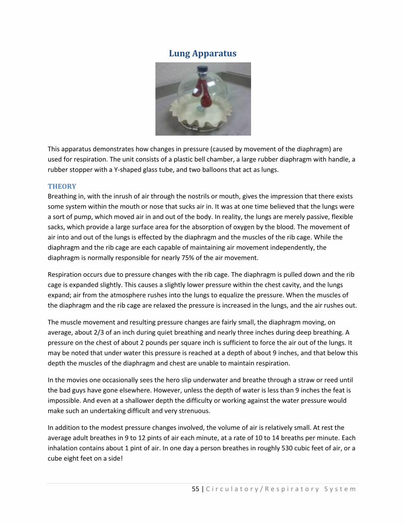
Lung Apparatus
This apparatus demonstrates how changes in pressure (caused by movement of the diaphragm) are
used for respiration. The unit consists of a plastic bell chamber, a large rubber diaphragm with handle, a
rubber stopper with a Y-shaped glass tube, and two balloons that act as lungs.
THEORY
Breathing in, with the inrush of air through the nostrils or mouth, gives the impression that there exists
some system within the mouth or nose that sucks air in. It was at one time believed that the lungs were
a sort of pump, which moved air in and out of the body. In reality, the lungs are merely passive, flexible
sacks, which provide a large surface area for the absorption of oxygen by the blood. The movement of
air into and out of the lungs is effected by the diaphragm and the muscles of the rib cage. While the
diaphragm and the rib cage are each capable of maintaining air movement independently, the
diaphragm is normally responsible for nearly 75% of the air movement.
Respiration occurs due to pressure changes with the rib cage. The diaphragm is pulled down and the rib
cage is expanded slightly. This causes a slightly lower pressure within the chest cavity, and the lungs
expand; air from the atmosphere rushes into the lungs to equalize the pressure. When the muscles of
the diaphragm and the rib cage are relaxed the pressure is increased in the lungs, and the air rushes out.
The muscle movement and resulting pressure changes are fairly small, the diaphragm moving, on
average, about 2/3 of an inch during quiet breathing and nearly three inches during deep breathing. A
pressure on the chest of about 2 pounds per square inch is sufficient to force the air out of the lungs. It
may be noted that under water this pressure is reached at a depth of about 9 inches, and that below this
depth the muscles of the diaphragm and chest are unable to maintain respiration.
In the movies one occasionally sees the hero slip underwater and breathe through a straw or reed until
the bad guys have gone elsewhere. However, unless the depth of water is less than 9 inches the feat is
impossible. And even at a shallower depth the difficulty or working against the water pressure would
make such an undertaking difficult and very strenuous.
In addition to the modest pressure changes involved, the volume of air is relatively small. At rest the
average adult breathes in 9 to 12 pints of air each minute, at a rate of 10 to 14 breaths per minute. Each
inhalation contains about 1 pint of air. In one day a person breathes in roughly 530 cubic feet of air, or a
cube eight feet on a side!
56 | C i r c u l a t o r y / R e s p i r a t o r y S y s t e m
OPERATION
Before using the demonstration unit, make sure that the balloons; are deflated when the diaphragm is
relaxed. To do this, remove the rubber stopper and pull the diaphragm out, increasing the volume of the
bell jar, while holding the diaphragm, and any air in the balloons will be forced out. To ensure a good
seal for the rubber stopper, twist it slowly while forcing it into the neck.
When the diaphragm is pulled down, the volume in the chamber is increased. While the volume is
larger, the amount of air in the chamber is the same, and the pressure drops. This is shown by
the ideal gas law, PV = nRT where P is pressure, V is volume, n is the number of moles of gas, R is the
universal gas constant, and T is the pressure and volume, so the equation can be simplified to
PV=constant. It follows that as the volume in increased the pressure must decrease, and as the volume
is decreased the pressure must increase. In this case the only changes are in the pressure and volume,
so the equation can be simplified to PV = constant. It follows that as the volume is increased the
pressure must decrease, and as the volume is decreased the pressure must increase. In this case
pressure and volume are inversely proportional to each other. As one increases the other decreases.
As the pressure within the chamber decreases, a pressure differential is created between the outside
atmosphere and the interior of the chamber. Air is forced into the balloons by atmospheric pressure,
and they expand. When the diaphragm is released, and allowed to return to the initial 'position, the
volume in the chamber is decreased, increasing the pressure, and forcing the air out of the balloons.
As the diaphragm is alternately pulled down and released, the balloons in the chamber will expand and
contract, just as the lungs within the chest cavity expand and contract when breathing.

57 | C i r c u l a t o r y / R e s p i r a t o r y S y s t e m
Lung (Basic Model)

58 | C i r c u l a t o r y / R e s p i r a t o r y S y s t e m

59 | N e r v o u s S y s t e m
Nervous System
Neuron (Somso BS 35)
A. Perikaryon of the nerve cell with dendrites B. Peripheral nerve with sheaths
1. Neurite cone 2. Nucleus of nerve cell nucleolus 3. Endoplasmic reticulum 4. Neurofibrils 5. Synaptic terminals 6. Neuraxon 7. Schwann cell with nucleus 8. Schwann sheath 9. Linked Schwann cells at node of Ranvier 10. Mitochondria 11. Medullary (Myelin) sheath 12. Perineural (Endoneural) sheath of connective tissue 13. Mesaxon 14. Dendrites 15. Lysosomes 16. Neurotubules 17. Golgi apparatus
Somso Model BS 35
Magnification approx. 2500X

60 | N e r v o u s S y s t e m
Model of a Synapse (BS 35/3)
(Somso model BS 35/3)
1. Presynapse 2. Presynaptic membrane 3. Neuroplasma 4. Endoplasmic reticulum 5. Mitochondrium 6. Neurotubule 7. Neurofilament 8. Presynaptic grid 9. Vesicle 10. Vesicle with neurotransmitter 11. Transmitter release 12. Transmitter gap products 13. Endocytic material absorption 14. Pores for endocytic material absorption 15. Synaptic gap 16. Postsynapse 17. Subsynaptic/postsynaptic membrane 18. Postsynaptic membrane 19. Transmitter receptor molecule reaction

61 | N e r v o u s S y s t e m
Striated Muscle Fiber with Motor End-Plate (Somso BS 36)
Somso Model BS 36
1. Sarcoplasm cytoplasmic matrix 2. Nuclei 3. Myofibrils 4. Cohnheim’s areas 5. Sarcolemma 6. Endomysium
7. Schwann cell
7a. Node of Ranvier
8. Axon
9. End of myelin sheath
10. Cell membrane of synaptic end bulb
11. Axonal terminal
12. Neurofibrils
13. Unknown
14. Synaptic end bulb (knob)
15. Nucleus
16. Mitochondria
17. Motor end plate (junctional folds)
**Hot pink line between #17 and #10 represents the Synaptic Cleft
18. Motor end plate
19. Synaptic vesicles

62 | N e r v o u s S y s t e m
Transparent Human Brain (BS 25)
Somso Model BS 25

63 | N e r v o u s S y s t e m
Transparent Human Brain Cont… Somso Model BS 25
1. Left Cerebral Hemisphere (Cortex) 2. Right Cerebral Hemisphere (Cortex) 3. Callous Corpus (Corpus Callosum) 4. Medial and Lateral Longitudinal Stria of Corpus Callosum 5. Lateral Ventricle 6. a. Anterior Horn of Lateral Ventricle
b. Inferior Horn of Lateral Ventricle 7. Posterior Horn of Lateral Ventricle 8. Third Ventricle 9. Cerebral Aqueduct 10. Fourth Ventricle 11. Body of the Fornix 12. Choroid Plexus 13. Temporal Lobe 14. Occipital Lobe 15. Pes Hippocampi 16. Choroid Plexus 17. Dentate Gyrus 18. Fimbria of Hippocampus 19. Gyrus Parahippocampus 20. Frontal Gyrus 21. Olfactory Bulb 22. Olfactory Tract 23. Cerebellar Hemisphere 24. Cerebellar Peduncles 25. Flocculus 26. Cerebellar Tonsil 27. Vermis of the Cerebellum 28. Left Island of Reil 29. Caudate Nucleus 30. Putamen 31. Openings for Projection Tracts 32. Projection Fiber Bundle of Internal Capsule 33. Pallidum 34. Right Island of Reil 35. Lentiform Nucleus 36. Internal Capsule 37. Striate Body 38. Thalamus 39. Optic Chiasma 40. Crus Cerebri 41. Optic Tract 42. Pulvinar of Thalamus
43. Oculomotor Nerve 44. Mamillary Body 45. Trigeminal Nerve 46. Adbucens Nerve 47. Vestibulocochlear Nerve 48. Glossopharyngeal Nerve, Vagus Nerve 49. Olives 50. Pons 51. Pyramids 52. Hypoglossal Nerve 53. Pineal Gland 54. Quadrigeminal Nerve 55. Trochlear Nerve 56. Rhomboid Fossa 57. Tubercle of Nucleus Gracilis 58. Tubercle of Nucleus Cuneatus

64 | N e r v o u s S y s t e m
Transparent Model of the Skull (Somso QS 65/7) (Including the Brain and the Cranial Nerves)
Cranial Nerves I. Olfactory bulb II. Optic nerve III. Oculomotor nerve IV. Tochlear nerve V. Trigeminal nerve VI. Abducens nerve VII. Facial nerve VIII. Vestibulocochlear nerve IX. Glossopharyngeal nerve X. Vagus nerve XI. Accessory nerve
XII. Hypoglossal nerve XIII. Intermediate nerve V. 1. Opthalmic nerve V. 2. Maxillary nerve V. 3. Mandibular nerve V. 4. Trigeminal ganglion V. 5. Buccal nerve V. 6. Deep temporal nerve V. 7. Lingual nerve V. 8. Inferior alveolar nerve V. 9. Auricolotemporal nerve
Cortical fields according to Brodmann
a. Motor speech center b. Frontal visual gaze center c. Motor center for writing d. Motor center for walking e. Precentral gyrus (primary motor cortical fields, area 4) f. Postcentral gyrus (primary sensory cortical fields, areas 1, 2, & 3) g. Primary center for taste h. Sensory fields for object & tactile recognition i. Sensory fields for taking action k. Fields for visual-spatial memory l. Primary auditory cortex (area 41) m. Secondary auditory cortex (area 42) n. Sensory center for speech according to Wernicke (area 22) o. Tertiary auditory cortical fields (comprehension of speech & music) (area 21) p. Tertiary visual cortical fields (area 19) s. Secondary visual cortical fields (image comprehension) (area 18) s. Primary center for vision (visual perception) (calcarine area, area 17) t. Cingulate gyrus – part of the limbic system (emotional coloration of sensory stimuli) u. Hippocampal cortex – part of the limbic system v. Prepiriform area – secondary olfactory centers for smell
Somso Model QS 65/7
65 | N e r v o u s S y s t e m
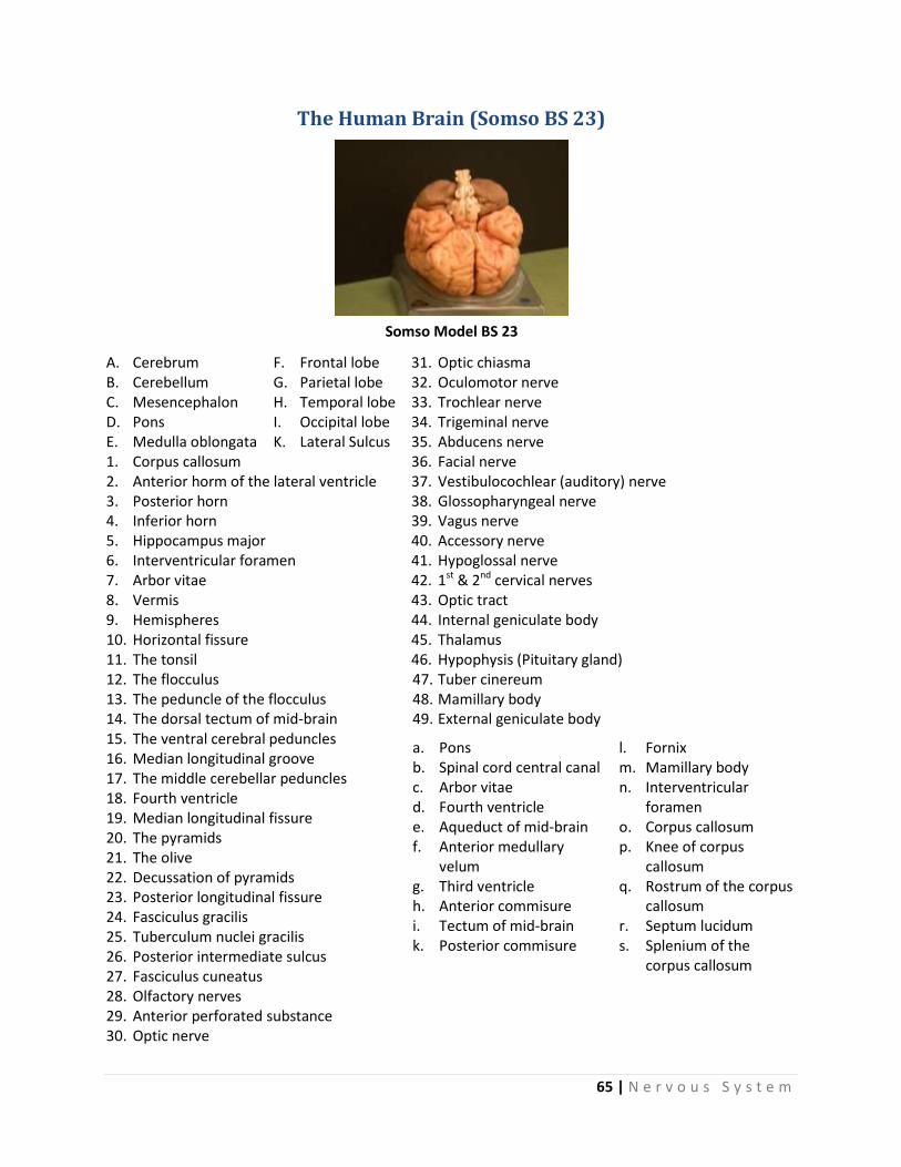
The Human Brain (Somso BS 23)
A. Cerebrum B. Cerebellum C. Mesencephalon D. Pons E. Medulla oblongata
F. Frontal lobe G. Parietal lobe H. Temporal lobe I. Occipital lobe K. Lateral Sulcus
31. Optic chiasma 32. Oculomotor nerve 33. Trochlear nerve 34. Trigeminal nerve 35. Abducens nerve
1. Corpus callosum 2. Anterior horm of the lateral ventricle 3. Posterior horn 4. Inferior horn 5. Hippocampus major 6. Interventricular foramen 7. Arbor vitae 8. Vermis 9. Hemispheres 10. Horizontal fissure 11. The tonsil 12. The flocculus 13. The peduncle of the flocculus 14. The dorsal tectum of mid-brain
36. Facial nerve 37. Vestibulocochlear (auditory) nerve 38. Glossopharyngeal nerve 39. Vagus nerve 40. Accessory nerve 41. Hypoglossal nerve 42. 1st & 2nd cervical nerves 43. Optic tract 44. Internal geniculate body 45. Thalamus 46. Hypophysis (Pituitary gland) 47. Tuber cinereum 48. Mamillary body 49. External geniculate body
15. The ventral cerebral peduncles 16. Median longitudinal groove 17. The middle cerebellar peduncles 18. Fourth ventricle 19. Median longitudinal fissure 20. The pyramids 21. The olive 22. Decussation of pyramids 23. Posterior longitudinal fissure 24. Fasciculus gracilis 25. Tuberculum nuclei gracilis 26. Posterior intermediate sulcus 27. Fasciculus cuneatus 28. Olfactory nerves 29. Anterior perforated substance 30. Optic nerve
a. Pons b. Spinal cord central canal c. Arbor vitae d. Fourth ventricle e. Aqueduct of mid-brain f. Anterior medullary
velum g. Third ventricle h. Anterior commisure i. Tectum of mid-brain k. Posterior commisure
l. Fornix m. Mamillary body n. Interventricular
foramen o. Corpus callosum p. Knee of corpus
callosum q. Rostrum of the corpus
callosum r. Septum lucidum s. Splenium of the
corpus callosum
Somso Model BS 23

66 | N e r v o u s S y s t e m
Brain Stem (Somso BS 23/2)
C. Mesencephalon D. Pons E. Medulla oblongata
6. Interventricular foramen 14. The dorsal tectum of mid-brain 15. The ventral cerebral peduncles 16. Median longitudinal groove 17. Middle cerebellar peduncles 18. Fourth ventricle 19. Median longitudinal fissure 20. The pyramids 21. The olive 22. Decussation of pyramids 23. Posterior longitudinal fissure 24. Fasciculus gracilis 25. Tuberculum nuclei gracilis 26. Posterior intermediate sulcus 27. Fasciculus cuneatus 29. Anterior perforated substance 30. Optic nerve 31. Optic chiasma
32. Oculomotor nerve 33. Trochlear nerve 34. Trigeminal nerve 35. Abducens nerve 36. Facial nerve 37. Vestibulocochlear) nerve 38. Glossopharyngeal nerve 39. Vagus nerve 40. Accessory nerve 41. Hypoglossal nerve 42. 1st & 2nd cervical nerves 43. Optic tract 45. Thalamus 46. Hypophysis (Pituitary gland) 47. Tuber cinereum 48. Mamillary body 49. External geniculate body
a. Grey substance of the pons b. Central canal of the spinal cord c. Arbor vitae d. Fourth ventricle e. Aqueduct of mid-brain f. Anterior medullary velum g. Third ventricle
Somso Model BS 23/2
67 | N e r v o u s S y s t e m
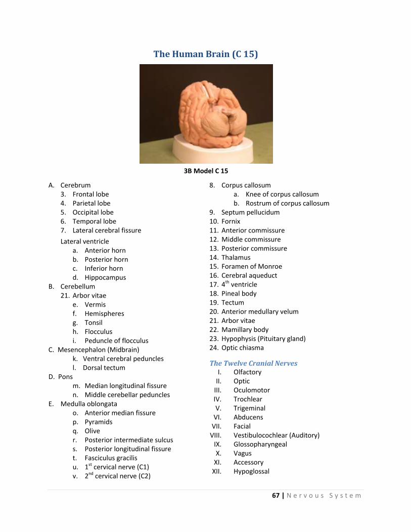
The Human Brain (C 15)
A. Cerebrum 3. Frontal lobe 4. Parietal lobe 5. Occipital lobe 6. Temporal lobe 7. Lateral cerebral fissure
Lateral ventricle a. Anterior horn b. Posterior horn c. Inferior horn d. Hippocampus
B. Cerebellum 21. Arbor vitae
e. Vermis f. Hemispheres g. Tonsil h. Flocculus i. Peduncle of flocculus
C. Mesencephalon (Midbrain) k. Ventral cerebral peduncles l. Dorsal tectum D. Pons
m. Median longitudinal fissure n. Middle cerebellar peduncles
E. Medulla oblongata o. Anterior median fissure p. Pyramids q. Olive r. Posterior intermediate sulcus s. Posterior longitudinal fissure t. Fasciculus gracilis u. 1st cervical nerve (C1) v. 2nd cervical nerve (C2)
8. Corpus callosum a. Knee of corpus callosum b. Rostrum of corpus callosum
9. Septum pellucidum 10. Fornix 11. Anterior commissure 12. Middle commissure 13. Posterior commissure 14. Thalamus 15. Foramen of Monroe 16. Cerebral aqueduct 17. 4th ventricle 18. Pineal body 19. Tectum 20. Anterior medullary velum 21. Arbor vitae 22. Mamillary body 23. Hypophysis (Pituitary gland) 24. Optic chiasma
The Twelve Cranial Nerves I. Olfactory
II. Optic III. Oculomotor IV. Trochlear V. Trigeminal
VI. Abducens VII. Facial
VIII. Vestibulocochlear (Auditory) IX. Glossopharyngeal X. Vagus
XI. Accessory XII. Hypoglossal
3B Model C 15

68 | N e r v o u s S y s t e m
Brain with Arteries
I. Cerebrum
1. Frontal lobe
2. Parietal lobe
3. Temporal lobe
4. Occipital lobe
5. Insula
6. Hippocampus
7. Lateral ventricles
8. Corpus callosum
9. Fornix
10. Internal carotid artery
11. Middle cerebral artery
12. Anterior cerebral artery
13. Posterior cerebral artery
14. Basilar artery
15. Vertebral artery
II. Cerebellum
16. Vermis of cerebellum
17. Hemisphere of cerebellum
18. Superior cerebellar artery
19. Anterior inferior cerebellar artery
20. Posterior inferior cerebellar artery
III. Diencephalon
21. Thalamus
22. Hypothalamus
23. Hypophysis
24. Optic nerve (II)
25. Pineal body
26. Medial geniculate body
27. Lateral geniculate body
IV. Mesencephalon
28. Superior colliculi
29. Inferior colliculi
30. Trochlear nerve (IV)
31. Oculomotor nerve (III)
V. Pons
32. Trigeminal nerve (V)
33. Abducens nerve (VI)
34. Facial nerve (VII)
35. Vestibulocochlear nerve (VIII)
VI. Medulla oblongata
36. Pyramid
37. Olive
38. Glossopharyngeal nerve (IX)
39. Vagus nerve (X)
40. Accessory nerve (XI)
41. Hypoglossal nerve (XII)
Altay Model 6160.14
69 | N e r v o u s S y s t e m
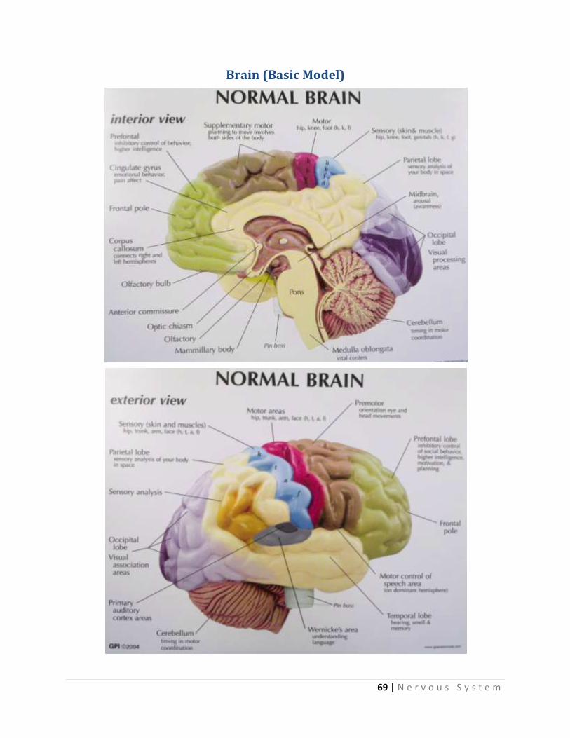
Brain (Basic Model)

70 | N e r v o u s S y s t e m
Fifth Cervical Vertebra with Spinal Cord (Somso BS 30)
1. Body of the vertebra 2. Transverse process 3. Vertebral arch 4. Spinose process 5. Yellow ligament 6. Posterior longitudinal ligament 7. Vertebral artery 8. Vertebral vein 9. Dura mater of the spinal cord 10. Epidural cavity 11. Subdural cavity 12. Arachnoid of the spinal cord 13. Subarachnoid cavity 14. Internal vertebral venous plexus 15. Denticulate ligament 16. Subarachnoid septum of spinal cord 17. Ventral root of cervical nerve 18. Dorsal root of cervical nerve 19. Spinal ganglion 20. Anterior ramus of nerve
21. Posterior ramus of nerve 22. Sympatic trunk 23. Ramus communicans between sympathetic
trunk & spinal nerve 24. Pia mater of the spinal cord 25. Anterior median fissure 26. Anterior lateral sulcus 27. Posterior lateral sulcus 28. Posterior median fissure 29. Posterior glial septum 30. Anterior funicle 31. Lateral funicle 32. Posterior funicle 33. White substance 34. Gray substance 35. Central canal 36. Anterior horn 37. Lateral horn 38. Posterior horn
Somso Model BS 30
Enlarged approx. 7 times

71 | N e r v o u s S y s t e m
Model of Spinal Cord with Spinal Canal (Somso BS 31) Somso Model BS 31
Bones
a) Temporal bone b) Occipital bone c) First cervical vertebra d) Seventh cervical vertebra e) First rib f) Twelfth rib g) First lumbar vertebra h) Fifth lumbar vertebra i) Sacrum
Brain, Cerebrum
A. Frontal lobe B. Insula C. Pons D. Cerebellum E. Spinal Cord
The Twelve Cerebral Nerves
I. Olfactory nerves II. Optic nerves
III. Oculomotor nerve IV. Trochlear nerve V. Trigeminal nerve
VI. Abducens nerve VII. Facial nerve
VIII. Auditory nerve IX. Glossopharyngeal nerve X. Vagus nerve
XI. Accessory nerve XII. Hypoglosal nerve
Spinal Cord
8 Cervical nerves
12 Thoracic nerves
5 Lumbar nerves
5 Saccral nerves
1 Coccygeal nerve
1. Cervical region of spinal cord 2. Cervical plexus 3. Brachial plexus 4. Thoracic plexus 5. Anterior tract of spinal cord
6. Anterior median fissure a) Iliohypogastric nerve b) Ilioinguinal nerve c) Genitofemoral nerve d) Lateral cutaneous nerve of the thigh
7. Anterior root of spinal nerve 8. Posterior root of spinal nerve 9. Nerve ganglion 10. Dura mater of the spinal cord 11. Denticulate ligament 12. Lumbar region of spinal cord 13. Lumbar plexus
e) Femoral nerve f) Obturator nerve
14. Sacral region of spinal cord 15. Terminal fiber 16. Sacral plexus

72 | N e r v o u s S y s t e m
Deluxe Spinal Cord
1. Dorsal column, white matter 2. Lateral column, white matter 3. Ventral column, white matter 4. Anterior white commissure 5. Dorsal gray horn, gray matter 6. Lateral gray horn, gray matter 7. Ventral gray horn, gray matter 8. Gray commissure 9. Central canal 10. Dorsal median sulcus 11. Dorsal intermediate sulcus 12. Dorsal lateral sulcus 13. Ventral lateral sulcus 14. Ventral median fissure 15. Ventral radicular filaments
16. Anterior spinal artery 17. Sulcal artery 18. Anterior spinal vein 19. Dorsal radicular filaments 20. Left posterolateral spinal vein 21. Left posterolateral spinal artery 22. Posterior spinal vein 23. Dorsal root 24. Spinal ganglion 25. Ventral root 26. Mixed spinal nerve 27. Dorsal ramus of spinal nerve 28. Ventral ramus of spinal nerve 29. Gray ramus communicates 30. White ramus communicates
Model #165, A65

73 | N e r v o u s S y s t e m
Spinal Cord with Nerve Branches (Somso BS 33)
A. Cross Section of Spinal Cord (10X) B. Piece of Spinal Cord w/Nerves
1. Gray matter 2. Ventral horn, gray matter 3. Lateral horn, gray matter 4. Gray Commissure 5. Anterior radicular filaments 6. Posterior radicular filaments 7. White matter 8. Anterior median fissure 9. Posterior median sulcus 10. Lateral sulcus
a. Anterior lateral sulcus b. Posterior lateral sulcus
11. Outlet of nerve bundles 12. Gray matter 13. Radicular filaments 14. Gray commissure 15. Central canal 16. White matter 17. Anterior column, white matter 18. Lateral column, white matter 19. Anterior median fissure 20. Posterior median sulcus 21. Dorsal root ganglion 22. Anterior/Ventral funicle 23. Lateral funicle 24. Posterior/Dorsal funicle
Somso BS 33

74 | N e r v o u s S y s t e m
Neuron (Altay 6160.27) Two Parts
1. Cell body
2. Dendrite
3. Axon
4. Cell membrane
5. Nuclear membrane
6. Nucleolus
7. Chromatin
8. Nuclear pores
9. Rough endoplasmic reticulum
10. Smooth endoplasmic reticulum
11. Golgi complex
12. Mitochondrion
13. Lipid droplet
14. Nissl’s body
15. Synapse
16. Myelin sheath
17. Ranvier node
18. Schwann cell

75 | S p e c i a l S e n s e s
Special Senses
The Human Eye (Somso CS 20)
1. Superior rectus muscle 2. Inferior rectus muscle 3. Medial rectus muscle 4. Lateral rectus muscle 5. Superior oblique muscle 6. Inferior oblique muscle 7. Sclera 8. Cornea 9. Choroid 10. Iris 11. Retina 12. Vitreous body 13. Aqueous humor 14. The sheath of the optic nerves passes into the sclera
at the 15. Entrance point of the ciliary nerves 16. The optic nerve spreads out as retina 17. Ciliary muscle 18. Ciliary crown 19. Venae vorticosae 20. Short & long ciliary nerves 21. Macula 22. Arteries of the retina 23. Veins of the retina
The Lacrimal Organs with Eyelid
supports
1. Lacrimal gland 2. Upper part of lacrimal gland 3. Lower part of lacrimal gland 4. Tendon of the levator palpebrae 5. Excretory ducts 6. Superior conjunctival fornix 7. Lacrimal papillas 8. Lacrimal canaliculus 9. Lacrimal sac 10. Superior end of lacrimal sac 11. Superior tarsus of eyelid 12. Inferior tarsus of eyelid 13. Sebaceous gland 14. Lateral palpebral ligament 15. Medial palpebral ligament 16. Palpebral conjunctiva 17. Bulbar conjunctiva 18. Inferior conjunctival fornix
Somso Model CS 20

76 | S p e c i a l S e n s e s
Eye in Orbit
1. Superior rectus muscle 2. Orbital fat 3. Tendon of superior oblique muscle 4. Lateral rectus muscle 5. Vorticose vein 6. Ciliary body 7. Ora serrata 8. Central vessels of retina passing into optic nerve 9. Central vessels of retina on retina 10. Optic disc 11. Retina 12. Choroid 13. Sclera 14. Fovea centralis 15. Cornea 16. Iris 17. Lens 18. Ciliary ganglion 19. Central artery of retina 20. Long posterior ciliary arteries 21. Short posterior ciliary arteries 22. Ciliary nerves 23. Inferior rectus muscle 24. Inferior oblique muscle 25. Medial rectus muscle
Model 256-6955
Dissectible in seven parts

77 | S p e c i a l S e n s e s
Giant Eye
1. Superior rectus muscle 2. Superior oblique tendon 3. Medial rectus muscle 4. Lateral rectus muscle 5. Inferior oblique muscle 6. Inferior rectus muscle 7. Anterior ciliary artery & vein 8. Pupillary margin of iris 9. Peripheral margin of iris 10. Cornea 11. Limbus of cornea 12. Choroid 13. Vorticose vein 14. Long posterior ciliary artery 15. Short posterior ciliary artery 16. Sclera 17. Optic nerve 18. Lens
19. Canal of Schlemm 20. Ciliary body 21. Ciliary process 22. Ora serrata 23. Retina 24. Choroid (capillary layer) 25. Choroid (vascular layer) 26. Optic disk 27. Superior temporal retinal artery & vein 28. Superior nasal retinal artery & vein 29. Inferior temporal retinal artery & vein 30. Inferior nasal retinal artery & vein 31. Macula lutea 32. Lower eyelid 33. Tarsal gland 34. Conjunctiva 35. Suspensory ligament 36. Orbital fat
Model 81-1156
Magnification ~ 6X

78 | S p e c i a l S e n s e s
The Ear (J 515)
1. Auricle 2. External auditory meatus (Auditory canal) 3. Tympanic membrane (Ear drum) 4. Fibrocartilaginous ring 5. Tympanic cavity 6. Malleus (Hammer)
a. Head b. Neck c. Inferior process d. Anterior process
7. Incus (Anvil) a. Body b. Short process c. Posterior process d. Lenticular process
8. Stapes (Stirrup) a. Head b. Anterior leg c. Posterior leg d. Base
9. Eustachian tube a. Tympanic end b. Pharyngeal end
10. Epitympanic recess (Attic) 11. Vestibule 12. Oval window 13. Lateral semicircular canal 15. Ampule of lateral canal 16. Superior semicircular canal 17. Ampule of superior canal 18. Posterior semicircular canal
19. Ampule of posterior canal 20. Cochlea 21. Vestibulocochlear nerve 22. Inner auditory canal 23. Internal carotid artery 24. Temple muscle 25. Styloid process
Model J 515

79 | S p e c i a l S e n s e s
Ear (P-2134)
A. Temporal bone 1. Petrous portion
B. External ear 2. External auditory meatus 3. Cartilaginous external auditory meatus 4. Auricle of pinna
C. Middle ear 5. Malleus (hammer) 6. Incus (anvil) 7. Stapes (stirrup) 8. Tympanic membrane 9. Tympanic ring 10. Tensor tympani 11. Tendon of tensor tympani 12. Chorda tympani 13. Eustachian (Auditory) tube 14. Tympanic cavity 15. Promontory
D. Internal ear 16. Superior semicircular canal 17. Posterior semicircular canal 18. Lateral semicircular canal 19. Ampulla of superior canal 20. Ampulla of posterior canal 21. Ampulla of lateral canal 22. Common crus 23. Vestibule (simple crus) 24. Utricle 25. Otoconia 26. Saccule 27. Round window (Cochlear opening) 28. Oval window (Vestibular opening)
Model P-2134

80 | S p e c i a l S e n s e s
The Organ of Hearing (Somso DS 3)
A. External Ear 1. External Auditory Canal/Meatus 2. Tympanic Membrane (Ear Drum) 3. Tympanic Ring
B. Middle Ear 4. Tympanum 5. Eustachian Tube 6. Eustachian Tube 7. Malleus (Hammer) 8. Incus (Anvil) 9. Stapes (Stirrup) 10. Lentiform Nodule of Incus 11. Cord of Tympanum 12. Tensor Muscle of Tympanum
C. Inner Ear 13. Vestibule 14. Fenestra Vestibuli 15. Promontory 16. Fenestra Cochlea 17. Lateral Semicircular Canal
a) Ampulla (Lateral) 18. Anterior Semicircular Canal
b) Ampulla (Anterior) 19. Posterior Semicircular Canal
c) Ampulla (Posterior) 20. Cochlea 21. Vestibulocochlear Nerve 22. Scala Vestibuli (Vestibular Canal) 23. Scala Tympani (Tympanic Canal) 24. Cochlea Duct 25. Organ of Corti
Somso Model DS 3

81 | S p e c i a l S e n s e s
The Labyrinth (Somso DS 6)
A. Osseous (bony) labyrinth B. Membranaceous (membranous) labyrinth
1. Vestibule a. Fenestra of vestibule b. Fenestra of cochlea c. Secondary tympanic membrane
2. Lateral semicircular canal 3. Anterior semicircular canal 4. Posterior semicircular canal
d. Ampulla 5. Cochlea
e. Basal turn f. Central turn g. Pointed turn h. Cupola i. Modiolus k. Osseous spiral lamina l. Spiral hamulus of lamina
6. Anterior semicircular duct 7. Posterior semicircular duct
m. Common crus of anterior & posterior semicircular duct 8. Utricle 9. Saccule 10. Endolymphatic duct 11. Utriculosaccular duct
n. Membranaceus spiral lamina o. Vestibular wall of cochlear duct p. Scala vestibule q. Scala tympani r. Cochlear duct
12. Vestibulocochlear nerve 13. Cochlear part 14. Vestibular part
Somso Model DS 6

82 | S p e c i a l S e n s e s
Spiral Organ of Corti (Somso DS 10)
1. Cochlea
2. Scala vestibule
3. Scala tympani
4. Vestibular wall of cochlear duct
5. Osseous spiral lamina
6. Secondary spiral lamina
7. Epithelium of internal spiral sulcus
8. Internal hair cells
9. External hair cells
10. Internal pillar cells
11. External pillar cells
12. Tunnel of Corti
13. Deiters’ supporting cells
14. Cells of Hensen
15. Claudius’ cells
16. Nuel’s space
17. Vestibular lip of limbus
18. Tectorial membrane
19. Cochlear duct
20. Spiral ligament of cochlea
21. Prominent vessel
22. Axis (central bony column of cochlea)
23. Lateral wall of cochlea
24. Spiral ganglion
25. Cochlear nerve
26. Cochlear arteries
27. Tympanic lip of limbus
Somso Model DS 10

83 | S p e c i a l S e n s e s
Nose & Nasal Cavity (Somso FS 6)
Osseous nasal cavity (left-handed model)
1. Frontal bone (green-blue) a. Frontal sinus
2. Ethmoid bone (dark pink) b. Crista galli c. Cribriform lamina d. Superior nasal concha e. Middle nasal concha f. Uncinate process g. Ethmoidal bulla h. Entrance to ethmoid cells i. Entrance to nasofrontal duct k. Hiatus maxillaris of maxillary sinus
3. Inferior nasal concha (pink) l. Artificial section of inferior nasal concha m. nasolacrimal duct
4. Lacrimal bone (pink)
5. Nasal bone (yellow)
6. Maxilla (yellow)
7. Palatine bone (blue) n. Sphenopalatine foramen o. Greater palatine foramen
8. Sphenoid bone (pink) p. Sphenoidal sinus
9. Nasal cartilages (blue)
Nasal cavity covered with mucous membrane
(right-handed model)
10. Nasal septum r. Vomer s. Perpendicular plate
11. Superior nasal concha
12. Middle nasal concha
13. Inferior nasal concha
14. Superior nasal meatus
15. Middle nasal meatus
16. Inferior nasal meatus
17. Pharyngeal opening of Eustachian (auditory) tube
18. Pharyngeal recess t. Sphenoidal sinus
19. Hard palate
20. Soft palate
21. Vestibule of nose
22. Pharyngeal tonsil
23. Palatine tonsil
24. Olfactory bulb
25. Olfactory nerve
Somso Model FS 6 A & B
Triple Magnification

84 | S p e c i a l S e n s e s

85 | U r i n a r y S y s t e m
Urinary System
Kidney (K 10)
A. Renal cortex
B. Medullary substance
1. Renal vein
2. Renal artery
3. Vein arciform of the kidney
4. Artery arciform of the kidney
5. Capillary bed
6. Veins interlobular of the kidney
7. Arteries interlobular of the kidney
8. Afferent vessel
9. Revelent vessel
10. Renal duct
11. Pelvis of ureter
12. Renal calyx
13. Renal papilla
14. Big papillary duct
15. Papillary duct
16. Curvature part of urinary tubules
17. Henle’s loop
18. Curvature part of urinary tubules
19. Renal corpuscles
20. Renal pyramid
3B Model K 10
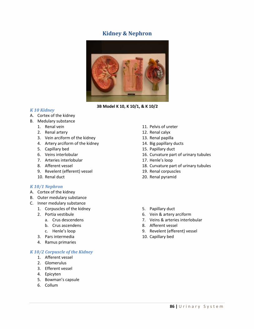
86 | U r i n a r y S y s t e m
Kidney & Nephron
K 10 Kidney A. Cortex of the kidney B. Medulary substance
1. Renal vein 2. Renal artery 3. Vein arciform of the kidney 4. Artery arciform of the kidney 5. Capillary bed 6. Veins interlobular 7. Arteries interlobular 8. Afferent vessel 9. Revelent (efferent) vessel 10. Renal duct
11. Pelvis of ureter 12. Renal calyx 13. Renal papilla 14. Big papillary ducts 15. Papillary duct 16. Curvature part of urinary tubules 17. Henle’s loop 18. Curvature part of urinary tubules 19. Renal corpuscles 20. Renal pyramid
K 10/1 Nephron A. Cortex of the kidney B. Outer medulary substance C. Inner medulary substance
1. Corpuscles of the kidney 2. Portia vestibule
a. Crus descendens b. Crus ascendens c. Henle’s loop
3. Pars intermedia 4. Ramus primaries
5. Papillary duct 6. Vein & artery arciform 7. Veins & arteries interlobular 8. Afferent vessel 9. Revelent (efferent) vessel 10. Capillary bed
K 10/2 Corpuscle of the Kidney 1. Afferent vessel 2. Glomerulus 3. Efferent vessel 4. Epicyten 5. Bowman’s capsule 6. Collum
3B Model K 10, K 10/1, & K 10/2

87 | U r i n a r y S y s t e m
Kidney (Somso LS 5)
A. Marrow of the Kidney
B. Cortex of the Kidney
1. Renal vein
2. Renal artery
3. Ureter
4. Pelvis of the kidney
5. Calyces of the kidney
6. Renal papillae
7. Renal pyramids
8. Collecting duct
9. Contort renal tubule II
10. Loop of Henle
11. Contort renal tubule I
12. Corpuscels of the kidney
13. Interlobular arteries
14. Afferent vessel
15. Efferent vessel
16. Arcuate arteries
17. Interlobular veins
18. Arcuate veins
19. Papillary ducts
20. Suprarenal gland
Somso LS 5

88 | U r i n a r y S y s t e m
Kidney (Somso K 12)
1. Cortex
2. Medulla
3. Renal artery
4. Renal vein
5. Renal pyramid
6. Interlobar arteries
7. Interlobar veins
8. Arcuate arteries
9. Interlobular arteries
10. Interlobular veins
11. Arcuate veins
12. Minor calyx
13. Major calyx
14. Columns of Bertini
15. Renal pelvis
16. Renal papilia
17. Ureter
18. Suprarenal gland
19. Suprarenal vein
20. Inferior suprarenal artery
3B Model K 12

89 | U r i n a r y S y s t e m
Glomerulus

90 | U r i n a r y S y s t e m
Renal Lobule

91 | U r i n a r y S y s t e m
Urinary System
1. Abdominal aorta
2. Celiac trunk
3. Superior mesenteric artery
4. Right renal artery
5. Left renal artery
6. Inferior vena cava
7. Right renal vein
8. Left renal vein
9. Right testicular vein
10. Left testicular vein
11. Right suprarenal (adrenal) gland
12. Right kidney
13. Cortex
14. Medulla
15. Adipose coat of renal sinus
16. Renal pelvis
17. Renal columns
18. Major calyces
19. Minor calyces
20. Ramification of renal artery & vein
21. Medullary rays of pyramids
22. Renal pailla
23. Renal helium
24. Ureters (yellow tubing)
25. Right testicular artery
26. Left testicular artery
27. Inferior mesenteric artery
28. Bifurcation of abdominal aorta
29. Right common iliac artery
30. Left common iliac artery
31. Right common iliac vein
32. Left common iliac vein
33. Right femoral artery
34. Left femoral artery
35. Right femoral vein
36. Left femoral vein
37. Urinary bladder
38. Plicae mucosae
39. Trigone
40. Ureteral orifices
41. Uvula vesica & internal urethral orifice
42. Urethral crest
43. Prostatic utricle
44. Ejaculatory ducts
45. Orifices of prostatic ducts
46. Prostate gland
47. Muscular layer of bladder
48. Serous coat of bladder
49. Ductus deferens
50. Seminal vesicles
Model #145, A55

92 | U r i n a r y S y s t e m
Kidney (Altay 6140.14) Kidney Section with Renal Nephron & Corpuscle
I. Kidney with suprarenal gland 1. Suprarenal gland 2. Cortex of suprarenal gland
a. Zone glomerulosa b. Zone fasciculata c. Zone reticularis
3. Medulla of suprarenal gland 4. Kidney 5. Anterior surface 6. Posterior surface 7. Renal cortex 8. Renal columns 9. Renal pyramids 10. Renal papilla 11. Minor renal calices 12. Major renal calices 13. Renal pelvis 14. Renal artery 15. Renal vein 16. Fatty tissue in renal sinus
II. Nephron 17. Renal corpuscle 18. Descending limb 19. Thin segment 20. Ascending limb 21. Collecting tubules 22. Papillary duct 23. Arcuate artery 24. Arcuate vein 25. Interlobar artery 26. Interlobar vein 27. Interlobular artery 28. Interlobular vein 29. Straight arterioles 30. Straight venules
III. Renal Corpuscle 31. Renal capsule 32. Glomerulus 33. Afferent glomerular arteriole 34. Efferent glomerular arteriole 35. Proximal convoluted tubule 36. Distal convoluted tubule 37. Macula densa 38. Juxtaglomerular cell 39. Podocyt

93 | R e p r o d u c t i v e S y s t e m
Reproductive System
Male Pelvis (Somso MS 2)
1. Pubis 2. Sacrum 3. Coccyx 4. Urinary bladder
a. Apex of urinary bladder b. Fundus of bladder c. Muscular coat of bladder d. Mucous membrane of bladder e. Ureter f. Trigone of urinary bladder
5. Prostate 6. Prostatic urethra
a. Urethral crest 7. Seminal vesicle 8. Ductus deferens 9. Ureter 10. Corpus cavernosum 11. Penis 12. Glans penis 13. External urethral orifice 14. Ischio-cavernosus muscle 15. Testicle
a. Superior end b. Inferior end
16. Epididymis a. Head of epididymis b. Pampiniform plexus
17. Testicular artery 18. Cremaster muscle 19. Spermatic Cord 20. Rectum 21. Common iliac artery 22. Common iliac vein 23. Peritoneum 24. Inguinal ligament 25. Pectineal part of the inguinal liagament 26. Femoral canal 27. Lancuna of the vessels 28. Interfoveolar ligament
Somso Model MS 2

94 | R e p r o d u c t i v e S y s t e m
Male Pelvis
1. Penis 2. Foreskin 3. Scrotum 4. Anus 5. Crest of ilium 6. 5th lumbar vertebra 7. Rectus abdominis muscle 8. Pyramidalis muscle 9. Internal oblique muscle 10. Transverse abdominis muscle 11. Multifidus muscle 12. Sacrospinalis muscle 13. Psoas muscle 14. Iliacus muscle 15. Spinal cord 16. Common iliac vein 17. Common iliac artery 18. External iliac artery 19. Internal iliac artery 20. External iliac vein 21. External iliac artery 22. Vesical artery 23. Lateral umbilical ligament 24. Obturator artery & vein 25. Inferior gluteal artery & vein 26. Internal pudendal artery & vein 27. Vesicle venous plexus 28. Prostatic venous plexus 29. Spermatic artery, vein, & nerve 30. Femoral nerve 31. Parietal lymph nodes 32. Ductus deferens
33. Spermatic cord 34. Cremaster muscle 35. External inguinal ring 36. Ureter 37. Urinary bladder 38. Seminal vesicle 39. Ampulla of ductus deferens 40. Prostate 41. Ejaculatory duct 42. Bulbourethral gland 43. Urogenital diaphragm 44. Prostatic urethra 45. Cavernous urethra 46. Bulbus penis 47. Corpus spongiosum 48. Corpus cavernosum 49. Glans penis 50. Bulbocavaernosus muscle 51. Septum of scrotum 52. Testis 53. Epididymis 54. Deep dorsal vein of penis 55. Ischiocavernosus muscle 56. Crus penis 57. Gracilis muscle 58. Pubis 59. Pubic symphysis 60. Suspensory ligament of penis 61. Sphincter ani externus muscle 62. Rectum 63. Coccyx 64. Sacrum
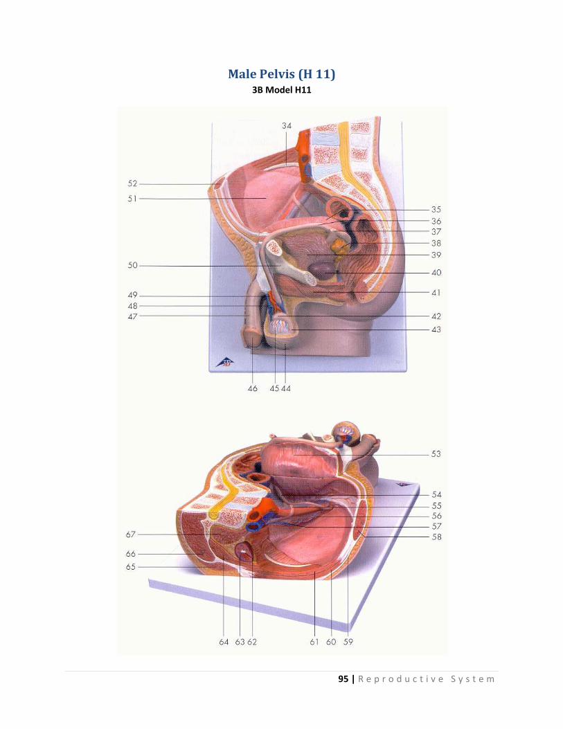
95 | R e p r o d u c t i v e S y s t e m
Male Pelvis (H 11) 3B Model H11

96 | R e p r o d u c t i v e S y s t e m
Male Pelvis Cont… 3B Model H11

97 | R e p r o d u c t i v e S y s t e m
Male Pelvis Cont… 3B Model H11
1. Intervertebral disc 2. Lumbar vertebra 3. Spinal marrow 4. Spinous process 5. Internal iliac artery 6. Sigmoid colon (pelvic colon) 7. Muscular coat of bladder 8. Fundus of bladder 9. Coccygeal bone 10. Internal urethral orifice 11. Prostatic utricle 12. Anococcygeal ligament 13. Sphincter ani externus muscle 14. Sphincter ani internus muscle 15. Rectal ampulla 16. Cauda of epididymus 17. Cremaster muscle 18. Testis 19. Head of epididymus 20. External opening of masculine urethra 21. Cavernous body of penis 22. Spongy part of masculine urethra 23. Spongy part of penis 24. Masculine urethra, prostatic part 25. Pubic symphysis 26. Transverse rectal fold 27. Apex of urinary bladder 28. External iliac artery 29. External iliac vein 30. Right ureter 31. Inferior mesenteric artery 32. Abdominal aorta 33. Renal artery 34. Crest of ilium
35. Bladder 36. Deferent duct (spermatic duct) 37. Left ureter 38. Seminal vesicle 39. Muscular coat of bladder 40. Prostate 41. Bulbospongiosus muscle 42. Head of epididymis 43. Cauda of epididymis 44. Scrotum 45. Testis 46. Glans of penis 47. Penis 48. Spermatic plexus 49. Testicular artery 50. Pubic bone 51. Visceral peritoneum 52. Rectus abdominis muscle 53. Bladder 54. Deferent duct (spermatic duct) 55. Right ureter 56. Right testicular artery 57. Right testicular vein 58. Rectus abdominis muscle 59. Aponeurosis of obliquus externus
abdominis muscle 60. Obliquus internus abdominis muscle 61. Transverse abdominis muscle 62. Renal pelvis 63. Renal cortex 64. Quadratus lumborum muscle 65. Latissimus dorsi muscle 66. Erector spinae muscle 67. Psoas major muscle

98 | R e p r o d u c t i v e S y s t e m
Female Pelvis 1. Mons pubis 2. Anus 3. Labium majus 4. Labium minus 5. Crista iliaca ilium 6. Lumbar vertebra (5th) 7. Rectus abdominis muscle 8. Oblique internus muscle 9. Transverse abdominis muscle 10. Sacrospinalis muscle 11. Multifidus muscle 12. Psoas major muscle 13. Iliacus muscle 14. Iliopsoas muscle 15. Spinal cord 16. Round ligament 17. Uterosacral ligament 18. Broad ligament 19. Ovarian ligament 20. Uterine tube 21. Ovary 22. Uterus 23. Cervix 24. Tunica serosa 25. Tunica muscularis 26. Tunica mucosa 27. Cavum uterum 28. Fornix 29. Vagina 30. Rectum 31. Urethra 32. Urinary bladder 33. Ureter 34. Sacrum 35. Coccyx 36. Pubic symphysis 37. Clitoris 38. Crus of clitoris 39. Bulbus vestibule (bulb of vestibule) 40. Greater vestibular gland 41. Ischiocavernosus muscle 42. Transverse perinea superficialis muscle 43. Sphincter ani externus muscle 44. Piriformis muscle 45. Coccygeal muscle
46. Levator ani muscle 47. Transversus perinea profundus muscle 48. Common iliac artery 49. Common iliac vein 50. Internal iliac artery 51. Internal iliac vein 52. External iliac artery 53. External iliac vein 54. Ovarian artery & vein 55. Obturator artery, vein, & nerve 56. Vesical artery & vein 57. Uterine artery & vein 58. Vaginal artery & vein 59. Superior gluteal artery & vein 60. Pudendal artery, vein, & nerve 61. Parietal lymph node 62. Middle hypogastric nerve plexus 63. Sympathetic nerve trunk 64. First sacral nerve 65. Second sacral nerve 66. Third sacral nerve 67. Fourth sacral nerve 68. Fifth sacral nerve 69. Sacral plexus 70. Lumbosacral nerve trunk 71. External inguinal ring 72. Superificial femoral lymph nodes 73. Inguinal ligament 74. Great saphenous vein 75. Pubis 76. Dorsal clitoral artery, vein, & nerve 77. Posterior femoral cutaneous nerve 78. Inferior epigastric artery & vein 79. Medial umbilical ligament 80. Lateral umbilical ligament

99 | R e p r o d u c t i v e S y s t e m
Female Pelvis (Somso MS 3)
1. Symphysis pubis 2. Saccrum 3. Coccyx 4. Labium majus 5. Labium minus 6. Clitoris 7. Urethra 8. Urinary bladder 9. Broad ligament 10. Vagina 11. Uterus 12. Fornix 13. Cervix 14. Infundibulum 15. Ovary 16. Ovarian ligament 17. Round ligament 18. Rectum 19. Anus
Somso Model MS 3

100 | R e p r o d u c t i v e S y s t e m
Human Female Breast
1. Areola 2. Areolar Glands 3. Papilla 4. Lactiferous Sinus & Duct 5. Parenchyma or Mammary Gland Proper 6. Fat 7. Retinaculum Cutis
Model 56-6600

101 | R e p r o d u c t i v e S y s t e m
Female Genital Organs (Somso MS 5)
1. Labium majus pudendi 2. Labium minus pudendi 3. Clitoris 4. Prepuce 5. Vagina 6. Body of uterus 7. Neck of uterus 8. Vaginal portion (cervix) 9. Uterine cavity 10. Uterine tube 11. Ovarian fimbria 12. Ovary 13. Ligament of the ovary 14. Round ligament of the uterus 15. Urinary bladder 16. Ureter 17. Rectum 18. Muscle sphincter ani externus 19. Muscle sphincter ani internus
Somso Model MS 5
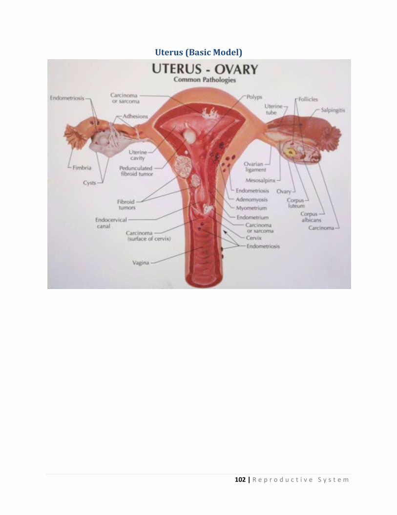
102 | R e p r o d u c t i v e S y s t e m
Uterus (Basic Model)

103 | R e p r o d u c t i v e S y s t e m
Female Pelvis (H 10) 3B Model H10

104 | R e p r o d u c t i v e S y s t e m
Female Pelvis Cont... 3B Model H10

105 | R e p r o d u c t i v e S y s t e m
Female Pelvis Cont... 3B Model H10
1. Intervertebral disc 2. Lumbar vertebra 3. Spinal marrow 4. Spinous process 5. Internal iliac artery 6. Sigmoid colon (pelvic colon) 7. Uterine cavity 8. Supravaginal part of cervix uteri 9. Transverse rectal fold 10. Coccygeal bone 11. Opening of uterus 12. Rectal ampulla 13. Anococcygeal ligament 14. Sphincter ani externus muscle 15. Sphincter ani internus muscle 16. Vagina 17. Lesser pudendal lip 18. Greater pudendal lip 19. Cavernous body of clitoris 20. Feminine urethra 21. Pubic symphysis 22. Bladder 23. Fundus of uterus 24. External iliac vein 25. External iliac artery 26. Ovarian vein 27. Ovarian artery 28. Inferior mesenteric artery 29. Renal artery 30. Abdominal aorta 31. Inferior vena cava 32. Crest of ilium 33. Ovary 34. Intestinal surface of uterus
35. Left ureter 36. Vagina 37. Sphincter ani externus muscle 38. Greater vestibular gland 39. Vestibula of vagina 40. Clitoris 41. Pubic bone 42. Bladder 43. Round ligament of uterus 44. External iliac vein 45. External iliac artery 46. Visceral peritoneum 47. Right ureter 48. Rectus abdominis muscle 49. Ovarian canal (uterine tube) 50. Uterus 51. Ampulla of uterine tube 52. Ovarian ligament 53. Round ligament of uterus 54. Rectus abdominis muscle 55. Ureter 56. Obliquus externus abdominis muscle 57. Obliquus internus abdominis muscle 58. Transverse abdominis muscle 59. Renal pelvis 60. Renal cortex 61. Latissimus dorsi muscle 62. Quadratus lumborum muscle 63. Psoas major muscle 64. Erector spinae muscle 65. Psoas minor muscle 66. Ovarian ligament 67. Ovary

106 | R e p r o d u c t i v e S y s t e m
Relief Model of the Ovary (Somso MS 51)
1. Mesovary 2. Germinal epithelium 3. Peritoneal epithelium 4. Tunica albuginae 5. Cortical zone 6. Medullary zone 7. Vessels 8. Smooth muscle cells 9. Primordial follicle 10. Follicle epithelium of primordial follicle 11. Secondary follicle 12. Atretic follicle 13. Cavum folliculi with liquor folliculi 14. Graffian follicle 15. Another follicle shown in section shortly after ovulation 16. Radiate corona 17. Ovum 18. Oolemma 19. Intercal theca of the follicle 20. External theca of the follicle 21. Corpus rubrum 22. Corpus luteum 23. Albicant corpus 24. Hilus with arterial & venous vascular plexus
Somso Model MS 51

107 | D i g e s t i v e S y s t e m
Digestive System
Molar Tooth (Somso ES 11/5)
1. Corona 2. Tubercles of the crown 3. Collum of the tooth 4. Root 5. Enamel 6. Dentin 7. Cement 8. Dental pulp
Somso Model ES 11/5
Molar with three roots

108 | D i g e s t i v e S y s t e m
Models of Teeth Somso Models
ES 8 1. Corona of tooth with carries 2. Tubercles of crown 3. Collum of tooth 4. Root of tooth 5. Enamel 6. Dentin 7. Cement 8. Dental pulp
ES 11/1, 2, 3 1. Corona of tooth 2. Tubercles of crown 3. Collum of tooth 4. Root of tooth 5. Enamel 6. Dentin 7. Cement 8. Dental pulp
ES 8 – Molar Tooth with carries ES 11/1, 2, 3

109 | D i g e s t i v e S y s t e m
Stomach (Basic Model)

110 | D i g e s t i v e S y s t e m
The Stomach (Somso JS 4)
1. Cardiac orifice 2. Fundus of the stomach 3. Small curvature 4. Big curvature 5. Pylorus 6. Layer of muscular tissue 7. Longitudinal muscular fibers 8. Circular muscular fibers 9. Oblique muscular fibers 10. Peritoneum 11. Left gastric artery 12. Right gastric artery 13. Left gastric epiploic artery 14. Right gastric epiploic artery 15. Left vagus nerve 16. Right vagus nerve 17. Gastric mucous membrane 18. Gastric folds 19. Submucous tunica 20. Section through a fundus gland microscopically enlarged
Somso Model JS 4

111 | D i g e s t i v e S y s t e m
Pancreas with Spleen & Part of the Duodenum (Somso JS 11)
A. Pancreas
B. Spleen
C. Duodenum
1. Pancreatic duct 2. Duct of Santorini 3. Choledochous duct 4. Longitudinal fold of the duodenum 5. Portal vein 6. Splenic vein 7. Superior mesenteric vein 8. Celiac trunk 9. Hepatic artery 10. Splenic artery 11. Gastric artery 12. Gastroduodenal artery 13. Superior mesenteric artery 14. Inferior pancreaticoduodenal artery
Somso Model JS 11

112 | D i g e s t i v e S y s t e m
Intestinal Villi (Wards Model 81 W 1332)

113 | D i g e s t i v e S y s t e m
Model of a Liver Cell (Somso JS 15)
1. Nucleoli 2. Chromatin 3. Nucleus membrane 4. Glycogen 5. Golgi apparatus 6. Endoplasmic reticulum 7. Mitochondria 8. Lysosomes 9. Microvilli 10. Bile-duct 11. Plasma membrane
Somso Model JS 15

114 | D i g e s t i v e S y s t e m
Internal Organs
1. Body of pancreas 2. Head of pancreas 3. Tail of pancreas 4. Pancreatic duct 5. Accessory pancreatic duct 6. Papilla major of duodenum 7. Papilla minor of duodenum 8. Duodenum 9. Descending portion of duodenum 10. Inferior portion of duodenum 11. Ascending portion of duodenum 12. Right suprarenal gland 13. Left suprarenal gland 14. Right kidney 15. Left kidney 16. Spleen 17. Hilus of spleen 18. Renal surface 19. Gastric surface 20. Diaphragmatic surface 21. Anterior margin 22. Upper extremity 23. Lower extremity 24. Gastrolienal ligament 25. Portion of inferior surface of liver 26. Gallbladder 27. Fundus of gallbladder 28. Neck of gallbladder 29. Cystic duct with spiral valve 30. Hepatic duct
31. Common bile duct 32. Coeliac artery 33. Gastroduodenal artery 34. Superior pancreatico-duodenal artery 35. Hepatic artery 36. Superior mesenteric artery 37. Intestinal arteries 38. Middle colic artery 39. Splenic artery 40. Suprarenal arteries & veins 41. Portal vein 42. Superior mesenteric vein 43. Intestinal veins 44. Middle colic vein 45. Splenic vein 46. Main trunk of portal vein 47. Left gastric artery 48. Splenic artery 49. Right renal artery 50. Right renal vein 51. Left renal artery 52. Left renal vein 53. Hilus of kidney 54. Right ureter 55. Left ureter 56. Branches of portal vein w/in liver 57. Branches of hepatic veins w/in liver 58. Branches of hepatic ducts w/in liver 59. Branches of hepatic artery w/in liver
Model #146, A56

115 | D i g e s t i v e S y s t e m
Liver & Gallbladder
1. Right lobe 2. Left lobe 3. Quadrate lobe 4. Caudate (spigelian) lobe 5. Papillary process 6. Caudate process 7. Inferior vena cava 8. Hepatic veins 9. Ramification of hepatic veins in liver 10. Portal vein 11. Ramification of portal vein 12. Hepatic artery 13. Ramification of hepatic artery 14. Hepatic duct 15. Ramification of hepatic duct 16. Cystic duct 17. Gallbladder 18. Round ligament 19. Falciform ligament 20. Coronary ligament, superior leaf 21. Coronary ligament, posterior leaf 22. Ligament of ductus venosus 23. Hepatic veins, branching off from inferior vena cava 24. Pyloric impression 25. Duodenal impression 26. Renal impression 27. Colic impression 28. Esophageal impression
Model #141, A51

116 | D i g e s t i v e S y s t e m
Liver
1. Falciform ligament 2. Round ligament of liver 3. Gallbladder 4. Common bile duct 5. Proper hepatic artery 6. Hepatic portal vein 7. Inferior vena cava 8. Lateral-superior area 9. Lateral-inferior area 10. Medial-superior area 11. Medial-inferior area 12. Anterior-superior area 13. Anterior-inferior area 14. Posterior-superior area 15. Posterior-interior area 16. Left segment of caudate lobe 17. Right segment of caudate lobe
Altay Model 6090.06

117 | D i g e s t i v e S y s t e m
Human Digestive System
1. oral lips 2. hard palate 3. soft palate 4. uvula 5. teeth 6. tongue 7. parotid gland 8. Steno’s duct 9. submandibular gland 10. submandibular duct 11. sublingual gland 12. sublingual caruncle 13. nasopharynx 14. pharyngeal opening - auditory tube 15. pharyngeal tonsil 16. oropharynx 17. palatine tonsil 18. laryngopharynx 19. cardia 20. pylorus 21. fornix of stomach 22. body of stomach 23. pyloric antrum 24. duodenum 25. major duodenal papilla 26. minor duodenal papilla 27. jejunum 28. ileum 29. cecum 30. ileocecal valve 31. vermicular appendage
32. ascending colon 33. transverse colon 34. descending colon 35. sigmoid colon 36. rectum 37. anal canal 38. dentate line 39. internal anal sphincter 40. external anal sphincter 41. falciform ligament 42. left lobe of liver 43. right lobe of liver 44. ligamentum teres 45. quadrate lobe 46. caudate lobe 47. gallbladder 48. cystic duct 49. common hepatic duct 50. common bile duct 51. proper hepatic artery 52. portal vein of the liver 53. head of pancreas 54. body of pancreas 55. tail of pancreas 56. pancreatic duct 57. accessory pancreatic duct
Altay Model 6090.01

118 | N e w M o d e l s - 2 0 1 1

119 | N e w M o d e l s - 2 0 1 1
Liver/Gallbladder (Basic Model)

120 | N e w M o d e l s - 2 0 1 1
Colon (Basic Model)

121 | N e w M o d e l s - 2 0 1 1
Rectum (Basic Model)


