Learning objective✓ To learn the anatomy of the eye, the orbit and the third, fourth and
sixth cranial nerves, as a background to the medical conditionsaffecting them.
IntroductionKnowledge of ocular anatomy and function is impor-tant to the understanding of eye diseases. A briefoutline is given below.
Surface anatomy of thefaceThe eyes are disposed symmetrically about the faceand their forward-looking arrangement permits alarge overlap in visual fields, the basis of stereopsis.Lying within the bony orbits, they are protected fromtrauma by the orbital walls and rims and by the eye-lids, by blinking and eye closure. With the eyes openand looking straight ahead, all but theupper and lowercornealmargins are exposed in thepalpebral aperture,together with two small white triangles of bulbarconjunctiva, overlying the sclera. The medial andlateral ends of the fissure are known as the medialand lateral canthi (Figure 1.1).The lids and the upper and lower orbital rims are
overlain by the orbicularis muscle which sweepsover these structures in an ellipse, from a region
just medial to the medial orbital rim. It acts as thepalpebral sphincter (Figure 1.2). Like all othermuscles of the face, it is supplied by the seventhcranial nerve. Contraction of its orbital part resultsin protective, forced eye closure, while contractionof its palpebral part is employed in the downstrokeof the upper lid during a blink. The levator palpe-brae muscle, the elevator of the upper lid (seebelow), is concerned with the upstroke of the blink(third cranial nerve). These synchronized contrac-tions are completed within just 300ms. The con-tents of the orbit are separated from those of the lidby a connective tissue sheet, or orbital septum,which extends from the orbital rim to the tarsalplate, deep to orbicularis.
Sensory innervation of theface: the fifth cranialnerveThe sensory innervation of each half of the face isprovided by the trigeminal nerve (Figure 1.3). Theeye, upper lid, eyebrow, forehead and nose aresupplied by its ophthalmic division (V1), via itslacrimal, frontal and nasociliary branches, whichenter the orbit through the superior orbital fissure.The maxillary division (V2), lying inferolaterally toV1 in the cavernous sinus, exits the cranial cavity viathe foramen rotundum and, at the inferior orbitalfissure, gives rise to the infraorbital and zygomaticnerves. These supply, chiefly, the lower lid and the
upper lip and cheek. The mandibular division (V3),exiting the skull via the foramen ovale, supplies thelower lip, chin and jaw and the preauricular skinand temporal region. It is also motor to the musclesof mastication.The neurons of the three divisions of the trigem-
inal nerve converge upon the trigeminal ganglion,whose sensory roots enter the pons to be distrib-uted to the trigeminal nuclei in the brainstem. Themesencephalic nucleus is concerned with propri-oception, the main sensory nucleus with touch andthe medullary nucleus of the spinal tract withpain and temperature sensibility. Fibres from theophthalmic division go to the lowest part of thisnucleus, those from the mandibular division to itshighest part.
Medial canthusLateral canthus
Figure 1.1 The eye, looking straight ahead.
SUPERFICIALCERVICAL PLEXUSMANDIBULAR AREAMAXILLARY AREA
OPHTHALAMIC AREA
Supratrochlear N.
Lacrimal N.
Infratrochlear nerve
Nasal nerve
Infraorbital nerveBuccal nerve
Supraorbital N.
CERVICAL NERVES(POSTERIOR DIVISIONS)
Mental nerve
Temporal BR.of temporo-malar
Malar BR. oftemporo-malar
Auriculo-temporalnerve
Figure 1.3 Sensory innervation of the face by the trigeminal nerve.
Orbicularis oculimuscle
Orbital part
Medial palpebral ligament
Palpebralpart
Lateral palpebralligament
Figure 1.2 Disposition of the orbicularis is muscle.
2 Anatomy
c01 11/13/2017 12:27:40 Page 3
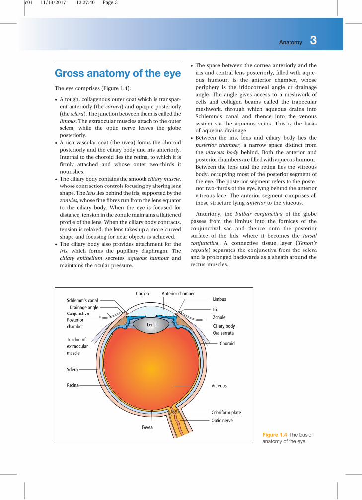
Gross anatomy of the eyeThe eye comprises (Figure 1.4):
• A tough, collagenous outer coat which is transpar-ent anteriorly (the cornea) and opaque posteriorly(the sclera). The junction between them is called thelimbus. The extraocular muscles attach to the outersclera, while the optic nerve leaves the globeposteriorly.
• A rich vascular coat (the uvea) forms the choroidposteriorly and the ciliary body and iris anteriorly.Internal to the choroid lies the retina, to which it isfirmly attached and whose outer two-thirds itnourishes.
• The ciliary body contains the smooth ciliary muscle,whose contraction controls focusing by altering lensshape. The lens lies behind the iris, supported by thezonules, whose fine fibres run from the lens equatorto the ciliary body. When the eye is focused fordistance, tension in the zonulemaintains aflattenedprofile of the lens. When the ciliary body contracts,tension is relaxed, the lens takes up a more curvedshape and focusing for near objects is achieved.
• The ciliary body also provides attachment for theiris, which forms the pupillary diaphragm. Theciliary epithelium secretes aqueous humour andmaintains the ocular pressure.
• The space between the cornea anteriorly and theiris and central lens posteriorly, filled with aque-ous humour, is the anterior chamber, whoseperiphery is the iridocorneal angle or drainageangle. The angle gives access to a meshwork ofcells and collagen beams called the trabecularmeshwork, through which aqueous drains intoSchlemm’s canal and thence into the venoussystem via the aqueous veins. This is the basisof aqueous drainage.
• Between the iris, lens and ciliary body lies theposterior chamber, a narrow space distinct fromthe vitreous body behind. Both the anterior andposterior chambers arefilledwith aqueous humour.Between the lens and the retina lies the vitreousbody, occupying most of the posterior segment ofthe eye. The posterior segment refers to the poste-rior two-thirds of the eye, lying behind the anteriorvitreous face. The anterior segment comprises allthose structure lying anterior to the vitreous.
Anteriorly, the bulbar conjunctiva of the globepasses from the limbus into the fornices of theconjunctival sac and thence onto the posteriorsurface of the lids, where it becomes the tarsalconjunctiva. A connective tissue layer (Tenon’scapsule) separates the conjunctiva from the scleraand is prolonged backwards as a sheath around therectus muscles.
CorneaSchlemm's canal
Conjunctiva
Tendon ofextraocularmuscle
Iris
Lens
Drainage angle
Posteriorchamber
Sclera
Retina
Anterior chamberLimbus
Ciliary body
Zonule
Ora serrata
Cribriform plate
Optic nerveFovea
Choroid
Vitreous
Figure 1.4 The basicanatomy of the eye.
Anatomy 3
c01 11/13/2017 12:27:41 Page 4
The orbitThe eye, or globe, lies within the bony orbit, which hasthe shape of a four-sided pyramid (Figure 1.5). At itsposterior apex is the optic canal, which transmits theoptic nerve to the chiasm, tract and lateral geniculatebody. The superior and inferior orbital fissures trans-mit the blood vessels and cranial nerves that supplythe orbital structures. The lacrimal gland lies anteri-orly in the superolateral aspect of the orbit. On theanterior part of the medial wall lies the fossa for thelacrimal sac.
The eyelids (the tarsus)The eyelids (Figure 1.6):
• offer mechanical protection to the globe;• spread the tears over the conjunctiva and corneawith each blink.
The levator muscle is the main elevator of theupper lid. It passes forwards from an attachmenton the sphenoid bone, above the optic foramen, toan aponeurosis which inserts into the tarsal plate.It is innervated by the third cranial nerve. Damage
Superior orbital fissure
Ant.Iacrimal crest
Post lacrimal crest
Infra orbital fissure
Sphenoid
Frontal bone
Optic canal
MaxillaZygoma
Nasal bone
Ethmoid
Lacrimal fossa
Lacrimal bone
Figure 1.5 The anatomy of the orbit.
Levator muscle and tendon
Müller'smuscle
Skin
Orbicularismuscle
Tarsal plate
Lash
Meibomiangland
Sclera
Cornea
Upper fornix
Conjunctiva
Figure 1.6 The anat-omy of the eyelids.
4 Anatomy
c01 11/13/2017 12:27:41 Page 5
to the nerve orweakening of the aponeurosis in old ageresults in drooping of the upper eyelid (ptosis). A flat,smooth muscle, (the superior tarsal, or Müller’smuscle) innervated by the sympathetic nervous sys-tem, arises from the deep surface of the levator andinserts into the tarsal plate. Müllers muscle also con-tributes to a lesser extent to elevation of the lid, and ifthe sympathetic supply is damaged, a slight ptosisresults as part of Horner’s syndrome.Each eyelid comprises:
• an anterior layer of skin;• the palpebral part of the orbicularis muscle;• a tough collagenous layer (the tarsal plate) whichhouses the meibomian oil glands;
• an epithelial lining, the tarsal conjunctiva;• the lash-bearing, lid margins.
The tarsal conjunctiva is reflected, via the fornices,onto the anterior surface of the globe, where itbecomes the bulbar conjunctiva. When the eyes areclosed, this lining forms the conjunctival sac, whichcontains the tears. When the eyes open, a tear film isformed which covers and protects the exposed corneaand conjunctiva. At the lid margins, the tear film isbordered by the tear menisci (Figure 1.7).The lid margins exhibit a narrow, posterior con-
junctival zone, continuous with the tarsal conjunctivaand a cutaneous zone anteriorly, which bears thelashes. These zones are separated by the muco-cutaneous junction which forms the anterior bound-ary of each tear meniscus (Figure 1.8). At the medialends of each lid margin, dipping into a lake of tears atthe nasal canthus, are the lacrimal puncta, throughwhich tears drain from the tear menisci into thelacrimal drainage system.The meibomian oil glands, embedded in the tarsal
plates (Figure 1.8), deliver their oil to the skin of the lid
margin, just anterior to the mucocutaneous junction.This oil spreads onto the anterior surface of the tearfilm with each blink, to form a lipid layer, whichretards evaporation and stabilizes the tear film.
The lacrimal drainagesystemTears drain into the upper and lower puncta and theninto the lacrimal sac via theupper and lower canaliculi(Figure 1.9). They form a common canaliculus beforeentering the lacrimal sac. The nasolacrimal ductpasses from the sac to the nasal cavity which entersat the inferior meatus. Failure of the distal part of the
Accessory lacrimalglands Main
lacrimal gland
Lower meniscus
Nasolacrimalduct
Upper meniscus
Meibomiangland
(a) (b)
Meniscus
Tear film
Cornea
Eyelid
Conjunctival/fornical sac
Lacrimalsac
CanaliculiFigure 1.7 Drawing of the eye: (a) in crosssection, (b) in frontal view to illustrate the distri-bution of the tears. (Source: Gaffney EA et al.Progress in Retinal and Eye Research 2010;29(1):59–78. Reproduced with permission ofElsevier.)
Black lineTear film
Tearmeniscus
Marx’sline
Occlusalconjunctiva
Meibomianorifices
Apex
TFLL
Occlusalskin
MG
Marginalconjunctiva
Figure 1.8 Diagram of lid margin to show meibomianorifices, meniscus and tear film lipid layer (TFLL). (Source:Bron et al. Ocul Surf 2011; 9(2):70–91. Reproduced withpermission of Elsevier.)
Anatomy 5
c01 11/13/2017 12:27:41 Page 6
nasolacrimal duct to fully canalize at birth is the usualcause of a watering, sticky eye in an infant. Teardrainage is an active process. Each blink helps topump tears through the system.
Detailed functionalanatomy
The tear filmThe eye is bathed constantly by the tears, secretedby thelacrimal gland into the upper fornix of the conjunctivalsac. There is a small contribution from the conjunctiva.Tears are lost from the surface in part by evaporationand in part by drainage via the nasolacrimal system.Lacrimal secretion is under parasympathetic controlthrough a feedback loop from the cornea, via the tri-geminal nerve, to the superior salivatory nucleus andthence to the lacrimal gland. This ensures that tearproduction is regulated reflexly in response to signalsfrom the ocular surface.The most superficial epithelial cells of the ocular
surface express amucin-rich glycocalyxwhich rendersthe surface wettable. When the eyes are open, theexposed ocular surface is covered by a tear film, 3 μmthick. This has two layers:
• Amucoaqueous layer containing gelmucin from theconjunctival goblet cells and aqueous tear fluid fromthe lacrimal gland, directly in contact with theocular surface.
• A thin surface oil layer (100 nm) produced by themeibomian glands and delivered to the tear filmfrom the lid margins.
Functions of the tear film• It moistens the eye, preventing dehydration of itssurface.
• It provides a smooth, air/tear, optical interface fordistortion-free refraction of light at the cornea.
• It transmits oxygen to the avascular cornea.• It removes debris and foreign particles from theocular surface through the flow and drainage of thetears and the action of the blink.
• It has antibacterial properties bymeansof lysozyme,lactoferrin, defensins and the immunoglobulins,particularly secretory IgA.
The tear film is replenished with each blink.
The corneaThe cornea is 0.5mm thick and comprises(Figure 1.10):
• The epithelium, an anterior, non-keratinised squa-mous layer,five cells thick, thickenedperipherally atthe limbus where it is continuous with the conjunc-tiva. The limbus houses the germinative stem cellswhich maintain the corneal epithelium. The basalcells of the epithelium are firmly attached to anunderlying basal lamina by hemidesmosomes andby anchoring fibrils which extend into Bowman’slayer.
• An underlying stroma which accounts for over90% of the corneal thickness. On its most anterioraspect is a tough, anterior limiting layer (Bow-man’s layer), 20 μm thick, which is free of cellsand composed of fine, short, tightly interwovencollagen fibrils. The main body of the stromaconsists of type I collagen fibrils arranged in
Upper canaliculus
Puncta
Lowercanaliculus
Tear sac
Common canaliculus
Nasolacrimal duct
Inferior turbinate
Inferior meatus
Nasal cavity
Nasal mucosa
Figure 1.9 The majorcomponents of thelacrimal drainagesystem.
6 Anatomy
c01 11/13/2017 12:27:41 Page 7
parallel within lamellae, each fibril surrounded by aground substance rich inproteoglycans.Between thelamellae are scattered keratocytes which, like fibro-blasts, engage in stromal maintenance and repair.The anterior lamellae lie in the plane of the cornea,while posteriorly they have a more woven arrange-ment. The regular packing of the collagen fibrils,their small diameter and narrow separation (in theregion of 200nm)accounts for corneal transparency.Backscattered light, towards the source, is obliter-ated by destructive interference and over 90% of thelight is transmitted. This orderly architecture ismaintained by regulating stromal hydration. Thestroma is bounded behind by the posterior limitinglayer (Descemet’s layer), the basal lamina of thecorneal endothelium. It is chiefly composed oftype IV collagen.
• The endothelium, a monolayer of hexagonal, non-regenerating cells (Figure 1.11) which activelypump ions from the stroma into the anterior cham-ber carrying water with them. This controls cornealhydration and thickness and hence transparency.
The difference between the regenerative capacity ofthe epithelium and endothelium is important. Dam-age to the epithelial layer, by an abrasion, for example,
is rapidly repaired by cell spreading and proliferation.Endothelial damage by disease or surgery is repairedby cell spreading alone, with a loss of cell density.When cell density falls below a critical level, a loss of
Iris
Epithelium
Mucoaqeouslayer
Basal cells
Stroma
Epithelium
Tear film
Descemet’slayer
Schlemm’scanal
Sclera
Ciliary body
Endothelium
Basal lamina
TrabecularmeshworkAnterior chamber
Bowman'slayer
Conjunctival epithelium
Lipid layer
Bowman'slayer
Figure1.10 Thestructureof the corneaandprecorneal tear film (schematic, not to scale– the stromaaccounts for 95%ofthe corneal thickness).
Figure 1.11 Normal corneal endothelium shown byconfocal microscopy. (Courtesy of Paula Hedges.)
Anatomy 7
c01 11/13/2017 12:27:41 Page 8
barrier and pumping functions leads to corneal over-hydration (oedema), stromal swelling, disruption ofthe regular packing of the collagen fibrils and tocorneal clouding. The effect on vision is compoundedby an associated epithelial oedema.The nutrition of the cornea is supplied almost
entirely by the aqueous humour, which circulatesthrough the anterior chamber and bathes the posteriorsurface of the cornea. The aqueous also supplies oxy-gen to the posterior stroma, while the anterior stromareceives its oxygen from the ambient air. The oxygensupply to the anterior cornea is reduced but still suffi-cient during lid closure; however, a too tightly fittingcontact lensmaydeprive the anterior corneaof oxygen,causing epithelial oedema and visual loss.
Functions of the cornea• It protects the internal ocular structures.• Together with the lens, it refracts and focuses lightonto the retina. The junction between the ambientair and the curved surface of the cornea, covered bythe optically smooth tear film, forms a powerfulrefractive interface.
The sclera
• The sclera is formed from interwoven collagenfibrils lying within a ground substance and main-tained by fibroblasts. Because of the coarse weaveand the variation in fibril width, the sclera scatterslight strongly and appears white and opaque.
• It is of variable thickness, 1 mm around the opticnerve head and 0.3mm just posterior to the rectusmuscle insertions.
The choroid
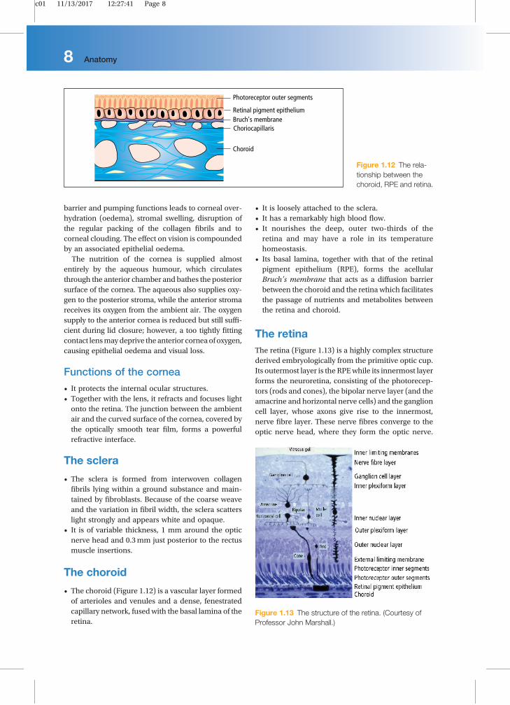
• The choroid (Figure 1.12) is a vascular layer formedof arterioles and venules and a dense, fenestratedcapillary network, fused with the basal lamina of theretina.
• It is loosely attached to the sclera.• It has a remarkably high blood flow.• It nourishes the deep, outer two-thirds of theretina and may have a role in its temperaturehomeostasis.
• Its basal lamina, together with that of the retinalpigment epithelium (RPE), forms the acellularBruch’s membrane that acts as a diffusion barrierbetween the choroid and the retina which facilitatesthe passage of nutrients and metabolites betweenthe retina and choroid.
The retinaThe retina (Figure 1.13) is a highly complex structurederived embryologically from the primitive optic cup.Its outermost layer is the RPEwhile its innermost layerforms the neuroretina, consisting of the photorecep-tors (rods and cones), the bipolar nerve layer (and theamacrine and horizontal nerve cells) and the ganglioncell layer, whose axons give rise to the innermost,nerve fibre layer. These nerve fibres converge to theoptic nerve head, where they form the optic nerve.
Photoreceptor outer segments
Retinal pigment epithelium
Choroid
Bruch's membraneChoriocapillaris
Figure 1.12 The rela-tionship between thechoroid, RPE and retina.
Figure 1.13 The structure of the retina. (Courtesy ofProfessor John Marshall.)
8 Anatomy
c01 11/13/2017 12:27:41 Page 9
Müller cells, the principal glial cells of the retina,extend across its thickness and are vital for the healthof the retinal neurons.
The retinal pigment epithelium (RPE)• It is consists of a single layer of cells.• It is loosely attached to theneuroretina, except at theperiphery (ora serrata) and around the optic disc.
• It formsmicrovilliwhichprojectbetweenandembracethe outer segment discs of the rods and cones.
• It phagocytoses the redundant, pigment-containingdiscs, which are replaced by new ones.
• It takes part in the regeneration of rhodopsin andcone opsin, the photoreceptor visual pigments andin recycling vitamin A.
• It contains melanin granules which absorb lightscattered by the sclera, thereby enhancing imageformation on the retina.
The photoreceptor layerThe photoreceptor layer is responsible for convert-ing light into electrical impulses. The initial integra-tion of these impulses is also performed by theretina.
• Cones (Figure 1.14) are responsible for daylight andcolour vision and have a relatively high threshold to
light. Different subgroups of cones are responsive toshort, medium and long wavelengths (red, greenand blue,). They are concentrated at the fovea,where they provide the high resolution requiredfor detailed vision, as in reading.
• Rods are responsible for night vision. They have alow light threshold and do not signal wavelengthinformation (colour). They form the large majorityof photoreceptors in the remaining retina.
The vitreous
• The vitreous is a clear gel occupying two-thirds ofthe globe.
• It is 98% water. The remainder is gel-forming hyal-uronic acid traversed by a fine collagen network.There are few cells.
• It is firmly attached anteriorly to the peripheralretina, pars plana and around the optic disc, andless firmly to the macula and retinal vessels.
• It has a physically supportive role and permits thepassage of nutrients and metabolites.
Loss of gel structure in later life, with collapse of thevitreous away from the retina (vitreous detachment),puts traction on points of attachment and may occa-sionally lead to a peripheral retinal break or hole, wherethe vitreous pulls off a flap of the underlying retina. Thisis a risk factor for subsequent retinal detachment.
Retinal pigmentepithelium
Outer segment
Inner segment
Outer nuclearlayer
Outer plexiformlayer
Cone Rod
Nucleus
Outerfibre
Ellipsoid
Cilium
Discs
External limitingmembrane
Cilium
Figure 1.14 The struc-ture of the retinal rodsand cones (schematic).
Anatomy 9
c01 11/13/2017 12:27:41 Page 10
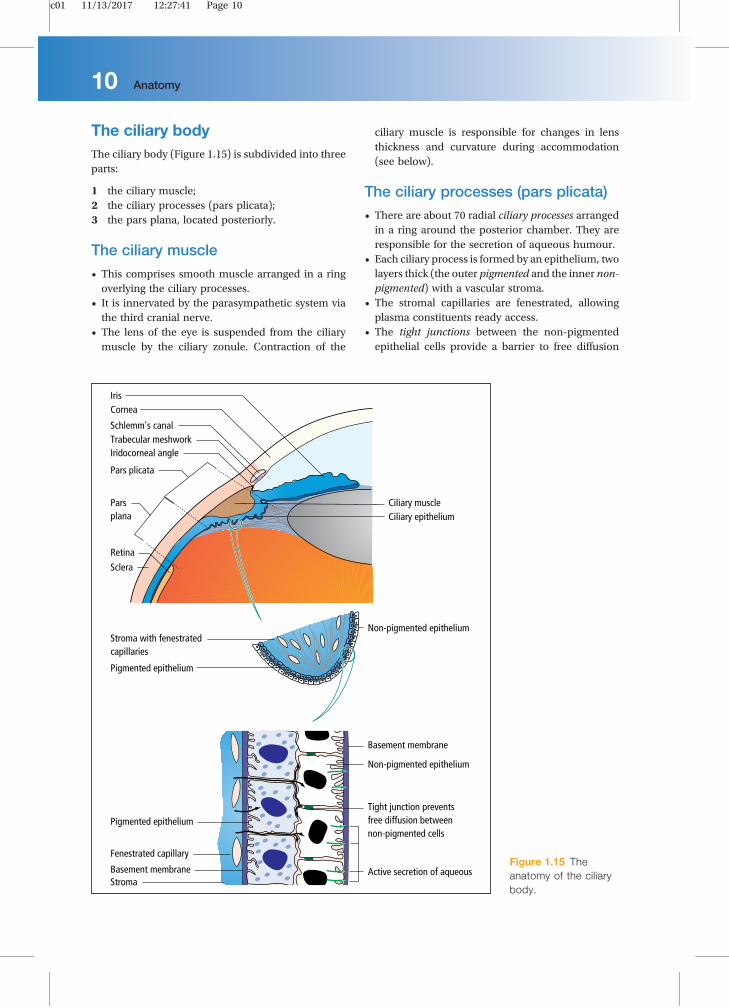
The ciliary bodyThe ciliary body (Figure 1.15) is subdivided into threeparts:
1 the ciliary muscle;2 the ciliary processes (pars plicata);3 the pars plana, located posteriorly.
The ciliary muscle• This comprises smooth muscle arranged in a ringoverlying the ciliary processes.
• It is innervated by the parasympathetic system viathe third cranial nerve.
• The lens of the eye is suspended from the ciliarymuscle by the ciliary zonule. Contraction of the
ciliary muscle is responsible for changes in lensthickness and curvature during accommodation(see below).
The ciliary processes (pars plicata)• There are about 70 radial ciliary processes arrangedin a ring around the posterior chamber. They areresponsible for the secretion of aqueous humour.
• Each ciliary process is formed by an epithelium, twolayers thick (the outer pigmented and the inner non-pigmented) with a vascular stroma.
• The stromal capillaries are fenestrated, allowingplasma constituents ready access.
• The tight junctions between the non-pigmentedepithelial cells provide a barrier to free diffusion
Pigmented epithelium
Fenestrated capillary
Stroma
Basement membrane
Pigmented epitheliumTight junction preventsfree diffusion between non-pigmented cells
Non-pigmented epithelium
Ciliary muscleCiliary epithelium
Non-pigmented epithelium
Basement membrane Active secretion of aqueous
Stroma with fenestratedcapillaries
Cornea
Trabecular meshworkSchlemm's canal
Sclera
Iridocorneal angle
Iris
Pars plicata
Pars plana
Retina
Figure 1.15 Theanatomy of the ciliarybody.
10 Anatomy
c01 11/13/2017 12:27:41 Page 11
into the posterior chamber. They are essential forthe active secretion of aqueous by these cells.
• The epithelial layers show marked infolding, whichincreases their surface area for fluid and solutetransport.
The pars plana• This comprises a relatively avascular stroma cov-ered by an epithelial layer, two cells thick.
• It is safe to make surgical incisions through thescleral wall in this region to gain access to thevitreous cavity.
The iris
• The iris diaphragm is attached peripherally to theanterior part of the ciliary body.
• It is perforated centrally by the pupil, which isconstricted or dilated by contraction of the circularsphincter or radial dilator muscles, respectively, tocontrol the amount of light entering the eye.
• It has an anterior border layer of fibroblasts andcollagen and a cellular stroma in which the sphinc-ter muscle is embedded at the pupil margin.
• The sphincter muscle is innervated by the para-sympathetic system.
• The smooth dilator muscle extends from the irisperiphery towards the sphincter. It is innervated bythe sympathetic system.
• Posteriorly, the iris is lined by a pigmented epithe-lium two layers thick.
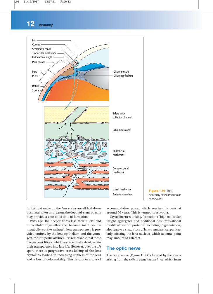
The iridocorneal (drainage) angleThis lies between the iris, the anterior tip of theciliary body and the cornea. It is the site of aqueousdrainage from the eye via the trabecular meshwork(Figure 1.16).
The trabecular meshworkThis overlies Schlemm’s canal and is composed of alattice of collagen beams covered by trabecular cells.The spaces between these beams become increas-ingly small as Schlemm’s canal is approached. Theoutermost zone of the meshwork accounts for mostof the resistance to aqueous outflow. Damage hereraises the resistance and increases intraocular pres-sure in primary open-angle glaucoma. Some of thespaces may be blocked and there is a reduction inthe number of cells covering the trabecular beams(see Chapter 10).
Fluid passes into Schlemm’s canal, both throughgiant vacuoles in its endothelial lining and throughintercellular spaces.
The lens
FunctionThe lens is the second major refractive element of theeye; thecorneabeing thefirst. It is aperfectly transparentstructure that lies directly behind the iris and pupil,suspended from the ciliary body by the fibres of theciliary zonule (Figure1.17). Thezonularfibres insert intothe lens equator, transmitting forces generated by theciliary muscle to the lens, to change its shape andrefractive power. This allows focusing to be adjustedfrom distance to near. At rest, during distance viewing,the zonular fibres are under tension, giving the lens aflattened profile. Contraction of the muscle duringaccommodation fornear, relaxes thezonuleandpermitsthe elasticity of the lens to increase its curvature andhence its refractive power. This may seem counter-intuitive, but it comes about because the muscle bulgesinwards and moves forwards during contraction.
Anatomy• The lens comprises an outer, tough, collagenouscapsule.
• A compact inner mass of lens fibre cells.
The capsule is the basal lamina of the lens epithe-lium, a monolayer of cells that lies between the cap-sule and the lens fibres anteriorly. The anterior part ofthe capsule increases in thickness throughout life butsynthesis of the posterior part ceases after birth, so it isthinner and more fragile. This is important duringcataract surgery.The epithelial cells at the lens equator form a
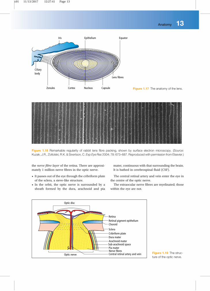
germinative zone, where cell division gives rise tothe lens fibres that make up the bulk of the lens.Fibres are elongated, spindle-shaped cells arrangedin layers which arch over the lens equator. Anteriorlyand posteriorly, their tips meet to form the lens whichincrease in complexity as the lens ages (Figure 1.18).The high concentration of lens-specific proteins
within the fibres (the lens crystallins), accounts forthe high refractive index of the lens. Their molecularorder, together with the regular packing of the lensfibres, accounts for its perfect transparency.The lens grows throughout life, as shells of new
fibres are laid down at the surface of the fibre mass.Thus, the oldest, central fibres that form the lensnucleus represent the fetal lens and the fibres external
Anatomy 11
c01 11/13/2017 12:27:41 Page 12
to this that make up the lens cortex are all laid downpostnatally. For this reason, the depth of a lens opacitymay provide a clue to its time of formation.With age, the deeper fibres lose their nuclei and
intracellular organelles and become inert, so themetabolic work to maintain lens transparency is pro-vided entirely by the lens epithelium and the youn-gest,most superficial fibres. It is remarkable that thesedeeper lens fibres, which are essentially dead, retaintheir transparency into late life. However, over the lifespan, there is progressive cross-linking of the lenscrystallins leading to increasing stiffness of the lensand a loss of deformability. This results in a loss of
accommodative power which reaches its peak ataround 50 years. This is termed presbyopia.Crystallin cross-linking, formation of highmolecular
weight aggregates and additional post-translationalmodifications to proteins, including pigmentation,also lead to a steady loss of lens transparency, particu-larly affecting the lens nucleus, which at some pointmay amount to cataract.
The optic nerveThe optic nerve (Figure 1.19) is formed by the axonsarising from the retinal ganglion cell layer, which form
Ciliary muscleCiliary epithelium
Cornea
Trabecular meshworkSchlemm's canal
Sclera
Iridocorneal angle
Iris
Pars plicata
Pars plana
Retina
Sclera withcollector channel
Schlemm's canal
Corneo-scleralmeshwork
Uveal meshwork
Anterior chamber
Endothelial meshwork
Figure 1.16 Theanatomyof the trabecularmeshwork.
12 Anatomy
c01 11/13/2017 12:27:41 Page 13
the nerve fibre layer of the retina. There are approxi-mately 1 million nerve fibres in the optic nerve.
• It passes out of the eye through the cribriform plateof the sclera, a sieve-like structure.
• In the orbit, the optic nerve is surrounded by asheath formed by the dura, arachnoid and pia
mater, continuous with that surrounding the brain.It is bathed in cerebrospinal fluid (CSF).
The central retinal artery and vein enter the eye inthe centre of the optic nerve.The extraocular nerve fibres are myelinated; those
within the eye are not.
body
Epithelium
eluspaCsuelcuNselunoZ
Lens fibres
Iris Equator
Cortex
Ciliary
Figure 1.17 The anatomy of the lens.
Figure 1.18 Remarkable regularity of rabbit lens fibre packing, shown by surface electron microscopy. (Source:Kuzak, J.R., Zoltolski, R.K. & Sivertson, C. Exp Eye Res 2004; 78: 673–687. Reproduced with permission from Elsevier.)
Retina
Central retinal artery and vein
Optic disc
Retinal pigment epitheliumChoroid
ScleraCribriform plateDura mater Arachnoid mater
Pia materSub-arachnoid space
Nerve fibresOptic nerve Figure 1.19 The struc-
ture of the optic nerve.
Anatomy 13
c01 11/13/2017 12:27:41 Page 14
The ocular blood supplyThe eye receives its blood supply from the ophthalmicartery (a branch of the internal carotid artery) via theretinal artery, ciliary arteries and muscular arteries(Figure 1.20). The conjunctival circulation anastomo-ses anteriorly with branches from the external carotidartery.The anterior optic nerve is supplied by branches
from the ciliary arteries. The inner retina is sup-plied by arterioles branching from the central reti-nal artery. These arterioles each supply an area ofretina, with little overlap. Obstruction results inischaemia of most of the area supplied by thatarteriole. The fovea is so thin that it requires nosupply from the retinal circulation. It is suppliedindirectly, as are the outer layers of the retina, bydiffusion of oxygen and metabolites across theretinal pigment epithelium from the choroidalcapillaries.The endothelial cells of the retinal capillaries are
joined by tight junctions so that the vessels areimpermeable to proteins. This forms an innerblood–retinal barrier, with properties similar tothose of the blood–brain barrier. The capillariesof the choroid, however, are fenestrated and leaky.The retinal pigment epithelial cells are also joinedby tight junctions and present an external blood–retinal barrier between the leaky choroid and theretina.Breakdown of these barriers is responsible
for the clinical features of many retinal vasculardiseases.
The third, fourth and sixthcranial nervesThe structures supplied by each of these nerves areshown in Table 1.1.
Table 1.1 The muscles and tissues suppliedby the third, fourth and sixth cranial nerves.
Third (oculomotor)Fourth(trochlear)
Sixth(abducens)
Medial rectus Superioroblique
Lateralrectus
Inferior rectus
Superior rectus (innervatedby the contralateralnucleus)
Inferior oblique
Levator palpebrae (bothlevators are innervated bya single midline nucleus)
Preganglionicparasympathetic fibresfrom the EdingerWestphäl nucleus run inthe third nerve and endin the ciliary ganglion.Here postganglionicfibres arise and pass inthe short ciliary nerves tothe sphincter pupillaeand the ciliary muscle
14 Anatomy
c01 11/13/2017 12:27:41 Page 15
Central originThe nuclei of the third (oculomotor) and fourth(trochlear) cranial nerves lie in themidbrain; the sixthnerve (abducens) nuclei lie in the pons. Figure 1.21shows some of the important relations of these nucleiand their fascicles.Nuclear and fascicular palsies of these nerves
are unusual. If they do occur, they are associated
with other neurological problems reflecting theaccompanying brainstem injury. For example, if thethird nerve fascicles are damaged as they pass throughthe red nucleus, the ipsilateral third nerve palsywill beaccompanied by a contralateral tremor. Also, anuclear third nerve lesion results in an ipsilateral palsyof the muscles supplied by the third nerve, bilateralptosis and a palsy of the contralateral superior rectus,
Figure 1.21 Diagramsto show the nuclei andinitial course of the (a)third, (b) fourth and (c)sixth cranial nerves.
Anatomy 15
c01 11/13/2017 12:27:41 Page 16
since both sets of crossing fibres from the subnucleusare affected.
Peripheral courseFigure 1.22 shows the intracranial course of the third,fourth and sixth cranial nerves.
Third nerveThe third nerve leaves themidbrain ventrally betweenthe cerebral peduncles. It then passes between theposterior cerebral and superior cerebellar arteries andthen lateral to the posterior communicating artery.Aneurysms of this artery may cause a third nervepalsy. The nerve enters the cavernous sinus in itslateral wall and enters the orbit through the superiororbital fissure.
Fourth nerveThe nerve decussates and leaves the dorsal aspect ofthe midbrain below the inferior colliculus. It firstcurves around the midbrain before passing like thethird nerve between the posterior cerebral and supe-rior cerebellar arteries to enter the lateral aspect of thecavernous sinus inferior to the third nerve. It enters theorbit via the superior orbital fissure.
Sixth nerveFibres leave from the inferior border of the pons. It hasa long intracranial course passing upwards along thepons to angle anteriorly over the petrous bone and intothe cavernous sinus, where it lies inferomedial to thefourth nerve in proximity to the internal carotid artery.It enters the orbit through the superior orbital fissure.
This long course is important because the nerve can beinvolved in numerous intracranial pathologies, includ-ing base of skull fractures, invasion by nasopharyngealtumours and raised intracranial pressure.
The seventh cranial nerveThe seventh cranial nerve arises from a nucleus in thepons, loops over that of the sixth cranial nerve (Figure1.21c) and leaves the brainstemat the cerebellopontineanglewhere it is joined by the nervus intermedius. Thetwo nerves travel together in the internal auditorycanal where they fuse to form the geniculate ganglion.From there, the somatic motor fibres issue from theskull through the stylomastoid foramen, to supply themuscles of the face and scalp. The nervus intermediuscarries secretomotor fibres to the lacrimal gland viathe nerve of the pterygoid canal, a mixed autonomicnerve which includes sympathetic fibres from thecarotid plexus. The preganglionic, parasympatheticfibres synapse with postganglionic fibres in the pter-ygopalatine ganglion and reach the lacrimal gland viathe lacrimal nerve. The nervus intermedius also car-ries taste fibres and secretomotor fibres to the sub-mandibular and sublingual glands, which run in thechorda tympani.
Assessment questionsTrue or False1. The cornea
a Has an endothelial layer that regenerates readily.b Has an epithelial layer that fails to regenerate.
Trochlear (IV) nerve
Trochlear (IV)nerve
Superiorcerebellar artery
Posterior cerebral artery
Trigeminal ganglionAbducent (VI) nerve
Superior orbital fissureAnterior clinoid process
Oculomotor (III) nerve
Posterior communicating arteryOptic nerve
Cavernous sinus
Figure 1.22 The intra-cranial course of thethird, fourth and sixthcranial nerves.
16 Anatomy
c01 11/13/2017 12:27:41 Page 17
c The endothelium actively pumps water from thestroma.
d Is an important refractive component of theeye.
e Has a stroma composed of randomly arrangedcollagen fibrils.
2. The retina
a Is ten layers thick.b Has ganglion cells whose axons form the optic
nerve.c Has three types of rods responsible for colour
vision.d The neuroretina is firmly attached to the retinal
pigment epithelium.e The RPE delivers vitamin A for rhodopsin
production.
3. The lens
a Grows throughout life.b Is surrounded by a collagenous capsule.c Cortex and nucleus are rich in organelles.d Has a high refractive index owing to its protein
content.e Shape becomes more curved during accommo-
dation for near.
4. Thesuspensory ligamentof the lens (thezonule)
a Attaches the lens to the ciliary body.b Is part of the iridocorneal angle.c Is composed of smooth muscle.d Transmits changes in tension to the lens capsule.
5. The posterior chamber
a Is another name for the vitreous body.b Lies between the iris, lens and ciliary body.c Contains aqueous humour, secreted by the ciliary
processes.d Is in communication with the anterior chamber.
6. The tear film
a Is 100 μm thick.b Tears are drained by the nasolacrimal system.c The mucoaqueous layer is in contact with the
cornea.d Is important in the refraction of light entering the
eye.e Contains lysozyme and secretory IgA.
7. The iridocorneal angle
a Is the site of aqueous production.b Lies between the cornea and the ciliary body.
c In primary open-angle glaucoma, there is areduction in the number of cells covering thetrabecular meshwork.
d Fluid passes through the trabecular meshwork toSchlemm’s canal.
8. The optic nerve
a Axons leave the eyeball through the cribriformplate.
b Is not bathed in CSF until it enters the cranial cavity.c Anteriorly is supplied by blood from the ciliary
arteries.d Axons are not myelinated in the retrobulbar part
of the nerve.e Is formed by axons of the nerve fibre layer of the
retina.
9. The third, fourth and sixth cranial nerves
a All originate in the midbrain.b A nuclear third nerve palsy will cause a contra-
lateral palsy of the superior rectus.c The fourth nerve supplies the lateral rectus.d The sixth nerve has a long intracranial course.e The third nerve may be affected by aneurysms of
the posterior communicating artery.
Answers1. The cornea
a False. The human endothelium does not regen-erate; dead cells are replaced by the spreading ofsurviving cells.
b False. The epithelial layer readily regenerates.c True. The endothelial cells pump out ions and the
water follows osmotically. Removal of watermaintains corneal transparency.
d True. The cornea is a more powerful refractiveelement than the natural lens of the eye.
e False. The fine, equally spaced, stromal collagenfibrils are arranged in parallel and packed in anorderly manner. This is a requirement fortransparency.
2. The retina
a True. See Figure 1.13.b True. The retinal ganglion cell axons form the
retinal nerve fibre layer and exit the eye at theoptic nerve head.
c False. The rods are responsible for night visionand three cone types are responsible for daylightand colour vision.
Anatomy 17
c01 11/13/2017 12:27:42 Page 18
d False. The attachment is loose; the neuroretinaseparates from the RPE in retinal detachment.
e True. Vitamin A is delivered by the RPE to thephotoreceptors and combined with opsin.
3. The lens
a True. It does grow throughout life.b True. This is of great importance in cataract surgery.c False. The older, deep cortical and nuclear fibres
lose their nuclei and other organelles.d True. The high protein content accounts for its
high refractive index.e True. See page 11.
4. Thesuspensory ligamentof the lens (thezonule)
a True. Zonular fibres extend from the pars plicataof the ciliary body to the lens equator.
b False. The zonule lies behind the iris and irido-corneal angle.
c False. The ciliary muscle contains smoothmuscle, not the zonule, which is acellular.
d True. Contraction of the ciliarymuscle relaxes thezonular fibres allowing the lens to increase itscurvature and thus its refractive power (this is‘accommodation’).
5. The posterior chamber
a False. The vitreous body is quite separate.b True. See Figure 1.4.c True. See page 10.d True. Communication is via the pupil, in the gap
between iris and lens at the pupil margin. If thisgap is narrowed or closed, pressure in the poste-rior chamber pushes the iris forward and mayclose the angle (acute closed-angle glaucoma).
6. The tear film
a False. The tear film is about 3 μm thick.b True. There is a punctum on the medial aspect of
both upper and lower eyelids. These allow tears todrain into the nasolacrimal drainage system.
c True. Themucin layer is produced by goblet cells.d True. It provides a smooth interface for the refrac-
tion of light.e True. These account for the antibacterial proper-
ties of the tear film.
7. The iridocorneal angle
a False. It is the site of aqueous drainage.b True. See Figure 1.15.c True. This may reduce aqueous drainage.d True. Flow depends on the pressure gradient
between the anterior chamber and Schlemm’scanal and there is also an active component.
8. The optic nerve
a True. This sieve-like structure provides supportfor the optic nerve as it leaves the eye.
b False. In the orbit, outside its pial sheath, theoptic nerve is surrounded by cerebrospinalfluid within the subarachnoid space. This isin continuity with that in the intracranialcavity.
c True. The supply to the anterior part of the opticnerve differs from the supply to the anterior layersof the retina.
d True. This is a most important blood supply forthe anterior optic nerve.
e False. They are usually not myelinated within theeye.
f True. It is made up from retinal ganglion cellaxons.
9. The third, fourth and sixth cranial nerves
a False. The nuclei of the sixth and seventh nerveslie in the pons.
b True. The superior rectus is innervated by thecontralateral nucleus.
c False. It supplies the superior oblique.d True. This makes the sixth nerve susceptible to
trauma, which may cause lateral rectus palsy.e True. It passes lateral to the artery.