André Tridon Jagendorf (Currently Liberty H. Bailey Prof Emeritus, Plant Biology , College of Agriculture and Life Sciences, Cornell University), co-recipient, with Wolfgang Junge , of 2012 Lifetime Achievement Award of The Rebeiz Foundation of Basic Biological Research A Tribute to My elder brother, Andre Bhaiya by Govindjee September 28, 2013 [email protected]URL: http://www.life.illinois.edu/govindjee Delivered by Govindjee at the Rebeiz Foundation for Basic Biology, Champaign, Illinois
Transcript
André Tridon Jagendorf (Currently Liberty H. Bailey Prof
Emeritus, Plant Biology , College of Agriculture and Life Sciences, Cornell
University), co-recipient, with Wolfgang Junge , of 2012 Lifetime Achievement Award of The Rebeiz Foundation of
Delivered by Govindjee at the Rebeiz Foundation for Basic Biology, Champaign, Illinois
Andre T. Jagendorf (ATJ) in 1946.. with his friend Avnet and his girl friends
A way of academic life: From one celebration to another, 67 years later ..From Cornell to Champaign
I have learned that ATJ used to listen to Gilbert and Sullivan’s operettas on the radio
Govindjee
Some facts about André
ò Born: October 21, 1926 , New York City
ò Son of Moritz Adolph and Sophie Sheba (Sokolosky); his father was a dentist and children’s book writer; mother was an accomplished and a wonderful person
ò He learned to type when he was very young; was an avid reader of books especially Science fiction
ò At young age, he was enamored by classical music; played “mandolin”; and then viola till 1966.
Govindjee
Some facts about André
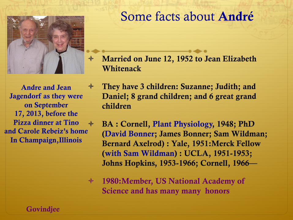
ò Married on June 12, 1952 to Jean Elizabeth Whitenack
ò They have 3 children: Suzanne; Judith; and Daniel; 8 grand children; and 6 great grand children
ò BA : Cornell, Plant Physiology, 1948; PhD (David Bonner; James Bonner; Sam Wildman; Bernard Axelrod) : Yale, 1951:Merck Fellow (with Sam Wildman) : UCLA, 1951-1953; Johns Hopkins, 1953-1966; Cornell, 1966—
ò 1980:Member, US National Academy of Science and has many many honors
Andre and Jean Jagendorf as they were
on September 17, 2013, before the Pizza dinner at Tino
and Carole Rebeiz’s home In Champaign,Illinois
Govindjee
André has followed the same rules from when he was 6 up to when he is 87
Take a hammer and hit it at the right place..
His ability to think for himself was obvious when he created, with
available blocks and boxes (big and small), a 2-story "ship", with
stairs going up the planks laid on top of a big box. His kindergarten
teacher had never seen anything like it!
Govindjee
I will not talk about his research. You can read about it in the two articles I had invited him to
write for “Photosynthesis Research”
ò Jagendorf, A.T. (1998) Chance, luck and photosynthesis research: An inside story. Photosynth Res 57: 215-229
ò Jagendorf, A.T. (2002) Photophosphorylation and the chemiosmotic perspective. Photosynth Res 73: 233-241
ò Andre recognized many, particularly the following three:
André Tridon Jagendorf(Courtesy of Division of Rare & ManuscriptCollections, Cornell University Library)
234
Figure 1. Mordhay Avron.
Rhodospirillum rubrum by Frenkel (1954), and fromthe anaerobic bacterium Chromatium by Williams(1956). With the ‘chromatophores’ from both bacteria,it was clear that a cyclic electron flow not involvingnet oxidation of any substrate supported the ATP syn-thesis. (A photograph of Daniel Arnon is shown in thearticle by R. Porra in this issue.)
With isolated chloroplasts (actually, thylakoids) aswell, the first discovered ATP synthesis seemed to besupported by a cyclic electron flow pattern. The ori-ginal rates were at the level of 3 µmol ATP/mg Chl/h.However adding FMN or menadione as ‘cofactors’permitted rates to be raised to almost 500 µmol/mgChl/h (Allen et al. 1958) and, as in bacterial extracts,methyl phenazinium sulfate (PMS) brought rates upto 900 (Avron and Jagendorf 1958; see Figure 1 for aphotograph of Mordhay Avron).
A little later, it was found that linear (uphill) elec-tron flow from water as donor to added NADP+,yielding O2 as a waste product, supported ATP syn-thesis (Arnon et al. 1958a). This also occurred usingnonphysiological electron acceptors such as potassiumferricyanide, although not with trichlorophenol in-dophenol (Avron et al. 1958).
A variant of this pattern was found in which theadded electron acceptor reduces the O2 that had beenevolved, that is, a net O2 exchange reaction whichcan also support ATP synthesis. This is similar to
the Mehler reaction (Mehler 1951; see Heber, thisissue, for further information), in which O2 is re-duced to H2O2. By a number of criteria [see Jagendorf(1962) for a full discussion], ATP synthesis by FMN,menadione, and many other ‘cofactors’ arouses thissort of O2 exchange pattern. This sequence was re-named ‘psuedocyclic photophosphorylation’ (Arnonet al. 1961).
Coupling, and uncouplers
It had been known earlier (Lardy and Wellman 1952)that electron flow in mitochondria depends almostentirely on added ADP and Pi, permitting simultan-eous ATP synthesis. A similar phenomenon, althoughnot so dramatic, was found for linear electron flowfrom H2O to NADP+ (Arnon et al. 1958b) and toferricyanide (Avron et al. 1958).
Uncoupling of mitochondrial oxidation by DNPwas demonstrated even earlier (Loomis and Lipmann,1948). For chloroplasts, ammonium chloride was thefirst uncoupler found (Krogmann et al. 1959). Thiswas followed very soon by the demonstration of un-coupling by a variety of organic amines (Good 1960).Thus, by 1959, the bioenergetics of chloroplasts hadachieved the position of mitochondrial research in1952.
CF1 and CFo
Discovery of the enzymatic machinery for ATP syn-thesis similarly benefited from prior work with mi-tochondria. Efraim Racker (see N. Nelson in thisissue for a photograph of Racker) and others, ex-tracted proteins from mitochondria so that oxidativephosphorylation was eliminated, and added them backto restore some phosphorylation (see Racker 1970).These were called coupling factors, and also had AT-Pase activity. The first demonstration of an extractablecoupling factor for chloroplasts was made by Avron(1963), using thylakoids uncoupled by treatment withethylene diamine tetraacetic acid, EDTA (Jagendorfand Smith 1962). It was nicknamed CF1(for Chloro-plast F1), to distinguish it from the analogous mito-chondrial F1 (for factor 1). Subsequently, Vambutasand Racker (1965) purified a Ca2+-requiring ATPasefrom chloroplasts, which was latent until activatedby trypsin. It could restore photophosphorylation todepleted thylakoids.
236
high-energy state, but not in the dark or if uncouplerswere present in light (Ryrie and Jagendorf 1971). Inanother approach, a series of chemical modifying re-agents were shown, over several years, to attack CF1if thylakoids were in light, but this did not occur inthe dark. The first of these was N-ethylmaleimide, at-taching to a sulfhydryl group on the ! subunit onlyin light (McCarty et al. 1972; McCarty and Fagan1973). Space does not permit a real review of thisinteresting area, but one outstanding study (Komatsu-Takaki 1989) showed accelerated attack by pyridoxal-5!-phosphate on a lysine of the epsilon subunit inlight.
Mechanism of CF1 action: chemiosmotic conceptstake over
From the time of discovery of oxidative phosphoryla-tion, the connection between electron flow and thechemistry of high-energy phosphate bond formationwas an intriguing mystery. Essentially all of the specu-lation about this connection relied on the paradigm ofATP synthesis performed by triose-P dehydrogenase.This was shown to include a low-energy phosphateaddition to an SH group on the enzyme, rising to ahigh-energy level due to oxidation. Therefore, bio-chemists kept looking for a high-energy intermediate,prior to ATP synthesis, involving one or more of theelectron transport enzymes of mitochondria (examplesin Chance and Williams 1956).
In the search for a chemical intermediate in pho-tophosphorylation, chloroplasts were illuminated in apipette (Shen and Shen 1962) or a syringe (Hind andJagendorf 1963), then injected into buffer containing32P, Mg2+, and ADP, where some ATP synthesis thenoccurred. (The two discoveries were quite independ-ent.) There was a problem with the results, however:the amount of ATP made in the dark was up to 50 timesmore than the concentration of any single electron car-rier in the thylakoid membrane. Thus there must havebeen actual turnover of the enzymatic machinery in thedark. For a more detailed description of this, and otherwork from this laboratory, see Jagendorf (1998).
Earlier, based on his understanding of thephysiology and physical chemistry of ion transport,Peter Mitchell (Figure 2) had proposed a drasticallydifferent idea for the mechanism. Realizing that onlyorganelles with membranes accomplished oxidativeand photosynthetic phosphorylation, he developed a‘chemiosmotic’ concept of the mechanism (Mitchell1961). In this, the connecting link is the active vec-
Figure 2. Peter Mitchell.
torial transport of protons across the membrane, aselectron flow proceeds alternately from metals (iron orcopper) handling only electrons to quinones requiringboth an electron and a proton to be reduced. The high-energy intermediate, therefore, would be a differencebetween the electrochemical activity (both chemicalconcentration and membrane potential) of protons onthe inside, with that on the outside of the membrane.The intermediate would drive a membrane-located,vectorial, reversible, proton-pumping ATPase.
Peter Mitchell’s terminology and writing were dif-ficult to understand. I was fortunate in having abrilliant postdoctoral from England, Geoffrey Hind(Figure 3), who could explain the concept to me. Indiscussing it, it occurred to us that it might be pos-sible to observe the uptake of protons into thylakoidsin light and their dissipation in the dark. It was anexciting moment to observe this happening using asimple glass electrode (Jagendorf and Hind 1963). Itsrelationship to photophosphorylation was further con-firmed by finding that uncouplers lead to rapid dissip-ation of the internal protons (Neumann and Jagendorf1964; Jagendorf and Neumann 1965).
Others realized that a massive movement of pro-tons had to be accompanied by some sort of counter-ion flux to keep the membrane potentials in a reas-onable range. The first counter-ion flux observed wasextrusion of K+ and Mg2+ in light (Dilley and Vernon
237
Figure 3. Geoffrey Hind.
1965). Somewhat later (Deamer and Packer 1969),the membrane potential was found to be controlledto an even greater extent by the uptake of Cl! ionswith the protons. This seems to occur through athylakoid chloride channel, discovered by Vambutuset al. (1984). A more complete discussion of iontransport is found in the review by Rottenberg (1985).
The rise in external pH in light implied that thepH of the thylakoid lumen would become more acid.Evidence for this came from the careful measure-ment of penetrating labeled amines and of cationicdyes in light (see Rottenberg 1985). The estimatedinternal pH dropped to 4 – 4.5, as the external pHrose to 7.5 – 8. The 3.5 pH unit difference betweenexternal and internal spaces was certainly high enoughto drive net synthesis of ATP at physiological substrateconcentrations.
The first direct indication of a membrane poten-tial was deduced from carotenoid absorption changes(the ‘electrochromic’ shift at 515 nm), related tothe coupling state of chloroplasts (Junge and Witt1968). A similar change in bacterial ‘chromatophores’was shown to serve as an intrinsic voltmeter for themembrane potential (Jackson and Crofts 1971). Otherestimates come from the change in distribution ofcharged anions or cations into the energized organelle(see Rottenberg 1985). A full discussion is found inJunge and Jackson (1982)
Acid to base induced ATP synthesis
In trying to find spectrophotometric evidence for theenergetic state, Hind found that pre-illumination of thethylakoids caused an increase in their light-scatteringability (Hind and Jagendorf 1965). This was espe-cially true at lower pH levels, which had providedhigher level of postillumination ATP synthesis in light(Hind and Jagendorf 1963). Looking for a correlationbetween light scattering and the dark ATP synthesis,Hind inserted a control in which thylakoids were neverexposed to light, but simply were shifted from pH 4.5to pH 8. To our surprise, this one protocol provideda very small amount of ATP synthesis (about 10% ofthat seen when light had been used).
In Hind’s experiments, the acid pH had been ad-justed using HCl. Worried about possible damage by astrong mineral acid, after Hind left I tried the sameexperiment using a divalent acid (phthallic) to ad-just the acid pH. This increased the amount of ATPmade in darkness a great deal; and work with otherdicarboxylic organic acids (succinic, maleic, etc.) in-creased the yield 10-fold, to twice that seen withpre-illumination (Jagendorf and Uribe 1966). Thisamounted to up to 100 times the level of any electroncarrier in the thylakoids. The fact that ATP was beingmade without any electron flow was further emphas-ized by the failure of electron transport inhibitors toprevent the acid-base ATP synthesis, and indeed someof them were able to increase the ATP yield (Miles andJagendorf 1970).
Other work further confirmed the validity ofMitchell’s chemiosmotic hypothesis. Mitochondrialparticles were shown to make ATP due to an acid/basetransition (Thayer and Hinkel 1975). Most spectacu-lar was the demonstration that photophosphorylationcould be done by liposomes into which had beeninserted both the light-activated, proton-pumping bac-teriorhodopsin from Halobacterium halobium, and theFoF1 complex from mitochondria (Racker and Stoeck-enius 1974). There was no evolutionary precedent forthe mating of those two components; so it must havebeen the protonmotive force generated by illuminatedrhodopsin that drove the F1 to turn over, catalyzing thesynthesis of ATP.
The question was raised whether the pH differ-ence or the membrane potential is most responsiblefor ATP synthesis. The pH jump-driven ATP synthesishad been done with swollen thylakoids, previouslywashed in 10 mm NaCl. This permitted the entry ofvery considerable levels of the divalent acid, whichcorrelated well with the extent of ATP synthesis (Uribe
Mordhay Avron Peter
Mitchell
Geoffrey Hind
Govindjee
Andre T. Jagendorf
e - H 2 0 + + +
+ + + + + +
Chloroplast
Jagendorf’s chloroplast work provided powerful support for Peter Mitchell’s theory for ATP generation
Chemiosmotic Mechanism for ATP Synthesis
A Proton Gradient Generates ATP in Chloroplasts
Some quotes from Jagendorf (1998)
ò “The probability of any one event occurring is amazingly small; in retrospect, each step seems like a minor miracle. Any person’s career has to be shaped by interactions with other people;”
ò “…doing science is fun”
ò “I had heard Peter Mitchell talk about chemiosmosis at a
bioenergetics meeting in Sweden. His words went into one of my
ears and out the other, leaving me feeling annoyed they had allowed
such a ridiculous and incomprehensible speaker in. But – Geoffrey
read Nature. Geoffrey was from England, both better trained and more intelligent than I was. He read Peter Mitchell’s paper, came to
me, and said ‘André. could this possibly explain XE [something that
preceded ATP formation]?’”
Some more quotes from Jagendorf (1998)
ò “At this point I began to communicate with Peter Mitchell himself... and invited me to spend a week there so he could educate me about the chemiosmotic hypothesis in more detail. I was happy to go, and enjoyed very much meeting his family and the family donkey, and …..I doubt that I learned enough about chemiosmosis, however. “
ò “Later that summer I did the experiment that convinced me …that we were really seeing a chemiosmotic mechanism at work. The amount of ATP that was made depended on the height of the pH difference between acid and base stages, more than on their absolute values (Jagendorf and Uribe 1966).”
1941--1951
1941: With Albert Novikoff (Fake bar)
A 1945 photo. Enlisted
In US Army In
1944; learned to be a
photographer
1950: At Yale
We can recognize him here
1951: Happy
Graduate From Yale
ATJ
1953-1966 Johns Hopkins University
Appointed in 1953 by Willian McElroy without any interview!
ò 1955 Photo: With the late Tony San Pietro and the late Mordhay Avron, major discoverers in biochemistry of
photosynthesis ATJ
A S-P
M A
1960s
1961: Johns Hopkins 1961: Maryland Academy
1962: Johns Hopkins.
ATJ 1963: Airlie House, Virginia ( B. Kok and A.T. Jagendorf): Nobel laureate James Franck was also there (standing, 2nd from left). Govindjee
Congratulations André
ò I thank Tino Rebeiz for giving me the opportunity to show my happiness, through my slides, at this gathering about André as a person–
ò The only request to André is that he holds back his “off-color” jokes (that many of us really love) for a few minutes—may be at least an hour! OK??
ò Thank you all who have gathered here today to honor two extraordinary persons (Andre and Wolfgang) . I will talk about Wolfgang a bit later. I am sure if Eugene Rabinowitch (my professor) was here, he will say “Well.. Let’s get some vodka.”


![New South Wales Supreme Court - … · New South Wales Supreme Court CITATION : ACD Tridon v Tridon Australia [2002] NSWSC 896 ... Cloe Z Shipping Co Inc v Odyssey Re (London) Ltd](https://static.documents.pub/doc/80x56/5b926c9e09d3f215288de47d/new-south-wales-supreme-court-new-south-wales-supreme-court-citation-acd.jpg)


