LEUKEMIA LECTURE IN INTERNAL MEDICINE FOR IV COURSE STUDENTS M. Yabluchansky, L. Bogun, L. Martymianova, O. Bychkova, N. Lysenko, N. Makienko V.N. Karazin National University Medical School’ Internal Medicine Dept.
Transcript
LEUKEMIA
LECTURE IN INTERNAL MEDICINE FOR IV COURSE STUDENTS
M. Yabluchansky, L. Bogun, L. Martymianova, O. Bychkova, N. Lysenko, N. Makienko V.N. Karazin National University Medical School’ Internal Medicine Dept.
• Leukemia (leukaemia) is a group of progressive, malignant neoplasms (cancers) of the blood-forming organs, marked by diffuse replacement of the bone marrow development of leukocytes and their precursors in the blood and bone marrow, accompanied by a reduced number of erythrocytes and blood platelets, and resulting in anemia, increased susceptibility to infection and hemorrhage with weakness and malaise, fever, pain in the joints and bones, swelling of the lymph nodes, spleen, and liver
MAJOR KINDS OF LEUKEMIA (ACUTE MYELOGENOUS & LYMPHOBLASTIC, CHRONIC
MYELOGENOUS & LYMPHOCYTIC)
Acute myelogenous leukemia (AML): definition
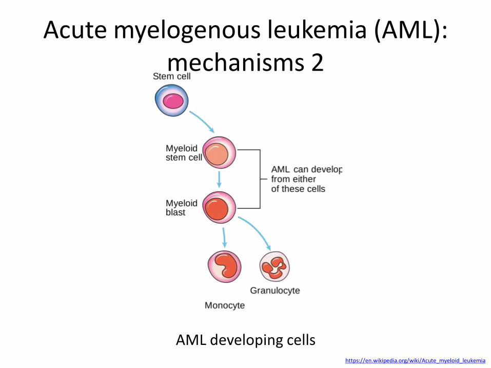
Acute myelogenous (myeloid, nonlymphocytic) leukemia (AML) represents a group of clonal hematopoietic stem cell disorders in which both a block in differentiation and unchecked proliferation result in the accumulation of myeloblasts at the expense of normal hematopoietic precursors
• AML is characterized by clonal expansion of hematopoietic stem and progenitor cells with blocked differentiation
• AML is the product of several functionally cooperating genetic alterations including chromosomal translocations leading to expression of leukemogenic fusion proteins
• Several AML-associated lesions target chromatin regulators like histone methyltransferases or histone acetyltransferases, including mixed-lineage leukemia 1 (MLL1) or CREB binding protein/p300
• Chromatin modulating mechanisms are mediating the transforming activity of key drivers of leukemogenesis by aberrant recruitment of corepressors (aberrant DNA, aberrantly expressed microRNAs)
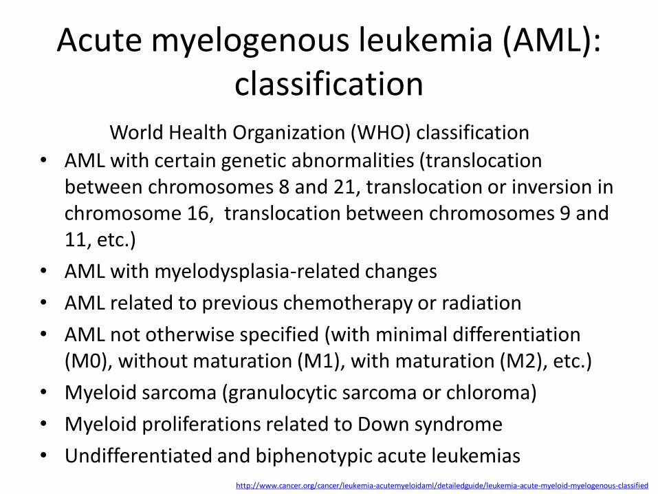
• AML with certain genetic abnormalities (translocation between chromosomes 8 and 21, translocation or inversion in chromosome 16, translocation between chromosomes 9 and 11, etc.)
• AML with myelodysplasia-related changes
• AML related to previous chemotherapy or radiation
• AML not otherwise specified (with minimal differentiation (M0), without maturation (M1), with maturation (M2), etc.)
• Myeloid sarcoma (granulocytic sarcoma or chloroma)
• Myeloid proliferations related to Down syndrome
• Undifferentiated and biphenotypic acute leukemias http://www.cancer.org/cancer/leukemia-acutemyeloidaml/detailedguide/leukemia-acute-myeloid-myelogenous-classified
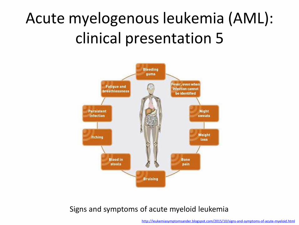
Initial clinical presentations are related to pancytopenia, the reduction of all cell counts, reflecting the leukemic cell replacement of bone marrow and include:
• Headache/disorientation due to abnormal white blood cells infiltrating the central nervous system (CNS)
• Anemia which is accompanied by pallor, fatigue, malaise, hypoxia, and bleeding, caused by rapidly proliferating leukocytes inhibiting formation of erythrocytes and thrombocytes
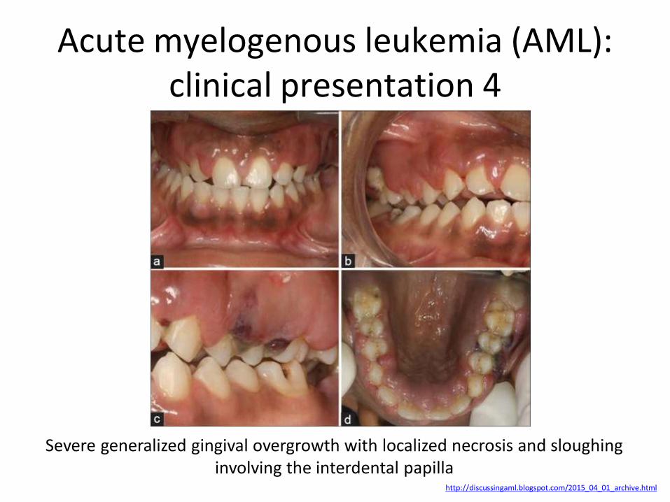
• Infections (pneumonia) and mouth/throat ulcerations, caused by increased numbers of immature or abnormal leukocytes which are unable to fight off infections
• Increased metabolic rate with weakness, pallor, and weight loss, caused by increased leukocyte production which require increased nutrient production; destruction of cells also increases metabolic waste
• Hyperuricemia which may lead to renal pain, obstruction, and infection (later development includes renal insufficiency with uremia), caused by a great number of leukocytes being destroyed which releases large amounts of uric acid; in late stages, leukocytes infiltrate the kidneys
• Enlarged organs (spleen, liver), caused by increased number of white blood cells accumulating within liver and spleen causing tissue distension
• Lymphadenopathy and bone pain, caused by excessive number of white blood cells accumulating in lymph nodes and bone marrow
• Bone discomfort (especially in ribs, sternum, and tibia)
• Older adults may experience delirium, and progressive weakness
• History and Physical Examination: clinical presentation
• Blood Test: an excess of abnormal white blood cells (leukocytosis), sometimes leukemic blasts, isolated decreases in platelets, red blood cells, or even with a low white blood cell count (leukopenia)
• Bone marrow biopsy and aspiration: to diagnose the presence of leukemia, to differentiate AML from other types of leukemia (e.g. acute lymphoblastic leukemia - ALL), and to identify the subtype of AML accordingly to its classifications
• Immunophenotyping: to determine the subtype of AML by comparing the cancer cells to normal cells in the immune system
The primary treatment is chemotherapy, which can be divided into three phases:
• The first (Induction) phase clears the blood of leukemia cells and reduces the number of blasts in the bone marrow with the goal to return blood counts to a normal level over time and to reach a complete remission
• The second (Consolidation) phase is administered after a rest period where the patient recovers from the first phase with the goal to kill the small number of leukemia cells that are still present
• The third (Maintenance) phase is necessary in only certain types of leukemia and includes giving low doses of a chemo drug for months or years after the consolidation phase
1) Chemotherapy: systemic, intrathecal, or regional drugs usage depends of the AML subtype to stop the growth either to kill cancer cells or to stop their dividing
2) Radiation: different types of radiation to kill cancer cells or keep them from growing through external and internal (radioactive material encapsulated in needles, seeds, wires, or catheters placed directly into the affected region) approaches which depend of the AML subtype
3) Stem Cell Transplant: method of administering chemo and replacing blood forming cells that are affected or destroyed by the cancer treatment
4) Targeted Therapy: monoclonal antibodies or other substances to destroy specific cancer cells without harming the patient's normal cells
• All FAB subtypes except M3 are usually given with cytarabine (ara-C) and an anthracycline (most often daunorubicin) in different regimens (3+7, etc.)
• The M3 subtype is almost universally treated with the drug all-trans-retinoic acid (ATRA) in addition to induction chemotherapy, usually an anthracycline
• Because of the toxic effects of therapy, including myelosuppression and an increased risk of infection, induction chemotherapy may not be offered to the very elderly, and the options may include less intense chemotherapy or palliative care
• Even after complete remission is achieved, leukemic cells likely remain in numbers too small to be detected with current diagnostic techniques, and more therapy is necessary to prevent relapse
• The specific type of consolidation therapy is individualized based on a patient's prognostic factors and general health
• For patients who are not eligible for a stem cell transplant, immunotherapy with a combination of histamine dihydrochloride (Ceplene) and interleukin 2 (Proleukin) after the completion of consolidation has been shown to reduce the absolute relapse risk by 14%, translating to a 50% increase in the likelihood of maintained remission
• When receiving chemotherapy, patients should avoid exposure to crowds and people with contagious illnesses, especially children with viral infections. Any patient with neutropenic fever or infection should immediately be treated with broad-spectrum antibiotics
• Appropriate transfusion support includes transfusion of platelets and clotting factors (fresh frozen plasma - FFP, cryoprecipitate) as guided by the patient’s blood test results and bleeding history
• For Patients with relapsed AML the only proven potentially curative therapy is a hematopoietic stem cell transplant, if one has not already been performed
Physical Therapy and exercise are aimed at symptom management, preservation of muscle function, pain control, and increased quality of life and include:
• Up to 15 minutes of walking 5x per week can reduce fatigue, symptom distress, anxiety, and depressive status while maintaining cardiovascular health
• Strength training combined with aerobic exercises 3x per week, twice daily, for 30 minutes improves cardiorespiratory endurance, reduces total fatigue and depression, maintains quality of life, and may reduce inflammatory markers
• Pain management (transcutaneous electrical nerve stimulation (TENS), hot packs, cold packs, massage, etc. )
• Stretching (sustained stretch, active and passive range of motion (ROM), splinting, etc.)
• Patients should be instructed to call their healthcare providers immediately if they are febrile or have signs of bleeding
• Patients are best treated at a center whose staff has significant experience in the treatment of leukemia
• Patients should be transferred to an appropriate (generally tertiary care) hospital if they are admitted to hospitals without appropriate blood product support, leukapheresis capabilities, or physicians and nurses familiar with the treatment of leukemia patients
• Acute myeloid leukemia is a curable disease, and the chance of cure for a specific patient depends on a number of prognostic factors
• Increasing age is an adverse factor, because older patients more frequently have a previous antecedent hematologic disorder along with comorbid medical conditions that compromise the ability to give full doses of chemotherapy
• A previous antecedent hematologic disorder is associated with a poor outcome to therapy
• Immunostaining and surface marker analysis revealed that the blast cells were positive for cytoplasmic myeloperoxidase, CD4, CD7, CD33, CD44, CD117, and HLA-DR, but negative for CD34 and CD56
• Karyotype was normal
• MS associated with AML was diagnosed
• Multidrug chemotherapy for AML was completely ineffective, and MS continued to progress
• Immunohistochemistry revealed that the blasts were negative for asparagine synthetase (AS); therefore, chemotherapy including L: -asparaginase was initiated
• After the first administration of L: -asparaginase, the patient's condition improved; however, she subsequently developed tumor lysis syndrome and sepsis, which eventually led to death
• Aggressive MS in childhood is rare and refractory to existing AML chemotherapy
• Chemotherapy including L -asparaginase may prove to be effective in such cases, especially those in which blast cells show negative AS expression
From: Department of Hematology/Oncology, Saitama Children's Medical Center, 2100 Magome, Iwatsuki-ku, Saitama, Japan
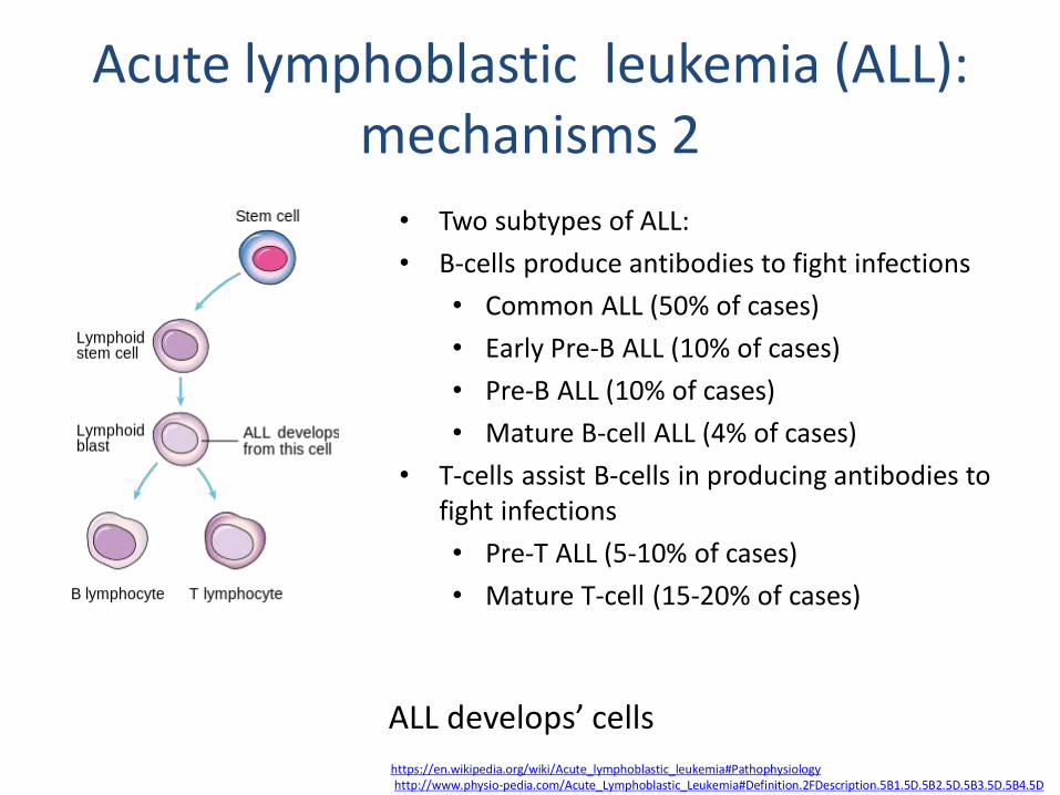
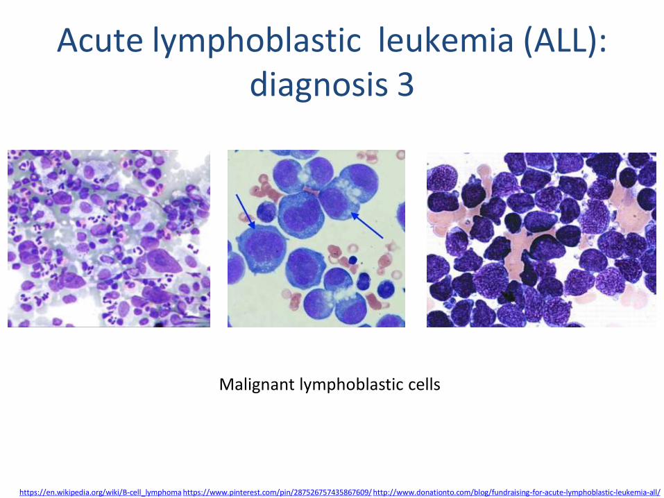
Acute lymphoblastic (lymphocytic, lymphoid) leukemia (ALL) represents a group of clonal hematopoietic stem cell disorders in which both a block in differentiation and unchecked proliferation result in the accumulation of lymphoblasts at the expense of normal hematopoietic precursors
• ALL is caused by damage to DNA that leads to uncontrolled cellular growth and spreads throughout the body, either by increasing chemical signals that cause growth or by interrupting chemical signals that control growth
• Damage can be caused through the formation of fusion genes, as well as the dysregulation of a proto-oncogene via juxtaposition of it to the promoter of another gene, e.g. the T-cell receptor gene by environmental factors such as chemicals, drugs or radiation, and occurs naturally during mitosis or other normal processes
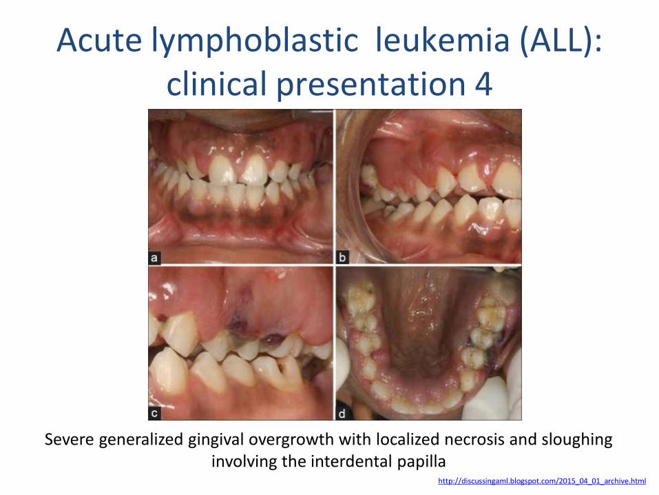
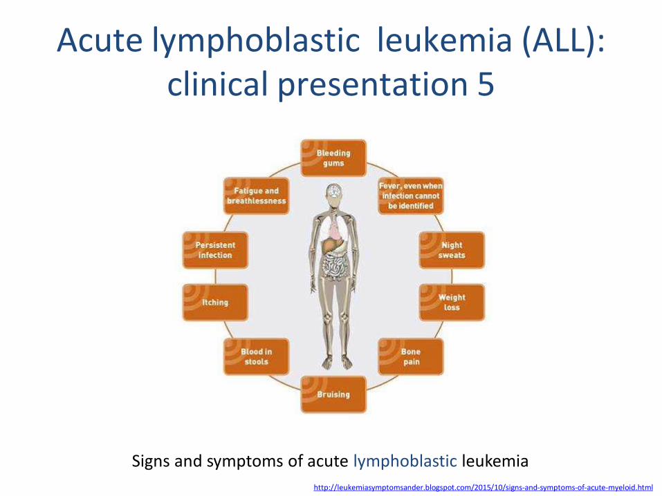
• History and Physical Examination: clinical presentation
• Blood Test: an excess of abnormal white blood cells (leukocytosis), sometimes leukemic blasts, isolated decreases in platelets, red blood cells, or even with a low white blood cell count (leukopenia)
• Bone marrow biopsy and aspiration: to diagnose the presence of leukemia, to differentiate ALL from other types of leukemia, and to identify the subtype of ALL accordingly to its classifications
• Immunophenotyping: to determine the subtype of ALL by comparing the cancer cells to normal cells in the immune system (may reveal terminal deoxynucleotidyl transferase (TdT -a specialized DNA polymerase expressed in immature, pre-B, pre-T lymphoid cells, and ALL/lymphoma cells or common acute lymphoblastic leukemia antigen (CALLA)
Example of immunophenotyping plots: A: Simply classification of T Cells [in blue] (CD3+) and B Cells [in purple] (CD19+) (i); with lymphocytes [in blue and purple] distinguished by Forward and Side Scatter (ii). B: Classification of eosinophils [in
red] as SiglecF+ (i) F4/80intCD11bint (ii) Ly6G-/lowCD11c– (iii) FSCloSSCint/hiSiglecF+ (iv).
Treatment can span 2 ½ - 3 ½ years depending on each individual situation and is broken down into the following 4 phases:
1. Induction therapy with the purpose to achieve remission by killing most of the cancer cells in the blood and bone marrow (chemotherapy drugs injected intrathecally, steroids, the anthracyclines)
2. Consolidation (post-remission) therapy with the goal to destroy any remaining leukemia cells in the central nervous system (4 - 8 weeks)
3. Maintenance (low dose) therapy is given to prevent cancer cell re-growth (4 weeks)
4. Preventive therapy to the spinal cord (chemotherapy drugs are injected directly into the spinal cord fluid)
There are 4 main types of the specific treatments :
• Chemotherapy (all patients need spinal taps to inject chemotherapy into the cerebrospinal fluid (CSF) to kill any leukemia cells that may have spread to the brain and spinal cord)
• Targeted drug therapy to attack specific abnormalities that cause the cancer cell growth
• Radiation therapy is typically used when the cancer has spread to the central nervous system
• Stem cell transplant (SCT) may be used for patients at risk or currently going through a relapse
Physical Therapy and exercise are aimed at symptom management, preservation of muscle function, pain control, and increased quality of life and include:
• Up to 15 minutes of walking 5x per week can reduce fatigue, symptom distress, anxiety, and depressive status while maintaining cardiovascular health
• Strength training combined with aerobic exercises 3x per week, twice daily, for 30 minutes improves cardiorespiratory endurance, reduces total fatigue and depression, maintains quality of life, and may reduce inflammatory markers
• Pain management (transcutaneous electrical nerve stimulation (TENS), hot packs, cold packs, massage, etc.
• Stretching (sustained stretch, active and passive range of motion (ROM), splinting, etc.)
• Chemotherapy drugs: L - asparaginase, Vincristine
• Steroid: Dexamethasone, Hydrocortisone
• Drugs for high-risk patients: Daunorubicin, Cytarabine
• Other drugs that may be given early: Methotrexate, 6-mercaptopurine
Novel approaches:
• For some subtypes of relapsed ALL, aiming at biological targets such as the proteasome, in combination with chemotherapy, has given promising results in clinical trials
• Chimeric antigen receptors (CARs) have been developed as a promising therapy for ALL
• Acute lymphoblastic leukemia is a curable disease, and the chance of cure for a specific patient depends on a number of prognostic factors (females tend to fare better than males; Caucasians are more likely to develop acute leukemia than African-Americans, Asians, or Hispanics; children 1–10 years of age are most likely to develop ALL and to be cured of it; cases in older patients are more likely to result from chromosomal abnormalities, etc.)
• The 5-year survival rate has improved from zero six decades ago, to 85% currently, largely because of clinical trials on new chemotherapeutic agents and improvements in SCT technology
Acute lymphoblastic leukemia (ALL): clinical case 1
• Donor cell leukemia (DCL) is a rare but severe complication after allogeneic stem cell transplantation
• The mechanisms of leukemogenesis are unclear, and multiple factors can contribute to the development of DCL
• In recent years, cord blood has emerged as an alternative source of hematopoietic progenitor cells, and at least 12 cases of DCL have been reported after unrelated cord blood transplantation
• A new case of DCL after unrelated cord blood transplantation in a 44-year-old woman diagnosed as having ALL with t(1;19) that developed AML with normal karyotype and nucleophosmin (NPM1) mutation in donor cells
• This is the first report of NPM1 mutation contributing to DCL development
Acute lymphoblastic leukemia (ALL): clinical case 2a
• Therapy-related ALL develops in patients after chemotherapy and/or radiotherapy for a prior cancer, and most cases are AML with a much lower frequency of ALL
• One unique feature of these therapy-related ALL (t-ALL) is an increased incidence of chromosome band 11q23 aberrations as compared with de novo ALL
• There was a report about the case of a 49-year-old Taiwanese lady who developed t-ALL with t(4;11)(q21;q23) 16 months after cyclophosphamide, epirubicin, and 5-fluorouracil chemotherapy for her breast cancer
• The unusual feature is that the t-ALL was heralded 4 months ago by marrow lymphocytosis comprising atypical small lymphocytes with condensed chromatin mimicking a B-cell chronic lymphoproliferative disorder
Acute lymphoblastic leukemia (ALL): clinical case 2b
• Retrospective studies using additional antibodies for immunophenotyping and PCR-based clonality study for immunoglobulin gene rearrangement showed that these atypical small lymphocytes shared similar features with the leukemic blasts at the frank leukemic stage
• The results suggest that these atypical small lymphocytes are lymphoblasts in disguise and that the clinicopathological correlations with ancillary pathological studies are important to reach a definitive diagnosis of such an unusual case
Chronic myelogenous (myeloid, myelocytic, granulocytic) leukemia (CML) is the clonal hematopoietic stem cell disorder with an abnormal increase in mature and immature granulocytes (as neutrophils, eosinophils, and myelocytes) especially in bone marrow and blood, that occurs especially in adults, and that is associated with the presence of the Philadelphia chromosome CML
• CML is a biphasic disease, initiated by the 'Philadelphia chromosome‘ expression of the BCR - ABLfusion gene product in self-renewing, haematopoietic stem cells (HSCs)
• HSCs can differentiate into common myeloid progenitors (CMPs), which then differentiate into granulocyte / macrophage progenitors (GMPs; progenitors of granulocytes (G) and macrophages (M)) and megakaryocyte / erythrocyte progenitors (MEPs; progenitors of red blood cells (RBCs) and megakaryocytes (MEGs), which produce platelets)
• HSCs can also differentiate into common lymphoid progenitors (CLPs), which are the progenitors of lymphocytes such as T cells and B cells
• The initial chronic phase (3-4 years) of CML (CML-CP) is characterized by a massive expansion of the granulocytic-cell series
• Acquisition of additional genetic mutations beyond expression of BCR-ABL causes the progression of CML from chronic phase to blast phase (CML-BP) by a block of cell differentiation that results in the presence of 30% or more myeloid or lymphoid blast cells in peripheral blood or bone marrow, or the presence of EXTRAMEDULLARY infiltrates of blast cells, characterized by an accumulation of myeloid (2/3 patients) or lymphoid blast cells (1/3 patients)
• Although the CML stem cell is multipotent, production of B cells from the neoplastic clone occurs only at low levels, and only rare T-cell precursors can be detected
• Chronic phase: there are few blast cells in the blood and bone marrow and there may be no symptoms of leukemia, this phase may last from several months to several years
• Accelerated phase: there are more blast cells in the blood and bone marrow, and fewer normal cells
• Blastic phase (the blast crisis): more than 30% of the cells in the blood or bone marrow are blast cells and the blast cells may form tumors outside of the bone marrow in places such as the bone or lymph nodes
• Refractory CML: leukemia cells do not decrease even though treatment is given
< 15 % blast cells in blood or bone marrow < 30 % – the sum of blast cells and promyelocytes in blood or bone marrow < 20 % basophile granulocytes in peripheral blood Thrombocyte count > 100 x 109/l
Normal or close to normal blood values without immature granulocytes in the blood
Blast count between 15–29 % in blood or bone marrow ≥ 30 %, the sum of blast cells and promyelocytes in blood or blood marrow ≥ 20 % basophile granulocytes in blood Thrombocyte count < 100 x 109/l, which cannot be explained by treatment
Often also increasing splenomegaly new chromosome changes in Ph+ clone
The way CML presents depends on the stage of the disease:
• Most patients (~90%) are diagnosed during the chronic stage which is most often asymptomatic, and may be diagnosed incidentally with an elevated white blood cell count on a routine laboratory test
• It can also present with symptoms indicative of enlarged spleen and liver and the resulting upper quadrant pain this causes
• The enlarged spleen may put pressure on the stomach causing a loss of appetite and resulting weight loss
• It may also present with mild fever and night sweats due to an elevated basal level of metabolism
• Some (<10%) are diagnosed during the accelerated stage which most often presents bleeding, petechiae and ecchymosis, when fevers are most commonly the result of opportunistic infections
• Some patients are initially diagnosed in the blast phase in which the symptoms are most likely fever, bone pain and an increase in bone marrow fibrosis
• CML is often suspected on the basis of a complete blood count, which shows increased granulocytes of all types, typically including mature myeloid cells
• Basophils and eosinophils are almost universally increased; this feature may help differentiate CML from a leukemoid reaction
• A bone marrow biopsy is often performed as part of the evaluation for CML, and CML is diagnosed by cytogenetics that detects the translocation t(9;22)(q34;q11.2) which involves the ABL1 gene in chromosome 9 and the BCR gene in chromosome 22: as a result of this translocation, the chromosome looks smaller than its homologue chromosome, and this appearance is known as the Philadelphia chromosome chromosomal abnormality
• Thus, CML can be detected by routine cytogenetics, and the involved genes BCR-ABL1 can be detected by fluorescent in situ hybridization, as well as by polymeric chain reaction (PCR)
• Controversy exists over so-called Ph-negative CML, or cases of suspected CML in which the Philadelphia chromosome cannot be detected: many such patients in fact have complex chromosomal abnormalities that mask the (9;22) translocation, or have evidence of the translocation in spite of normal routine karyotyping
• The small subset of patients without detectable molecular evidence of bcr-abl fusion may be better classified as having an undifferentiated myelodysplastic/myeloproliferative disorder, as their clinical course tends to be different from patients with CML
• CML must be distinguished from a leukemoid reaction, which can have a similar appearance on a blood smear
This chronic myelomonocytic leukemia-2 was morphologically composed of (A, Wright, ×400) 14% immature monocytic cells (blasts and promonocytes) and 26% mature monocytes. The flow cytometric immunophenotypic data revealed 54% of cells within the "monocytic" region (B; orange), which revealed uniform bright expression of CD11b (C), CD14 (D), and CD33 (E) with moderate expression of CD64 (F), partial loss of expression of CD15 (C), complete loss of expression of CD13 (D), and aberrant dim expression of CD2 (G). FITC, fluorescein isothiocyanate; PE, phycoerythrin
• In the past, antimetabolites (e.g., cytarabine, hydroxyurea), alkylating agents, interferon alfa 2b, and steroids were used as treatments of CML in the chronic phase, but since the 2000s have been replaced by Bcr-Abl tyrosine-kinase inhibitor drugs that specifically target BCR-ABL, the constitutively activated tyrosine kinase fusion protein caused by the Philadelphia chromosome translocation
• Despite the move to replacing cytotoxic antineoplastics (standard anticancer drugs) with tyrosine kinase inhibitors sometimes hydroxyurea is still used to counteract the high WBCs encountered during treatment with tyrosine kinase inhibitors like imatinib; in these situations it may be the preferred myelosuppressive agent due to its relative lack of leukemogenic effects and hence the relative lack of potential for secondary haematologic malignancies to result from treatment
• The first of this new class of drugs was imatinib mesylate, approved by the U.S. Food and Drug Administration (FDA) in 2001
• Imatinib was found to inhibit the progression of CML in the majority of patients (65–75%) sufficiently to achieve regrowth of their normal bone marrow stem cell population (a cytogenetic response) with stable proportions of maturing white blood cells
• Since the advent of imatinib, CML has become the first cancer in which a standard medical treatment may give to the patient a normal life expectancy
• To overcome imatinib resistance and to increase responsiveness to TK inhibitors, three novel agents were later developed
• Dasatinib, blocks several further oncogenic proteins, in addition to more potent inhibition of the BCR-ABL protein, and was initially approved in 2007 by the US FDA to treat CML in patients who were either resistant to or intolerant of imatinib
• Nilotinib, was also approved by the FDA for the same indication in 2010
• Radotinib joined the class of novel agents in the inhibition of the BCR-ABL protein and was approved in South Korea in 2012 for patients resistant to or intolerant of imatinib
• While capable of producing significantly improved responses compared with the action of imatinib, neither dasatinib nor nilotinib could overcome drug resistance caused by one particular mutation found to occur in the structure of BCR-ABL known as the T315I mutation
• Two approaches were developed to the treatment of CML as a result:
• In September 2012, the FDA approved a non BCR-ABL targeted agent omacetaxine, administered subcutaneously (under the skin) in patients who had failed with imatinib and exhibited T315I kinase domain mutation
• In December 2012 , the FDA approved a new pan-BCR-ABL inhibitor Ponatinib which showed (for the first time) efficacy against T315I, as well as all other known mutations of the oncoprotein
In 2005, encouraging but mixed results of vaccination were reported with the BCR/abl p210 fusion protein in patients with stable disease, with GM-CSF as an adjuvant
• Before the advent of tyrosine kinase inhibitors, the median survival time for CML patients had been about 3–5 years from time of diagnosis
• With the use of tyrosine kinase inhibitors, survival rates have improved dramatically
• A 2006 follow up of 553 patients using imatinib found an overall survival rate of 89% after five years
• A 2011 follow up of 832 patients using imatinib who achieved a stable cytogenetic response found an overall survival rate of 95.2% after 8 years, which is similar to the rate in the general population. Less than 1% of patients died because of leukemia progression
Chronic myelogenous leukemia (CML): clinical case 1b
• Even though initial cytogenetic studies could not be performed because of 'dry tap' aspirate, persistent efforts for cytogenetic studies were made, including a 'squeeze preparation' from the core biopsy, which revealed t(9;22)(q34;q11.2) and trisomy 19
• The patient was treated with tyrosine kinase inhibitors, chemotherapy, and subsequently an allogeneic stem cell transplant
• She is in persistent remission
• This case illustrates a complex presentation of chronic myelogenous leukemia and provides an overview of morphologic cues and the importance of performing cytogenetic studies that led to the diagnosis
Chronic myelogenous leukemia (CML): clinical case 2
• Chronic myelogenous leukemia (CML) is very rare in the pediatric population
• Doctors report the case of a 2-year-old female with CML and concurrent myelodysplastic syndrome (MDS) associated cytogenetic abnormalities
• The co-existence of t(9;22) and chromosomal deletions that are associated with MDS poses a unique diagnostic challenge
• Given the reported association of t(9;22) and genomic instability, doctors hypothesize that the chromosomal deletions represent clonal evolution of the CML
Chronic myelogenous leukemia (CML): clinical case 3a
• A 68-year-old man complained of dizziness and was referred to hospital by his primary physician for evaluation of an elevated leukocyte count
• In April 2002, soon after the chronic phase of chronic myeloid leukemia had been diagnosed, he was treated with imatinib
• In March 2010, imatinib treatment was completed and the BCR/ABL fusion gene had become undetectable by real time quantitative PCR
• Subsequently, leukocyte counts and the hematocrit gradually rose
• In August 2012, a bone marrow aspirate showed hypercellular marrow with marked erythroid hyperplasia and the presence of the JAK2 gene V617F mutation
Chronic lymphocytic leukemia (CLL) is a monoclonal disorder characterized by a progressive accumulation of small, mature-appearing functionally incompetent lymphocytes in blood, bone marrow, and organs
• Trisomy 12 is associated with atypical lymphocyte morphology and immunophenotype (CD5–, FMC7+, strong immunoglobulin expression) and disease progression
• Patients with 11q deletions tend to be younger, with an advanced clinical stage at presentation associated with extensive peripheral, abdominal, and mediastinal lymphadenopathy, and a treatment-free interval of nine months in contrast to 43 months for those without the deletion
• Chromosome 17 abnormalities have been associated with p53 mutation, fludarabine-resistance therapy, and therapy failure in patients with Richter's syndrome, atypical CLL morphology, and prolymphocytic leukemia (PLL)
• The presence of IgV gene mutations is associated with the lack of CD38 expression in a group of patients with good clinical outcome and better survival
• CLL is characterized by accumulation of small, mature-appearing lymphocytes in blood, bone marrow, and organs
• Recently, much attention is focused on the CLL B cell receptor (BCR) and on chemokine receptors that enable CLL cells to home to lymphoid tissues and to establish the leukemia microenvironment
• Agents that can interfere with BCR signaling or chemokine–receptor signaling, or that target surface antigens selectively expressed on CLL cells, promise to have significant therapeutic benefit in patients with this disease
0 - characterized by absolute lymphocytosis (>15,000/mm3) without adenopathy, hepatosplenomegaly, anemia, or thrombocytopenia
I - characterized by absolute lymphocytosis with lymphadenopathy without hepatosplenomegaly, anemia, or thrombocytopenia
II - characterized by absolute lymphocytosis with either hepatomegaly or splenomegaly with or without lymphadenopathy
III - characterized by absolute lymphocytosis and anemia (hemoglobin <11 g/dL) with or without lymphadenopathy, hepatomegaly, or splenomegaly
IV - characterized by absolute lymphocytosis and thrombocytopenia (<100,000/mm3) with or without lymphadenopathy, hepatomegaly, splenomegaly, or anemia
• About 40%-60% of patients with CLL are diagnosed in the absence of disease-related symptoms, even with very high numbers of circulating lymphocytes >100 × 109/l
• Frequently, the presence of lymphadenopathy or an abnormal CBC performed during a routine medical examination is the only reason to consider the diagnosis
• The remaining patients may present with weakness, fatigue, night sweats, fever, and may be with or without infections or autoimmune diseases
• Physical examination generally reveals nontender, painless, and mobile lymphadenopathy, splenomegaly, or hepatomegaly
• Metabolic abnormalities (e.g., hyperuricemia) or mechanical disorders (e.g., airway obstruction) related to the tumor burden, may also be present
• Any part of the body, including skin and meninges may be infiltrated by CLL cells; however, such findings are uncommon
• Manifestations of bone marrow (BM) involvement, particularly significant anemia (hemoglobin <11g/dl) or thrombocytopenia (platelets count <100 × 109/l), are noted at presentation in 15% of CLL patients
• A positive direct antiglobulin test (DAT) is present in about 20% of patients at diagnosis but is not commonly associated with hemolytic anemia
The National Cancer Institute-Sponsored Working Group diagnosis and response criteria for CLL
• The peripheral blood should exhibit an increase in the number of small mature-appearing lymphocytes to >5,000/μl
• The bone marrow (BM) aspirate smear must show >30% of all nucleated cells to be lymphoid
• Although a BM examination is rarely required to make the diagnosis of CLL in general practice, it may be valuable prior to the start of treatment in order to define prognostic factors
• Subsequently, a BM examination is indicated primarily to evaluate response to treatment or to assess normal elements if there is an unexplained anemia or thrombocytopenia
Clonal B cells (orange) in bone marrow with an immunophenotype characteristic of CLL: CD5+, CD10-, CD20dim+, CD23+, FMC7-, and dim+ surface λ light chain.
The cells also are CD19+ and CD38-. Clonal B cells represent 2.2% of events; benign polytypic B cells (blue), 4.9%; and normal precursor B cells (hematogones;
• CLL treatment focuses on controlling the disease and its symptoms rather than on an outright cure
• CLL is treated by chemotherapy, radiation therapy, biological therapy, or bone marrow transplantation
• Symptoms are sometimes treated surgically (splenectomy removal of enlarged spleen) or by radiation therapy ("de-bulking" swollen lymph nodes)
• Initial CLL treatments vary depending on the exact diagnosis and the progression of the disease, and even with the preference and experience of the health care practitioner
• An initial treatment regimen that contains fludarabine, cyclophosphamide, and rituximab (known as FCR) has demonstrated higher overall response rates and complete response rates
• Physicians use a “watchful waiting” mode until the disease progressed, but the novel prognostic markers indicate that 50% of patients have a poor outcome
• An early start of therapy may be justified in patients with a poor prognosis
• Since CLL cells must interact with the stroma in bone marrow or lymphoid tissues to survive, these interactions need to be explored as targets of innovative therapies, and specific inhibition of the B-cell receptor signaling pathway, as targeting the actively proliferating cells that maintain the CLL clone by a cell-cycle–active agent may be an option
• Since as 20% of patients with the worst prognostic markers have stereotypic antigen receptors, they may be valuable points of attack
Chronic lymphocytic leukemia (CML): clinical case 1
• Granulomatous cutaneous reactions are well described in association with T-cell non-Hodgkin lymphoma and Hodgkin lymphoma, but are rarely seen in association with B-cell non-Hodgkin lymphoma or leukemia
• Doctors report a case of a 65-year-old woman with B-cell chronic lymphocytic leukemia (CLL)/small lymphocytic lymphoma (SLL) who presented with multiple, tender, firm pink papules on the face, upper trunk and upper extremities 6 years after diagnosis of CLL
• Biopsy revealed both palisading granulomatous dermatitis consistent with actinic granuloma and a dense perivascular lymphocytic infiltrate consistent with the patient's known history of leukemia
• This is an unusual manifestation of cutaneous B-cell CLL that is rarely seen
Patients with leukemia in periods of severe granulocytopenia and thrombocytopenia related to the specific antileukemic therapies administered demand secondary prophylaxis with antifungals and antibiotics
• ALL - acute lymphoblastic leukemia • AML - acute myelogenous leukemia • BM – bone marrow • CALLA - common acute lymphoblastic leukemia antigen • CARs - Chimeric antigen receptors • CLL - chronic lymphocytic leukemia • CML - chronic myelogenous leukemia • CNS - central nervous system • DCL - donor cell leukemia • FAB - French-American-British classification • FFP - fresh frozen plasma • ROM - range of motion • SCT - stem cell transplant • TdT - terminal deoxynucleotidyl transferase • TENS - transcutaneous electrical nerve stimulation • WHO - World Health Organization • PCR - polymeric chain reaction • FDA - U.S. Food and Drug Administration
Diagnostic guidelines
Europe • Peripheral T-Cell Lymphomas: ESMO Clinical Practice Guidelines • Diffuse Large B-Cell Lymphoma: ESMO Clinical Practice Guidelines • Chronic Lymphocytic Leukaemia: ESMO Clinical Practice Guidelines • Hairy Cell Leukaemia: ESMO Clinical Practice Guidelines • Philadelphia Chromosome-Negative Chronic Myeloproliferative
Neoplasms: ESMO Clinical Practice Guidelines • Myelodysplastic Syndromes: ESMO Clinical Practice Guidelines • New aspects of the updated guidelines for the diagnosis and
treatment of chronic lymphocytic leukemia North America • Leukemia: An Overview for Primary Care


![RESEARCH ARTICLE Open Access Cytological maps of …dspace.univer.kharkov.ua/bitstream/123456789/10105/2/Dedukh_ea... · west to Volga River in the east [6,10], ... P. ridibundus](https://static.documents.pub/doc/80x56/5a879df57f8b9afc5d8debd8/research-article-open-access-cytological-maps-of-to-volga-river-in-the-east.jpg)


