An Innovative Paradigm: Coordinating Anesthetic Care for Complex PediatricPatients requiring Multiple ProceduresJill E. Kilkelly1, MD and Jill Kinch2*
1Clinical Director, Pediatric Perioperative Care, Assistant Professor, Division of Pediatric Anesthesiology, Monroe Carell Jr.
*Corresponding author: Jill Kinch, MSN, MMHC, APRN, CPNP-PC/AC, Assistant Director Advanced Practice Nursing, Ambulatory and Acute Care, Division ofPediatric Anesthesiology, Monroe Carell Jr. Children's Hospital at Vanderbilt, 2200 Children's Way Suite 3010, Nashville, TN 37232; E-mail: [email protected]
Received date: Nov 27th, 2014, Accepted date: Dec 20th, 2014, Published date: Jan 20th, 2015
Background: The goal of the anesthesia coordination of care team is to optimize safety for children requiringmultiple procedures under general anesthesia, by providing a single continuous anesthetic for imaging and surgicalrequests.
Methods: We developed an inter-professional team to create a pathway for providers to request multipleprocedures with one anesthetic. Data collected includes patient and family satisfaction and a growing expertise ofbest practices for planning such coordinated care.
Results: The program began in December 2011, with over 300 cases completed to date. Through thedevelopment of this program, we have evolved our clinical expertise to provide optimal combinations andsequencing of procedures under one continuous anesthetic.
Conclusions: After a review of the literature, our team has not identified another care organization thatconsistently and prospectively plans for one continuous anesthetic for multiple procedures for children. Evidencesupports this necessary planning, as there is a growing body of scientific research suggesting a possible risk of longterm neurocognitive deficits related to anesthetic exposure at an early age. Certainly this approach is the right thingto do for patient safety, and it also is very appreciated by families.
Introduction and BackgroundFrequently, both a surgical procedure and an anesthetized
radiological procedure are requested, but often they are not arrangedto occur with one continuous anesthetic. Unfortunately, theseprocedures are planned by schedulers from multiple subspecialties,leading to uncoordinated care. Previously, hospital faculty and staffknew some patients were scheduled for more than one sedatedprocedure, such as an MRI and a minor surgery, often within the samemonth, but this was frequently discovered too late to coordinate thecare. For patients, this means multiple trips to the hospital for twoseparate sedations and procedures or imaging. This results in twoseparate fasting periods, two separate intravascular access attempts,two intubations and two anesthetic exposures. To decrease risk for thepatient, care can be coordinated to proactively plan for one continuousanesthetic that allows for all requested imaging and surgicalprocedures to be sequenced and completed in one visit.
Initiative DescriptionThe goal of the coordination of care team is to optimize care for
patients requiring multiple procedures under general anesthesia, oftenin multiple hospital locations, by providing a single continuousanesthetic for all requested procedures/imaging. In order to define the
most efficient process for planning this care, we collaborated withpediatric surgical, medical, procedural, nursing, scheduling andadmitting services in perioperative and radiology departments. Theinitial request is sent to the designated pediatric anesthesiologist whoserves as a consultant to provide guidance and recommendationregarding optimal procedural sequence, procedural room reservationand timing details. When arrangements for care are finalized, thecoordination of care plan is saved to the patient's medical record.
InnovationAfter a review of the literature, our team has not identified another
care organization that consistently and prospectively plans for onecontinuous anesthetic for multiple procedures for children.Recommendations for planning and sequencing combinationsinclude:
a. As a general rule, non-invasive procedure or imaging should bescheduled first, then the procedure or surgery should follow. Forsequence guidelines and examples see Figure 1.
b. Some procedural combinations are not recommended, such aselective imaging that must be read prior to the procedure.
Anesthesia & Clinical Research Kilkelly and Kinch, J Anesth Clin Res 2014, 5:11http://dx.doi.org/10.4172/2155-6148.1000490
Research Article Open Access
J Anesth Clin ResISSN:2155-6148 JACR, an open access journal
Figure 1: Template for Sequencing Coordination of Anesthetic Care.
For Example: MRI needs to be read and reviewed with theInterventional Radiology (IR) attending prior to performing jointinjections, so taking the patient direct to IR leads to wasted time in theIR suite, and extra anesthesia time, so these must be purposefullyplanned to be separated. For additional examples see Figure 2.
Types of Procedures/Imaging
Clinical Significance
Dental rehabilitation andadenoidectomy
nasal endotracheal tube required for dentalprocedure obstructs ability to excise adenoidtissue
T&A and pH probe the pH probe cannot be in place during immediateT&A postop period due to risk of irritation andensuing hemorrhage to newly cauterized tonsillarbed
Any invasive procedureswith cardiac catheterization
MRI Any non-urgent imaging that is required to be readprior to the procedure
Figure 2: Procedural/Imaging Combinations that are NotRecommended.
Actual Results and OutcomesPositive outcomes include both significant patient satisfaction with
the approach and better understanding of best practice for sequencingand planning coordinated anesthetic care. Families of childrenrequiring combined care procedures can now expect a well-executedhospital experience (see Figures 3 and 4), improving satisfaction.Confirmation of enhanced patient and family satisfaction areevidenced by responses to our patient satisfaction survey (IRB #
120682). Families shared with us their impressions of their experiencewith this program.
“I am extremely pleased with the communication level and thecompassion shown in trying to coordinate these procedures. My childdoes not handle anesthesia well and it helped my peace of mindknowing that this was going to be a one-time anesthesia.”
Parent described the coordination of care as "lifesaving" for herautistic child. “It decreased my child’s aggression and anxiety.”
“Improved my child's safety”
“With him being 2 years old we wanted to limit his exposure toanesthesia”
“Appreciate the opportunity for multiple procedures in the sameday since we live so far away”
“Initial communication from both clinics was a little confusing, butonce the nurse (pre-operative clinic) became involved, everythingbecame clear. Great experience overall!”
Citation: Kilkelly J and Kinch J (2014) An Innovative Paradigm: Coordinating Anesthetic Care for Complex Pediatric Patients requiring MultipleProcedures . J Anesth Clin Res 5: 490. doi:10.4172/2155-6148.1000490
Page 2 of 4
J Anesth Clin ResISSN:2155-6148 JACR, an open access journal
Volume 5 • Issue 11 • 1000490
Figure 3: Prior State: One Patient, Two Anesthetics.
Figure 4: Current process: patient Centered perioperative and anesthetic care, One coordinated visit.
In addition to improving satisfaction, we hypothesize that patientrisk is likely reduced by well-planned care that minimizes the numberof required preoperative fasting experiences, preoperative anxiety,intubations, venipunctures and multiple discrete anesthetics. This isespecially important for medically complex children who are at highrisk for complications. Furthermore, this program allows fewerseparate hospital trips, resulting in cost savings to families who travel asignificant distance for each visit, and reduces absences from school orwork days.
The program began in December 2011. To date, we have completedover 300 cases. Development of this program did not add expense forthe hospital. Instead, the program grew as a result of two committedcolleagues, who invested in seeing this concept become a real andsustainable program. One of our pediatric anesthesiologists has actedas physician champion of this program, with partnership from one ofour perioperative nurse practitioner leaders. Ultimately,communicating the intent of the program to colleagues throughout theinstitution has helped create increasing enthusiasm for the process
supporting coordination of services. We are experiencing an increasein requests now that we have a well-defined care pathway. As theservice continues to grow, we recognize that sustainability will only beachieved through further team work, and we are beginning to engageand train a dedicated perioperative advance practice nursing team tohelp plan and coordinate these requests in the future.
Methods
IRB/Consent:The study was approved and evaluated by the IRB and determined
to be non-research and consent therefore is not necessary.
RE: IRB# 120682 "Patient/Family Satisfaction with Coordination ofMultiple Procedures Under Single Continuous Anesthetic." A designeeof the Institutional Review Board reviewed the research studyidentified above. The designee determined the project does not qualifyas "research" per 45 CFR §46.102(d). Family satisfaction is discussed
Citation: Kilkelly J and Kinch J (2014) An Innovative Paradigm: Coordinating Anesthetic Care for Complex Pediatric Patients requiring MultipleProcedures . J Anesth Clin Res 5: 490. doi:10.4172/2155-6148.1000490
Page 3 of 4
J Anesth Clin ResISSN:2155-6148 JACR, an open access journal
via phone call and documented in a secure de-identified database,following their perioperative care.
To improve our process, we de-briefed with stakeholders andreviewed a number of early cases, and three key issues were identified.First, communication flow and a clear process was needed to supportthe team work. Second, an informatics access point for coordinationand accountability for the plan was also necesarry. Finally, identifyingteam leaders for aligning resources and scheduling was also essentialfor success. This was an iterative process, and required ongoingcommunication to achieve a well-coordinated team approach.Bringing key stakeholders together to debrief was the most efficientform of designing and re-designing our approach. Repeating thisinvestigation at other care organizations would start with identifying aphysician champion who can lead the process development. First stepswould include engaging key personnel, including admitting,registration, surgical schedulers, and anesthesia, surgical andinformatics leaders, to start the discussion on how best to accomplishthis coordinated care in their individual institution’s setting.
DiscussionThis coordination supports a culture of personalized care that
children and families appreciate. Additionally this methodologystreamlines the patient’s care experience, specifically including theopportunity to decrease repeated anesthetic exposure. As a growingbody of scientific research suggests a possible risk of long termneurocognitive deficits related to anesthetic exposure at an early age,this work is clinically relevant. A 2013 article by Bong et al. inAnesthesia and Analgesia, describes an observational cohort studyundertaken to determine whether children exposed to generalanesthesia for minor surgery during infancy exhibited differences inacademic achievement at age twelve years, compared with childrenwho were never exposed to anesthesia or sedation. Findings include a4.5 times greater odds of a formal diagnosis of a learning disability byage 12 years in children who had been exposed to general anesthesia.Although further research is needed to sort out this clinical question ofcausality, it seems prudent to proactively minimize the number ofexposures to general anesthesia for infants and children. Also, socialmedia is actively bombarding parents of young children with theseconcerns and thus parents and families are seeking opportunities tominimize repeated anesthetic exposure.
ConclusionsAfter a review of the literature, our team has not identified another
care organization that consistently and prospectively plans for onecontinuous anesthetic for multiple procedures for children. Evidencesupports this planning as necessary, due to potential neurologicalsequelae related to anesthetic exposure at an early age.
Our experiences thus far have guided our development of thetemplate for sequencing coordination of anesthetic care (see figure 1and 2). Evaluation of our program has included both patientsatisfaction feedback and successful streamlining of the perioperativeprocess (see figure 3 and 4). We are enthusiastic to further exploreother potential positive effects of this approach, including cost savingsfor the perioperative service line. This is especially relevant as ourhealth care industry is challenged to shift from a fee-for-service modelto a value based health care purchasing model.
This paradigm is dedicated to patient centered care and emphasizesthe ethic of making the patient the first priority, rather than schedulingat the convenience of the operating room or surgical schedule.Although identifying the best process for coordinating this care will besomewhat different at each organization due to scheduling andregistration systems, we hope that this article shares a framework onhow to begin and thus encourages other organizations to developsimilar programs.
References:1. Bong C, Allen JC, Kim JTS (2013) The Effects of exposure to general
anesthesia in infancy on academic performance at age 12. Anesthesia andAnalgesia 117 (6): 1419-1428.
2. Ing C, DiMaggio C, Whitehouse A, Hegarty M, Brady J, Von Ungern-Sternber B, et al (2012) Long-term differences in language and cognitivefunction after childhood exposure to anesthesia. Pediatrics 130 (6):476-485.
3. Loepke AW, Soriano, SG (2008) An Assessment of the Effects of GeneralAnesthetics on Developing Brain Structure and NeurocognitiveFunction. Anesthesia and Analgesia 106 (6): 1681-1707.
4. Velayudha, R (2012) Effect of general anesthetics on the developingbrain. Journal of Anaesthesiology Clinical Pharmacology 28(1): 6-10.
Citation: Kilkelly J and Kinch J (2014) An Innovative Paradigm: Coordinating Anesthetic Care for Complex Pediatric Patients requiring MultipleProcedures . J Anesth Clin Res 5: 490. doi:10.4172/2155-6148.1000490
Page 4 of 4
J Anesth Clin ResISSN:2155-6148 JACR, an open access journal
A Multispecialty Pediatric Neurovascular Conference: A Modelfor Interdisciplinary Management of Complex Disease
Travis R. Ladner BA a, Jasia Mahdi BA, BS a, Albert Attia MDb,Michael T. Froehler MD, PhD c,d, Truc M. Le MDe, Amanda N. Lorinc MD f,J. Mocco MD, MS d, Robert P. Naftel MDd, Allen T. Newton PhD g, Sumit Pruthi MDg,Todd Tenenholz MD, PhDb, E. Haley Vance DNP d, Curtis A. Wushensky MDg,John C. Wellons III MD, MSPHd, Lori C. Jordan MD, PhDh,*
aVanderbilt University School of Medicine, Vanderbilt University Medical Center, Nashville, TennesseebDepartment of Radiation Oncology, Vanderbilt University Medical Center, Nashville, TennesseecDepartment of Neurology, Vanderbilt University Medical Center, Nashville, TennesseedDepartment of Neurological Surgery, Vanderbilt University Medical Center, Nashville, TennesseeeDivision of Critical Care Medicine, Department of Pediatrics, Vanderbilt University Medical Center, Nashville, TennesseefDepartment of Anesthesiology, Vanderbilt University Medical Center, Nashville, TennesseegDepartment of Radiology, Vanderbilt University Medical Center, Nashville, TennesseehDivision of Pediatric Neurology, Department of Pediatrics, Vanderbilt University Medical Center, Nashville, Tennessee
Article HistReceived S* Commu
Pediatric NMedical Ce
E-mail a
0887-8994/$http://dx.doi
abstract
INTRODUCTION: In 2013, our institution established a
multidisciplinary pediatric neurovascular conference forcoordination of care. Here, we review our initial experience. METHODS: Clinical and demographic data were ob-tained from medical records for patients presented to the pediatric neurovascular conference from April 2013 toJuly 2014. Patient descriptive characteristics were described by mean and standard deviation for continuousmeasures and by number and percent for categorical measures. Patients were secondarily stratified by lesion/disease type, and descriptive statistics were used to measure demographic and clinical variables. RESULTS: Thepediatric neurovascular conference met 26 times in the study period. Overall, 75 children were presented to theconference over a 15-month period. The mean age was 9.8 (standard deviation, 6.3) years. There were 42 (56%)male patients. These 75 children were presented a total of 112 times. There were 28 (37%) patients with history ofstroke. Complex vascular lesions were the most frequently discussed entity; of 62 children (83%) with a diagnosedvascular lesion, brain arteriovenous malformation (29%), cavernous malformation (15%), and moyamoya (11%)were most common. Most discussions were for review of imaging (35%), treatment plan formulation (27%), theneed for additional imaging (25%), or diagnosis (13%). Standardized care protocols for arteriovenous malformationand moyamoya were developed. CONCLUSION: A multidisciplinary conference among a diverse group of providersguides complex care decisions, helps standardize care protocols, promotes provider collaboration, and supportscontinuity of care in pediatric neurovascular disease.
ory:eptember 30, 2014; Accepted in final form January 2, 2015nications should be addressed to: Dr. Jordan; Division ofeurology; Department of Pediatrics; Vanderbilt Universitynter; Nashville, TN 37232.ddress: [email protected]
- see front matter � 2015 Elsevier Inc. All rights reserved..org/10.1016/j.pediatrneurol.2014.10.010
Introduction
Pediatric neurovascular diseases are uncommon andheterogeneous in nature. With the exception of stroke,there are no evidence-based management guidelinesavailable for these conditions in children.1-3 Care issimply based on extrapolation of the adult literature,
T.R. Ladner et al. / Pediatric Neurology 52 (2015) 165e173166
when applicable, and on expert opinion. Many of theseconditions are found in childrenwith rare diseases that maypredispose them to neurovascular abnormalities4-9
(Table 1).Given the paucity of guidelines for pediatric neuro-
vascular disease management, in 2013, our institutionestablished a multispecialty pediatric neurovascular con-ference (PNVC) to address complex cases that we believedwould benefit from a coordinated, multidisciplinaryapproach to patient care. Our objective was to create acollaborative forum where providers could meet to discusscases, review diagnoses and imaging, and develop jointmanagement plans at no cost to the patient.
The PNVC is composed of attending physicians and nursepractitioners representing seven specialties: pediatricneurology, pediatric neurosurgery, neurointerventional(endovascular) surgery, anesthesiology, neuroradiology,radiation oncology, and pediatric critical care. The PNVC isheld every 2 weeks, and trainees are invited to attend aswell. Cases of known or suspected neurovascular lesions aresubmitted by participants for discussion.
Here, we review our initial experience and examine howthe PNVC has helped foster multispecialty collaboration andcoordinated care protocols for pediatric neurovascular dis-ease management.
Methods
Study procedure
A record of patients presented to the PNVC was established in April2013 and prospectively populated for quality assurance. After institu-tional review board approval, clinical records for these listed patientspresented from April 2013 to July 2014 were retrospectively reviewed.Demographic variables (age, sex, and race/ethnicity) and clinical vari-ables (presenting symptoms, stroke history, diagnosis, neuroimaging,and all interventions) were collected. PNVC records were reviewed to
determine reason for referral to PNVC and clinical decisions after PNVCdiscussion.
A seven-item survey regarding usefulness of the PNVC was devel-oped and administered to PNVC participants (all of whom are co-authorson this report) for quality improvement purposes. The survey wasadministered by the senior author, who did not complete a survey.Respondent specialty was collected. Items #1-6 were scored on a 5-pointLikert scale, with choices ranging from “strongly agree” ¼ 5 to “stronglydisagree” ¼ 1. Item #7 was a four-option multiple-choice questionregarding what the respondent felt was the most useful aspect of PNVC.
Data analysis
Statistical analyses were computed using Microsoft Excel 2011(Microsoft Corp, Bellevue, WA). Patient descriptive characteristics weredescribed by mean and standard deviation for continuous measures andby number and percent for categorical measures. Patients weresecondarily stratified by lesion/disease type, and descriptive statisticswere used to measure demographic and clinical variables.
Results
Patient population
Overall, 75 children were presented to the PNVC over a15-month period. The mean age was 9.8 (standard devia-tion, 6.3) years. There were 42 (56%) males. There were 53(71%) caucasian, non-hispanic/latino; 14 (19%) African-American, non-hispanic/latino; and 8 (11%) caucasian, his-panic/latino patients. Headache (21 of 75, 28%), seizure (15of 75, 20%), altered mental status/loss of consciousness (10of 75, 13%), and focal weakness (9 of 75, 12%) were the mostcommon presenting symptoms. Twenty-eight (37%) chil-dren had a history of stroke, 15 of 75 (20%) hemorrhagic and13 of 75 (17%) ischemic. Of 62 children (83%) with a diag-nosed vascular lesion, arteriovenous malformation (AVM;18 of 62, 29%), cavernous malformation (9 of 62, 15%), andmoyamoya (7 of 62, 11%) were most common (Table 2).
Initial PNVC discussions
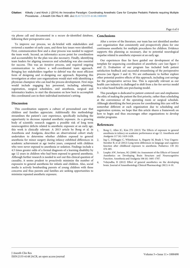
The PNVC met 26 times in the study period. Seventy-fivechildren were presented a total of 112 times. Initial dis-cussions were primarily for review of imaging (26 of 75,35%), treatment plan (20 of 75, 27%), need for additionalimaging (19 of 75, 25%), or diagnosis (10 of 75, 13%; Table 3).Discussion of treatment planwasmore common in AVM (10of 18, 55%) and large vessel occlusion or dissection (3 of 6,50%). Review of imaging was a common reason for moya-moya (6 of 7, 86%) and aneurysm (3 of 5, 60%) cases. Forcases where there was a concern for vascular lesion on MRI(n ¼ 4), concern for an infectious aneurysm (n ¼ 3) oranother uncategorized vascular lesion (n ¼ 9), need foradditional vascular imaging was the primary question in 6of 16 (38%) cases. Forty-two (56%) patients had a magneticresonance imaging (MRI)/magnetic resonance angiography(MRA), and 10 (13%) patients had a computed tomographyangiography after discussion at PNVC.
Interventions for 75 patients
Follow-up conventional angiography was performed in18 of 75 (24%) cases, particularly for AVM (9 of 18, 50%)and aneurysm (3 of 5, 60%; Table 3). A diagnostic catheter
TABLE 2.Baseline Demographic and Clinical Characteristics of 75 Patients Presented to PNVC
Condition N Age, years* Sex Number Prior Stroke Type Number
Arteriovenous malformation 18 12.8 (4.7) Male 14 Hemorrhage 8Female 4
Cavernous malformation 9 9.3 (6.8) Male 7 Hemorrhage 2Female 2
Moyamoya 7 14.7 (4.5) Male 3 AIS 2Female 4
Large artery occlusion or dissection 6 7.8 (7.3) Male 2 AIS 5Female 4 Hemorrhage 1
angiogram was performed in 13 individuals, and in theother 5 cases (all AVMs), embolization was performed.Considering all cases, a surgical operation was employedin 17 of 75 (23%); 12 of 75 (16%) underwent endovascularintervention; and 9 of 75 (12%) received stereotacticradiosurgery. Thirteen (72%) AVM patients had undergonetreatment before presentation at conference; treatmentswere a combination of embolization in 7 patients, ste-reotactic radiosurgery in 4 patients, and resection in 5patients. Five underwent treatment or retreatment afterPNVC (a combination of embolization in 4 patients,resection 2 patients, and stereotactic radiosurgery 4 pa-tients). Embolization was attempted in all individualswith vein of Galen malformation (n ¼ 3). Three childrenunderwent pial synangiosis after PNVC discussion(moyamoya in 2 patients, craniometaphyseal dysplasiawith bilateral internal carotid occlusions and transientischemic attack in 1 patient). One aneurysm was clipped,and three cavernous malformations were resected afterPNVC discussion. All cases of developmental venousanomaly were managed conservatively.
Post-PNVC repeat discussions for 26 patients
Twenty-six patients (35%) were discussed more thanonce, a total of 36 repeated discussions. The most common
reasons for rediscussion were imaging follow-up (17 of 36,47%), need for additional imaging/timing of angiography (9of 36, 25%), and treatment (8 of 36, 22%). Fourteen (39%) ofthe repeat discussions were for patients with AVMs(Table 3).
Illustrative cases
The following two cases represent the unique coordina-tion of care offered by the PNVC, synthesizing expertiseacross disciplines to arrive at a joint management plan forcomplex cerebrovascular lesions.
Case #1A 13-year-old girl presented with progressive myelop-
athy, with weakness and spasticity of both legs and herleft arm. This progression was thought to be because of apersistent cervicomedullary AVM (previously coiled threetimes, with partial resection at age 11 years and treat-ments at three different medical institutions), causingvascular steal from the spinal cord. Discussions resulted indiagnostic cerebral angiography, which revealed aSpetzler-Martin grade 4 AVM, with reversed flow in theanterior spinal artery (Fig 1). Cervical spine MRI revealeda thin cervical cord with changes suggestive ofchronic ongoing ischemia and prominent dural venouschannels. The risk of surgical resection was thought to be
TABLE 3.Management of 75 Patients at First Presentation to PNVC
ArteriovenousMalformation
CavernousMalformation
Moyamoya Large ArteryOcclusion orDissection
Aneurysm
N 18 9 7 6 5Question for PNVC Review imaging 4 2 6 2 3
Abbreviations:angio ¼ catheter angiogramAVM ¼ Arteriovenous malformationCEA ¼ Carotid endarterectomyCTA ¼ Computed tomography angiographyDSA ¼ Digital subtraction angiographyembo ¼ embolizationF/U ¼ Follow-upICA ¼ Internal carotid arteryMRA ¼ Magnetic resonance angiographyMRI ¼ Magnetic resonance imagingMT ¼ Mechanical thrombectomyPHACE ¼ Posterior fossa malformations, hemangioma, arterial lesions, cardiac abnormalities, and eye abnormalitiesPNVC ¼ Pediatric neurovascular conferencepost-op = post operativePS ¼ Pial synangiosiss/p ¼ status post or afterSRS ¼ Stereotactic radiosurgery
* Follow-up discussions: percentage is out of total number of repeat PNVCdiscussions (n ¼ 34), as some patients had two or more repeat discussions.
y Prior AVM treatments: embolization ¼ 7, SRS ¼ 5, and resection ¼ 5.z Other vascular lesions: PHACE, sinus pericranii, congenitally absent ICAs, radiation vasculopathy, carotid occlusions from craniometaphyseal dysplasia, intracranial
venous drainage abnormalities, and tumor encasing ICA.
T.R. Ladner et al. / Pediatric Neurology 52 (2015) 165e173168
extraordinarily high, and stereotactic radiosurgery wasrecommended. Three dimentional angiography withpossible repeat embolization is planned for radiosurgerytargeting.
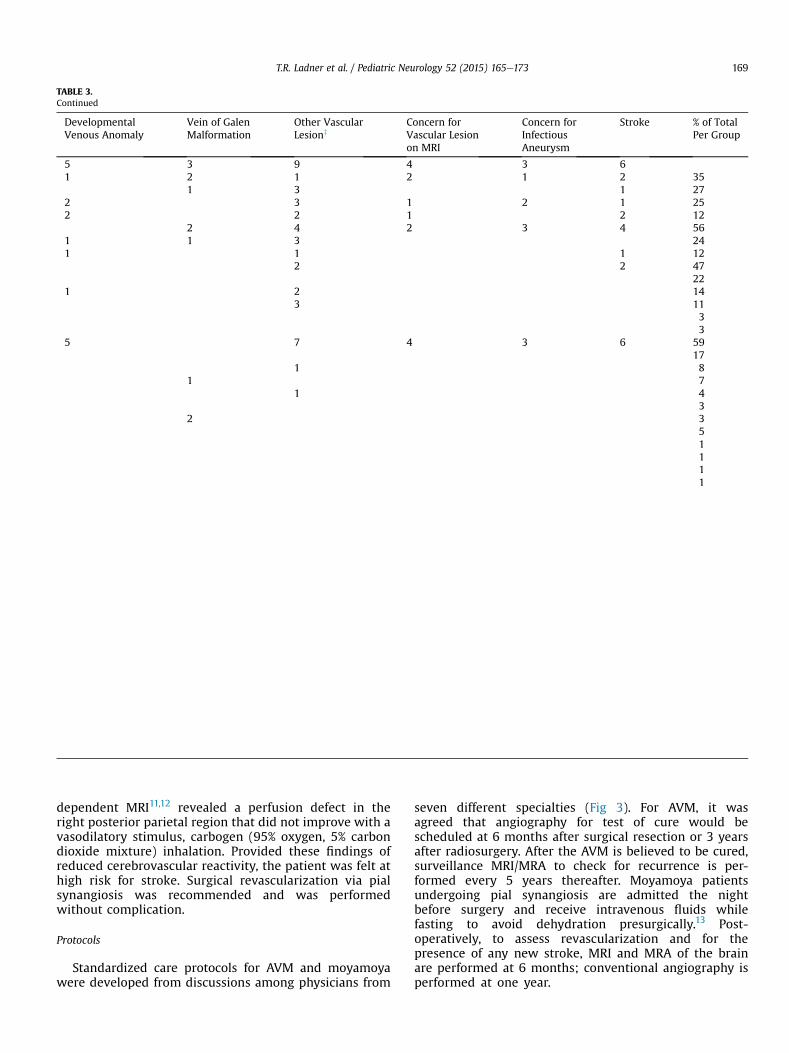
Case #2A 15-year-old girl with a remote history of facial
hemangioma presented with migraine. Brain MRI per-formed for worsening headaches revealed a 2-cm right-sided vascular anomaly within the perimesencephaliccistern. She was diagnosed with PHACE syndrome (pos-terior fossa malformations, hemangioma, arterial lesions,
cardiac abnormalities, and eye abnormalities)10 based onhemangioma and arterial abnormalities. Brain MRI/MRAwas obtained and reviewed at PNVC (Fig 2). Thisconfirmed abnormal flow voids in the right peri-mesencephalic space and indicated a right internal ca-rotid artery occlusion. Digital subtraction angiographywas obtained, which demonstrated near-complete occlu-sion of the distal right internal carotid artery and prox-imal right middle cerebral artery, with moyamoya-likecollaterals. In the absence of transient ischemic attack orother stroke symptoms, observation with hemodynamicMRI was recommended. Blood oxygenation level-
dependent MRI11,12 revealed a perfusion defect in theright posterior parietal region that did not improve with avasodilatory stimulus, carbogen (95% oxygen, 5% carbondioxide mixture) inhalation. Provided these findings ofreduced cerebrovascular reactivity, the patient was felt athigh risk for stroke. Surgical revascularization via pialsynangiosis was recommended and was performedwithout complication.
Protocols
Standardized care protocols for AVM and moyamoyawere developed from discussions among physicians from
seven different specialties (Fig 3). For AVM, it wasagreed that angiography for test of cure would bescheduled at 6 months after surgical resection or 3 yearsafter radiosurgery. After the AVM is believed to be cured,surveillance MRI/MRA to check for recurrence is per-formed every 5 years thereafter. Moyamoya patientsundergoing pial synangiosis are admitted the nightbefore surgery and receive intravenous fluids whilefasting to avoid dehydration presurgically.13 Post-operatively, to assess revascularization and for thepresence of any new stroke, MRI and MRA of the brainare performed at 6 months; conventional angiography isperformed at one year.
FIGURE 1.Case #1: 13-year-old girl with cervicomedullary dural arteriovenous malformation (AVM). (A) Magnified digital subtraction angiography (DSA)vertebral artery injection, early arterial phase. There is significant supply from the anterior spinal artery, with reversed flow ascending to supply thelesion (black arrows). The anterior spinal artery is already visualized filling, without opacification of its normal origin at the distal vertebral artery(arrowhead). (B) DSA reveals that the AVM, at the site of prior embolization, is supplied by bilateral vertebral arteries and bilateral posterior inferiorcerebellar arteries. There is rapid shunting through the right sigmoid sinus, a contralateral cerebellar vein, and the spinal venous plexus (white arrows).(C) DSA three dimentional reconstruction reveals a 3.5-cm Spetzler-Martin grade 4 AVM, with a diffuse nidus. (D) Sagittal T2-weighted magneticresonance images of the cervical spine (D, E) redemonstrate the cervicomedullary AVM. (E) Also visible are prominent dural venous channels pos-teriorly (arrow).
T.R. Ladner et al. / Pediatric Neurology 52 (2015) 165e173170
Respondent survey results
Eight PNVC participants completed the survey (Fig 4).Rated highest were both PNVC’s utility in informing a betterunderstanding of patients’ cases (5.0 of 5.0) and facilitatingbetter communication across specialties (5.0 of 5.0). Re-spondents from the pediatric intensive care unit andanesthesia reported, appropriately, that PNVC would notsignificantly change their management plan (3.0 of 5.0),whereas other specialties represented felt that it did (5.0 of5.0). Respondents unanimously cited PNVC’s greatest utilityas facilitation of a collaborative approach to patient care.
Discussion
The beneficial role tumor boards play in promotingmultidisciplinary review of care for adults and childrenwith
cancer has been historically affirmed.14 Although we areaware of boards similar to the PNVC at other centers, nonehave yet been presented in the literature. However, thedevelopment of primary pediatric stroke centers has beendescribed for the first time recently.15 The PNVC is anextension of the idea that children with neurovasculardisease deserve coordinated, multispecialty care. Therefore,we have examined our initial experience with such a con-ference as an instrument for the coordination of complexneurovascular disease care.
The paucity of established guidelines for pediatric neu-rovascular conditions served as the impetus for institutingthe PNVC at our center. Accordingly, issues discussed pri-marily concerned the best imaging and treatment plans forcomplex pediatric neurovascular cases. The most commonreason that patients were presented was to review imaging;however, in reviewing images, the fundamental objective
FIGURE 2.Case #2: A 15-year-old girl with PHACE and moyamoya (A). Magnified image from an axial gradient echo MRI demonstrates prominent right peri-mesencephalic flow voids. (B) Sagittal maximum intensity projection. Three dimentional magnetic resonance angiographic image demonstrates distalinternal carotid artery (ICA) occlusion (arrow). (C) Right ICA digital subtraction angiogram (DSA) image demonstrates distal ICA occlusion (black arrowhead)and proximal middle cerebral artery (MCA) occlusion with reconstitution via several lenticulostriate collaterals (white arrow). (D) Left ICA DSA imagereveals brisk filling across the anterior communicating artery to supply the right anterior cerebral artery (ACA) and the right MCA via leptomeningeal andlenticulostriate collaterals (black arrows). (E) Blood oxygenation level dependent (BOLD) MRI demonstrates impaired cerebrovascular reactivity on the rightside, particularly in the MCA territory (white arrowheads). PHACE, posterior fossa malformations, hemangioma, arterial lesions, cardiac abnormalities, andeye abnormalities. (The color version of this figure is available in the online edition.)
was to determine how best to manage these patients. Ul-timately, 41% of patients had an operative intervention ofsome kind at some point, and 24% had follow-upangiography.
The PNVC promoted the standardization of pediatricneurovascular disease management at our institution,most clearly demonstrated by the development of careprotocols for AVM and moyamoya perioperative andpostoperative management. Recognizing that most casesof AVM were presented to discuss treatment, we wereprompted to develop a standardized care protocol forAVM, informed by guidance from multifaceted trainingbackgrounds. No clear protocol emerged from less com-mon diseases. The PNVC helped providers better under-stand their patients’ cases, and also informed providers’medical decision-making process, while establishing agroup consensus on a case-by-case basis. Providers wereable to counsel patients and families that the PNVC hadspecifically discussed their case and that the chosenmanagement plan was reached after considerable thoughtand deliberation by the group. This conference has heldsignificant educational benefit for trainees as well. Bymaking the decision-making process more transparent,the PNVC provided a platform to develop clinicalreasoning skills, whereas exposing trainees to different
specialties, and more specifically to the way differentspecialties interface with one another, to provide coordi-nated care.
We believe that a PNVC can be useful in helping pro-viders better understand and address the complex casesthat arise in patient management, and as the study’s surveyresults espouse, the PNVC holds particular power in itsability to foster collaboration across the specialties. How-ever, there remain children with complex cerebrovascularconditions for whom there is no good treatment. It isdifficult to assess from the manner in which discussionswere recorded in the medical record whether PNVC con-ference discussion truly made a major change in clinicalmanagement for most patients. Group discussion seemed tobring up new ideas for evaluation and treatment in a fewcases. More typically, the attending physician would have arange of evaluation or treatment options in mind, and thegroup discussion would help to confirm or solidify theseplans.
Limitations
This study is limited in its single-center, nonblinded,retrospective nature. The corollary is that content of PNVCdiscussions in their entirety were not available for review,
FIGURE 3.Protocols for arteriovenous malformation (AVM) and moyamoya developed by the pediatric neurovascular conference. (A) For AVM, it was agreed thatangiography for test of cure would be scheduled for 6 months after resection or 3 years after radiosurgery. After this, an MRI/MRA to check for recurrencewould occur every 5 years thereafter. (B) For moyamoya, patients undergoing pial synangiosis would be admitted the night before surgery and administeredfluids while fasting. Postoperatively, there would be an MRI/MRA at 6 months and angiography at 1 year. (The color version of this figure is available in theonline edition.)
T.R. Ladner et al. / Pediatric Neurology 52 (2015) 165e173172
and information used in this study was largely derived fromretrospective chart review and PNVC records. We alsoacknowledge that survey participants co-authored thismanuscript, which could bias qualitative findings regardingthe utility of the PNVC. However, participants were notinformed that these data would be used in this report attime of survey completion. The survey was intended as a
FIGURE 4.Results from survey of pediatric neurovascular conference (PNVC) respondents (agree” ¼ 5. (The color version of this figure is available in the online edition.)
means for feedback for quality improvement/qualityassurance purposes.
Conclusion
A multidisciplinary conference held among a diversegroup of providers is a powerful resource that helps
n ¼ 8). Scored on Likert scale from 1 to 5. “Strongly disagree” ¼ 1, “Strongly
physicians better address complex cases, leads to thedevelopment of standardized care protocols, promotesfaculty collaboration, and supports continuity of care inpediatric neurovascular disease management.
The authors would like to thank Melissa Gindville for invaluable help in organizingthe pediatric neurovascular conference.
References
1. Roach ES, Golomb MR, Adams R, et al. Management of stroke ininfants and children: a scientific statement from a Special WritingGroup of the American Heart Association Stroke Council and theCouncil on Cardiovascular Disease in the Young. Stroke. 2008;39:2644-2691.
2. Singhal AB, Biller J, Elkind MS, et al. Recognition and management ofstroke in young adults and adolescents. Neurology. 2013;81:1089-1097.
3. Monagle P, Chan AKC, Goldenberg NA, et al. Antithrombotic therapyin neonates and children: Antithrombotic Therapy and Preventionof Thrombosis, 9th ed: American College of Chest PhysiciansEvidence-Based Clinical Practice Guidelines. Chest. 2012;141:e737S-e801S.
4. Vanaman MJ, Hervey-Jumper SL, Maher CO. Pediatric and inheritedneurovascular diseases. Neurosurg Clin N Am. 2010;21:427-441.
5. Krings T, Ozanne A, Chng SM, Alvarez H, Rodesch G, Lasjaunias PL.Neurovascular phenotypes in hereditary haemorrhagic telangiec-tasia patients according to age. Review of 50 consecutive patientsaged 1 day-60 years. Neuroradiology. 2005;47:711-720.
6. Ganesan V, Kirkham FJ, ebrary I. Stroke and Cerebrovascular Diseasein Childhood. London: Mac Keith Press; 2011.
7. Dashti SR, Hoffer A, Hu YC, SelmanWR.Molecular genetics of familialcerebral cavernous malformations. Neurosurg Focus. 2006;21:e2.
8. Edwards MSB, Hoffman HJ. Cerebral Vascular Disease in Children andAdolescents. Baltimore: Williams & Wilkins; 1989.
9. Alberts MJ. Genetics of Cerebrovascular Disease. Armonk, NY: Futura;1999.
10. Hess CP, Fullerton HJ, Metry DW, et al. Cervical and intracranialarterial anomalies in 70 patients with PHACE syndrome. AJNR Am JNeuroradiol. 2010;31:1980-1986.
11. Donahue MJ, Dethrage LM, Faraco CC, et al. Routine clinical evalu-ation of cerebrovascular reserve capacity using carbogen in patientswith intracranial stenosis. Stroke. 2014;45:2335-2341.
12. Arteaga DF, Strother MK, Faraco CC, et al. The vascular steal phe-nomenon is an incomplete contributor to negative cerebrovascularreactivity in patients with symptomatic intracranial stenosis. J CerebBlood Flow Metab. 2014;34:1453-1462.
13. Smith ER, Scott RM. Surgical management of moyamoya syndrome.Skull Base. 2005;15:15-26.
14. Newman EA, Guest AB, Helvie MA, et al. Changes in surgical man-agement resulting from case review at a breast cancer multidisci-plinary tumor board. Cancer. 2006;107:2346-2351.
15. Bernard TJ, Rivkin MJ, Scholz K, et al. Emergence of the primarypediatric stroke center: impact of the thrombolysis in pediatricstroke trial. Stroke. 2014;45:2018-2023.