Anesthesia Monitoring Latin: Monere means to warn, remind Pre Residents: Raina Gaoiran Britten Norman Santiago University of Santo Tomas Hospital Department Of Anesthesiology España Boulevard, Manila 1008 Philippines
Transcript
Anesthesia Monitoring
Latin: Monere means to warn, remind
Pre Residents:Raina Gaoiran
Britten Norman Santiago
University of Santo Tomas HospitalDepartment Of AnesthesiologyEspaña Boulevard, Manila 1008
Philippines
Monitoring
• Monitoring equipment is used to heighten situational awareness of the anesthesiologist.
• Detecting clinical problems more rapidly• Addressing the problems timely. • However no monitoring equipment can
replace judgment
Standard for Anesthesia monitoring
• Standard I– Qualified anesthesia personnel shall be present in
the room throughout the conduct of all general anesthetics, regional anesthetics and monitored anesthesia care.
Standard for Anesthesia monitoring
• Sytandard II– During all anesthetics, the patient’s oxygenation,
ventilation, circulation and temperature shall be continually evaluated.
Oxygenation
• Inspired gas: – Oxygen Analyzer with Low oxygen concentration
limit alarm.– Mandatory to prevent administration of hypoxic
gas mixture
Oxygenation
• Blood Oxygenation– Pulse oximetry: Non-invasive method to detect
hypoxemia.– Pitch pulse tone and low treshold alarm– But it is a poor indicator of adequate ventilation– Desaturation: late sign of apnea or respiratory
insufficiency
• Pulse Oximetry is based on the premises:– Color of blood is a function of oxygen saturation– The change in color results from optical properties
of Hgb in relation to oxygen– The ratio of oxyhemoglobin and hemoglobin can
be determined by spectrophotometry
spectophotometry
• 2 wavelengths are used to distinguish HbO2 from Hb.
• The light emiting diodes of the pulse oximeter emit red and near infrared light.
• The pulse oximeter uses plethysmography to differentiate arterial and venous blood. – absence of a pulsatile waveform during extreme
hypothermia or hypoperfusion can limit the ability of a pulse oximeter to calculate the Spo2.
Pulse oximetry
• Measures functional saturation
• Unlike Laboratory Co-Oximeters use multiple wavelengths to distinguish other types of Hb
• Co-Oximeter measure fractional saturation
Factors affecting Pulse Oximetry
• Dyshemoglobins• Dyes • Nail polish • Ambient light• Light emitting diode variability• Motion artifact• Background noise• Electrocautery • Surgical stereotactic positioning systems that make use
of infrared position sensors
Ventilation
• Qualitative clinical signs:– Chest excursion– Observation of reservoir breathing bag– Auscultation of breath sounds
• Expired CO2 – Recommended for procedures involving moderate
to deep sedation
Expired CO2
• Capnometry – numeric representation of CO2 concentration
• Capnogram – continuous concentration–time display of the CO2 concentration sampled at a patient’s airway during ventilation.
Capnogram
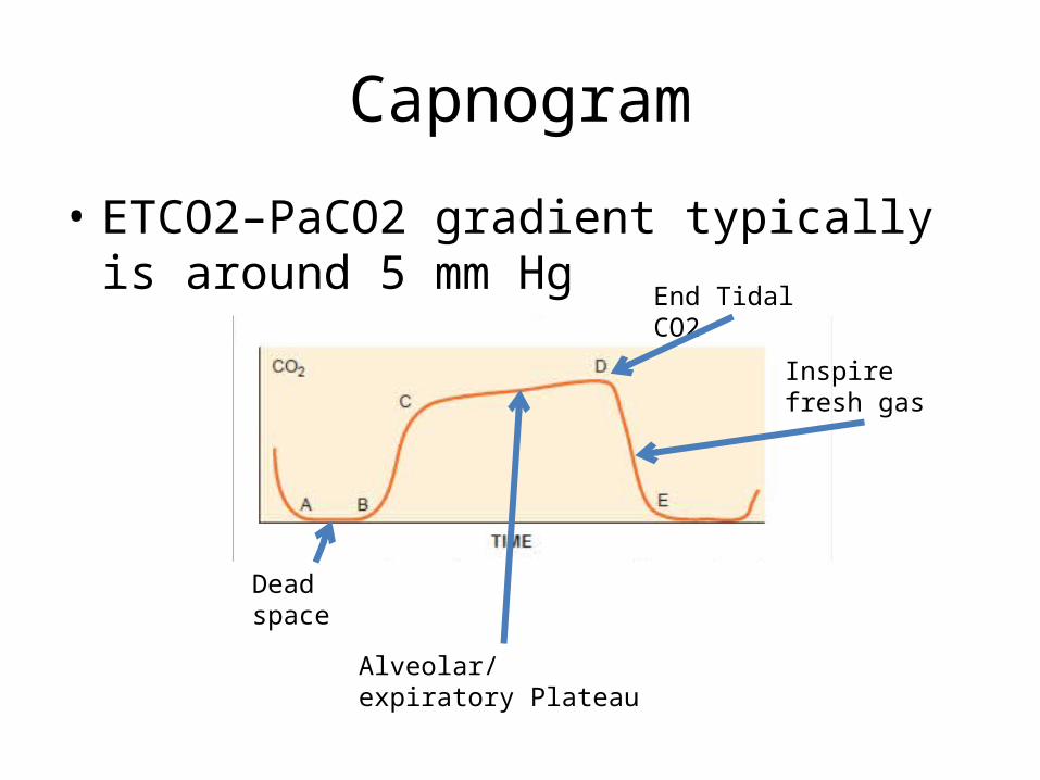
• ETCO2–PaCO2 gradient typically is around 5 mm Hg
Dead space
Alveolar/expiratory Plateau
End Tidal CO2
Inspire fresh gas
Capnography
• Stable ETCO2 for 3 consecutive breaths:– Tube is not in the esophagus
• Stable ETCO2 ensures the presence of alveolar ventilation
• But does not indicate it is placed properly– It may be either endobronchial (must auscultate
breath ssounds) or placed proximally in the vocal cord (may easily be dislodged
Capnography
• Slow rate of upstroke– COPD– Acute airway obstruction
• Normally shaped but increased ETCO2– Alveolar hypoventilation– Increased CO2 production
• Transient increase in ETCO2– Tourniquet release– Bicarbonate administration– Insufflation of CO2 during laparoscopy
• Failure of baseline to return to 0– Rebreathing
Sudden drop in ETCO2
• May be due to:– Malposition of the ET tube into pharynx/
esophagus– Disruption of airway integrity– Disruption of sampling line– Pulmonary embolism– Severe hypotension– Cardiac arrest
Circulation
• Methods for Assessing Circulatory Function– ECG– Blood pressure and heart rate determined every 5
minutes– Contiunally evaluated by atleast:
Palpable pulse, Heart sounds, monitoring of a tracing of intraarterial pressure, ultrasound peripheral pulse monitoring, or pulse plethysmography or oximetry.
Blood Pressure Monitoring
• Indirect measurement of Arterial BP– Systolic BP– Diastolic BP– Mean Arterial Pressure
• Automated Oscillometry
Blood presssure Monitoring
• Mechanical errors (auscultatory)• Falsely High BP– Cuff too small (must be 40% of circumference)– Cuff too loose– Uneven compression of artery– Extremity is below heart level
Indwelling artrerial cannulationContinuous BP monitoringAccess for arterial Blood Sampling
Uses fluid filled tubing to transmit a pressure pulse wave to a pressure transducer.
BP Monitoring (Invasive)
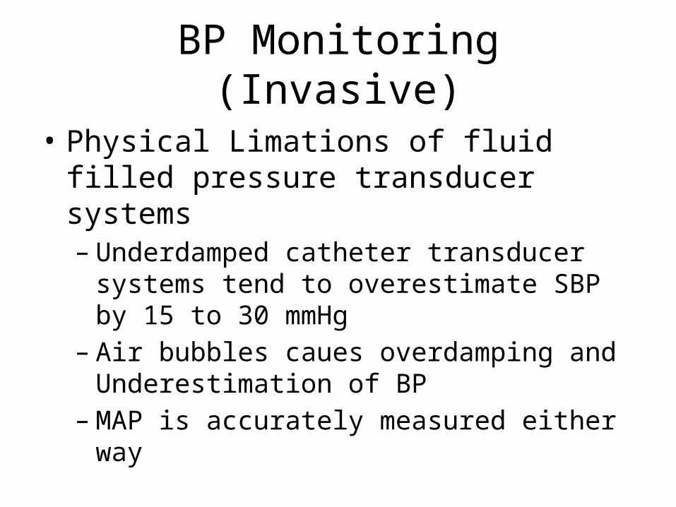
• Physical Limations of fluid filled pressure transducer systems– Underdamped catheter transducer systems tend
to overestimate SBP by 15 to 30 mmHg– Air bubbles caues overdamping and
Underestimation of BP– MAP is accurately measured either way
BP Monitoring (Invasive)
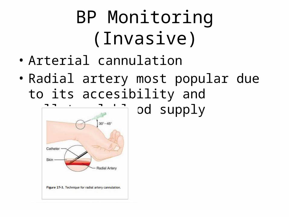
• Arterial cannulation• Radial artery most popular due to its
accesibility and collateral blood supply
BP Monitoring (Invasive)
• Complications– Traumatic: median nerve dysfunction, hematoma
formation and thrombosis– Abnormal radial artery blood flow after removal of
catheter ( normalization after 3 to 7 days)
BP Monitoring (Invasive)
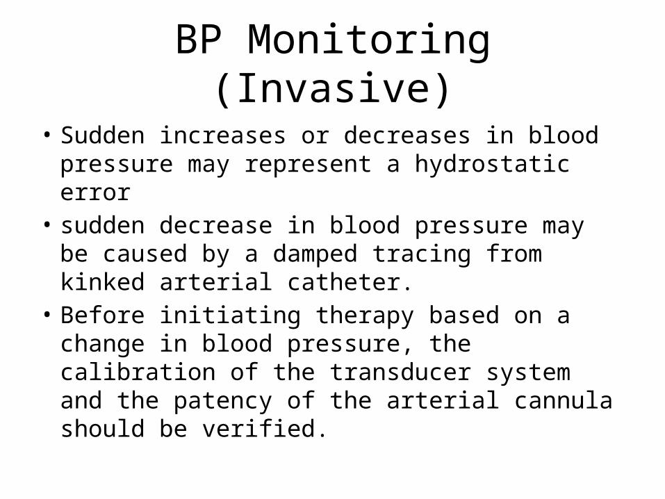
• Sudden increases or decreases in blood pressure may represent a hydrostatic error
• sudden decrease in blood pressure may be caused by a damped tracing from kinked arterial catheter.
• Before initiating therapy based on a change in blood pressure, the calibration of the transducer system and the patency of the arterial cannula should be verified.
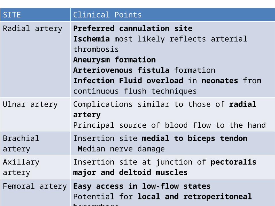
SITE Clinical Points
Radial artery Preferred cannulation site Ischemia most likely reflects arterial thrombosisAneurysm formation Arteriovenous fistula formation Infection Fluid overload in neonates from continuous flush techniques
Ulnar artery Complications similar to those of radial arteryPrincipal source of blood flow to the hand
Brachial artery Insertion site medial to biceps tendon Median nerve damage
Axillary artery Insertion site at junction of pectoralis major and deltoid muscles
Femoral artery Easy access in low-flow states Potential for local and retroperitoneal hemorrhage Catheter with increased length preferred