Lemke_Chap16.indd 508Lemke_Chap16.indd 508 12/9/2011 4:14:08 AM12/9/2011 4:14:08 AM
CHAPTER 16 / ANESTHETIC AGENTS: GENERAL AND LOCAL ANESTHETICS 509
INTRODUCTIONAnesthesia, defi ned as a loss of sensation with or with-out loss of consciousness, can be effectively achieved with a wide range of drugs with very diverse chemical struc-tures. The list of such compounds includes not only the classic anesthetic agents, such as the general and local anesthetics, but also many central nervous system (CNS) depressants, such as analgesics, sedative/hypnotics (bar-biturates and benzodiazepines), anticonvulsants, and skeletal muscle relaxants. Although various mechanisms of action are attributed to these agents, ultimately they all produce their anesthetic actions by interfering with con-duction in sensory neurons and sometimes also motor neurons. Many of these agents are routinely used today in clinical practice to facilitate surgical and medical pro-cedures. This chapter will focus on those agents typically classifi ed as “general” and “local” anesthetics.
GENERAL ANESTHETICSPrior to the mid-1800s, pain-producing surgical and dental procedures typically were undertaken without the aid of effective anesthetic agents. Chemical methods available at the time included intoxication with ethanol, hashish (cannabis), or opium, whereas physical meth-ods included packing a limb in ice, creating ischemic conditions with tourniquets, inducing unconsciousness by a blow to the head, or the most common technique, employing strong-armed assistants to hold down the help-less patient during the entire painful surgical procedure. Additionally, at this time, many practicing physicians had been erroneously taught that pain was a requirement for effective healing; therefore, the observation of a patient in terrible pain was viewed as part of the normal healing process. These factors, along with the lack of knowledge regarding aseptic techniques or the availability of suit-able infection-fi ghting agents, made surgical procedures a last resort approach to treating disease.
There have been many accounts of the fi rst dem-onstration by the Hartford dentist Horace Wells of the use of nitrous oxide as a general anesthetic for surgery in 1844. Wells fi rst observed the anesthetic actions of
nitrous oxide at a public demonstration of “laughing gas.” One of the volunteers, a pharmacy clerk named Samuel Cooley, injured his leg while under the infl uence of this gas and appeared to experience no pain. The next day, Wells inhaled the gas himself and, with the aid of a colleague, had one of his own teeth extracted without any sensation of pain. Wells then began routinely using nitrous oxide for dental procedures in his own practice. In 1845, he attempted to demonstrate the anesthetic effects of nitrous oxide at the Massachusetts General Hospital in Boston. This demonstration was considered to be a failure, however, because the patient cried out in the middle of the procedure. Following this unfortu-nate incident, the use of nitrous oxide was minimal until it resurfaced in dental practice during the mid-1860s, when it was combined with oxygen and made available in steel cylinders. This gas is still commonly used today, especially in combination with other anesthetic and anal-gesic agents.
The general anesthetic that gained greatest popularity shortly after the failed demonstration of Wells was diethyl ether. William Morton, a Boston dentist, was familiar at the time with the use of nitrous oxide by Wells. He also had heard of the interesting effects of diethyl ether and began to experiment on animals and himself with this volatile liquid. In 1846, he was allowed an opportunity to demonstrate the anesthetic actions of diethyl ether at, again, the Massachusetts General Hospital. In the famed “Ether Dome,” which still stands today, Morton admin-istered diethyl ether with a specially designed delivery device to the nervous patient, and the surgical proce-dure was performed without apparent pain. Following this demonstration, word of its success spread quickly, and soon, dental and medical practices throughout the United States and Europe were employing diethyl ether as a general anesthetic agent. Today, diethyl ether is no longer used in procedures because of its toxicity and dangerous physical properties (e.g., it is fl ammable and explosive!).
Other general anesthetic agents that enjoyed early pop-ularity were chloroform and cyclopropane. Chloroform vapor depresses the CNS of a patient, allowing a doctor to perform various otherwise painful surgical procedures.
SCENARIOPaul Arpino, R.Ph.
CDL is a 70-year-old obese man scheduled for carpal tunnel sur-gery. A review of his medical file indicates a history of obstruc-tive sleep apnea and benign prostatic hypertrophy (BPH). CDL sleeps with a continuous positive pressure airway device and his BPH is treated with tamsulosin, 0.4 mg daily. Given that patients with sleep apnea are at high risk for respiratory depression, the clinical team decides that a peripheral nerve block would be a better alternative to both neuraxial and general anesthesia.
During the preoperative assessment before the scheduled day of surgery, the team discovers that CDL has an undefined allergy to procaine (Novocain) and that he experienced severe blistering after a dental procedure many years ago and was told he cannot receive “drugs like Novocain again.”
(The reader is directed to the clinical solution and chemical analy-sis of this case at the end of the chapter).
Lemke_Chap16.indd 509Lemke_Chap16.indd 509 12/9/2011 4:14:09 AM12/9/2011 4:14:09 AM
510 PART III / PHARMACODYNAMIC AGENTS
In 1847, the Scottish obstetrician James Young Simpson fi rst used chloroform for general anesthesia during child-birth. The use of chloroform during surgery expanded rapidly thereafter in Europe. In the United States, chlo-roform replaced diethyl ether as an anesthetic at the beginning of the 20th century; however, it was quickly aban-doned due to its cardio and CNS toxicity. Cyclopropane is a hydrocarbon with anesthetic properties like diethyl ether, except it is also explosive and is no longer used. As described later in this chapter, the inhalational general anesthetic agents used today are typically hydrocarbons and halogenated ethers (Cl, Br, or F); nitrous oxide is the exception. Table 16.1 lists the characteristics of the “ideal” general anesthetic agent. Unfortunately, the agent that fulfi lls all these characteristics is currently unknown.
Stages of General AnesthesiaThe ideal general anesthetic state is characterized by a loss of all sensations and includes analgesia and muscle relaxation. Neuronal depression in specifi c areas of the CNS is believed to be largely responsible for such an anesthetic state. The areas involved include many corti-cal regions that are represented by excitatory pyramidal cells and inhibitory/excitatory stellate cells. Excitation of the pyramidal cells helps to maintain consciousness, whereas the degree of inhibition or excitation of stellate cells determines the overall activity level of the pyrami-dal cells with which they synapse. As the concentration of the anesthetic agent increases in the brain, the degree of overall neuronal depression also increases, resulting in progressively deeper stages of anesthesia. Based on observations using diethyl ether, Guedel in 1920 origi-nally described this progression as four distinct stages,
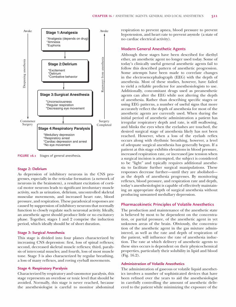
and Gillespie subsequently further subdivided these stages (Fig. 16.1), as described in the following sections.
Stage 1: AnalgesiaCharacterized by a mild depression of higher cortical neurons, this stage is suitable for minor surgical pro-cedures that do not require signifi cant neuromuscular relaxation. Depression of thalamic centers probably accounts for the observed analgesia, because many of the neuronal systems that mediate pain sensation traverse through this anatomic area. Some general anesthetic agents do not possess signifi cant analgesic activity, but they all produce a loss of consciousness that, in turn, can produce some degree of insensitivity to painful stimuli.
Anesthetics are a structurally diverse class of medications that
enable clinicians to perform sur-gery and other noxious procedures.
Understanding the essential components of the anesthetic state (i.e., immobilization, analgesia, and amnesia) as well as the medicinal chemistry of the various anesthetic agents allows the clinician to optimize therapy to meet patient specific needs. The patient undergoing a minimally invasive ambulatory surgical procedure may only require a local anesthetic with adjunc-tive pain control. Alternatively, patients undergoing a major surgi-cal procedure may require general anesthesia with several different classes of anesthetics as well as several adjunctive medications to counteract deleterious emergence reactions related to the anesthet-ics. In both cases, a thorough understanding of the basic chemical properties of the drugs and their respective mechanisms of action will prove invaluable to making appropriate clinical decisions.
The practice of anesthesia is typically not considered to be therapeutic; therefore, the practice as well as the development
of new agents is aimed at reducing adverse reactions, main-taining optimal physiologic conditions during procedures, and minimizing postoperative complications related to the proce-dure itself. The study of medicinal chemistry gives us hope for future treatment options, and knowledge of structure-activity relationships fosters the development of new medi-cations and administration techniques. New generations of drugs are being created by modifying the structures of exist-ing compounds to improve the side effect and pharmacoki-netic profiles. Clinicians will have to stay up to date with new developments in anesthesia practices, the molecular actions of anesthetics, and the pharmacokinetic properties of the drugs to provide the best therapeutic outcomes for patients.
Paul Arpino, RPhHarvard Medical SchoolDepartment of PharmacyMassachusetts General HospitalBoston, MA
CLINICAL SIGNIFICANCE
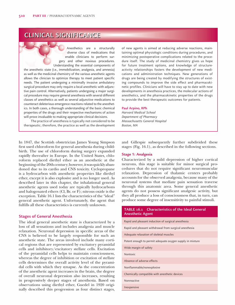
TABLE 16.1 Characteristics of the Ideal General Anesthetic Agent
Rapid and pleasant induction of surgical anesthesia
Rapid and pleasant withdrawal from surgical anesthesia
Adequate relaxation of skeletal muscles
Potent enough to permit adequate oxygen supply in mixture
Wide margin of safety
Nontoxic
Absence of adverse effects
Nonflammable/nonexplosive
Chemically compatible with anesthetic devices
Nonreactive
Inexpensive
Lemke_Chap16.indd 510Lemke_Chap16.indd 510 12/9/2011 4:14:09 AM12/9/2011 4:14:09 AM
CHAPTER 16 / ANESTHETIC AGENTS: GENERAL AND LOCAL ANESTHETICS 511
Stage 2: DeliriumAs depression of inhibitory neurons in the CNS pro-gresses, especially in the reticular formation (a network of neurons in the brainstem), a resultant excitation of corti-cal motor neurons leads to signifi cant involuntary muscle activity, such as urination, delirium, uncontrolled skeletal muscular movements, and increased heart rate, blood pressure, and respiration. These paradoxical responses are caused by suppression of inhibitory neurons that normally function to closely regulate such neuronal activity. Ideally, an anesthetic agent should produce little or no excitatory phase. Together, stages 1 and 2 comprise the induction period, which ideally should be of short duration.
Stage 3: Surgical AnesthesiaThis stage is divided into four planes characterized by increasing CNS depression: fi rst, loss of spinal refl exes; second, decreased skeletal muscle refl exes; third, paraly-sis of intercostal muscles; and fourth, loss of most muscle tone. Stage 3 is also characterized by regular breathing, a loss of many refl exes, and roving eyeball movements.
Stage 4: Respiratory ParalysisCharacterized by respiratory and vasomotor paralysis, this stage represents an overdose or toxic level that should be avoided. Normally, this stage is never reached, because the anesthesiologist is careful to monitor abdominal
respiration to prevent apnea, blood pressure to prevent hypotension, and heart rate to prevent asystole (a state of no cardiac electrical activity).
Modern General Anesthetic AgentsAlthough these stages have been described for diethyl ether, an anesthetic agent no longer used today. Some of today’s clinically useful general anesthetic agents fail to follow this described pattern of anesthetic progression. Some attempts have been made to correlate changes in the electroencephalograph (EEG) with the depth of anesthesia. Most of these studies, however, have failed to yield a reliable predictor for anesthesiologists to use. Additionally, concomitant drugs used as preanesthetic agents can alter the EEG while not altering the depth of anesthesia. Rather than describing specifi c stages or using EEG patterns, a number of useful signs that more accurately refl ect the depth of anesthesia for most of the anesthetic agents are currently used. When during the initial period of anesthetic administration a patient has irregular respiratory depth and rate, is still swallowing, and blinks the eyes when the eyelashes are touched, the desired surgical stage of anesthesia likely has not been reached. However, when a loss of the eyelash refl ex occurs along with rhythmic breathing, however, a level of adequate surgical anesthesia has generally begun. If a patient at this stage exhibits elevations in blood pressure, increased respiration rate, or increased jaw tension when a surgical incision is attempted, the subject is considered to be “light” and typically requires additional anesthe-sia to facilitate further surgical manipulations. These responses decrease further—until they are abolished—as the depth of anesthesia progresses. By monitoring refl exes, blood pressure, and respiration rate and depth, today’s anesthesiologist is capable of effectively maintain-ing an appropriate depth of surgical anesthesia without producing unwanted medullary depression.
Pharmacokinetic Principles of Volatile AnestheticsThe production and maintenance of the anesthetic state is believed by most to be dependent on the concentra-tion, or partial pressure, of the anesthetic agent in yet unknown areas of the brain. Obviously, the concentra-tion of the anesthetic agent in the gas mixture admin-istered, as well as the rate and depth of respiration of the patient, will infl uence the rate of anesthesia induc-tion. The rate at which delivery of anesthetic agents to these sites occurs is dependent on their physicochemical properties, particularly their solubility in lipid and blood (Fig. 16.2).
Administration of Volatile AnestheticsThe administration of gaseous or volatile liquid anesthet-ics involves a number of sophisticated devices that have been refi ned over the years to aid the anesthesiologist in carefully controlling the amount of anesthetic deliv-ered to the patient while minimizing the exposure of the
Stage 1:Analgesia
*Analgesia (depends on drug)*Amnesia*Euphoria
Stage 2:Delirium
*Excitement*Delirium*Combative behavior
Stage 3:Surgical Anesthesia
*Unconsciousness*Regular respiration*Decreasing eye movement
Stage 4:Respiratory Paralysis
*Medullary depression*Respiratory arrest*Cardiac depression and arrest*No eye movement
Awake
Commence Surgery
SurgeryCompleted
Awake
Dee
peni
ng a
nest
hesia
Rec
over
yt fro
m a
nest
hesia
FIGURE 16.1 Stages of general anesthesia.
Lemke_Chap16.indd 511Lemke_Chap16.indd 511 12/9/2011 4:14:09 AM12/9/2011 4:14:09 AM
512 PART III / PHARMACODYNAMIC AGENTS
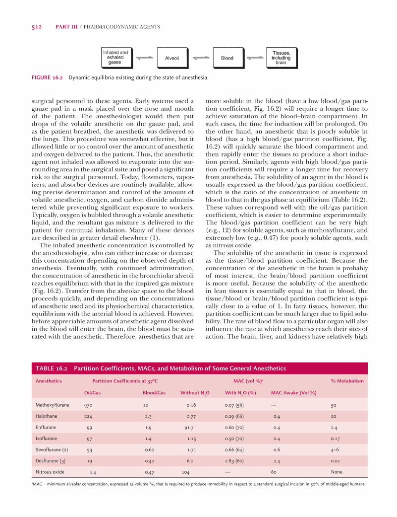
more soluble in the blood (have a low blood/gas parti-tion coeffi cient, Fig. 16.2) will require a longer time to achieve saturation of the blood–brain compartment. In such cases, the time for induction will be prolonged. On the other hand, an anesthetic that is poorly soluble in blood (has a high blood/gas partition coeffi cient, Fig. 16.2) will quickly saturate the blood compartment and then rapidly enter the tissues to produce a short induc-tion period. Similarly, agents with high blood/gas parti-tion coeffi cients will require a longer time for recovery from anesthesia. The solubility of an agent in the blood is usually expressed as the blood/gas partition coeffi cient, which is the ratio of the concentration of anesthetic in blood to that in the gas phase at equilibrium (Table 16.2). These values correspond well with the oil/gas partition coeffi cient, which is easier to determine experimentally. The blood/gas partition coeffi cient can be very high (e.g., 12) for soluble agents, such as methoxyfl urane, and extremely low (e.g., 0.47) for poorly soluble agents, such as nitrous oxide.
The solubility of the anesthetic in tissue is expressed as the tissue/blood partition coeffi cient. Because the concentration of the anesthetic in the brain is probably of most interest, the brain/blood partition coeffi cient is more useful. Because the solubility of the anesthetic in lean tissues is essentially equal to that in blood, the tissue/blood or brain/blood partition coeffi cient is typi-cally close to a value of 1. In fatty tissues, however, the partition coeffi cient can be much larger due to lipid solu-bility. The rate of blood fl ow to a particular organ will also infl uence the rate at which anesthetics reach their sites of action. The brain, liver, and kidneys have relatively high
surgical personnel to these agents. Early systems used a gauze pad in a mask placed over the nose and mouth of the patient. The anesthesiologist would then put drops of the volatile anesthetic on the gauze pad, and as the patient breathed, the anesthetic was delivered to the lungs. This procedure was somewhat effective, but it allowed little or no control over the amount of anesthetic and oxygen delivered to the patient. Thus, the anesthetic agent not inhaled was allowed to evaporate into the sur-rounding area in the surgical suite and posed a signifi cant risk to the surgical personnel. Today, fl owmeters, vapor-izers, and absorber devices are routinely available, allow-ing precise determination and control of the amount of volatile anesthetic, oxygen, and carbon dioxide adminis-tered while preventing signifi cant exposure to workers. Typically, oxygen is bubbled through a volatile anesthetic liquid, and the resultant gas mixture is delivered to the patient for continual inhalation. Many of these devices are described in greater detail elsewhere (1).
The inhaled anesthetic concentration is controlled by the anesthesiologist, who can either increase or decrease this concentration depending on the observed depth of anesthesia. Eventually, with continued administration, the concentration of anesthetic in the bronchiolar alveoli reaches equilibrium with that in the inspired gas mixture (Fig. 16.2). Transfer from the alveolar space to the blood proceeds quickly, and depending on the concentrations of anesthetic used and its physiochemical characteristics, equilibrium with the arterial blood is achieved. However, before appreciable amounts of anesthetic agent dissolved in the blood will enter the brain, the blood must be satu-rated with the anesthetic. Therefore, anesthetics that are
FIGURE 16.2 Dynamic equilibria existing during the state of anesthesia.
TABLE 16.2 Partition Coefficients, MACs, and Metabolism of Some General Anesthetics
Anesthetics Partition Coefficients at 37°C MAC (vol %)a % Metabolism
Oil/Gas Blood/Gas Without N2O With N2O (%) MAC-Awake (Vol %)
Methoxyflurane 970 12 0.16 0.07 (56) — 50
Halothane 224 2.3 0.77 0.29 (66) 0.4 20
Enflurane 99 1.9 91.7 0.60 (70) 0.4 2.4
Isoflurane 97 1.4 1.15 0.50 (70) 0.4 0.17
Sevoflurane (2) 53 0.60 1.71 0.66 (64) 0.6 4–6
Desflurane (3) 19 0.42 6.0 2.83 (60) 2.4 0.02
Nitrous oxide 1.4 0.47 104 — 60 None
aMAC = minimum alveolar concentration, expressed as volume %, that is required to produce immobility in respect to a standard surgical incision in 50% of middle-aged humans.
Lemke_Chap16.indd 512Lemke_Chap16.indd 512 12/9/2011 4:14:10 AM12/9/2011 4:14:10 AM
CHAPTER 16 / ANESTHETIC AGENTS: GENERAL AND LOCAL ANESTHETICS 513
responses to verbal commands are lost in 50% of the patients tested. At this concentration, amnesia and a loss of awareness are evident, and the patient is said to be in a state of hypnosis. The MAC-Awake occurs at concentra-tions signifi cantly lower (e.g., 50% to 75% lower) than those required for surgical anesthesia.
Theories About the Mechanisms of AnesthesiaMeyer-Overton TheoryIn the early 1900s, Hans Meyer and Charles Overton sug-gested that the potency of a substance as an anesthetic was directly related to its lipid solubility, or oil/gas partition coeffi cient (Table 16.2) (3–5). This has commonly been referred to as the “unitary theory of anesthesia.” They used olive oil, octanol, and other “membrane-like” lipids to determine the lipid solubility of the agents available at that time. Compounds with high lipid solubility required lower concentrations (i.e., lower MAC) to produce anes-thesia. It was later postulated that the interaction of the anesthetic molecules with a hydrophobic portion of the nerve membrane caused a distortion of the nerve mem-brane near the channels that conducted Na+, those that mediated the fast action potentials and neuronal cell fi r-ing. The presence of this critical volume of anesthetic dissolved within the membrane caused the membrane to “bloat” and cause a “squeezing in” on the Na+ channel to interfere with Na+ conductance and normal neuronal depolarization. In support of this theory, it was found that at high pressures (40 to 100 atmospheres), the anes-thetic actions of many of these agents could be partially reversed, presumably by compressing membranes back to their original conformation. Arguing against this theory, however, is the fi nding that not all highly lipid-soluble substances are capable of producing anesthesia. Additionally, more recent work involving protein–drug interactions has seriously challenged this theory. Today, more than 150 years after the fi rst demonstration of the use of a volatile anesthetic agent, most theories about the mechanisms of anesthesia suggest that multiple selective
blood fl ows, whereas skeletal muscle at rest and fat tissues have relatively poor blood fl ows and would be expected to accumulate less of the anesthetic agent.
Reversal of the anesthetic state and recovery requires a reduction in the concentration of the anesthetic in the brain. This is achieved by stopping the delivery of the anesthetic through the lungs. As the patient continues to breathe, the anesthetic is continually removed, which favors diffusion from the brain to the blood, to the alve-oli, and fi nally, to the expired air. The rate at which this occurs generally parallels that of induction, because the solubility of an agent in the brain and in the blood deter-mines how quickly these compartments will return to a preanesthetic state. The main route of elimination is via the expired air, which can be mostly captured by gas-scavenging devices with absorbers, but some metabolism of these agents does take place (discussed below).
Minimum Alveolar ConcentrationThe minimum alveolar concentration (MAC) is defi ned as the concentration at 1 atmosphere of anesthetic in the alveoli that is required to produce immobility in 50% of adult patients subjected to a standard surgical incision. A further increase to 1.3 MAC will frequently cause immo-bility in 99% of patients. At equilibrium, the concentra-tion (or partial pressure) of an anesthetic in the alveoli is equal to that in the brain, and it is this concentration in the brain that most closely refl ects the concentration at the site responsible for the anesthetic actions. Thus, the MAC is often used as a measure of the potency of individual anesthetic agents. The MAC of many of the volatile and gaseous anesthetics in use today is shown in Table 16.2.
When anesthetic agents are used in combination, the MACs for inhaled anesthetics are simply additive. For instance, the anesthetic depth achieved with 0.5 MAC of enfl urane plus 0.5 MAC of nitrous oxide is equiva-lent to that produced by 1.0 MAC of either agent alone. The combination of two anesthetics is a very common practice, because this technique allows for a reduction in the patient exposure to any one of the individual agents, thereby decreasing the likelihood of adverse reactions.
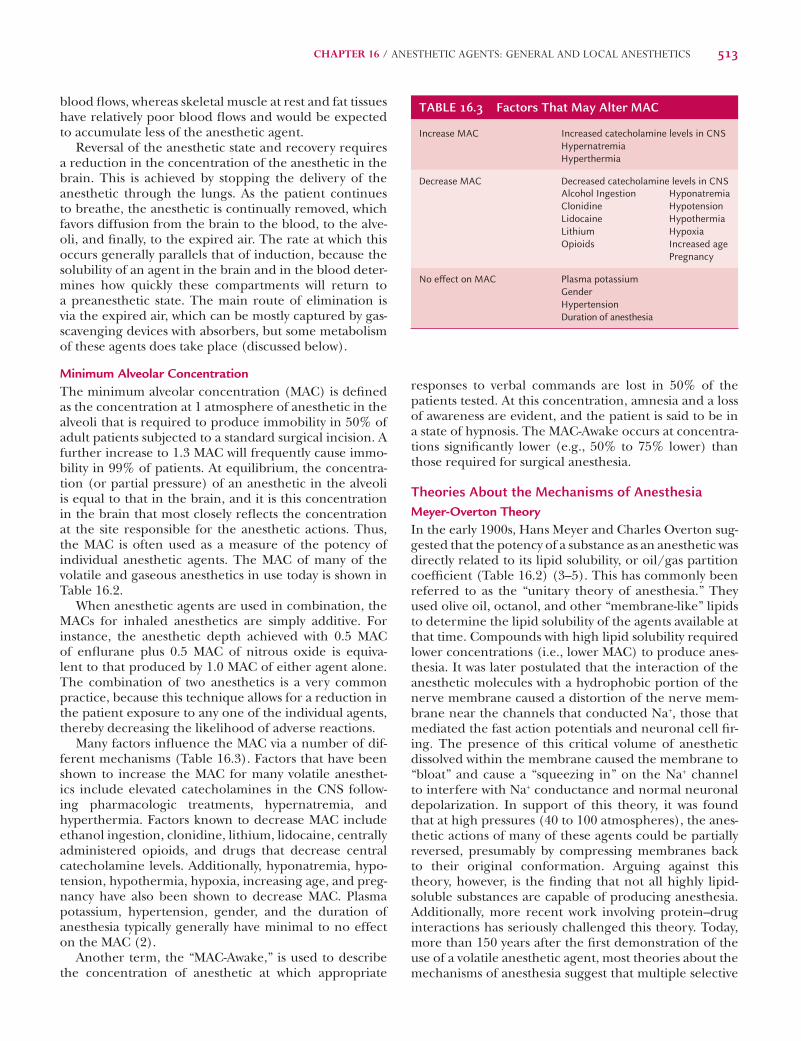
Many factors infl uence the MAC via a number of dif-ferent mechanisms (Table 16.3). Factors that have been shown to increase the MAC for many volatile anesthet-ics include elevated catecholamines in the CNS follow-ing pharmacologic treatments, hypernatremia, and hyperthermia. Factors known to decrease MAC include ethanol ingestion, clonidine, lithium, lidocaine, centrally administered opioids, and drugs that decrease central catecholamine levels. Additionally, hyponatremia, hypo-tension, hypothermia, hypoxia, increasing age, and preg-nancy have also been shown to decrease MAC. Plasma potassium, hypertension, gender, and the duration of anesthesia typically generally have minimal to no effect on the MAC (2).
Another term, the “MAC-Awake,” is used to describe the concentration of anesthetic at which appropriate
TABLE 16.3 Factors That May Alter MAC
Increase MAC Increased catecholamine levels in CNSHypernatremiaHyperthermia
Decrease MAC Decreased catecholamine levels in CNSAlcohol IngestionClonidineLidocaineLithiumOpioids
No effect on MAC Plasma potassiumGenderHypertensionDuration of anesthesia
Lemke_Chap16.indd 513Lemke_Chap16.indd 513 12/9/2011 4:14:10 AM12/9/2011 4:14:10 AM
514 PART III / PHARMACODYNAMIC AGENTS
curves observed, 2) the stereochemical requirements of various anesthetics, 3) the fi nding that increasing the molecular weight and corresponding lipid solubility of an anesthetic can actually decrease or abolish anesthetic activity, and 4) the fi nding that specifi c ion channels and neurotransmitter receptor systems are required for most of the observed effects of the anesthetics. What appears to be emerging as a central theme for the mechanism of action of general anesthetics involves the interaction of the anesthetics with receptors that allosterically modulate the activity of ion channels (e.g., chloride and potassium) or with the ion channel directly (e.g., sodium). Many other mechanisms are also emerging to help explain the mechanisms of action of the general anesthetics.
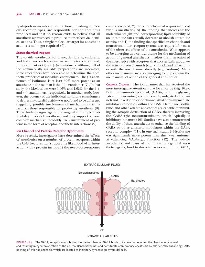
CHLORIDE CHANNEL The ion channel that has received the most investigative attention is that for chloride (Fig. 16.3). Both the g-aminobutyric acidA (GABAA) and the glycineA (strychnine-sensitive) receptors are ligand-gated ion chan-nels and linked to chloride channels that normally mediate inhibitory responses within the CNS. Halothane, isofl u-rane, and other volatile anesthetics are capable of inhibit-ing the synaptic destruction of GABA, thereby increasing the GABAergic neurotransmission, which typically is inhibitory in nature (10). Studies have also demonstrated the ability of these anesthetics to enhance the binding of GABA or other allosteric modulators within the GABA receptor complex (11). In one such study, (+)-isofl urane was signifi cantly more potent than the (−)-enantiomer at enhancing GABAergic function (12). The volatile anesthetics, and many of the intravenous general anes-thetic agents, bind to discrete cavities within the GABAA
lipid–protein membrane interactions, involving numer-ous receptor types, are responsible for the anesthesia produced and that no reason exists to believe that all anesthetic agents need to produce their effects via identi-cal actions. Thus, a single molecular target for anesthetic actions is no longer required (6).
Stereochemical AspectsThe volatile anesthetics isofl urane, desfl urane, enfl urane, and halothane each contain an asymmetric carbon and, thus, can exist as (+)- or (−)-enantiomers. Although all of the commercially available preparations are racemates, some researchers have been able to determine the anes-thetic properties of individual enantiomers. The (+)-enan-tiomer of isofl urane is at least 50% more potent as an anesthetic in the rat than is the (−)-enantiomer (7). In that study, the MAC values were 1.06% and 1.62% for the (+)- and (−)-enantiomers, respectively. In another study, how-ever, the potency of the individual isofl urane enantiomers to depress myocardial activity was not found to be different, suggesting possible involvement of mechanisms dissimi-lar from those responsible for producing anesthesia (8). These fi ndings argue against the original and simple lipid-solubility theory of anesthesia, and they support a more complex mechanism, probably likely involvement of pro-teins in the form of receptor–anesthetic interactions (9).
Ion Channel and Protein Receptor HypothesesMore recently, investigators have determined the effects of anesthetics on a number of protein receptors within the CNS. Features that support the likelihood of an inter-action with a protein include 1) the steep dose–response
EXTRACELLULAR FLUID
INTRACELLULAR FLUID
Cl-
GABA
Barbituates
Benzodiazepines
FIGURE 16.3 The GABAA receptor controls the chloride ion channel. GABA binds to its receptor, opening the chloride ion channel and resulting in hyperpolarization of the neuron. Benzodiazepines and barbiturates can produce anesthesia by allosterically enhancing GABA opening of chloride channels, which are located at inhibitory synapses on pyramidal cells.
Lemke_Chap16.indd 514Lemke_Chap16.indd 514 12/9/2011 4:14:10 AM12/9/2011 4:14:10 AM
CHAPTER 16 / ANESTHETIC AGENTS: GENERAL AND LOCAL ANESTHETICS 515
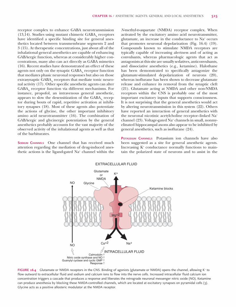
N-methyl-d-aspartate (NMDA) receptor complex. When activated by the excitatory amino acid neurotransmitter, glutamate, an increase in the conductance to Na+ occurs that promotes neuronal depolarization (Fig. 16.4) (19). Compounds known to stimulate NMDA receptors are typically capable of increasing alertness and of acting as convulsants, whereas pharmacologic agents that act as antagonists at this site are usually sedatives, anticonvulsants, and dissociative anesthetics (e.g., ketamine). Halothane has been demonstrated to specifi cally antagonize the glutamate-stimulated depolarization of neurons (20), whereas isofl urane has been shown to decrease glutamate release and enhance its removal from the synaptic cleft (21). Glutamate acting at NMDA and other non-NMDA receptors within the CNS is probably one of the most important excitatory inputs that supports consciousness. It is not surprising that the general anesthetics would act by altering neurotransmission in this system (22). Others have reported an interaction of general anesthetics with the neuronal nicotinic acetylcholine receptor–linked Na+ channel (23). Voltage-gated Na+ channels in small, nonmy-elinated hippocampal axons also appear to be inhibited by general anesthetics, such as isofl urane (24).
POTASSIUM CHANNELS Potassium ion channels have also been suggested as a site for general anesthetic agents. Increasing K+ conductance normally functions to main-tain the polarized state of neurons and to assist in the
receptor complex to enhance GABA neurotransmission (13,14). Studies using mutant chimeric GABAA receptors have identifi ed a specifi c binding site for general anes-thetics located between transmembrane segments 2 and 3 (15). At therapeutic concentrations, just about all of the inhalational general anesthetics are capable of enhancing GABAergic function, whereas at considerably higher con-centrations, many also can act directly as GABA mimetics (16). Recent studies have demonstrated an effect of these agents not only on the synaptic GABAA receptor function that mediates phasic neuronal responses but also on those extrasynaptic GABAA receptors that mediate tonic neuro-nal activity (17). Other specifi c anesthetic agents can alter GABAA receptor function via different mechanisms. For instance, propofol, an intravenous general anesthetic, appears to slow the desensitization of the GABAA recep-tor during bouts of rapid, repetitive activation at inhibi-tory synapses (18). Most of these agents also potentiate the actions of glycine, the other important inhibitory amino acid neurotransmitter (16). The combination of GABAergic and glycinergic potentiation by the general anesthetics probably accounts for the vast majority of the observed activity of the inhalational agents as well as that of the barbiturates.
SODIUM CHANNELS One channel that has received much attention regarding the mediation of drug-induced anes-thetic actions is the ligand-gated Na+ channel within the
Ketamine blocks
Glycine
Glutamateor
NMDA
EXTRACELLULAR FLUID
NO
NO Na+
K+
Ca+2
Mg+2
INTRACELLULAR FLUIDCalmodulin
Nitric oxide synthase and NOGuanylyl cyclase and cyclic GMP
Response
FIGURE 16.4 Glutamate or NMDA receptors in the CNS. Binding of agonists (glutamate or NMDA) opens the channel, allowing K+ to flow outward to extracellular fluid and sodium and calcium ions to flow into the nerve cells. Increased intracellular fluid calcium ion concentration triggers a cascade that produces a response and liberates the retrograde neuronal messenger nitric oxide (NO). Ketamine can produce anesthesia by blocking these NMDA-controlled channels, which are located at excitatory synapses on pyramidal cells (3). Glycine acts as a positive allosteric modulator at the NMDA receptor.
Lemke_Chap16.indd 515Lemke_Chap16.indd 515 12/9/2011 4:14:10 AM12/9/2011 4:14:10 AM
516 PART III / PHARMACODYNAMIC AGENTS
agents to be introduced as a general anesthetic, has high potency with signifi cant analgesic and neuromuscular relaxing effects. This agent is extremely fl ammable, and when mixed with air, oxygen, or nitrous oxide, is explo-sive. Induction with diethyl ether is very slow; signifi cant time is spent progressing through the delirium stage. Irritation of the respiratory tract by diethyl ether can lead to excessive bronchial secretions, complicating adequate ventilation. In addition to its unpleasant induction and adverse effects, recovery is similarly prolonged and can be accompanied by vomiting. These pharmacologic and physical characteristics of diethyl ether have limited the utility of this anesthetic in humans.
SHORT-CHAIN HYDROCARBONS Many of the short-chain alkanes, alkenes, and alkynes are capable of producing an anesthetic state when administered to patients. Potency generally increases as chain length increases. However, because of their fl ammability and increased propensity to cause cardiovascular toxicity, these nonsubstituted hydrocarbons are not useful as anesthetic agents.
CHLOROFORM Another of the earlier anesthetic agents to be used was chloroform (CHCl3). This halogenated hydro-carbon was fi rst offi cially used in the United States in 1847; however, its toxicity seriously limited its utility. The addi-tion of halogens to the hydrocarbon backbone increases potency and volatility, as well as decreases fl ammability. Similar effects are also observed with such substitutions on ethers. As an anesthetic agent, chloroform is very potent and possesses signifi cant analgesic and neuromuscular
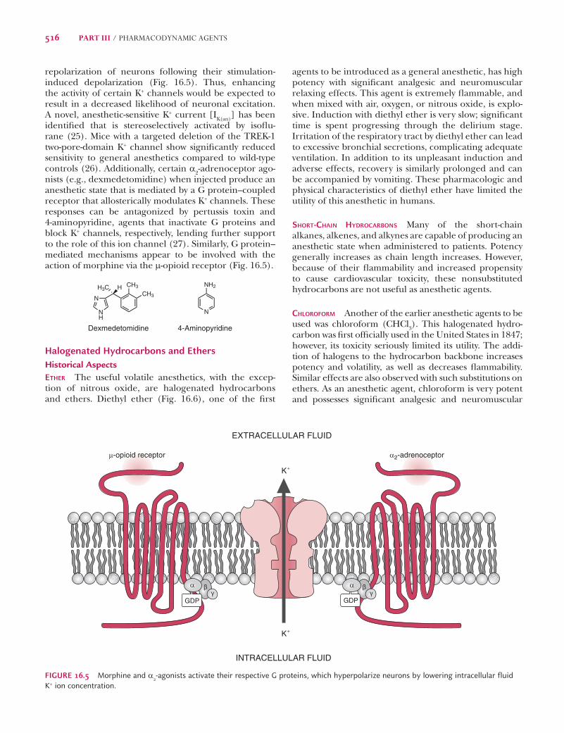
repolarization of neurons following their stimulation-induced depolarization (Fig. 16.5). Thus, enhancing the activity of certain K+ channels would be expected to result in a decreased likelihood of neuronal excitation. A novel, anesthetic-sensitive K+ current [IK(an)] has been identifi ed that is stereoselectively activated by isofl u-rane (25). Mice with a targeted deletion of the TREK-1 two-pore-domain K+ channel show signifi cantly reduced sensitivity to general anesthetics compared to wild-type controls (26). Additionally, certain a2-adrenoceptor ago-nists (e.g., dexmedetomidine) when injected produce an anesthetic state that is mediated by a G protein–coupled receptor that allosterically modulates K+ channels. These responses can be antagonized by pertussis toxin and 4-aminopyridine, agents that inactivate G proteins and block K+ channels, respectively, lending further support to the role of this ion channel (27). Similarly, G protein–mediated mechanisms appear to be involved with the action of morphine via the m-opioid receptor (Fig. 16.5).
CH3
CH3N
NH
HH3C
Dexmedetomidine
N
NH2
4-Aminopyridine
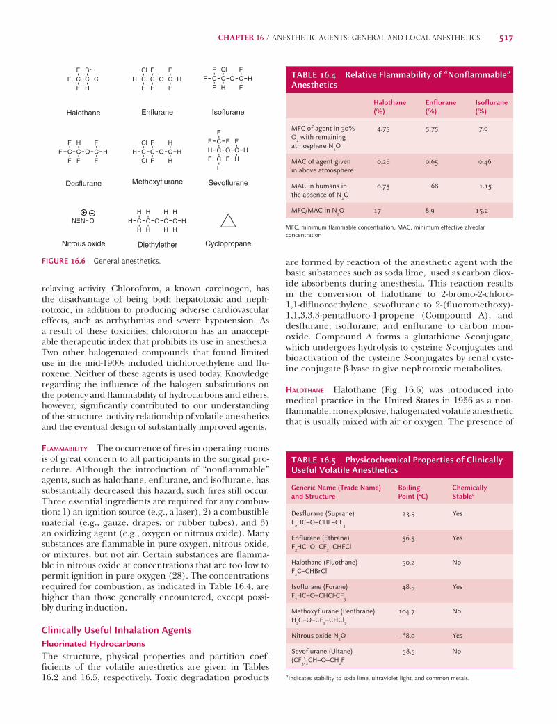
Halogenated Hydrocarbons and EthersHistorical AspectsETHER The useful volatile anesthetics, with the excep-tion of nitrous oxide, are halogenated hydrocarbons and ethers. Diethyl ether (Fig. 16.6), one of the fi rst
GDPGDP
K+
K+
EXTRACELLULAR FLUID
INTRACELLULAR FLUID
μ-opioid receptor α2-adrenoceptor
α α
FIGURE 16.5 Morphine and a2-agonists activate their respective G proteins, which hyperpolarize neurons by lowering intracellular fluid K+ ion concentration.
Lemke_Chap16.indd 516Lemke_Chap16.indd 516 12/9/2011 4:14:10 AM12/9/2011 4:14:10 AM
CHAPTER 16 / ANESTHETIC AGENTS: GENERAL AND LOCAL ANESTHETICS 517
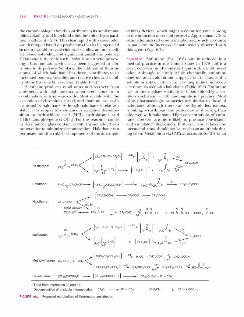
are formed by reaction of the anesthetic agent with the basic substances such as soda lime, used as carbon diox-ide absorbents during anesthesia. This reaction results in the conversion of halothane to 2-bromo-2-chloro-1,1-difl uoroethylene, sevofl urane to 2-(fl uoromethoxy)-1,1,3,3,3-pentafl uoro-1-propene (Compound A), and desfl urane, isofl urane, and enfl urane to carbon mon-oxide. Compound A forms a glutathione S-conjugate, which undergoes hydrolysis to cysteine S-conjugates and bioactivation of the cysteine S-conjugates by renal cyste-ine conjugate b-lyase to give nephrotoxic metabolites.
HALOTHANE Halothane (Fig. 16.6) was introduced into medical practice in the United States in 1956 as a non-fl ammable, nonexplosive, halogenated volatile anesthetic that is usually mixed with air or oxygen. The presence of
relaxing activity. Chloroform, a known carcinogen, has the disadvantage of being both hepatotoxic and neph-rotoxic, in addition to producing adverse cardiovascular effects, such as arrhythmias and severe hypotension. As a result of these toxicities, chloroform has an unaccept-able therapeutic index that prohibits its use in anesthesia. Two other halogenated compounds that found limited use in the mid-1900s included trichloroethylene and fl u-roxene. Neither of these agents is used today. Knowledge regarding the infl uence of the halogen substitutions on the potency and fl ammability of hydrocarbons and ethers, however, signifi cantly contributed to our understanding of the structure–activity relationship of volatile anesthetics and the eventual design of substantially improved agents.
FLAMMABILITY The occurrence of fi res in operating rooms is of great concern to all participants in the surgical pro-cedure. Although the introduction of “nonfl ammable” agents, such as halothane, enfl urane, and isofl urane, has substantially decreased this hazard, such fi res still occur. Three essential ingredients are required for any combus-tion: 1) an ignition source (e.g., a laser), 2) a combustible material (e.g., gauze, drapes, or rubber tubes), and 3) an oxidizing agent (e.g., oxygen or nitrous oxide). Many substances are fl ammable in pure oxygen, nitrous oxide, or mixtures, but not air. Certain substances are fl amma-ble in nitrous oxide at concentrations that are too low to permit ignition in pure oxygen (28). The concentrations required for combustion, as indicated in Table 16.4, are higher than those generally encountered, except possi-bly during induction.
Clinically Useful Inhalation AgentsFluorinated HydrocarbonsThe structure, physical properties and partition coef-fi cients of the volatile anesthetics are given in Tables 16.2 and 16.5, respectively. Toxic degradation products
FCFF
CBr
ClH
Halothane
ClCHF
CF
OF
CF
FH
Enflurane
FCFF
CCl
OH
CF
FH
Isoflurane
FCFF
CH
OF
CF
FH
Desflurane
ClCHCl
CF
OF
CH
HH
Methoxyflurane
C O CF
HH
C
CH
FF F
FF
F
Sevoflurane
N N
Nitrous oxide
OHCHH
CH
OH
CH
HCH
HH
Diethylether Cyclopropane
FIGURE 16.6 General anesthetics.
TABLE 16.4 Relative Flammability of “Nonflammable” Anesthetics
Halothane (%)
Enflurane (%)
Isoflurane (%)
MFC of agent in 30% O2 with remaining atmosphere N2O
TABLE 16.5 Physicochemical Properties of Clinically Useful Volatile Anesthetics
Generic Name (Trade Name) and Structure
Boiling Point (°C)
Chemically Stablea
Desflurane (Suprane) F2HC–O–CHF–CF3
23.5 Yes
Enflurane (Ethrane) F2HC–O–CF2–CHFCl
56.5 Yes
Halothane (Fluothane) F2C–CHBrCl
50.2 No
Isoflurane (Forane) F2HC–O–CHCl-CF3
48.5 Yes
Methoxyflurane (Penthrane) H2C–O–CF2–CHCl2
104.7 No
Nitrous oxide N2O –*8.0 Yes
Sevoflurane (Ultane) (CF3)2CH–O–CH2F
58.5 No
aIndicates stability to soda lime, ultraviolet light, and common metals.
Lemke_Chap16.indd 517Lemke_Chap16.indd 517 12/9/2011 4:14:11 AM12/9/2011 4:14:11 AM
518 PART III / PHARMACODYNAMIC AGENTS
delivery devices, which might account for some slowing of the induction onset and recovery. Approximately 20% of an administered dose is metabolized, which accounts, in part, for the increased hepatotoxicity observed with this agent (Fig. 16.7).
ENFLURANE Enfl urane (Fig. 16.6) was introduced into medical practice in the United States in 1973 and is a clear, colorless, nonfl ammable liquid with a mild, sweet odor. Although relatively stable chemically, enfl urane does not attack aluminum, copper, iron, or brass and is soluble in rubber, which can prolong induction/recov-ery times, as seen with halothane (Table 16.5). Enfl urane has an intermediate solubility in blood (blood/gas par-tition coeffi cient = 1.9) and signifi cant potency. Most of its pharmacologic properties are similar to those of halothane, although there can be slightly less nausea, vomiting, arrhythmias, and postoperative shivering than observed with halothane. High concentrations of enfl u-rane, however, are more likely to produce convulsions and circulatory depression. Enfl urane also relaxes the uterus and, thus, should not be used as an anesthetic dur-ing labor. Metabolism via CYP2E1 accounts for 2% of an
the carbon–halogen bonds contributes to its nonfl amma-bility, volatility, and high lipid solubility (blood/gas parti-tion coeffi cient = 2.3). This clear liquid with a sweet odor was developed based on predictions that its halogenated structure would provide chemical stability, an intermedi-ate blood solubility, and signifi cant anesthetic potency. Halothane is the only useful volatile anesthetic possess-ing a bromine atom, which has been suggested to con-tribute to its potency. Similarly, the addition of fl uorine atoms, of which halothane has three, contributes to its increased potency, volatility, and relative chemical stabil-ity of the hydrocarbon skeleton (Table 16.5).
Halothane produces rapid onset and recovery from anesthesia with high potency when used alone or in combination with nitrous oxide. Most metals, with the exception of chromium, nickel, and titanium, are easily tarnished by halothane. Although halothane is relatively stable, it is subject to spontaneous oxidative decompo-sition to hydrochloric acid (HCl), hydrobromic acid (HBr), and phosgene (COCl2). For this reason, it comes in dark, amber glass containers with thymol added as a preservative to minimize decomposition. Halothane can permeate into the rubber components of the anesthetic
CF3
F
OCHF2
CF3 CFH O CF2OHF3C H
O
F F
O+
**
F3C OCHF2
F
OHF3C F
O**
+
CF2CHClFO
F2HC CF2CClFOHO
F2HC F2HCO
CF2F
O
HC
+e-/-Br-
Minor pathway
+e-CF3 C
ClCl
OCO
OH C CO
OHHOO
HOCH2 CO
OH
F3C OCHF2
Cl
F3C OCHF2
Cl
OH
F3C H
O
F F
O
F3C Cl
O
+**
** -Cl-
H2O
C CO
OHHOO
Cl2HC CF2 O CH3
(CF3)2CHOH + F + CO2-
**Decomposition of unstable intermediates: 2F + CO2 ; 2F + HCOOH ;- -
F3C CHCl O CF2OH
Desflurane:
Enflurane:
Halothane:
Isoflurane:
Methoxyflurane:
Sevoflurane:
+
+
CHCl2COOHH2CO +-HF
-HCl
-F-
-2F--Cl-
H2O
H2O
-HF
-F-
H2O
-HCl -F-
-2F-
-Cl-
-HCl
*Data from references 28 and 29.
CF3COOH
CHF2OH
CHF2OCF2COOH
CF3CH2Cl
CF3CHBrCl
CF2=CHCl
CF3COOHCF3CBrClOH CF3COCl
-F-
CHF2OH
[O]
CF3COOH
CHCl2CF2OCH2OH
HOCCl2CF2OCH3
CHCl2COF
CH3OCF2COCl CH3OCF2COOH
(CF3)2CHOCH2F (CF3)2CHOCHFOH
CF2O CHF2OH
[O]
FIGURE 16.7 Proposed metabolites of fluorinated anesthetics.
Lemke_Chap16.indd 518Lemke_Chap16.indd 518 12/9/2011 4:14:11 AM12/9/2011 4:14:11 AM
CHAPTER 16 / ANESTHETIC AGENTS: GENERAL AND LOCAL ANESTHETICS 519
of desfl urane is close to room temperature, a specially designed, heated vaporizer is used to deliver the anes-thetic with appropriate concentrations of oxygen either alone or in combination with nitrous oxide. Recovery from the anesthetic state is also rapid, being approxi-mately twice as rapid as that with isofl urane. Because of the rapid induction and recovery associated with desfl urane, this anesthetic has gained popularity in outpatient surgical procedures. Desfl urane is rather pun-gent, so patients often are induced with an intravenous anesthetic agent and then maintained with desfl urane. Desfl urane is not metabolized to any great extent and, therefore, has not been associated with hepatotoxicity or nephrotoxicity (31). Metabolites, mostly trifl uoroac-etate, account for less than 0.02% of the administered dose (Fig. 16.7). Although desfl urane can react with soda lime or Baralyme to form carbon monoxide, no reports of adverse outcomes in patients have appeared.
SEVOFLURANE Sevofl urane (Fig. 16.6) is a nonfl ammable, nonirritating, pleasant-odored volatile anesthetic avail-able for use in the United States. Similar to desfl urane in many of its pharmacologic actions, except sevofl urane which has low blood solubility (blood/gas partition coef-fi cient = 0.60), higher potency, and the advantage of not being irritating to the respiratory tract. Induction and recovery are rapid. Sevofl urane undergoes signifi cantly more metabolism (CYP2E1) than desfl urane, however, and as much as 3% of an administered dose can be recov-ered as hexafl uoroisopropanol (Fig. 16.7). Some fl uoride ion can also be produced, but the incidence of nephro-toxicity or hepatotoxicity appears low, especially when used infrequently for short periods of time. There have been concerns regarding the reactivity of sevofl urane with soda lime or Baralyme, in which a potentially toxic olefi n byproduct termed “Compound A” (2-(fl uoromethoxy)-1,1,3,3,3-pentafl uoro-1-propene) can be formed. With appropriate precautions, however, sevofl urane can be used safely in both children and adults.
METHOXYFLURANE Methoxyfl urane (Fig. 16.6) is seldom used because of its propensity to cause renal toxicity. It is the most potent of the agents discussed here, and it has high solubility in blood (blood/gas partition coeffi -cient = 12). Induction and recovery would be expected to be slow. Chemically, it is rather unstable, and as much as 50% of an administered dose can be metabolized. Toxic metabolites signifi cantly limit its utility as a general anes-thetic (Fig. 16.7).
TOXICITY OF FLUORINATED GENERAL ANESTHETICS Although few signs of toxicity usually are observed during the short-term, infrequent administration of general anesthetics, a few well-defi ned toxic effects have been noted. For instance, halothane and methoxyfl urane are known to produce hepatotoxicity and nephrotoxicity, respectively. Both of these toxic reactions are believed to result from highly reactive metabolites of the parent compound.
inhaled dose and includes metabolism to form a fl uoride ion and fl uoromethoxydifl uoroacetic acid (Fig. 16.7) (30). During recovery, enfl urane leaves the fatty tissues rapidly and, therefore, is not available for a prolonged period of time for signifi cant metabolism to proceed.
ISOFLURANE Isofl urane (Fig. 16.6) was introduced in the United States in 1981 and is a potent anesthetic agent with many similarities to its isomer enfl urane (potent, nonfl ammable, and intermediate blood solubility; with blood/gas partition coeffi cient = 1.4). However, it does produce signifi cantly fewer cardiovascular effects than enfl urane and can be used safely with epinephrine with-out a concern for arrhythmia production. Isofl urane has a more pungent odor than halothane and, thus, can cause irritation to the throat and respiratory tract, triggering coughing and laryngospasm. To overcome this prob-lem, it is often supplemented with intravenous agents. Less than 0.2% of an administered dose is metabolized, mostly to fl uoride and trifl uoroacetic acid (Fig. 16.7). As discussed below, some minimal potential for hepatotoxic-ity is associated with a trifl uoroacetyl halide metabolite.
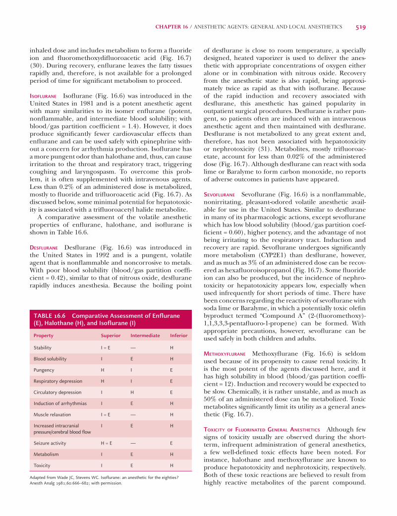
A comparative assessment of the volatile anesthetic properties of enfl urane, halothane, and isofl urane is shown in Table 16.6.
DESFLURANE Desfl urane (Fig. 16.6) was introduced in the United States in 1992 and is a pungent, volatile agent that is nonfl ammable and noncorrosive to metals. With poor blood solubility (blood/gas partition coeffi -cient = 0.42), similar to that of nitrous oxide, desfl urane rapidly induces anesthesia. Because the boiling point
TABLE 16.6 Comparative Assessment of Enflurane (E), Halothane (H), and Isoflurane (I)
Adapted from Wade JC, Stevens WC. Isoflurane: an anesthetic for the eighties? Anesth Analg 1981;60:666–682; with permission.
Lemke_Chap16.indd 519Lemke_Chap16.indd 519 12/9/2011 4:14:11 AM12/9/2011 4:14:11 AM
520 PART III / PHARMACODYNAMIC AGENTS
potential to produce damage to the renal tubular cells. Of the fl uorinated anesthetics, methoxyfl urane is the only agent commonly associated with nephrotoxicity. Methoxyfl urane is metabolized (Fig. 16.7) to produce plasma fl uoride ion levels in excess of the threshold value for renal damage of 40 mmol/L. Others, such as sevofl u-rane, have only very rarely been associated with nephro-toxicity—and then usually in patients with severe renal compromise. Plasma levels of fl uoride only reach 15 to 20 mmol/L following 2.5 MAC-hour exposure to enfl urane (33). The rates of metabolic defl uorination of the useful anesthetic agents are as follows: methoxyfl urane > enfl u-rane = sevofl urane > isofl urane > desfl urane = halothane.
Low-level Chronic Exposure Typically, patients are exposed to greater-than-MAC concentrations of the vola-tile anesthetics for limited periods of time, such as a num-ber of hours during a surgical procedure, and not for extended periods of time (e.g., days or weeks). Because surgical and dental personnel, however, can be exposed to low levels of the general anesthetics for prolonged peri-ods over many years or even decades, the ability of such agents to produce chronic toxicity is of paramount con-cern. Although the occupational exposure to these agents has been minimized with improved waste gas–scavenging devices, some epidemiologic studies have demonstrated increased levels of spontaneous abortions, congenital birth defects in offspring, and increased rates of certain cancers in chronically exposed medical personnel (34).
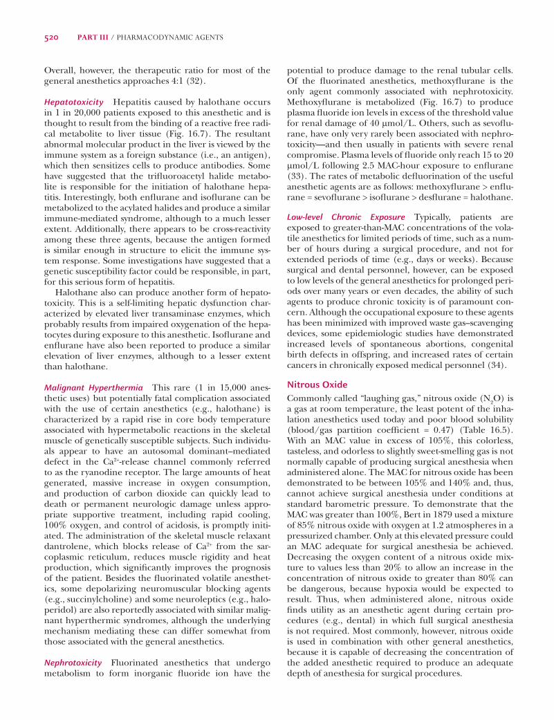
Nitrous OxideCommonly called “laughing gas,” nitrous oxide (N2O) is a gas at room temperature, the least potent of the inha-lation anesthetics used today and poor blood solubility (blood/gas partition coeffi cient = 0.47) (Table 16.5). With an MAC value in excess of 105%, this colorless, tasteless, and odorless to slightly sweet-smelling gas is not normally capable of producing surgical anesthesia when administered alone. The MAC for nitrous oxide has been demonstrated to be between 105% and 140% and, thus, cannot achieve surgical anesthesia under conditions at standard barometric pressure. To demonstrate that the MAC was greater than 100%, Bert in 1879 used a mixture of 85% nitrous oxide with oxygen at 1.2 atmospheres in a pressurized chamber. Only at this elevated pressure could an MAC adequate for surgical anesthesia be achieved. Decreasing the oxygen content of a nitrous oxide mix-ture to values less than 20% to allow an increase in the concentration of nitrous oxide to greater than 80% can be dangerous, because hypoxia would be expected to result. Thus, when administered alone, nitrous oxide fi nds utility as an anesthetic agent during certain pro-cedures (e.g., dental) in which full surgical anesthesia is not required. Most commonly, however, nitrous oxide is used in combination with other general anesthetics, because it is capable of decreasing the concentration of the added anesthetic required to produce an adequate depth of anesthesia for surgical procedures.
Overall, however, the therapeutic ratio for most of the general anesthetics approaches 4:1 (32).
Hepatotoxicity Hepatitis caused by halothane occurs in 1 in 20,000 patients exposed to this anesthetic and is thought to result from the binding of a reactive free radi-cal metabolite to liver tissue (Fig. 16.7). The resultant abnormal molecular product in the liver is viewed by the immune system as a foreign substance (i.e., an antigen), which then sensitizes cells to produce antibodies. Some have suggested that the trifl uoroacetyl halide metabo-lite is responsible for the initiation of halothane hepa-titis. Interestingly, both enfl urane and isofl urane can be metabolized to the acylated halides and produce a similar immune-mediated syndrome, although to a much lesser extent. Additionally, there appears to be cross-reactivity among these three agents, because the antigen formed is similar enough in structure to elicit the immune sys-tem response. Some investigations have suggested that a genetic susceptibility factor could be responsible, in part, for this serious form of hepatitis.
Halothane also can produce another form of hepato-toxicity. This is a self-limiting hepatic dysfunction char-acterized by elevated liver transaminase enzymes, which probably results from impaired oxygenation of the hepa-tocytes during exposure to this anesthetic. Isofl urane and enfl urane have also been reported to produce a similar elevation of liver enzymes, although to a lesser extent than halothane.
Malignant Hyperthermia This rare (1 in 15,000 anes-thetic uses) but potentially fatal complication associated with the use of certain anesthetics (e.g., halothane) is characterized by a rapid rise in core body temperature associated with hypermetabolic reactions in the skeletal muscle of genetically susceptible subjects. Such individu-als appear to have an autosomal dominant–mediated defect in the Ca2+-release channel commonly referred to as the ryanodine receptor. The large amounts of heat generated, massive increase in oxygen consumption, and production of carbon dioxide can quickly lead to death or permanent neurologic damage unless appro-priate supportive treatment, including rapid cooling, 100% oxygen, and control of acidosis, is promptly initi-ated. The administration of the skeletal muscle relaxant dantrolene, which blocks release of Ca2+ from the sar-coplasmic reticulum, reduces muscle rigidity and heat production, which signifi cantly improves the prognosis of the patient. Besides the fl uorinated volatile anesthet-ics, some depolarizing neuromuscular blocking agents (e.g., succinylcholine) and some neuroleptics (e.g., halo-peridol) are also reportedly associated with similar malig-nant hyperthermic syndromes, although the underlying mechanism mediating these can differ somewhat from those associated with the general anesthetics.
Nephrotoxicity Fluorinated anesthetics that undergo metabolism to form inorganic fl uoride ion have the
Lemke_Chap16.indd 520Lemke_Chap16.indd 520 12/9/2011 4:14:11 AM12/9/2011 4:14:11 AM
CHAPTER 16 / ANESTHETIC AGENTS: GENERAL AND LOCAL ANESTHETICS 521
rapidly via hepatic conversion to its glucuronide and sul-fate conjugates, with less than 0.3% excreted unchanged. Because this agent produces a rapid induction and recov-ery and is infrequently associated with episodes of vomit-ing, propofol has found utility as an anesthetic agent in outpatient surgical environments.
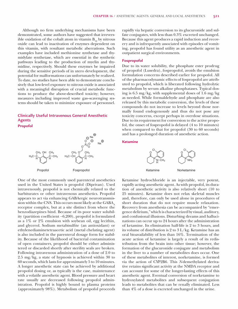
FospropofolDue to its water solubility, the phosphate ester prodrug of propofol (Lusedra), fospropofol, avoids the emulsion formulation concerns described earlier for propofol. All of the pharmacodynamic effects of fospropofol are attrib-uted to propofol, which is liberated following hydrolytic metabolism by serum alkaline phosphatases. Typical dos-ing is 6.5 mg/kg, with supplemental doses of 1.6 mg/kg as needed. While formaldehyde and phosphate are also released by this metabolic conversion, the levels of these compounds do not increase to levels beyond those nor-mally found endogenously and thus do not pose any toxicity concerns, except perhaps in overdose situations. Due to its requirement for conversion to the active propo-fol, the onset of fospropofol is delayed (4 to 10 minutes) when compared to that for propofol (30 to 60 seconds) and has a prolonged duration of anesthetic action.
Ketamine
O
NH
Cl
CH3
Ketamine
O
NH2
Cl
Norketamine
Ketamine hydrochloride is an injectable, very potent, rapidly acting anesthetic agent. As with propofol, its dura-tion of anesthetic activity is also relatively short (10 to 25 minutes). Ketamine does not relax skeletal muscles and, therefore, can only be used alone in procedures of short duration that do not require muscle relaxation. Recovery from anesthesia can be accompanied by “emer-gence delirium,” which is characterized by visual, auditory, and confusional illusions. Disturbing dreams and halluci-nations can occur up to 24 hours after the administration of ketamine. Its elimination half-life is 2 to 3 hours, and its volume of distribution is 2 to 3 L/kg. Ketamine has an oral bioavailability of less than 16%. Termination of the acute action of ketamine is largely a result of its redis-tribution from the brain into other tissue; however, the formation of the glucuronide conjugate and metabolism in the liver to a number of metabolites does occur. One of these metabolites of interest, norketamine, is formed via the action of CYP2B6. This N-demethylated deriva-tive retains signifi cant activity at the NMDA receptor and can account for some of the longer-lasting effects of this anesthetic agent. Eventual conversion of norketamine to hydroxylated metabolites and subsequent conjugation leads to metabolites that can be renally eliminated. Less than 4% of a dose is excreted unchanged in the urine.
Although no fi rm underlying mechanisms have been demonstrated, some authors have suggested that irrevers-ible oxidation of the cobalt atom in vitamin B12 by nitrous oxide can lead to inactivation of enzymes dependent on this vitamin, with resultant metabolic aberrations. Such examples have included methionine synthetase and thy-midylate synthetase, which are essential in the synthetic pathways leading to the production of myelin and thy-midine, respectively. Should these enzymes be impaired during the sensitive periods of in utero development, the potential for malformations can unfortunately be realized. To date, no studies have been able to demonstrate conclu-sively that low-level exposure to nitrous oxide is associated with a meaningful disruption of crucial metabolic func-tions to produce the above-described toxicity; however, measures including improved waste gas–scavenging sys-tems should be taken to minimize exposure of personnel.
Clinically Useful Intravenous General Anesthetic AgentsPropofol
OHCH
CCH3
CH3
CH3
H3C
Propofol
OCH
CHCH3
CH3
CH3
H3C
Fospropofol
OPO
OO Na
Na
H
One of the most commonly used parenteral anesthetics used in the United States is propofol (Diprivan). Used intravenously, propofol is not chemically related to the barbiturates or other intravenous anesthetics. Propofol appears to act via enhancing GABAergic neurotransmis-sion within the CNS. This occurs most likely at the GABAA receptor complex, but at a site distinct from where the benzodiazepines bind. Because of its poor water solubil-ity (partition coeffi cient ∼6,200), propofol is formulated as a 1% or 2% emulsion with soybean oil, egg lecithin, and glycerol. Sodium metabisulfi te (an antioxidant) or ethylenediaminetetraacetic acid (metal chelating agent) is also included in the parenteral dosage form for stabil-ity. Because of the likelihood of bacterial contamination of open containers, propofol should be either adminis-tered or discarded shortly after sterility seals are broken. Following intravenous administration of a dose of 2.0 to 2.5 mg/kg, a state of hypnosis is achieved within 30 to 60 seconds, which lasts for approximately 5 to 10 minutes. A longer anesthetic state can be achieved by additional propofol dosing or, as typically is the case, maintenance with a volatile anesthetic agent. Blood pressure and heart rate usually are decreased following propofol admin-istration. Propofol is highly bound to plasma proteins (approximately 98%). Metabolism of propofol proceeds
Lemke_Chap16.indd 521Lemke_Chap16.indd 521 12/9/2011 4:14:11 AM12/9/2011 4:14:11 AM
522 PART III / PHARMACODYNAMIC AGENTS
Ultrashort-Acting Barbiturates
HN N
O O
S
H3CCH3 CH3
Na
Thiopental
Thiopental, an ultrashort-acting barbiturate (partition coeffi cient ∼390), is used intravenously to produce a rapid unconsciousness for surgical and basal anesthesia. This agent is used initially to induce anesthesia, which then can be maintained during the surgical procedure with a general anesthetic agent. The induction typically is very rapid and pleasant. (The ultrashort-acting barbitu-rates are discussed in Chapter 15.)
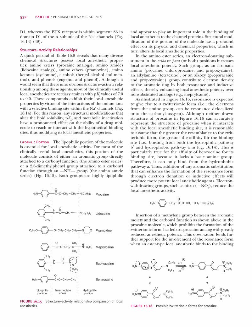
LOCAL ANESTHETICSLocal anesthetic agents are drugs that, when given either topically or administered directly into a localized area, produce a state of local anesthesia by reversibly blocking nerve conductances that transmit the sensations of pain from this localized area to the brain. Unlike the anes-thesia produced by general anesthetics, the anesthesia produced by local anesthetics is without loss of conscious-ness or impairment of vital central cardiorespiratory functions. Local anesthetics block nerve conductance by binding to selective sites on the Na+ channels in the excit-able membranes, thereby reducing Na+ passage (i.e., con-ductance) through the pores and, thus, interfere with the generation of action potentials. Although local anes-thetics decrease the excitability of nerve membranes, they do not affect the neuron’s resting potential. Local anesthetics, in contrast to analgesic compounds, do not interact with the pain receptors or inhibit the release or the biosynthesis of pain mediators.
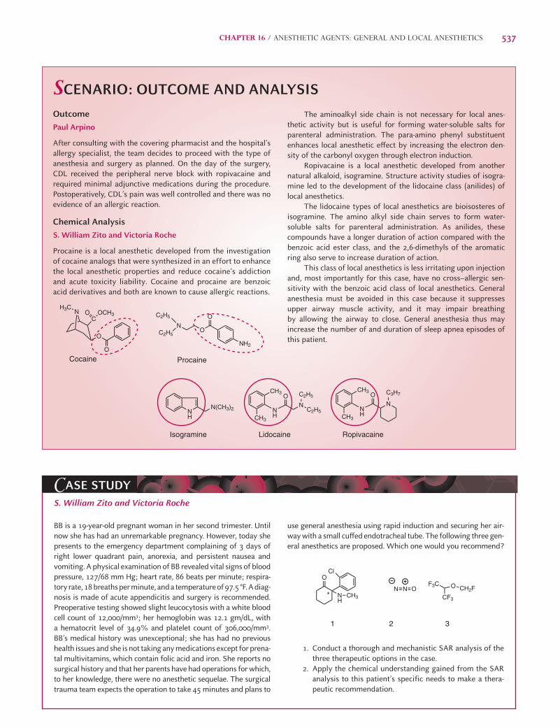
The Discovery of Local AnestheticsAs with many modern drugs, the initial leads for the design of clinically useful local anesthetics originated from nat-ural sources. As early as 1532, the anesthetic properties of coca leaves (Erythroxylon coca Lam) became known to Europeans from the natives of Peru, who chewed the leaves for a general feeling of well-being and to reduce hunger. Saliva from chewing the leaves was often used by the natives to relieve painful wounds. The active principle of the coca leaf, however, was not discovered until 1860 by Niemann, who obtained a crystalline alkaloid from the leaves, to which he gave the name cocaine, and who noted the anesthetic effect on the tongue (see Fig. 16.8 for structure of cocaine). Although Moréno y Maiz in 1868 fi rst asked the question of whether cocaine could be used as a local anesthetic, Von Anrep in 1880, after many animal experiments, recommended that cocaine
Ketamine is capable of producing a “dissociative” anes-thesia, which is characterized by EEG changes indicating a dissociation between the thalamocortical and limbic sys-tems (35). These neuronal systems, which normally are associated with one another, help to maintain the neuro-nal connections required for consciousness. When disas-sociated, the subject will appear to be cataleptic, with the eyes open in a slow, nystagmic gaze (oscillating movement of the eyeball) (1). A potent analgesic and amnesic effect is produced, as is an increase in muscle tone in some areas. Although patients can appear to be awake, they are inca-pable of communicating and do not remember the event or the people around them. Blood pressure and heart rate usually are increased following ketamine administration.
Ketamine appears to act similarly to phencyclidine (PCP; also known as Angel Dust), which acts as an antago-nist within the cationic channel of the NMDA receptor complex (36). By preventing the fl ow of cations through this channel, ketamine prevents neuronal activation, which normally is required for the conscious state. The analgesic activity of ketamine, however, is more likely the result of an interaction with an opioid receptor or the less well-understood non-opioid sigma receptor. Other studies have suggested a possible involvement of serotonin recep-tors and muscarinic receptors (37). Ketamine, like PCP, has a signifi cant potential for abuse.
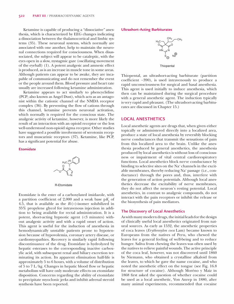
Etomidate
N
NO
O
H3C
R-Etomidate
(R )
Etomidate is the ester of a carboxylated imidazole, with a partition coeffi cient of 2,000 and a weak base pKa of 4.5, that is available as the R-(+)-isomer solubilized in 35% propylene glycol for intravenous injection in addi-tion to being available for rectal administration. It is a potent, short-acting hypnotic agent (<3 minutes) with-out analgesic activity and with a rapid onset of action. This agent is useful for the induction of anesthesia in hemodynamically unstable patients prone to hypoten-sion because of hypovolemia, coronary artery disease, or cardiomyopathies. Recovery is similarly rapid following discontinuance of the drug. Etomidate is hydrolyzed by hepatic esterases to the corresponding inactive carbox-ylic acid, with subsequent renal and biliary excretion ter-minating its action. Its apparent elimination half-life is approximately 5 to 6 hours, with a volume of distribution of 5 to 7 L/kg. Changes in hepatic blood fl ow or hepatic metabolism will have only moderate effects on etomidate disposition. Concerns regarding the ability of etomidate to precipitate myoclonic jerks and inhibit adrenal steroid synthesis have been reported.
Lemke_Chap16.indd 522Lemke_Chap16.indd 522 12/9/2011 4:14:12 AM12/9/2011 4:14:12 AM
CHAPTER 16 / ANESTHETIC AGENTS: GENERAL AND LOCAL ANESTHETICS 523
Although the intrinsic potency of procaine was low and its duration of action relatively short compared with that of cocaine, it was found that these defi ciencies could be remedied when procaine was combined with a vaso-constrictor, such as epinephrine. Vasoconstrictor agents reduce the local blood supply and, thereby, prolong the residence time of the local anesthetic at the injection site.
Following the introduction of procaine, hundreds of structurally related analogs were prepared and their local anesthetic properties examined in an attempt to identify agents with enhanced potency and duration of action compared to the weak and short-acting procaine. Among these compounds, tetracaine remains the most potent, long-acting, ester-type local anesthetic agent, which is used in spinal anesthesia.
C4H9HN
ON(CH3)2
O
H2N
OCH2CH3
O
Tetracaine Benzocaine
The topical anesthetic agent benzocaine was synthe-sized by Ritsert in 1890 and found to have good anes-thetizing properties and low toxicity. However, due to its limited water solubility, except at low pH values as a result of the lack of a basic aliphatic amino group, the prepara-tion of pharmaceutically acceptable parenteral solutions could not be achieved.
NH
N(CH3)2N
ON(C2H5)2
Isogramine Lidocaine
CH3
CH3
H
The serendipitous discovery of the local anesthetic activity of another natural alkaloidal product, isogra-mine, in 1935 by von Euler and Erdtman was the next major turning point in the development of clinically use-ful local anesthetic agents. This observation led to the synthesis of lidocaine (Xylocaine) by Löfgren in 1946; lidocaine was the fi rst nonirritating, amide-type local anesthetic agent with good local anesthetic properties yet less prone to allergenic reactions than procaine analogs, and was found to be stabile in aqueous solution due to its more stable amide functionality. Structurally, lidocaine can be viewed as an open-chain analog of isogramine and, thus, is a bioisosteric analog of isogramine.
Since the discovery of lidocaine in the 1940s, much more progress has been achieved in the fi elds of neuro-physiology and neuropharmacology than in the synthesis of local anesthetics by medicinal chemists. Most of this research has signifi cantly increased our understanding of how nerve conduction occurs and how compounds interact with the neuronal membranes to produce local anesthesia. It should be noted, however, that although a number of current clinically useful local anesthetic
be used clinically as a local anesthetic. The fi rst report of successful surgical use of cocaine appeared in 1884 by Koller, an Austrian ophthalmologist. This discovery led to the rapid development of new local anesthetic agents and anesthetic techniques (38).
Cocaine dependence (or addiction) is psychologi-cal dependency on the regular use of cocaine. The use of cocaine, depending on the severity, can cause mood swings, paranoia, insomnia, psychosis, high blood pres-sure, tachycardia, panic attacks, cognitive impairments, and drastic changes in the personality that can lead to aggressive, compulsive, criminal, and/or erratic behaviors. The symptoms of cocaine withdrawal range from moder-ate to severe: dysphoria, depression, anxiety, psychological and physical weakness, pain, and compulsive craving.
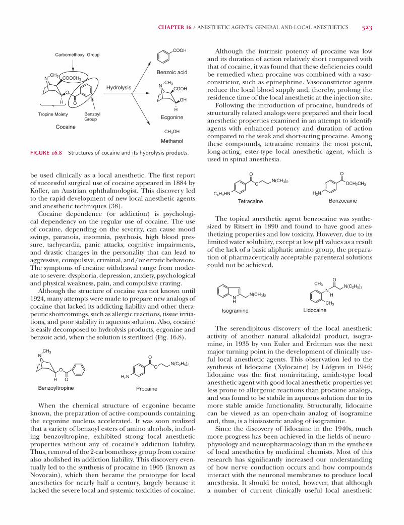
Although the structure of cocaine was not known until 1924, many attempts were made to prepare new analogs of cocaine that lacked its addicting liability and other thera-peutic shortcomings, such as allergic reactions, tissue irrita-tions, and poor stability in aqueous solution. Also, cocaine is easily decomposed to hydrolysis products, ecgonine and benzoic acid, when the solution is sterilized (Fig. 16.8).
NCH3
O
H OH2N
ON(C2H5)2
O
Benzoyltropine Procaine
When the chemical structure of ecgonine became known, the preparation of active compounds containing the ecgonine nucleus accelerated. It was soon realized that a variety of benzoyl esters of amino alcohols, includ-ing benzoyltropine, exhibited strong local anesthetic properties without any of cocaine’s addiction liability. Thus, removal of the 2-carbomethoxy group from cocaine also abolished its addiction liability. This discovery even-tually led to the synthesis of procaine in 1905 (known as Novocain), which then became the prototype for local anesthetics for nearly half a century, largely because it lacked the severe local and systemic toxicities of cocaine.
NCH3 COOCH3
O
H O
NCH3
COOH
OH
H
Cocaine
Hydrolysis
Ecgonine
COOH
Benzoic acid
Methanol
Benzoyl Group
Carbomethoxy Group
Tropine Moiety
CH3OH
FIGURE 16.8 Structures of cocaine and its hydrolysis products.
Lemke_Chap16.indd 523Lemke_Chap16.indd 523 12/9/2011 4:14:12 AM12/9/2011 4:14:12 AM
524 PART III / PHARMACODYNAMIC AGENTS
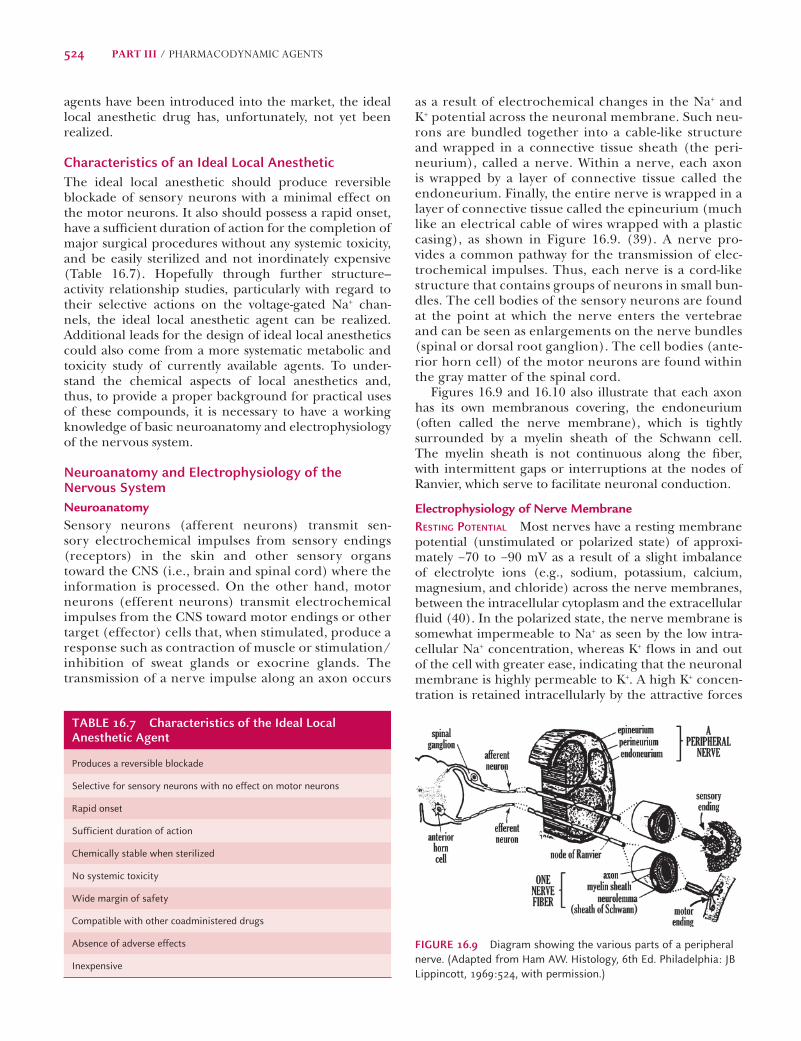
as a result of electrochemical changes in the Na+ and K+ potential across the neuronal membrane. Such neu-rons are bundled together into a cable-like structure and wrapped in a connective tissue sheath (the peri-neurium), called a nerve. Within a nerve, each axon is wrapped by a layer of connective tissue called the endoneurium. Finally, the entire nerve is wrapped in a layer of connective tissue called the epineurium (much like an electrical cable of wires wrapped with a plastic casing), as shown in Figure 16.9. (39). A nerve pro-vides a common pathway for the transmission of elec-trochemical impulses. Thus, each nerve is a cord-like structure that contains groups of neurons in small bun-dles. The cell bodies of the sensory neurons are found at the point at which the nerve enters the vertebrae and can be seen as enlargements on the nerve bundles (spinal or dorsal root ganglion). The cell bodies (ante-rior horn cell) of the motor neurons are found within the gray matter of the spinal cord.
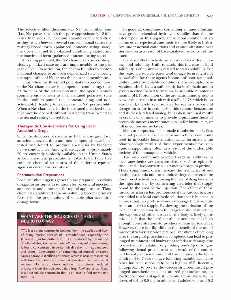
Figures 16.9 and 16.10 also illustrate that each axon has its own membranous covering, the endoneurium (often called the nerve membrane), which is tightly surrounded by a myelin sheath of the Schwann cell. The myelin sheath is not continuous along the fi ber, with intermittent gaps or interruptions at the nodes of Ranvier, which serve to facilitate neuronal conduction.
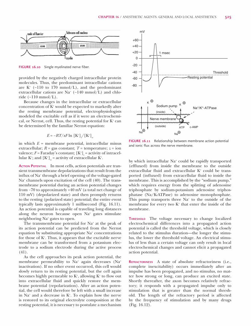
Electrophysiology of Nerve MembraneRESTING POTENTIAL Most nerves have a resting membrane potential (unstimulated or polarized state) of approxi-mately −70 to −90 mV as a result of a slight imbalance of electrolyte ions (e.g., sodium, potassium, calcium, magnesium, and chloride) across the nerve membranes, between the intracellular cytoplasm and the extracellular fl uid (40). In the polarized state, the nerve membrane is somewhat impermeable to Na+ as seen by the low intra-cellular Na+ concentration, whereas K+ fl ows in and out of the cell with greater ease, indicating that the neuronal membrane is highly permeable to K+. A high K+ concen-tration is retained intracellularly by the attractive forces
agents have been introduced into the market, the ideal local anesthetic drug has, unfortunately, not yet been realized.
Characteristics of an Ideal Local AnestheticThe ideal local anesthetic should produce reversible blockade of sensory neurons with a minimal effect on the motor neurons. It also should possess a rapid onset, have a suffi cient duration of action for the completion of major surgical procedures without any systemic toxicity, and be easily sterilized and not inordinately expensive (Table 16.7). Hopefully through further structure–activity relationship studies, particularly with regard to their selective actions on the voltage-gated Na+ chan-nels, the ideal local anesthetic agent can be realized. Additional leads for the design of ideal local anesthetics could also come from a more systematic metabolic and toxicity study of currently available agents. To under-stand the chemical aspects of local anesthetics and, thus, to provide a proper background for practical uses of these compounds, it is necessary to have a working knowledge of basic neuroanatomy and electrophysiology of the nervous system.
Neuroanatomy and Electrophysiology of the Nervous SystemNeuroanatomySensory neurons (afferent neurons) transmit sen-sory electrochemical impulses from sensory endings (receptors) in the skin and other sensory organs toward the CNS (i.e., brain and spinal cord) where the information is processed. On the other hand, motor neurons (efferent neurons) transmit electrochemical impulses from the CNS toward motor endings or other target (effector) cells that, when stimulated, produce a response such as contraction of muscle or stimulation/inhibition of sweat glands or exocrine glands. The transmission of a nerve impulse along an axon occurs
TABLE 16.7 Characteristics of the Ideal Local Anesthetic Agent
Produces a reversible blockade
Selective for sensory neurons with no effect on motor neurons
Rapid onset
Sufficient duration of action
Chemically stable when sterilized
No systemic toxicity
Wide margin of safety
Compatible with other coadministered drugs
Absence of adverse effects
Inexpensive
FIGURE 16.9 Diagram showing the various parts of a peripheral nerve. (Adapted from Ham AW. Histology, 6th Ed. Philadelphia: JB Lippincott, 1969:524, with permission.)
Lemke_Chap16.indd 524Lemke_Chap16.indd 524 12/9/2011 4:14:12 AM12/9/2011 4:14:12 AM
CHAPTER 16 / ANESTHETIC AGENTS: GENERAL AND LOCAL ANESTHETICS 525
by which intracellular Na+ could be rapidly transported (effl uxed) from inside the membrane to the outside extracellular fl uid and extracellular K+ could be trans-ported (infl uxed) from extracellular fl uid to inside the membrane. This is accomplished by the “sodium pump,” which requires energy from the splitting of adenosine triphosphate by sodium-potassium adenosine triphos-phatase (Na/K-ATPase) to adenosine monophosphate. This pump transports three Na+ to the outside of the membrane for every two K+ that enter the inside of the membrane.
THRESHOLD The voltage necessary to change localized electrochemical differences into a propagated action potential is called the threshold voltage, which is closely related to the stimulus duration—the longer the stimu-lus, the lower the threshold voltage. An electrical stimu-lus of less than a certain voltage can only result in local electrochemical changes and cannot elicit a propagated action potential.
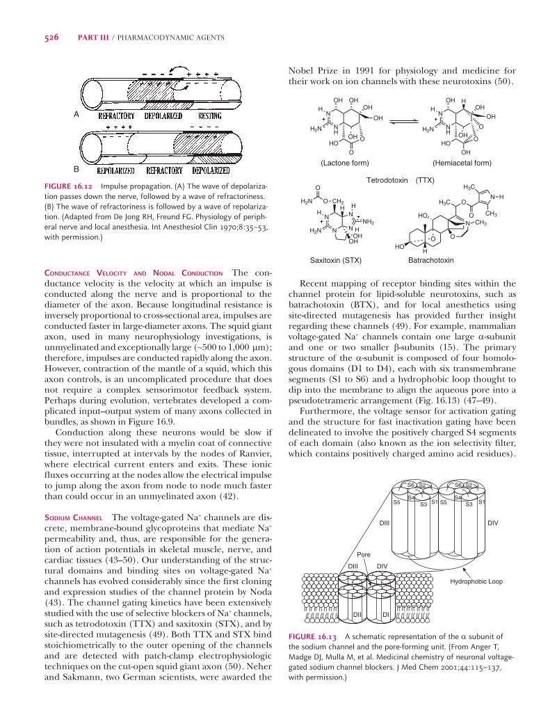
REFRACTORINESS A state of absolute refractoriness (i.e., complete inexcitability) occurs immediately after an impulse has been propagated, and no stimulus, no mat-ter how strong or long, can produce an excited state. Shortly thereafter, the axon becomes relatively refrac-tory; it responds with a propagated impulse only to stimulation that is greater than the normal thresh-old. The length of the refractory period is affected by the frequency of stimulation and by many drugs (Fig. 16.12).
provided by the negatively charged intracellular protein molecules. Thus, the predominant intracellular cations are K+ (∼110 to 170 mmol/L), and the predominant extracellular cations are Na+ (∼140 mmol/L) and chlo-ride (∼110 mmol/L).
Because changes in the intracellular or extracellular concentration of K+ would be expected to markedly alter the resting membrane potential, electrophysiologists modeled the excitable cell as if it were an electrochemi-cal, or Nernst, cell. Thus, the resting potential for K+ can be determined by the familiar Nernst equation:
E = −RT/zF ln [K+]i/[K+]o
in which E = membrane potential, intracellular minus extracellular; R = gas constant; T = temperature; z = ion valence; F = Faraday’s constant; [K+]i = activity of intracel-lular K+; and [K+]o = activity of extracellular K+.
ACTION POTENTIAL In most cells, action potentials are tran-sient transmembrane depolarizations that result from the infl ux of Na+ through a brief opening of the voltage-gated Na+ channels upon excitation of the cell (40). The trans-membrane potential during an action potential changes from −70 to approximately +40 mV (a total net change of 110 mV) (depolarized state) and then promptly returns to the resting (polarized state) potential; the entire event typically lasts approximately 1 millisecond (Fig. 16.11). An action potential is capable of traveling long distances along the neuron because open Na+ gates stimulate neighboring Na+ gates to open.
The transmembrane potential for Na+ at the peak of its action potential can be predicted from the Nernst equation by substituting appropriate Na+ concentrations for those of K+. Thus, it appears that the excitable nerve membrane can be transformed from a potassium elec-trode to a sodium electrode during the active process (41).
As the cell approaches its peak action potential, the membrane permeability to Na+ again decreases (Na+ inactivation). If no other event occurred, this cell would slowly return to its resting potential, but the cell again becomes highly permeable to K+, allowing K+ to fl ow out into extracellular fl uid and quickly restore the mem-brane potential (repolarization). After an action poten-tial, the cell would therefore be left with a small increase in Na+ and a decrease in K+. To explain how the nerve is restored to its original electrolyte composition at the resting potential, it is necessary to postulate a mechanism
FIGURE 16.10 Single myelinated nerve fiber.
Nerve membrane
Na
K
Na
K
ATP c-AMP
Na+/K+-ATPaseSodium pump
(outside)
(inside)
0
+20
+40
+60
-20
-40
-60
-80
mV
Threshold
1 msec
Resting potential
FIGURE 16.11 Relationship between membrane action potential and ionic flux across the nerve membrane.
Lemke_Chap16.indd 525Lemke_Chap16.indd 525 12/9/2011 4:14:13 AM12/9/2011 4:14:13 AM
526 PART III / PHARMACODYNAMIC AGENTS
Nobel Prize in 1991 for physiology and medicine for their work on ion channels with these neurotoxins (50).
N
NH
OHO
O
OH OHOH
OH
H2N
H
OH
N
NH
OHO
OHOH
OH
H2N
H
OH
OH
H
O
N
N
N
NH2N
NH2
HH
CH2H
(Lactone form) (Hemiacetal form)
Saxitoxin (STX)
Tetrodotoxin (TTX)
H
H3C O
O
N
CH3
H3C
H
HHO
O
N CH3
O
HO
Batrachotoxin
OH2N
O
OHOH
Recent mapping of receptor binding sites within the channel protein for lipid-soluble neurotoxins, such as batrachotoxin (BTX), and for local anesthetics using site-directed mutagenesis has provided further insight regarding these channels (49). For example, mammalian voltage-gated Na+ channels contain one large a-subunit and one or two smaller b-subunits (15). The primary structure of the a-subunit is composed of four homolo-gous domains (D1 to D4), each with six transmembrane segments (S1 to S6) and a hydrophobic loop thought to dip into the membrane to align the aqueous pore into a pseudotetrameric arrangement (Fig. 16.13) (47–49).
Furthermore, the voltage sensor for activation gating and the structure for fast inactivation gating have been delineated to involve the positively charged S4 segments of each domain (also known as the ion selectivity fi lter, which contains positively charged amino acid residues).
CONDUCTANCE VELOCITY AND NODAL CONDUCTION The con-ductance velocity is the velocity at which an impulse is conducted along the nerve and is proportional to the diameter of the axon. Because longitudinal resistance is inversely proportional to cross-sectional area, impulses are conducted faster in large-diameter axons. The squid giant axon, used in many neurophysiology investigations, is unmyelinated and exceptionally large (∼500 to 1,000 mm); therefore, impulses are conducted rapidly along the axon. However, contraction of the mantle of a squid, which this axon controls, is an uncomplicated procedure that does not require a complex sensorimotor feedback system. Perhaps during evolution, vertebrates developed a com-plicated input–output system of many axons collected in bundles, as shown in Figure 16.9.
Conduction along these neurons would be slow if they were not insulated with a myelin coat of connective tissue, interrupted at intervals by the nodes of Ranvier, where electrical current enters and exits. These ionic fl uxes occurring at the nodes allow the electrical impulse to jump along the axon from node to node much faster than could occur in an unmyelinated axon (42).
SODIUM CHANNEL The voltage-gated Na+ channels are dis-crete, membrane-bound glycoproteins that mediate Na+ permeability and, thus, are responsible for the genera-tion of action potentials in skeletal muscle, nerve, and cardiac tissues (43–50). Our understanding of the struc-tural domains and binding sites on voltage-gated Na+ channels has evolved considerably since the fi rst cloning and expression studies of the channel protein by Noda (43). The channel gating kinetics have been extensively studied with the use of selective blockers of Na+ channels, such as tetrodotoxin (TTX) and saxitoxin (STX), and by site-directed mutagenesis (49). Both TTX and STX bind stoichiometrically to the outer opening of the channels and are detected with patch-clamp electrophysiologic techniques on the cut-open squid giant axon (50). Neher and Sakmann, two German scientists, were awarded the
A
B
FIGURE 16.12 Impulse propagation. (A) The wave of depolariza-tion passes down the nerve, followed by a wave of refractoriness. (B) The wave of refractoriness is followed by a wave of repolariza-tion. (Adapted from De Jong RH, Freund FG. Physiology of periph-eral nerve and local anesthesia. Int Anesthesiol Clin 1970;8:35–53, with permission.)
Pore
Hydrophobic Loop
DIV
DIV
DIII
DIII
DII DI
S5 S5S4 S4
S3 S3S1 S1
S2 S2S6 S6
FIGURE 16.13 A schematic representation of the a subunit of the sodium channel and the pore-forming unit. (From Anger T, Madge DJ, Mulla M, et al. Medicinal chemistry of neuronal voltage-gated sodium channel blockers. J Med Chem 2001;44:115–137, with permission.)
Lemke_Chap16.indd 526Lemke_Chap16.indd 526 12/9/2011 4:14:13 AM12/9/2011 4:14:13 AM
CHAPTER 16 / ANESTHETIC AGENTS: GENERAL AND LOCAL ANESTHETICS 527
In general, compounds containing an amide linkage have greater chemical hydrolytic stability than do the ester types. In this regard, an aqueous solution of an amino ester–type local anesthetic is more likely to hydro-lyze under normal conditions and cannot withstand heat sterilization as a result of base-catalyzed hydrolysis of the ester.
Local anesthetic activity usually increases with increas-ing lipid solubility. Unfortunately, this increase in lipid solubility is often inversely related to water solubility. For this reason, a suitable parenteral dosage form might not be available for these agents because of poor water sol-ubility under acceptable conditions. For example, ben-zocaine, which lacks a suffi ciently basic aliphatic amino group needed for salt formation, is insoluble in water at neutral pH. Protonation of the aromatic amino group in benzocaine results in a salt with a pKa of 2.78, which is too acidic and, therefore, unsuitable for use as a parenteral dosage form for injection. For this reason, benzocaine and its closely related analog, butamben, are used mostly in creams or ointments to provide topical anesthesia of accessible mucous membranes or skin for burns, cuts, or infl amed mucous surfaces.
Many attempts have been made to substitute oils, fats, or fl uid polymers for the aqueous vehicle commonly used in injectable local anesthetics. Unfortunately, the pharmacologic results of these experiments have been quite disappointing, often as a result of the undesirable toxicity of the nonaqueous vehicle.
The only commonly accepted organic additives to local anesthetics are vasoconstrictors, such as epineph-rine and levonordefrin (a-methylnorepinephrine). These compounds often increase the frequency of suc-cessful anesthesia and, to a limited degree, increase the duration of activity by reducing the rate of drug loss from the injection site, by constricting arterioles that supply blood to the area of the injection. The effect of these vasoconstrictors is less pronounced if the vasoconstrictors are added to a local anesthetic solution that is injected in an area that has profuse venous drainage but is remote from an arterial supply. By slowing the diffusion of the local anesthetic away from the targeted site of injection, the exposure of other tissues in the body is likely mini-mized such that the local anesthetic never reaches high enough concentrations to produce unwanted toxicities. However, there is a fl ip slide to the benefi t of the use of vasoconstrictors: A prolonged local anesthetic effect long after the surgical procedure is completed can lead to pro-longed numbness and inadvertent soft tissue damage due to mechanical irritation (e.g., biting one’s lip or tongue following dental procedures) as a result of the contin-ued loss of pain sensations. Soft tissue injury to the lip in children 4 to 7 years of age following mandibular nerve block has been reported to be as high as 16%. Recently, an approach to reverse the vasoconstrictor-induced pro-longed anesthetic state has utilized phentolamine, an a-adrenoceptor antagonist. Phentolamine mesylate in doses of 0.4 to 0.8 mg in adults and adolescents and 0.2
The selective fi lter discriminates Na+ from other ions (i.e., Na+ passes through this pore approximately 12-fold faster than does K+). Sodium channels open and close as they switch between several conformational states: the resting/closed form (polarized nonconducting state), the open channel (depolarized conducting state), and the inactivated form (polarized nonconducting state).
At resting potential, the Na+ channels are in a resting/closed polarized state and are impermeable to the pas-sage of Na+. On activation, the channels undergo confor-mational changes to an open depolarized state, allowing the rapid infl ux of Na+ across the neuronal membrane.
Thus, when the threshold potential is exceeded, most of the Na+ channels are in an open, or conducting, state. At the peak of the action potential, the open channels spontaneously convert to an inactivated polarized state by the “sodium pump” (i.e., nonconducting and non-activatable), leading to a decrease in Na+ permeability. When a Na+ channel is in the inactivated polarized state, it cannot be opened without fi rst being transformed to the normal resting/closed form.
Therapeutic Considerations for Using Local Anesthetic DrugsSince the discovery of cocaine in 1880 as a surgical local anesthetic, several thousand new compounds have been tested and found to produce anesthesia by blocking nerve conductance. Among these agents, approximately 20 are currently clinically available in the United States as local anesthetic preparations (Table 16.8). Table 16.9 contains chemical structures of the different types of agents in current or recent use.
Pharmaceutical PreparationsLocal anesthetic agents generally are prepared in various dosage forms: aqueous solutions for parenteral injection, and creams and ointments for topical applications. Thus, chemical stability and aqueous solubility become primary factors in the preparations of suitable pharmaceutical dosage forms.
TTX is a potent neurotoxin isolated from the ovaries and liver of many marine species of Tetraodontidae, especially the Japanese fugu (or puffer fish). STX, produced by the marine dinoflagellates, Gonyaulax catenella or Gonyaulax tamarensis, is found concentrated in certain bivalve shellfish (e.g., mussels and clams). Consumption of contaminated mussels or clams causes paralytic shellfish poisoning, which is usually associated with toxic “red tide” environmental episodes in various coastal regions. BTX, a cardiotoxic and neurotoxic steroid isolated originally from the poisonous dart frog, Phyllobates terribilis, is a lipid-soluble neurotoxin that is at least 10-fold more toxic than TTX.
WHAT ARE THE SOURCES OF THESE NEUROTOXINS?
Lemke_Chap16.indd 527Lemke_Chap16.indd 527 12/9/2011 4:14:14 AM12/9/2011 4:14:14 AM
528 PART III / PHARMACODYNAMIC AGENTS
of the classic local anesthetic agents that can result in sig-nifi cant clinical advantages.
A Eutectic Mixture of a Local Anesthetic (EMLA) cream containing 2.5% lidocaine and 2.5% prilocaine (or etidocaine) is used for the topical application of local anesthetic through the keratinized layer of the intact skin to provide dermal or epidermal analgesia. This mode of administration allows the use of higher concentrations of local anesthetic with minimal local irritation and lower systemic toxicity. The use of EMLA creams, especially those containing prilocaine, on mucous membranes is not recommended, however, because of the faster absorption of the drugs and, therefore, the increasing
to 0.4 mg in children reverses the vasoconstriction and allows for a more rapid diffusion of the local anesthetic from the injection site and a recovery of sensation (51).
Administration of a local anesthetic in a carbonic acid–carbon dioxide aqueous solution rather than the usual solution of a hydrochloride salt appreciably improves the time of onset and duration of action without causing increased local or systemic toxicity.
Carbon dioxide is believed to potentiate the action of local anesthetics by initial indirect depression of the axon, followed by diffusion trapping of the active form of the local anesthetic within the nerve. Use of the carbon-ate salt appears to be one pharmaceutical modifi cation
Lemke_Chap16.indd 528Lemke_Chap16.indd 528 12/9/2011 4:14:14 AM12/9/2011 4:14:14 AM
CHAPTER 16 / ANESTHETIC AGENTS: GENERAL AND LOCAL ANESTHETICS 529
neuromuscular junctions and the CNS are more suscep-tible than the cardiovascular system to the toxic effects of local anesthetics.
The actions on skeletal muscles tend to be transient and reversible, whereas the CNS side effects can be more deleterious. The primary effect of the toxicity seems to be convulsions, followed by severe CNS depression, par-ticularly of the respiratory and cardiovascular centers. This can be related to an initial depression of inhibitory
risk of systemic toxicity, such as methemoglobinemia, specifi cally with prilocaine (52).
Toxicity and Side EffectsThe side effects and toxicity of local anesthetics seem to be related to their actions on other excitable membrane proteins, such as in the Na+ and K+ channels in the heart, the nicotinic acetylcholine receptors in the neuromuscu-lar junctions, and the nerve cells in the CNS. In general,
TABLE 16.9 Structures of Local Anesthetics
Lemke_Chap16.indd 529Lemke_Chap16.indd 529 12/9/2011 4:14:14 AM12/9/2011 4:14:14 AM
530 PART III / PHARMACODYNAMIC AGENTS
fl exibility and responsiveness to changes in electrical fi elds.
The nonspecifi c membrane actions of local anesthet-ics can be easily ruled out, because most clinically use-ful agents, in contrast to general anesthetics, possess a defi ned set of structure–activity relationships. At much higher drug concentrations, local anesthetics also bind and block K+ channels.
INTERACTION WITH PHOSPHOLIPIDS AND CALCIUM Calcium exists in the membrane in a bound state. Many inves-tigators believe that the release of the bound calcium is the fi rst step in membrane depolarization and that this release leads to the changes in ionic permeability described previously. It has been suggested that local anesthetics displace the bound calcium from these sites and form more stable bonds, thereby inhibiting ionic fl uxes. The following evidence has been offered in sup-port of this theory: Both calcium and local anesthetics bind to phospholipids in vitro, reducing their fl exibil-ity and responsiveness to changes in electrical fi elds (56–58). Also, membrane excitability and instability increase in calcium-defi cient solutions. Local anesthet-ics counteract this abnormal increase in excitability, and more local anesthetic is necessary to block excita-tion in calcium-poor solutions (59). Direct proof of this hypothesis, however, is lacking because of the diffi culty in measuring temporal Ca2+ movements in vivo. It is also possible that the aforementioned cause-and-effect relationship between intracellular free Ca2+ and mem-brane excitability is the result of an Na+–Ca2+ exchange reaction; that is, the infl ux of Na+ displaces the mem-brane-bound calcium, which leads to an increase of intracellular free Ca2+ and, thereby, increases cellular excitability.
Local anesthetics interact differently, however, with neuronal phospholipids with or without the presence of cholesterol. Thus, the interactions of local anesthetics with the cellular membranes actually may help to explain some of the observed differences in toxicity of the indi-vidual local anesthetic agents (60).
ACTION ON VOLTAGE-SENSITIVE SODIUM CHANNELS As mentioned, the voltage-sensitive Na+ channels are membrane-bound glycoproteins that mediate Na+ per-meability. On excitation, these channels undergo con-formational changes from a closed to an open state, thus allowing a rapid infl ux of sodium. The movement of Na+ is blocked by the neurotoxins TTX and STX and by local anesthetics (61). Most electrophysiologists and neuropharmacologists now agree that the mechanism of action of local anesthetics results primarily from their binding to one or more sites within the Na+ chan-nels, thus blocking Na+ conductance (62). However, the exact location of these binding sites and whether all local anesthetics interact with a common site remain matters of dispute.
neurons, such as GABAergic systems, causing convul-sions, followed by depression of other neurons, leading to general depression of the CNS.
The amino amide–type local anesthetics (i.e., lido-caine derivatives) are, in general, more likely to produce CNS side effects than the amino ester–type compounds (procaine analog). However, it should be noted that the toxic effects observed depend heavily on the route and site of administration as well as on the lipid solubility and metabolic stability of a given local anesthetic mol-ecule. For example, most amide-type local anesthetics, such as lidocaine, are fi rst degraded via N-dealkylation by hepatic enzymes (see Fig. 16.17). Unlike lidocaine, however, the initial metabolic degradation of prilocaine in humans is hydrolysis of the amide linkage to give o-toluidine and N-propylalanine. Formation of o-tolu-idine and its metabolites can cause methemoglobin-emia in some patients (52). For this reason, prilocaine is much more likely than other local anesthetics to cause methemoglobinemia.
In contrast, allergic reactions to local anesthetics, although rare, are known to occur exclusively with p-ami-nobenzoic acid (PABA) ester-type local anesthetics (53). Whether the formation of PABA upon ester hydrolysis is solely responsible for this hypersensitivity remains to be determined. However, the preservative compounds, such as methylparaben, used in the preparation of amide-type local anesthetics are metabolized to the PABA-like sub-stance, p-hydroxybenzoic acid. Thus, patients who are allergic to amino ester–type local anesthetics should be treated with a preservative-free amino amide–type local anesthetic.
Amide-type local anesthetics (e.g., procainamide and lidocaine) also possess antiarrhythmic activity when given parenterally and at a subanesthetic dos-age. Although this action is likely an extension of their effects on Na+ channels in cardiac tissues, some evidence suggests a distinctly different mechanism of action with respect to the modulation of channel receptors and the location of binding sites for these compounds (54,55).
Chemical and Pharmacodynamic Aspects of Local AnestheticsMechanism of ActionLocal anesthetics act by decreasing the excitability of nerve cells without affecting the resting potential. Because the action potential, or the ability of nerve cells to be excited, is associated with the movement of Na+ across the nerve membranes, anything that interferes with the movement of these ions will interfere with cell excitability. For this reason, many hypotheses have been suggested to explain how local anesthetics regulate the changes in Na+ permeability that underlie the nerve impulse. These hypotheses include direct action on ionic channels that interfere with ionic fl uxes and interaction with phospholipids and calcium that reduces membrane
Lemke_Chap16.indd 530Lemke_Chap16.indd 530 12/9/2011 4:14:15 AM12/9/2011 4:14:15 AM
CHAPTER 16 / ANESTHETIC AGENTS: GENERAL AND LOCAL ANESTHETICS 531
forms, this result again suggests that the onium ions are required for binding to the channel receptors. Narahashi and Frazier (68) further estimated that approximately 90% of the blocking actions of lidocaine can be attrib-uted to onium forms of the drug molecule, whereas only approximately 10% can result from un-ionized molecule and, perhaps, at a hydrophobic binding site other than the primary binding site. Benzocaine, because of its lack of a basic amine group (pKa = 2.78), and other neutral anesthetics, such as benzyl alcohol, have been suggested to bind to this hydrophobic binding site.
In 1984, Hille (69) proposed a unifi ed theory involv-ing a single binding site in the Na+ channels for both onium ions (protonated tertiary amines and quaternary ammonium compounds) and un-ionized forms of local anesthetics. As depicted in Figure 16.14, a number of pathways are available, depending on the size, pKa, and lipid solubility of the drug molecules as well as the volt-age and frequency-dependent modulation of the chan-nel states, for a drug to reach its binding sites. Protonated anesthetic molecules [BH+] and quaternary ammonium compounds reach their target sites via the hydrophilic pathway externally (pathway b in Fig. 16.14), which is available only during channel activation.
The lipid-soluble anesthetic molecules, on the other hand, diffuse across the neuronal membrane in their un-ionized forms. They can interact with the same binding sites from either the hydrophilic pathway (pathway b′ in Fig. 16.14) on reprotonation to their onium ions [BH+] or via the hydrophobic pathway (pathway a in Fig. 16.14) in their un-ionized forms [B]. Benzocaine and other nonbasic local anesthetic molecules use this hydropho-bic pathway and, thus, bind in the hydrophobic domain to produce their actions. Site-directed mutagenesis stud-ies (70–73) suggest that local anesthetics bind to the hydrophobic amino acid residues near the center and the intracellular end of the S6 segment in the domain
ACTION ON SODIUM CONDUCTANCE Local anesthetics block Na+ conductance by two possible modes of action: tonic inhibition and phasic inhibition (63,64). Tonic inhibi-tion results from the binding of local anesthetics to non-activated closed Na+ channels and, thus, is independent of channel activation. Phasic inhibition is accomplished when local anesthetics bind to activated, open states (conducting) or to inactivated states (nonconducting) of the Na+ channels. Thus, it is not surprising that a greater phasic inhibition usually is obtained with repetitive depo-larization and is referred to as use-dependent blockade.
Two reasons have been suggested to explain this obser-vation. First, channel inactivation during depolarization increases the number of binding sites that normally are inaccessible to local anesthetics at resting potential. Second, both the open and the inactivated channels possess bind-ing sites with a higher affi nity; therefore, local anesthetics bind more tightly and result in a more stable nerve block.
Furthermore, it is generally agreed that most of the clinically useful local anesthetics exert their actions by binding to the inactivated forms of the channels and, thus, prevent their transition to the original resting state (64). Because most of these drugs exhibit both tonic and phasic inhibitions, whether tonic and phasic block results from drug interaction at the same or different sites remains unclear.
LOCAL ANESTHETICS BINDING TO SODIUM CHANNELS Most of the clinically useful local anesthetics are tertiary amines with a pKa of 7.0 to 9.0. Thus, under physiologic conditions, both protonated forms (onium ions) and the un-ionized, molecular forms are available for binding to the channel proteins. In fact, the ratio between the onium ions [BH+] and the un-ionized molecules [B] can be easily calcu-lated based on the pH of the medium and the pKa of the drug molecule by the Henderson-Hasselbalch equation:
pH = pKa − log [BH+]/[B]
The effect of pH changes on the potency of local anes-thetics has been extensively investigated (65). Based on these studies, it was concluded that local anesthetics block the action potential by fi rst penetrating the nerve mem-brane in their un-ionized forms and then binding to a site within the channels in their onium forms. Perhaps the most direct support for this hypothesis comes from the experi-mental results of Narahashi et al. (66,67), who studied the effects of internal and external perfusion of local anesthet-ics (both tertiary amines and quaternary ammonium com-pounds), at different pH values, on the Na+ conductance of the squid giant axon. The observation that both tertiary amines and quaternary ammonium compounds produce greater nerve blockage when applied internally indicates an axoplasmic site of action for these compounds.
Furthermore, only the tertiary amines exhibit a reduc-tion in their local anesthetic activities when the inter-nal pH is raised from 7.0 to 8.0. Because the increase of internal pH to 8.0 favors the existence of the un-ionized
TTX,STX binding site
External membrane
Internal membrane
selectivefilter
B + H BH b
B
B + H BHb'
a
Local anesthetic binding site
a
FIGURE 16.14 Model of a sodium channel, as suggested by Hille (69), depicting a hydrophilic pathway (denoted by b and b′) and a hydrophobic pathway (denoted by a) by which local anesthetics can reach their binding sites.
Lemke_Chap16.indd 531Lemke_Chap16.indd 531 12/9/2011 4:14:16 AM12/9/2011 4:14:16 AM
532 PART III / PHARMACODYNAMIC AGENTS
and appear to play an important role in the binding of local anesthetics to the channel proteins. Structural mod-ifi cation of this portion of the molecule has a profound effect on its physical and chemical properties, which in turn alters its local anesthetic properties.
In the amino ester series, an electron-donating sub-stituent in the ortho or para (or both) positions increases local anesthetic potency. Such groups as an aromatic amino (procaine, chloroprocaine, and propoxycaine), an alkylamino (tetracaine), or an alkoxy (proparacaine and propoxycaine) group contribute electron density to the aromatic ring by both resonance and inductive effects, thereby enhancing local anesthetic potency over nonsubstituted analogs (e.g., meprylcaine).
As illustrated in Figure 16.16, resonance is expected to give rise to a zwitterionic form (i.e., the electrons from the amino group can be resonance delocalized onto the carbonyl oxygen). Although neither drawn structure of procaine in Figure 16.16 can accurately represent the structure of procaine when it interacts with the local anesthetic binding site, it is reasonable to assume that the greater the resemblance to the zwit-terionic form, the greater the affi nity for the binding site (i.e., binding from both the hydrophilic pathway b′ and hydrophobic pathway a in Fig. 16.14). This is particularly true for the affi nity of benzocaine for its binding site, because it lacks a basic amine group. Therefore, it can only bind from the hydrophobic pathway a. Thus, addition of any aromatic substitution that can enhance the formation of the resonance form through electron donation or inductive effects will produce more potent local anesthetic agents. Electron-withdrawing groups, such as nitro (—NO2), reduce the local anesthetic activity.
H2N CO
O CH2 CH2 N(C2H5)2CH2
Insertion of a methylene group between the aromatic moiety and the carbonyl function as shown above in the procaine molecule, which prohibits the formation of the zwitterionic form, has led to a procaine analog with greatly reduced anesthetic potency. This observation lends fur-ther support for the involvement of the resonance form when an ester-type local anesthetic binds to the binding
D4, whereas the BTX receptor is within segment S6 in domain D1 of the a subunit of the Na+ channels (Fig. 16.14) (49).
Structure–Activity RelationshipsA quick perusal of Table 16.9 reveals that many diverse chemical structures possess local anesthetic proper-ties: amino esters (procaine analogs), amino amides (lidocaine analogs), amino ethers (pramoxine), amino ketones (dyclonine), alcohols (benzyl alcohol and men-thol), and phenols (eugenol and phenol). Although it would seem that there is no obvious structure–activity rela-tionship among these agents, most of the clinically useful local anesthetics are tertiary amines with pKa values of 7.0 to 9.0. These compounds exhibit their local anesthetic properties by virtue of the interactions of the onium ions with a selective binding site within the Na+ channels (Fig. 16.14). For this reason, any structural modifi cations that alter the lipid solubility, pKa, and metabolic inactivation have a pronounced effect on the ability of a drug mol-ecule to reach or interact with the hypothetical binding sites, thus modifying its local anesthetic properties.
LIPOPHILIC PORTION The lipophilic portion of the molecule is essential for local anesthetic activity. For most of the clinically useful local anesthetics, this portion of the molecule consists of either an aromatic group directly attached to a carbonyl function (the amino ester series) or a 2,6-dimethylphenyl group attached to a carbonyl function through an —NH— group (the amino amide series) (Fig. 16.15). Both groups are highly lipophilic
H2N
HN
CH3
CH3
CO
O CH2 CH2 N(C2H5)2
CO
CH2 N(C2H5)2
C O CH2 CHN
Procaine
Lidocaine
Meprylcaine
Lipophilic portiion
Intermediate chain
Hydrophilic portion
O CH3
CH3
C3H7
C4H7O CH2 CH2 CH2 N OO Pramoxine
HN
CH3
CH3
CO
NC4H9
H2N CO
O CH2 CH3
Bupivacaine
Benzocaine
FIGURE 16.15 Structure–activity relationship comparison of local anesthetics.
H2N NO
OH2N N
O
OH
H2NO
ON H2N
O
ON
H
+HC2H5
C2H5
C2H5
C2H5
C2H5
C2H5
C2H5
C2H5
-H
FIGURE 16.16 Possible zwitterionic forms for procaine.
Lemke_Chap16.indd 532Lemke_Chap16.indd 532 12/9/2011 4:14:16 AM12/9/2011 4:14:16 AM
CHAPTER 16 / ANESTHETIC AGENTS: GENERAL AND LOCAL ANESTHETICS 533
reduces local anesthetic potency as a result of a reduction of onium ions under physiologic conditions. As men-tioned earlier, the onium ions are required for effective binding of the amino amide–type local anesthetics to the channel binding sites.
HYDROPHILIC PORTION Most clinically useful local anesthetics have a tertiary alkylamine, which readily forms water-solu-ble salts with mineral acids, and this portion is commonly considered to be the hydrophilic portion of the molecule (Fig. 16.9). The necessity of this portion of the molecule for amino ester–type local anesthetics remains a matter of debate. The strongest opposition for requiring a basic amino group for local anesthetic action comes from the observation that benzocaine, which lacks the basic ali-phatic amine function, has potent local anesthetic activ-ity. For this reason, it is often suggested that the tertiary amine function in procaine analogs is needed only for the formation of water-soluble salts suitable for pharmaceuti-cal preparations. With the understanding of the voltage-activated Na+ channel and the possible mechanism of action of local anesthetics previously discussed, however, it is quite conceivable that the onium ions produced by protonation of the tertiary amine group are also required for binding in the voltage-gated Na+ channels (Fig. 16.14).
From Table 16.9, the hydrophilic group in most of the clinically useful drugs can be in the form of a secondary or tertiary alkyl amine or part of a nitrogen heterocycle (e.g., pyrrolidine, piperidine, or morpholine). As men-tioned earlier, most of the clinically useful local anesthet-ics have pKa values of 7.5 to 9.0. The effects of an alkyl substituent on the pKa depend on the size, length, and hydrophobicity of the group; and thus, it is diffi cult to see a clear structure–activity relationship among these structures. It is generally accepted that local anesthetics with higher lipid solubility and lower pKa values appear to exhibit more rapid onset and lower toxicity.
StereochemistryAre there any stereochemical requirements of local anes-thetic compounds when they interact with the Na+ channel binding sites? A number of clinically used local anesthet-ics do contain a chiral center (i.e., bupivacaine, etido-caine, mepivacaine, and prilocaine) (Table 16.9), but in contrast to other classes of drugs (e.g., cholinergics), the effect of optical isomerism on isolated nerve preparations revealed a lack of stereospecifi city. In a few cases (e.g., prilocaine, bupivacaine, and etidocaine), however, small differences in the total pharmacologic profi le of optical isomers have been noted when administered in vivo (76–78). Whether these differences result from differences in uptake, distribution, and metabolism or from direct binding to the Na+ channel have not been determined. When structural rigidity has been imposed on the mol-ecule, however, as in the case of some aminoalkyl spirot-etralin succinimides (79), differences in local anesthetic potency of the enantiomers have been observed (range, 1:2 to 1:10). Although these differences in enantiomers
site. When an amino or an alkoxy group is attached to the meta position of the aromatic ring, however, no resonance delocalization of their electrons is possible. The addition of this function only increases (alkoxy group) or decreases (amino group) the lipophilicity of the mole-cule (e.g., benoxinate, logDpH 7.4 = 3.19; and proparacaine, logDpH 7.4 = 2.05).
Furthermore, tetracaine is approximately 50-fold more potent than procaine. Experimentally, this increase in potency cannot be correlated solely with the 2,500-fold increase of lipid solubility by the n-butyl group (logDpH 7.4 = 2.73 vs. procaine logDpH 7.4 = −0.32). Perhaps part of this potentiation of local anesthetic activity can be attributed to the electron-releasing property of the n-butyl group via the inductive effect, which indirectly enhances the electron density of the p-amino group, which in turn increases the formation of the zwitterionic form available for interaction with the binding site pro-teins via both the hydrophobic and the hydrophilic path-ways of the receptor.
Another important aspect of aromatic substitution has been observed from structure–activity relationship studies. In the amino amides (lidocaine analogs), the o,o′-dimethyl groups are required to provide suitable protection from amide hydrolysis to ensure a desirable duration of action. Similar conclusions can be made to rationalize the increase in the duration of action of pro-poxycaine by the o-propoxy group. The shorter duration of action, however, observed with chloroprocaine when compared with that of procaine can only be explained by the inductive effect of the o-chloro group, which pulls the electron density away from the carbonyl function, thus making it more susceptible to nucleophilic attack by the plasma cholinesterases.
INTERMEDIATE CHAIN The intermediate chain almost always contains a short alkyl chain of one to three carbons in length linked to the aromatic ring via several possible organic functional groups. The nature of this intermedi-ate chain determines the chemical stability of the drug, which also infl uences the duration of action and relative toxicity. In general, amino amides are more resistant to metabolic hydrolysis than the amino esters and, thus, have a longer duration of action. The placement of small alkyl groups (i.e., branching), especially around the ester function (e.g., meprylcaine) or the amide function (e.g., bupivacaine, etidocaine, prilocaine, or ropivacaine), also hinders esterase- or amidase-catalyzed hydrolysis, prolonging the duration of action (Fig. 16.15 and Table 16.9). It should be mentioned, however, that prolonging the duration of action of a compound usually increases its systemic toxicities unless it is more selective toward the voltage-gated Na+ channel, as in the case of levobupiva-caine (74,75).
In the lidocaine series, lengthening of the alkyl chain from one to two or three increases the pKa of the terminal tertiary amino group from 7.7 to 9.0 or 9.5, respectively. Thus, lengthening of the intermediate chain effectively
Lemke_Chap16.indd 533Lemke_Chap16.indd 533 12/9/2011 4:14:16 AM12/9/2011 4:14:16 AM
534 PART III / PHARMACODYNAMIC AGENTS
The amino amide–type local anesthetics, however, are metabolized primarily in the liver, involving CYP1A2 iso-zymes (84). A general metabolic scheme for lidocaine is shown in Figure 16.17.
Marked species variations occur in the quantitative urinary excretion of these metabolites. For example, rats produce large quantities of the 3-hydroxy deriva-tives of both lidocaine and monoethylglycinexylidide, which are subsequently conjugated and recycled in the bile. Signifi cant quantities of these two metabolites, how-ever, are not produced by guinea pigs, dogs, or humans. Therefore, it is unlikely that biliary excretion is a major pathway for excretion in humans. Species variability is important primarily when the acute and chronic tox-icity of nonester-type local anesthetic agents is being evaluated.
Although the exact mechanism for the CNS toxicity of lidocaine remains unclear, the metabolic studies of lido-caine provide some insight for future studies. Of all the metabolites of lidocaine, only monoethylglycinexylidide (and not glycinexylidide) contributes to some of the CNS side effects of lidocaine. This observation suggests that the toxicities of lidocaine are, perhaps, related to the
clearly are not as pronounced as those with other phar-macologic agents, such as adrenergic antagonists or anti-cholinergic drugs, steric requirements are necessary for effective interaction between a local anesthetic agent and its proposed channel binding sites.
NO
O
(CH2)n-NRR'
n = 2 to 4
R and R' = alkyl or hydroxyethyl group
N-Aminoalkyl spirotetralinsuccinimides
Stereochemistry of the local anesthetics, however, plays an important role in their observed toxicity and pharmacokinetic properties. For example, ropivacaine and levobupivacaine, the only optically active local anes-thetics currently being marketed, have considerably lower cardiac toxicities than their close structural analog, bupivacaine (80). Furthermore, the degree of separation between motor and sensory blockade is more apparent with ropivacaine and levobupivacaine relative to bupi-vacaine at a lower end of the dosage scale (81). Thus, the observed cardiac toxicity of bupivacaine has been attributed to the R-(+)-bupivacaine enantiomer (76–78). The exact mechanisms for this enantiomeric difference remain unknown. Longobardo and colleagues observed a stereoselective blockade on the cardiac hKv1.5 channels by the R-(+)-enantiomers of bupivacaine, ropivacaine, and mepivacaine (82,83). It should be noted that S-(−)-bupivacaine, which is approved by the U.S. Food and Drug Administration and marketed under the name of Chirocaine, has even less CNS toxicity than ropivacaine.
Metabolism of Local AnestheticsAn understanding of the metabolism of local anesthetics is important in clinical practice because the overall toxic-ity of a drug depends not only on its uptake and tissue distribution but also on how it is deactivated in vivo. The amino ester–type local anesthetics are rapidly hydrolyzed by plasma cholinesterase (also known as pseudocholines-terase), which is widely distributed in body tissues. These compounds can therefore be metabolized in the blood, kidneys, and liver and, to a lesser extent, at the site of administration. For example, both procaine and benzo-caine are easily hydrolyzed by cholinesterase into PABA and the corresponding N,N′-diethylaminoethyl alcohol.
It is not surprising that potential drug interactions exist between the amino ester–type local anesthetics and other clinically important drugs, such as cholinesterase inhibitors or atropine-like anticholinergic drugs (see Chapter 9). These compounds either inhibit or compete with local anesthetics for cholinesterases, therefore pro-longing local anesthetic activity and/or toxicity. Another potential drug interaction with clinical signifi cance can be envisioned between benzocaine and sulfonamides; that is, the hydrolysis of benzocaine to PABA can antago-nize the antibacterial activity of sulfonamides.
NH
ON
CH3
CH3
NH
ON
CH3
OH
NH
ONH
CH3
OH
NH
ONH
CH3
CH3
CH3
NH2
CH3
CH3
NH
H3C O
NH2
3-Hydroxylidocaine
LidocaineCNS toxicities
3-Hydroxylmonoethylglycinexylidide
Monoethylglycinexylidide (CNS toxicities)
2,6-Xylidine
CH3
NH2
COOH
Glycinexylidide(No CNS toxicity)
CH3 CH3
Conguates
Conguates Conguates
CYP1A2CYP1A2
O
CH3H
N-dealkylation (CYP1A2)
CH3
NH2
CH3
HO
O
CH3H
FIGURE 16.17 Metabolic scheme for lidocaine.
Lemke_Chap16.indd 534Lemke_Chap16.indd 534 12/9/2011 4:14:16 AM12/9/2011 4:14:16 AM
CHAPTER 16 / ANESTHETIC AGENTS: GENERAL AND LOCAL ANESTHETICS 535
prone to being infl uenced by enzyme induction or inhi-bition due to other coadministered medications (e.g., cimetidine and barbiturates).
Common Agents Used for Local AnesthesiaLocal anesthetics are widely used in many primary care settings. Techniques for their administration in these set-tings include topical application, local infi ltration, fi eld block, and peripheral nerve block. Their use can be max-imized by an understanding of their potencies, durations of action, routes of administration, and pharmacokinetic and side effect profi les. The generic name, trade name, and recommended application are given in Table 16.8, and the chemical structures of these agents can be found in Table 16.9.
ArticaineArticaine (Table 16.9) has been widely used in dentistry since its U.S. Food and Drug Administration approval in 2000 due to its quick onset and short duration of action. The structure of articaine differs from the structures of all other amino amide–type local anesthetics in that it contains the bioisosteric thiophene ring instead of a ben-zene ring and a carbomethoxy group. This renders the molecule more lipophilic and, thus, makes it easier to cross lipoidal membranes.
Its local anesthetic potency is approximately 1.5-fold that of lidocaine, even though it has similar pKa (7.8) and smaller log DpH 7.4 (1.65 vs. logDpH 7.4 of 2.26 for lido-caine) and plasma protein binding (76%) properties. Articaine is metabolized primarily by plasma cholines-terases because of the presence of an ester group and, therefore, has a much shorter duration of action than lidocaine (i.e., only approximately one-fourth that of lidocaine). Articaine undergoes rapid hydrolysis of the carbomethoxy group to give articainic acid, which is eliminated either unchanged (75%) or as its glucuro-nides (25%). Compared with other short-acting, amino amide–type local anesthetics, such as mepivacaine, lido-caine, or prilocaine, articaine is said to be a much safer drug for regional anesthesia and is the drug of choice for dental procedures.
BenzocaineBenzocaine (Table 16.9) is used topically by itself or in combination with menthol or phenol in nonprescription dosage forms such as gels, creams, ointments, lotions, aerosols, and lozenges to relieve pain or irritation caused by such conditions as sunburn, insect bites, toothache, teething, cold sores or canker sores in or around the mouth, and fever blisters. Benzocaine is a lipophilic local anesthetic agent with a short duration of action.
Like most amino ester–type local anesthetics, it is easily hydrolyzed by plasma cholinesterase. However, because of its low pKa, it is un-ionized under most physi-ologic conditions and, therefore, can only bind to the lipid site in the sodium channel (logDpH 7.4 = 1.91) (Fig. 16.14). When administered topically to abraded skin, it
removal of the N-ethyl groups of lidocaine after crossing the blood–brain barrier. Support for this hypothesis can be obtained from the fact that reaction of a tryptophan derivative with formaldehyde under physiologic condi-tions gives rise to a b-carboline derivative, which is a CNS convulsant (Fig. 16.18). Advances in the GABAA recep-tor–benzodiazepine receptor–chloride ion channels and their role in the mechanism of action of benzodiazepine anticonvulsants lends further support to this hypothesis (i.e., many b-carbolines are inverse agonists at the benzo-diazepine binding site).
HNCH3H3C
CO
HNCOOCH3H3C
ON
Tolycaine
CH3
CH3
Tocainide
CH3
NH2
To minimize these unwanted side effects of lido-caine, tocainide and tolycaine have been prepared and found to possess good local anesthetic activity without any appreciable CNS side effects. Tocainide, which lacks the vulnerable N-ethyl group but has an a-methyl group to prevent degradation of the primary amine group from amine oxidase, has desirable local anesthetic properties. Tolycaine has an o-carbomethoxy substituted for one of the o-methyl groups of lidocaine. The carbomethoxy group is fairly stable in tissues but is rapidly hydrolyzed in the blood to the polar carboxyl-ate group and, thus, is unable to cross the blood–brain barrier.
For this reason, tolycaine lacks any CNS side effects, even though it still contains the N-ethyl group. It should be noted, however, that both tocainide and tolycaine are primarily used clinically as antiarrhythmic agents.
Furthermore, the metabolism of nonester-type drugs, especially lidocaine derivatives, is also known to be more
NH
COOR
HH2N
X
NH
X N
R'
COOR
R'
O
H
+
Tryptophan derivative (e.g., serotonin)
Acetaldehyde (R' = CH3)
-Carboline derivative(known CNS convulsants when
R'= small alkyl groups)
FIGURE 16.18 Reaction of a tryptophan derivative with acetalde-hyde under physiologic conditions.
Lemke_Chap16.indd 535Lemke_Chap16.indd 535 12/9/2011 4:14:17 AM12/9/2011 4:14:17 AM
536 PART III / PHARMACODYNAMIC AGENTS
with prilocaine or etidocaine as a eutectic mixture that is very popular with pediatric patients. The use of lido-caine–epinephrine mixtures should be avoided, however, in the areas with limited vascular supply to prevent tis-sue necrosis. Lidocaine is also frequently used as a class IB antiarrhythmic agent for the treatment of ventricular arrhythmias, both because it binds and inhibits Na+ chan-nels in the cardiac muscle and because of its longer dura-tion of action than amino ester–type local anesthetics (Chapter 21).
CNS changes are the most frequently observed systemic toxicities of lidocaine. The initial manifesta-tions are restlessness, vertigo, tinnitus, slurred speech, and eventually, seizures. Subsequent manifestations include CNS depression with a cessation of convul-sions and the onset of unconsciousness and respira-tory depression or cardiac arrest. This biphasic effect occurs because local anesthetics initially block the inhibitory GABAergic pathways, resulting in stimula-tion, and eventually block both inhibitory and excit-atory pathways (i.e., block the Na+ channels associated with the NMDA receptors, resulting in overall CNS inhibition) (85).
Lidocaine is extensively metabolized in the liver by CYP3A4 N-dealkylation and aromatic hydroxylation cata-lyzed by CYP1A2 (Fig. 16.17). Lidocaine also possesses a weak inhibitory activity toward the CYP1A2 isozymes and, therefore, can interfere with metabolism of other medi-cations (86).
MepivacaineMepivacaine hydrochloride (Fig. 16.8) is an amino amide–type local anesthetic agent widely used to provide regional analgesia and anesthesia by local infi ltration, peripheral nerve block, and epidural and caudal blocks. The pharmacologic and toxicologic profi le of mepiva-caine is quite similar to that of lidocaine, except that mepivacaine is less lipophilic (logDpH 7.4 = 1.95) and has a slightly longer duration of action but lacks the vasodila-tor activity of lidocaine. For this reason, it serves as an alternate choice for lidocaine when addition of epineph-rine is not recommended in patients with hypertensive vascular disease.
Mepivacaine undergoes extensive hepatic metabo-lism catalyzed by CYP3A4 and CYP1A2, with only a small percentage of the administered dosage (<10%) being excreted unchanged in the urine. The major metabolic biotransformations of mepivacaine are N-dealkylation (to give the N-demethylated compound 2′,6′-pipecolox-ylidide) and aromatic hydroxylations. These metabolites are excreted as their corresponding glucuronides.
RopivacaineS-Ropivacaine hydrochloride (Fig. 16.8) is the fi rst optically active, amino amide–type local anesthetic marketed in recent years. It combines the anesthetic potency and long duration of action of (±)-bupivacaine with a side effect profi le intermediate between those of
can easily cross membranes into the systemic circulation to cause systemic toxicities. Furthermore, being a PABA derivative, it has similar allergenic properties to procaine and is contraindicated with sulfonamide antibacterial agents.
Bupivacaine and LevobupivacaineBupivacaine hydrochloride (Table 16.8) is a race-mic mixture of the S-(−)- and R-(+)-enantiomers. Bupivacaine has higher lipid solubility (logDpH 7.4 = 2.54) and a much decreased rate of hepatic degra-dation compared with lidocaine. For this reason, bupivacaine has signifi cantly greater tendency than lidocaine to produce cardiotoxicity. Because of its greater affi nity for voltage-gated Na+ channels, the R-(+)-enantiomer confers greater cardiotoxicity than racemic bupivacaine.
It was not surprising to see the approval of levobupiva-caine, the S-(-)-enantiomer of (±)-bupivacaine, as the sec-ond optically active, amino amide–type local anesthetic for parenteral applications. Like ropivacaine, levobupiva-caine has a lower cardiotoxicity than bupivacaine, but it also has a lower CNS toxicity than both ropivacaine and lidocaine.
Possible pathways for metabolism of bupivacaine include CYP1A2 aromatic 3-hydroxylation, CYP3A4 N-dealkylation, and to a minor extent, the amide hydro-lysis. Only the N-dealkylated product, however, has been identifi ed in urine after epidural or spinal anesthesia.
ChloroprocaineChloroprocaine (Fig. 16.8) is a very short-acting, amino ester–type local anesthetic used to provide regional anesthesia by infi ltration as well as by peripheral and central nerve block, including lumbar and caudal epi-dural blocks. The presence of a chlorine atom ortho to the carbonyl of the ester function increases its lipophilic-ity (logDpH 7.4 = 0.95) and its rate of hydrolysis by plasma cholinesterase at least threefold compared to procaine and benzocaine. Thus, chloroprocaine can be used in maternal and neonatal patients with minimal placental passage of chloroprocaine. The lower plasma cholines-terase activity in the maternal epidural space must still have suffi cient activity for degrading chloroprocaine and, thus, not allowing it to cross the placental barrier. Like PABA, the hydrolysis product of chloroprocaine, 4-amino-2-chlorobenzoic acid, also inhibits the action of sulfonamides. Therefore, its use with sulfonamides should be avoided.
LidocaineLidocaine (Fig. 16.8) is the most commonly used amino amide–type local anesthetic. Lidocaine is very lipid solu-ble (logDpH 7.4 = 2.26) and, thus, has a more rapid onset and a longer duration of action than most amino ester–type local anesthetics, such as procaine and tetracaine. It can be administered parenterally (with or without epi-nephrine) or topically either by itself or in combination
Lemke_Chap16.indd 536Lemke_Chap16.indd 536 12/9/2011 4:14:17 AM12/9/2011 4:14:17 AM
CHAPTER 16 / ANESTHETIC AGENTS: GENERAL AND LOCAL ANESTHETICS 537
OutcomePaul Arpino
After consulting with the covering pharmacist and the hospital’s allergy specialist, the team decides to proceed with the type of anesthesia and surgery as planned. On the day of the surgery, CDL received the peripheral nerve block with ropivacaine and required minimal adjunctive medications during the procedure. Postoperatively, CDL’s pain was well controlled and there was no evidence of an allergic reaction.
Chemical AnalysisS. William Zito and Victoria Roche
Procaine is a local anesthetic developed from the investigation of cocaine analogs that were synthesized in an effort to enhance the local anesthetic properties and reduce cocaine’s addiction and acute toxicity liability. Cocaine and procaine are benzoic acid derivatives and both are known to cause allergic reactions.
NC
H3C
O
O
Cocaine
NH2
O
O
Procaine
N
C2H5
C2H5
O OCH3
The aminoalkyl side chain is not necessary for local anes-thetic activity but is useful for forming water-soluble salts for parenteral administration. The para-amino phenyl substituent enhances local anesthetic effect by increasing the electron den-sity of the carbonyl oxygen through electron induction.
Ropivacaine is a local anesthetic developed from another natural alkaloid, isogramine. Structure activity studies of isogra-mine led to the development of the lidocaine class (anilides) of local anesthetics.
The lidocaine types of local anesthetics are bioisosteres of isogramine. The amino alkyl side chain serves to form water-soluble salts for parenteral administration. As anilides, these compounds have a longer duration of action compared with the benzoic acid ester class, and the 2,6-dimethyls of the aromatic ring also serve to increase duration of action.
This class of local anesthetics is less irritating upon injection and, most importantly for this case, have no cross– allergic sen-sitivity with the benzoic acid class of local anesthetics. General anesthesia must be avoided in this case because it suppresses upper airway muscle activity, and it may impair breathing by allowing the airway to close. General anesthesia thus may increase the number of and duration of sleep apnea episodes of this patient.
SCENARIO: OUTCOME AND ANALYSIS
NH
N(CH3)2
Isogramine Lidocaine
CH3
CH3
NH
ON
C2H5
C2H5
CH3
CH3
NH
ON
C3H7
Ropivacaine
S. William Zito and Victoria Roche
BB is a 19-year-old pregnant woman in her second trimester. Until now she has had an unremarkable pregnancy. However, today she presents to the emergency department complaining of 3 days of right lower quadrant pain, anorexia, and persistent nausea and vomiting. A physical examination of BB revealed vital signs of blood pressure, 127/68 mm Hg; heart rate, 86 beats per minute; respira-tory rate, 18 breaths per minute, and a temperature of 97.5 °F. A diag-nosis is made of acute appendicitis and surgery is recommended. Preoperative testing showed slight leucocytosis with a white blood cell count of 12,000/mm3; her hemoglobin was 12.1 gm/dL, with a hematocrit level of 34.9% and platelet count of 306,000/mm3. BB’s medical history was unexceptional; she has had no previous health issues and she is not taking any medications except for prena-tal multivitamins, which contain folic acid and iron. She reports no surgical history and that her parents have had operations for which, to her knowledge, there were no anesthetic sequelae. The surgical trauma team expects the operation to take 45 minutes and plans to
use general anesthesia using rapid induction and securing her air-way with a small cuffed endotracheal tube. The following three gen-eral anesthetics are proposed. Which one would you recommend?
O
NH
Cl
CH3
1
*N N O
2
O CH2FCF3
F3C
3
1. Conduct a thorough and mechanistic SAR analysis of the three therapeutic options in the case.
2. Apply the chemical understanding gained from the SAR analysis to this patient’s specific needs to make a thera-peutic recommendation.
CASE STUDY
Lemke_Chap16.indd 537Lemke_Chap16.indd 537 12/9/2011 4:14:17 AM12/9/2011 4:14:17 AM
538 PART III / PHARMACODYNAMIC AGENTS
29. Eger EI II. Anesthetic Uptake and Action. Baltimore: Williams & Wilkins, 1974.
30. Christ DD, Kenna JG, Kammerer W, et al. Enfl urane metabolism produces covalently bound liver adducts recognized by antibodies from patients with halothane hepatitis. Anesthesiology 1988;69:833–888.
31. Koblin DD. Characteristics and implications of desfl urane metabolism and toxicity. Anesth Analg 1992;75:S10–S16.
32. Dodds C. General anesthesia: practical recommendations and recent advances. Drugs 1999;58:453–467.
33. Mazze RI, Calverely RK, Smith NT. Inorganic fl uoride nephrotoxicity: pro-longed enfl urane and halothane anesthesia in volunteers. Anesthesiology 1977;46:265–271.
34. Lane GA Nahrwold ML, Tait AR. Anesthetics as teratogens: nitrous oxide is fetotoxic, xenon is not. Science 1980;210:899–901.
35. Reich DL, Silvay G. Ketamine: an update on the fi rst twenty-fi ve years of clini-cal experience. Can J Anaesth 1989;36:186–197.
36. Yamamura T, Harada K, Okamura A, et al. Is the site of action of ketamine anes-thesia the N-methyl-D-aspartate receptor? Anesthesiology 1990;72:704–710.
37. Toro-Matos A, Redon-Platas AM, Avila-Valdez E, et al. Physostigmine antago-nizes ketamine. Anesth Analg 1980;59:764–767.
38. Liljestrand G. The historical development of local anesthesia in local anes-thetics. In: Lechat P, ed. International Encyclopedia of Pharmacology and Therapeutics. Oxford: Pergamon Press, 1971:1–38.
39. Arey LB. Developmental Anatomy, 7th Ed. Philadelphia: WB Saunders, 1965.40. De Jong RH, Freund FG. Physiology of peripheral nerve and local anesthesia.
Int Anesthesiol Clin 1979;8:35–53.41. Hodgkin AL, Huxley AF. The dual effect of membrane potential on sodium
conductance in the giant axon of Loligo. J Physiol (Lond) 1952;116:497–506.42. Tasaki I. Nerve Transmission. Springfi eld, IL: Charles C Thomas, 1953.43. Noda M, Shimizy S, Tanabe T, et al. Primary structure of Electrophorus electri-
cus sodium channel deduced from complimentary DNA sequence. Nature 1984;312:121–127.
44. Catterall WA. Structure and function of voltage-sensitive ion channels. Science 1988;242:50–61.
45. Catterall WA. Cellular and molecular biology of voltage-gated sodium chan-nels. Physiol Rev 1992;72(Suppl):15–48.
46. Balser JR. The molecular interaction between local anesthetic/antiarrhyth-mic agents and voltage-gated sodium channels. Trends Cardiovasc Med 1998;8:83–88.
47. Clare JJ, Tate SN, Nobbs M, et al. Voltage-gated sodium channels as therapeu-tic targets. Drug Discov Today 2000;5:506–520.
48. Anger T, Madge DJ, Mulla M, et al. Medicinal chemistry of neuronal voltage-gated sodium channel blockers. J Med Chem 2001;44:115–137.
49. Yang S-Y, Wang GK. Voltage-gated sodium channels as primary targets of diverse lipid-soluble neurotoxins. Cell Signal 2003;15:151–159.
50. Hamill OP, Marty A, Neher E, et al. Improved patch-clamp techniques for high-resolution current recording from cells and cell-free membrane patches. Pfl ügers Arch 1981;391:85–100.
51. Hersch EV, Lindenmyere RG. Phentolamine mesylate for accelerating recov-ery from lip and tongue anesthesia. Dent Clin North Am 2010;54:631–642.
52. Arthur GR. Pharmacokinetics of local anesthetics. In: Strichartz GR, ed. Local Anesthetics. Handbook of Experimental Pharmacology. Berlin: Springer, 1987;81:165–186.
53. Eggleston ST, Lush LW. Understanding allergic reactions to local anesthetics. Ann Pharmacother 1996;30:851–857.
54. Bean BP, Cohen CJ, Tsien RW. Lidocaine block of cardiac sodium channels. J Gen Physiol 1983;81:613–642.
55. Makielski JC, Sheets MF, Hanck DA, et al. Sodium current in voltage clamped internally perfused canine Purkinje cells. Biophys J 1987;52:1–11.
56. Welin-Berger K, Neelissen JAM. Engblom J. Physicochemical interaction of local anesthetics with lipid model systems—correlation with in vitro perme-ation and in vivo effi cacy. J Control Release 2002;81:33–43.
57. Feistein MB. Reaction of local anesthetics with phospholipids. A possible chemical basis for anesthesia. J Gen Physiol 1964;48:357–374.
58. Blaustein MP, Goldman DE. Competitive action of calcium and procaine on lobster axon. J Gen Physiol 1966;49:1043–1063.
59. Ritchie JM, Greengard P. On the mode of action of local anesthetics. Annu Rev Pharmacol 1966;6:405–430.
60. Pardo L, Blanck TJJ, Recio-Pinto E. The neuronal lipid membrane permeabil-ity was markedly increased by bupivacaine and mildly affected by lidocaine and ropivacaine. Eur J Pharmacol 2002;455:81–90.
61. Tamkun MM, Talvenheimo JA, Catterall WA. The sodium channel from rat brain: reconstitution of neurotoxin-activated ion fl ux and scorpion toxin binding from purifi ed components. J Biol Chem 1984;259:1676–1688.
62. Butterworth JF IV, Strichartz GR. Molecular mechanisms of local anesthetics: a review. Anesthesiology 1990;72:711–734.
63. Strichartz GR. The inhibition of sodium currents in myelinated nerve by qua-ternary derivatives of lidocaine. J Gen Physiol 1973;62:37–57.
64. Courtney KR. Mechanism of frequency-dependent inhibition of sodium currents in frog myelinated nerve by the lidocaine derivative GEA 968. J Pharmacol Exp Ther 1975;195:225–236.
65. Narahashi T, Frazier DT. Site of action and active form of local anesthetics. Neurosci Res (NY) 1971;4:65–99.
bupivacaine and lidocaine. Although ropivacaine has a pKa nearly identical to that of bupivacaine, it is two- to threefold less lipid soluble (logDpH 7.4 = 2.06) and has a smaller volume of distribution, a greater clearance, and a shorter elimination half-life than bupivacaine in humans.
The metabolism of ropivacaine in humans is mediated by hepatic CYP1A2 and, to a minor extent, by CYP3A4 (87). The major metabolite is 3-hydroxyropivacaine, and the minor metabolite is S-2′,6′-pipecoloxylidide (an N-dealkylated product).
3. Harrison NL, Flood P. Molecular mechanisms of general anesthetic action. Sci Med 1998;50:18–27.
4. Meyer HH. The theory of narcosis. JAMA 1906;26:1499–1502. 5. Overton E. Studien ueber die narkose, zugleich ein beitrag zur allgemeinem
pharmakologie. Jena, Germany: Gustav Fischer, 1901. 6. Hemming HC, Akabas MH, Goldstein PA, et al. Emerging molecular
mechanisms of general anesthetic action. Trends Pharmacol Sci 2005;26:503–510.
7. Lysco GS, Robinson JL, Casto R, et al. The stereospecifi c effects of isofl urane isomers in vivo. Eur J Pharmacol 1994;263:25–29.
8. Graf BM, Boban M, Stowe DF, et al. Lack of stereospecifi c effects of isofl u-rane and desfl urane isomers in isolated guinea pig hearts. Anesthesiology 1994;81:129–136.
9. Sidebotham DA, Schug SA. Stereochemistry in anesthesia. Clin Exp Pharmacol Physiol 1997;24:126–130.
10. Cheng S-C, Brunner EA. Effects of anesthetic agents on synaptosomal GABA disposal. Anesthesiology 1981;55:34–40.
11. Olsen RW. The molecular mechanism of action of general anesthet-ics: structural aspects of interactions with GABAA receptors. Toxicol Lett 1998;100–101:193–201.
12. Moody EJ, Harris BD, Skolnick P. Stereospecifi c actions of the inhala-tion anesthetic isofl urane at the GABAA receptor complex. Brain Res 1993;615:101–106.
13. Tomlin SL. Stereoselective effects of etomidate optical isomers on g-aminobu-tyric acid type A receptors and animals. Anesthesiology 1998;88:708–717.
14. Krasowski MD, Harrison NL. General anesthetic actions at ligand-gated ion channels. Cell Mol Life Sci 1999;55:1278–1303.
15. Koltchine VV. Agonist gating and isofl urane potentiation in the human GABAA receptor determined by the volume of a TM2 residue. Mol Pharmacol 1999;56:1087–1093.
16. Belelli D, Pistis M, Peters JA, et al. General anesthetic action at transmitter-gated inhibitory amino acid receptors. Trends Pharmacol Sci 1999;20:496–502.
17. Bai D. Distinct functional and pharmacological properties of tonic and quan-tal inhibitory postsynaptic currents mediated by g-aminobutyric acid A recep-tors in hippocampal neurons. Mol Pharmacol 2001;59:814–824.
18. Bai D. The general anesthetic propofol slows deactivation and desensitization of GABAA receptors. J Neurosci 1999;19:10635–10646.
19. Flohr H, Glade U, Motzko D. The role of the NMDA synapse in general anes-thesia. Toxicol Lett 1998;100–101:23–29.
20. Perouansky M, Kirson ED, Yaari Y. Mechanism of action of volatile anes-thetics: effects of halothane on glutamate receptors in vitro. Toxicol Lett 1998;100–101:65–69.
21. Larsen M, Langmoen IA. The effect of volatile anesthetics on synaptic release and uptake of glutamate. Toxicol Lett 1998;100–101:59–64.
22. Hudspith MJ. Glutamate: a role in normal brain function, anesthesia, analge-sia and CNS injury. Br J Anaesth 1997;78:731–747.
23. Narahashi T, Aistrup GL, Lindstrom JM, et al. Ion modulation as the basis for general anesthetics. Toxicol Lett 1998;100–101:185–191.
24. Shiraishi M, Harris RA. Effects of alcohol and anesthetics on recombinant voltage-gated Na+ channels. J Pharmacol Exp Ther 2004;309:987–994.
25. Franks NP, Lieb WR. Stereospecifi c effects of inhalational general anesthetic optical isomers on nerve ion channels. Science 1991;25:427–430.
26. Heurteaux C. TREK-1, a K+ channel involved in neuroprotection and general anesthesia. EMBO J 2004;23:2684–2695.
27. Doze VA, Chen BX, Ticklenberg JA, et al. Pertussis toxin and 4-aminopyridine differentially affect the hypnotic-anesthetic action of dexmedetomidine and pentobarbital. Anesthesiology 1990;73:304–307.
28. Perry LB, Gould AB, Leonord PE. Case history number 82: “nonfl ammable” fi res in the operating room. Anesth Analg 1975;54:152–154.
Lemke_Chap16.indd 538Lemke_Chap16.indd 538 12/9/2011 4:14:17 AM12/9/2011 4:14:17 AM
CHAPTER 16 / ANESTHETIC AGENTS: GENERAL AND LOCAL ANESTHETICS 539
85. Castaneda-Castellanos DR, Nikonorov I, Kallen RG, et al. Lidocaine stabilizes the open state of CNS voltage-dependent sodium channels. Mol Brain Res 2002;99:102–113.
86. Wei X, Dai R, Zhai S, et al. Inhibition of human liver cytochrome P-450 1A2 by the class 1B antiarrhythmics mexiletine, lidocaine, and tocainide. J Pharmacol Exp Ther 1999;289:853–858.
87. Arlander E, Ekstrom G, Alm C, et al. Metabolism of ropivacaine in humans is mediated by CYP1A2 and to a minor extent by CYP3A4: an interaction study with fl uvoxamine and ketoconazole as in vivo inhibitors. Clin Pharmacol Ther 1998;64:484–491.
Suggested ReadingsAkeson M, Deamer DW. Anesthetics and membranes: a critical review. In: Aloia
RC, Curtain CC, Gordon LM, eds. Drug and Anesthetic Effects on Membrane Structure and Function. New York: Wiley-Liss, 1991:71–89.
Arthur GR. Pharmacokinetics of local anesthetics. In: Strichartz GR, ed. Local Anesthetics. Handbook of Experimental Pharmacology. Berlin: Springer, 1987;81:165–186.
Brunton LL, Chabner BA, Knollmann BC. The Pharmacological Basis of Therapeutics, 12th Ed. New York: McGraw Hill, 2011.
Cooper JR, Bloom FE, Roth RH. The Biochemical Basis of Neuropharmacology, 8th Ed. New York: Oxford University Press, 2002.
Courtney KR, Strichartz GR. Structural elements which determine local anesthetic activity. In: Strichartz GR, ed. Local Anesthetics. Handbook of Experimental Pharmacology. Berlin: Springer, 1987;81:53–94.
Covino BG. Local anesthetics. In: Feldman SA, Scurr CF, Paton W, eds. Drugs in Anesthesia: Mechanisms of Action. London: Edward Arnold, 1987:261–291.
Covino BG. Toxicity and systemic effects of local anesthetic agents. In: Strichartz GR, ed. Local Anesthetics. Handbook of Experimental Pharmacology. Berlin: Springer, 1987;81:187–212.
Ezekiel MR. Handbook of Anesthesiology. 2007–2008 Edition. Laguna Hills, CA: CCS Publishing, 2007.
Franks NP. General anaesthesia: from molecular targets to neuronal pathways of sleep and arousal. Nat Rev Neurosci 2008;9:370–386.
Mashour GA, Forman GA, Campagna SA, et al. Mechanisms of general anesthesia: from molecules to mind. Best Pract Res Clin Anesthesiol 2005;19:349–364.
Stoelting RK, Hillier, SC. Pharmacology and Physiology of Anesthetic Practice, 4th Ed. Philadelphia: Lippincott Williams & Wilkins, 2005.
Stoelting RK, Miller RD. Basics of Anesthesia, 5th Ed. Philadelphia: Elsevier Health Sciences, 2007.
Strichartz R, Ritchie JM. The action of local anesthetics on ion channels of excitable tissues. In: Strichartz GR, ed. Local Anesthetics. Handbook of Experimental Pharmacology. Berlin: Springer, 1987;81:21–52.
Tung A. New anesthesia techniques. Thorac Surg Clin 2005;15:27–38.Vandam LD. Some aspects of the history of local anesthesia. In: Strichartz GR,
ed. Local Anesthetics. Handbook of Experimental Pharmacology. Berlin: Springer, 1987;81:1–19.
66. Narahashi T, Yamada M, Frazier DT. Cationic forms of local anesthetics block action potentials from inside the nerve membrane. Nature 1969;223:748–749.
67. Narahashi T, Frazier DT, Yamada M. The site of action and active form of local anesthetics. I. Theory and pH experiments with tertiary compounds. J Pharmacol Exp Ther 1970;171:32–44.
68. Narahashi T, Frazier DT. Site of action and active form of procaine in squid giant axons. J Pharmacol Exp Ther 1975;194:506–513.
69. Hille B. Mechanisms of blocks. In: Hille B, ed. Ionic Channels of Excitable Membranes. Sunderland, MA: Sinauer Associates, 1984:272–302.
70. Ragsdale DR, McPhee JC, Scheuer T, et al. Molecular determinants of state-dependent block of Na+ channels by local anesthetics. Science 1994;265:1724–1728.
71. Wang GK, Quan C, Wang SY. Local anesthetic block of batrachotoxin-resistant muscle Na+ channels. Mol Pharmacol 1998;54:389–396.
72. French RJ, Zamponi GW, Sierralta IE. Molecular and kinetic determinants of local anesthetic action on sodium channels. Toxicol Lett 1998;100–101:247–254.
73. Scheuer T. A revised view of local anesthetic action: what channel state is really stabilized? J Gen Physiol 1999;113:3–6.
74. Mather LE, Chang DH. Cardiotoxicity with modern local anesthetics. Is there a safer choice? Drugs 2001;61:333–342.
75. Rood JP, Coulthard P, Snowdon AT, et al. Safety and effi cacy of levobupiva-caine for postoperative pain relief after the surgical removal of impacted third molars: a comparison with lignocaine and adrenaline. Br J Oral Maxillofac Surg 2002;40:491–496.
76. Rutten AJ, Mather LE, McLean CF. Cardiovascular effects and regional clearances of IV bupivacaine in sheep: enantiomeric analysis. Br J Anaesth 1991;67:247–256.
77. Denson DD, Behbehani MM, Gregg RV. Enantiomer-specifi c effects of an intravenously administered arrhythmogenic dose of bupivacaine on neurons of the nucleus tractus solitarius and the cardiovascular system in the anesthe-tized rat. Reg Anesth 1992;17:311–316.
78. Rutten AJ, Mather LE, McLean CF, et al. Tissue distribution of bupivacaine enantiomers in sheep. Chirality 1993;5:485–491.
79. Aåkerman SBA, Camougis G, Sandburg RV. Stereoisomerism and dif-ferential activity in excitation block by local anesthetics. Eur J Pharmacol 1969;8:337–347.
80. McClure JH. Ropivacaine. Br J Anesth 1996;76:300–307.81. Markham A, Faulds D. Ropivacaine. A review of its pharmacology and thera-
peutic use in regional anesthesia. Drugs 1996;52:429–449.82. Longobardo M, Delpon E, Caballero R, et al. Structural determinants of
potency and stereoselective block of hKv1.5 channels induced by local anes-thetics. Mol Pharmacol 1998;54:162–169.
83. Franqueza L, Longobardo M, Vicente J, et al. Molecular determinants of stere-oselective bupivacaine block of hKv1.5 channels. Circ Res 1997;81:1053–1064.
84. Imaoka S, Enomotl K, Oda Y, et al. Lidocaine metabolism by human cyto-chrome P-450s purifi ed from hepatic microsomes: comparison of those with rat hepatic cytochrome P-450s. J Pharmacol Exp Ther 1990;255:1385–1391.
Lemke_Chap16.indd 539Lemke_Chap16.indd 539 12/9/2011 4:14:17 AM12/9/2011 4:14:17 AM