Page 1
Animal Disease Emergencies – Local Preparedness Animal Diseases of Concern
IHSEMD, IDALS, CFSPH 1 2008
S
li
d
e
1
Animal Disease EmergenciesDiseases of Concern
S
l
id
e
2
African Horse Sickness
• Viral infection• Horses, mules, donkeys
– Death rate up to 95%
• Spread by insects– Biting midges (Culicoides)
• Occurs in Africa– Outbreaks in other countries– Not found in U.S.
• Late summer – early autumn– Droughts followed by heavy rains
• Does not affect humans
Animal Disease Emergencies, 2008 - IHSEMD, IDALS, CFSPH
African horse sickness (AHS) is a serious, often fatal, disease of equines (e.g.,
horses, mules, donkeys). AHS is considered one of the most lethal horse
diseases. The disease is spread by insect vectors (primarily Culicoides species–
biting midges), with mortality in horses as high as 95%. The disease primarily
occurs in Africa (see map) but outbreaks have occurred in parts of the Middle
East, Egypt, Spain, Portugal, Morocco, Pakistan, and India. The disease is not
found in the U.S. but potential insect vectors for the disease exist in the United
States. Since the disease has never occurred in the Americas, our Equidae species
are naïve and highly susceptible to the virus. The disease most commonly occurs
in the late summer and early autumn and is related to climatic conditions that
favor insect breeding (periods of drought followed by heavy rains). No natural
human cases of AHS have been reported. [Photos: (top) Distribution of most
AHS cases from Government of United Arab Emirates at
http://www.uae.gov.ae/uaeagricent/livestock/img/
horse_Sickness1.jpg; (bottom) A biting midge from the USDA]
Sl
i
de
3
AHS: The Disease
• Incubation period
– 2–14 days
– Clinical signs in 5–7 days
• Respiratory and cardiac disease
– Fever
– Difficulty breathing, foaming from nostrils, swelling of head and neck
Animal Disease Emergencies, 2008 - IHSEMD, IDALS, CFSPH
The incubation period for AHS can range from 2-14 days. Most signs of illness
are seen within 5 to 7 days after infection. The virus causes damage to the
respiratory and circulatory system, resulting in increased permeability (leaking)
of blood vessels. There are four different forms of the disease: the pulmonary
form results in sudden fever and coughing and frothy nasal discharge; the death
rate for this form can reach 95%. If the virus affects the heart and circulatory
system (cardiac form), fever is also seen as is swelling (edema) of the face, head
and neck; the death rate for this form is 50-70%. Some animals may have both
pulmonary and cardiac signs of illness. This mixed form has a death rate of 70 to
over 80%. Horsesickness fever, is another, less severe form of the disease. It is
rarely fatal and has mild symptoms that wax and wane throughout the day
(remission of fever in the morning and exacerbation in the afternoon). Death
from this form is rare. The horse in this photo is suffering from the pulmonary
form of AHS and is frothing at the nares. (Photos: [top] horse with frothy nasal
discharge due to pulmonary edema courtesy of USDA Plum Island Animal
Disease Center; [bottom] swelling above the eye, courtesy of USAHA The Grey
book)
S
li
de
4
AHS: Impact & Response
• Impact– 1989: Portugal outbreak
• Eradication cost $1.9 million
– U.S. Horse Industry (1998)• 5.25 million horses• Sales: $1.75 billion
• Prevention and Response– Import restrictions and quarantines– Vector control– Stabling in insect-proof housing– Monitor animals for fever– Vaccine available in endemic areas
Animal Disease Emergencies, 2008 - IHSEMD, IDALS, CFSPH
The impact of AHS in loss of animals as well as control measures is highlighted
by the outbreak of AHS in Portugal in 1989. The disease was found on 104
farms; over 200 equines died or were destroyed and an estimated 170,000
equines were vaccinated. It took over one year, for Portugal to successfully
eradication the disease and be declared free of AHS, at an estimated cost of US
$1.9 million. In the U.S., the equine industry (in 1998) has an estimated 5.25
million horses ( sales value of $1.75 billion) and employes over 7 million
Americans (i.e., horse owners, service providers, employees or volunteers)
(USDA, National Agricultural Statistics Service).
Current efforts to prevent the introduction of AHS into the U.S. include import
restrictions or mandatory 60-day (minimum) quarantine of any equids from
endemic countries in an insect-proof facility at the point of entry. Additional
prevention and control measures (that you can do) for AHS include vector
control measures (e.g., destroying biting midge habitat, pesticides) and stabling
horses in insect-proof housing (especially during dusk to dawn when insect
vectors are most active). Closely monitor animals for fever and contact your
Page 2
Animal Disease Emergencies – Local Preparedness Animal Diseases of Concern
IHSEMD, IDALS, CFSPH 2 2008
veterinarian, so that the cause can be determined; this will also aid in early
detection should the disease be introduced into the U.S. Although a vaccine has
been developed, it is only used in endemic areas; vaccinated horses must be
permanently identified as vaccinated according to international trade standards
(OIE).
S
l
id
e
5
African Swine Fever
• Viral infection– Highly contagious
• Direct and indirect contact, ingestion (meat products),ticks, biting flies
• Persists in environmentand swine products
• Distribution– Africa; outbreaks in other countries
– Eradicated from Western Hemisphere
Animal Disease Emergencies, 2008 - IHSEMD, IDALS, CFSPH
African swine fever (ASF) is a highly contagious viral disease affecting domestic
and wild pigs; the disease is usually fatal. The virus (ASFV) is spread by direct
contact (oronasal) with infected animals, ingestion of contaminated animal by-
products, indirectly by contaminated equipment, vehicles, footwear, feed or
clothing. The virus can also be spread by certain ticks [Ornithodoros sp. (soft
ticks)] and possibly by biting flies. ASFV can be found in all tissues and body
fluids of infected swine, with particularly high levels in blood, which may lead to
environmental contamination; the virus can persist for up to a month in
contaminated pig pens and in some pork products for over 4-1/2 months. ASF
has primarily spread between countries through the feeding of uncooked garbage
containing ASFV-infected pork scraps. ASF is endemic in most of sub-Saharan
Africa, including the island of Madagascar, with the highest area of incidence
seen from the Equator to northern South Africa. Outbreaks have also occurred in
Europe, South America, and the Caribbean. ASF has been eradicated from the
Western Hemisphere, and has never been found in the U.S., however increasing
globalization increases the risk of introducing ASF into North America. There is
no known risk to humans. [Photo (top) shows endemic countries (red) and those
with sporadic outbreaks and infected wild pigs (orange); yellow indicates areas
where ASF has been eradicated following incursion (from Institute for Animal
Health at http://www.iah.bbsrc.ac.uk/ASF_Georgia_12jun07_copy(1).htm).
Photo (bottom): Ornithodoros spp. (soft tick) from
http://www.nhc.ed.ac.uk/images/collections/ticks/soft/image019.jpg)]
Sl
id
e
6
ASF: The Disease
• Incubation period: 5-19 days
• Asymptomatic (carriers)
• Sudden or chronic – Fever, reddened skin,
pneumonia,swollen joints
– Recumbency, death
– Abortion
• Illness rate up to 100%
• Death rate varies up to 100%
Animal Disease Emergencies, 2008 - IHSEMD, IDALS, CFSPH
ASF affects all ages of pigs. Signs of disease can be acute (sudden) or chronic
(longer duration) and develop 5-19 days after exposure; less than 5 days when
exposed to an infected tick. The virus may spread rapidly with 100% morbidity
(illness) and up to 100% mortality (death), depending on the virulence of the
virus and the naïveté of the herd. Animals that recover can serve as carriers of the
virus for months. Sudden death with no signs of illness is also possible,
especially in naive herds. Initial signs seen usually include fever and reddening
of the skin (due to the fever) and reduced appetite. The skin of affected animals
may be blotchy or have diffuse reddish-discoloration of the skin, especially the
ears, tail and legs. Pneumonia, labored breathing and coughing may be noted.
Other possible signs include painless swelling of the joints, emaciation and
stunting, mucoid or bloody diarrhea; abortions are frequently seen in pregnant
sows. Some swine infected with ASFV may remain in good condition. African
swine fever can resemble other systemic diseases of swine, such as PRRS or
salmonellosis. [Photo shows multiple areas of hemorrhage and necrosis on the
skin. Photo from Plum Island Animal Disease Center].
Page 3
Animal Disease Emergencies – Local Preparedness Animal Diseases of Concern
IHSEMD, IDALS, CFSPH 3 2008
S
li
d
e
7
ASF: Impact and Response
• Huge economic impact– Import/export ban
– Movement restrictions
– Depopulation
– Disinfection
• No treatment or vaccine
• Virus killed by high temperatures
• Many disinfectants ineffective
• Humans not affected
Animal Disease Emergencies, 2008 - IHSEMD, IDALS, CFSPH
Any suspected cases of ASF need to be reported immediately to the state and/or
federal veterinarian and a strict quarantine of the area must be implemented. A
confirmed case of ASF would lead to severe economic consequences. The
disease is reportable to the OIE (World Organization of Animal Health).
Consequently, a ban on the export and import of pigs to and from many different
countries, with obvious economic impact, would follow. For successful
eradication of the disease to occur, affected farms will need to be quarantined
and affected animals depopulated since the disease is highly contagious and no
treatment or vaccine currently exists for this disease. Disinfection of affected
premises will also be required. Although many disinfectants are ineffective
against the ASF virus, sodium hypochlorite (bleach), some iodine and quaternary
products have been found to be effective. The virus is also killed by high
temperatures. Humans are not susceptible to ASF. [Photo courtesy of Alex
Ramirez, DVM, MPH, DACVPM, Iowa State University]
Sl
i
de
8
ASF: Prevention
• Do not feed uncooked garbage• Biosecurity
– Isolate animals before introductioninto herd
– Restrict and monitor visitors– Cleaning and disinfection protocols
• Vehicles, trailers, equipment, footwear
• Tick and fly control• Prevent contact between domesticated and
feral swine
Animal Disease Emergencies, 2008 - IHSEMD, IDALS, CFSPH
Several prevention measures can be taken to minimize the risk of introduction of
African swine fever to your premises. Garbage, especially uncooked or
undercooked pork products, should never be fed to pigs. Implementing
biosecurity measures on your farm can also be helpful. Monitor visitors entering
your farm by using a visitor log sheet; keep visitors away from animal areas,
unless absolutely necessary. Visitors from ASF-endemic areas should not be
allowed on your farm or to have contact with your animal. Newly purchased or
returning (e.g., following shows or breeding) animals should be isolated for
several weeks before introducing them into the herd; this allows time for any
incubating diseases to become apparent and thereby minimizes risk to your herd.
Implement strict cleaning and disinfection protocols on your farm; be sure to
include any vehicles, trailers, or equipment in contact with animals. Additionally,
footwear should be disinfected before and after entry into animal areas. ASF can
be spread by certain species of ticks, and possibly mechanically by biting flies,
therefore implement tick and fly control measures on your farm. Finally, prevent
contact between your pigs and feral swine.
S
li
d
e
9
Anthrax: The Agent
• Gram positive, spore-forming bacteria – Bacillus anthracis
• Forms spores
• Human disease – Skin
– Intestinal
– Inhalational
• Animal disease– Septicemia and rapid death
Animal Disease Emergencies, 2008 - IHSEMD, IDALS, CFSPH
Anthrax results from infection by Bacillus anthracis, a spore forming, Gram
positive, aerobic rod. Anthrax can be found as a spore in the soil worldwide; it is
particularly common in parts of Africa, Asia, and the Middle East. In the United
States, foci of infection occur in the Dakotas, northwest Minnesota, Texas, and
Nevada, with smaller areas in other states. Spores can remain viable for decades
in the soil or animal products, such as dried or processed hides and wool. Spores
can also survive for 2 years in water, 10 years in milk, and up to 71 years on silk
threads; however, the vegetative organisms are thought to be destroyed within a
few days during the decomposition of unopened carcasses (exposure to oxygen
induces spore formation). There are three forms of the disease in humans: 1)
Cutaneous anthrax which develops after skin infections. This form is
characterized by a papular skin lesion, which becomes surrounded by a ring of
fluid-filled vesicles (as shown in picture). Most lesions (malignant carbuncle) are
non-painful and resolve spontaneously, but disseminated, fatal infections occur
in approximately 20% of cases. 2) Gastrointestinal anthrax develops after
eating contaminated meat. The initial signs may be mild malaise and
gastrointestinal symptoms. Severe symptoms can develop and rapidly progress to
shock, coma, and death. 3) Inhalational anthrax occurs after inhaling spores in
contaminated dust. Natural infections are mainly seen among workers who
handle infected hides, wool, and furs (wool sorter’s disease). Symptoms may
include fever, tiredness, and malaise; a nonproductive cough and mild chest pain
may be present. Then follows an acute onset of severe respiratory distress with
fatal septicemia and shock within one to two days. Fatalities may be prevented if
treated early, however, when symptoms are flu-like and non-specific, early
treatment is not usually sought. In animals, sheep, cattle, and horses are very
susceptible, while dogs, rats, and chickens are more resistant to disease. In
ruminants, sudden death may be the only sign; however, the disease may
manifest as flu-like symptoms. Chronic infections often have edema. (Top photo:
Eschars and edema of anthrax lesions on upper left arm; Bottom photo: A
Page 4
Animal Disease Emergencies – Local Preparedness Animal Diseases of Concern
IHSEMD, IDALS, CFSPH 4 2008
healing cutaneous anthrax lesion on the neck. [Images from CDC:
http://www.bt.cdc.gov/agent/anthrax/anthrax-images/cutaneous.asp])
S
l
id
e
10
Anthrax: The Bioweapon
• History
• Available & easily produced
• Spores infective
• Aerosolization
• Low lethal dose
• High mortality
• Person-to-person transmission rare
Animal Disease Emergencies, 2008 - IHSEMD, IDALS, CFSPH
In the 1950s and 1960s, B. anthracis was part of the U.S. bioweapons research
program. In 1979, there was an accidental release of aerosol anthrax from a
military compound in the Soviet Union. The neighboring residents experienced
high fevers, difficulty breathing, and a large number died. Fatality estimates
ranged from 200-1,000. In 1992, Russian President Boris Yeltsin finally
acknowledged that the release occurred from a large scale military research
facility. In 1991, Iraq admitted it had done research on B. anthracis as a
bioweapon. There are several characteristics of B. anthracis that make it
attractive as a bioweapon. It is widely available and relatively easy to produce.
The spores are infective, resistant, and remain infective when aerosolized. The
lethal dose for inhalation of spores is low and mortality is high; the case-fatality
rate for inhalational anthrax could approach 100%. Untreated pulmonary and
gastrointestinal infections are almost always fatal, especially if recognized too
late for effective treatment. Person-to-person transmission of anthrax is very rare
and has been reported only in cases of cutaneous anthrax. (Photo courtesy of D.
Bickett-Weddle, DVM, ISU.)
Sl
id
e
1
1
Anthrax: The Response
• Vaccine for Livestock
• Personal Protective Equipment
– When handling sick animals
• Antibiotics
– Treatment
– Prophylaxis
• Disinfection
– Sporicidal agents, sterilization
Animal Disease Emergencies, 2008 - IHSEMD, IDALS, CFSPH
Modified live vaccines are available for livestock, and should be used annually to
protect animals in endemic areas. Natural strains of B. anthracis are usually
susceptible to a variety of antibiotics, but effective treatment depends on early
recognition of the symptoms. Treatment for cutaneous anthrax is usually
effective, but pulmonary and gastrointestinal forms are difficult to recognize and
mortality rates are much higher. Prophylactic antibiotics are appropriate for all
exposed humans. Anthrax spores are resistant to heat, sunlight, drying, and many
disinfectants, but are susceptible to sporicidal agents (5% formaldehyde, 2%
glutaraldehyde, 10% sodium hydroxide) or sterilization (chlorine dioxide,
formaldehyde gas, heating to 121oC for at least 30 minutes).
Sl
i
de
1
2
Aujeszky’s Disease(Pseudorabies)
• Highly contagious viral disease– Reproductive
– Nervous system
• Primarily pigs– Other mammals
– Not humans
• Persistent in the environment
• Disease eradicated from most countries– Still occurs in parts of world
• Humans not affected
Animal Disease Emergencies, 2008 - IHSEMD, IDALS, CFSPH
Last Updated April 2008
Aujeszky’s disease, also known as pseudorabies or mad itch, is a highly
contagious viral disease of swine that causes reproductive and severe
neurological disease in affected animals; death is common. Pigs are the natural
host for Aujeszky’s disease virus and the only animals to become latent carriers.
The virus can infect nearly all domesticated and wild mammals, including cattle,
sheep, goats, cats, and dogs. It does not affect humans and infections in horses
are rare. The virus is somewhat persistent in the environment and may survive
for several days in contaminated bedding and water. Aujeszky’s disease still
occurs in parts of Europe, Southeast Asia, and Central and South America,
including Mexico, and has also been reported in Cuba, Samoa, and Rwanda.
Successful eradication of the disease has occurred in several countries of Europe,
Canada and New Zealand. Additional countries are conducting eradication
programs. Until recently, Aujeszky’s disease was endemic in the United States;
however, a successful eradication campaign has eliminated the virus from
domesticated swine as of December 2004. The virus remains present in feral pigs
in the U.S.; this remains a concern due to the potential for transmission to
domesticated herds. A surveillance program continues to monitor domestic herds
for the disease. As of Feb 2008, all U.S. states were classified as status 5 (free of
pseudorabies). Disease from Aujeszky's disease has not been seen in humans.
[Photo: Distribution of Aujeszky's disease from Jan-June 2007. Red indicates
Page 5
Animal Disease Emergencies – Local Preparedness Animal Diseases of Concern
IHSEMD, IDALS, CFSPH 5 2008
confirmed cases, dark green represents countries that have not reported the
disease during this period, light green indicates countries that have never
reported the disease. Source: www.oie.int]
S
l
id
e
13
Aujeszky’s: The Disease
• Transmission– Direct contact, reproductive,
fomites, aerosol, ingestion
• Incubation period: 2-6 days– Neurological
• tremors, seizures, paralysis
– Respiratory
– Intense itching
– Abortions and stillbirths
• Illness and death up to 100%– Especially in neonates and other species
Animal Disease Emergencies, 2008 - IHSEMD, IDALS, CFSPH
The Aujeszky’s disease virus is spread between pigs by direct contact,
reproductive routes, fomites, aerosol or oral routes as well as by sexual
transmission (considered a principal route of transmission between feral swine)
or from infected sows to their fetuses. Under suitable conditions (relative
humidity at least 55%), the aerosolized virus may travel up to two kilometers or
remain infectious for up to seven hours. Fomites (contaminated objects such as
equipment, vehicles, footwear) or infected carcasses can serve to spread the
virus. Other animal species usually become infected following direct contact
with infected pigs or following ingestion of contaminated raw meat. Disease
occurs 2-6 days after exposure. Piglets less than a week old will have fever,
listlessness, and anorexia, followed quickly by nervous system signs (e.g.,
tremors, paddling, seizures, hind leg paralysis). Death within 24-36 hours is
common. In older piglets, the death rate is lower; vomiting and respiratory signs
are seen. Adult pigs usually have mild or inapparent infections; respiratory signs
are the most common manifestation, but neurologic signs can occur. Pregnant
sows may resorb infected fetuses, abort, or give birth to weak neonates; a litter
can contain a mixture of normal piglets, stillborn piglets, and weak piglets. Feral
swine usually show no signs of disease. Other animal species (e.g., cattle, sheep,
goats, dogs, cats) are severely affected and typically die within a few days.
Affected animals will have intense itching concentrated in a patch of skin and is
manifested as severe licking, rubbing, or gnawing. Self-mutilation, convulsions,
bellowing, teeth grinding, cardiac irregularities, and rapid, shallow breathing are
common. In dogs and cats,neurological signs, such as pharyngeal paralysis and
profuse salivation may resemble rabies. [Photos: (top) Sow and piglets from
USDA ARS; (bottom) Calf with Aujeszky’s disease licking “itchy” area from
www.vetmed.uni-muenchen.de/med2/skripten/b8-5.html].
S
li
de
1
4
Aujeszky’s: Impact and Response
• Reportable disease
• Trade restrictions
• Treatment not recommended
– Depopulation and repopulation
– Test and removal
– Offspring segregation
• Vaccine available in some countries
Animal Disease Emergencies, 2008 - IHSEMD, IDALS, CFSPH
Aujeszky’s disease is a reportable disease (in the U.S. and internationally).
Occurrence of the disease can result in trade restrictions and consequential
economic impacts for the country. Eradication programs are underway or have
been successful in many countries. Treatment is usually not recommended due to
the possible development of latent infections. In the U.S., following
identification of a confirmed case, all movement of swine within a five-mile
radius of the case and from exposed herds must be stopped until testing is
completed. Disease eradication strategies include depopulation and repopulation,
test and removal, use of a marker vaccine and companion diagnostic test, and
offspring segregation strategies. The premises are cleaned, disinfected, and left
empty of pigs for 30 days. Vaccines are available in some countries. [Photo:
Feral pigs from USDA Wildlife Services].
Page 6
Animal Disease Emergencies – Local Preparedness Animal Diseases of Concern
IHSEMD, IDALS, CFSPH 6 2008
S
li
d
e
15
Aujeszky's: Prevention
• Isolate new or returning animals before entryinto the herd
• Disinfect vehicles, equipment, premises, footwear
• Keep pigs away fromferal swine
• U.S. surveillance program
– All states free as of April 2008
Animal Disease Emergencies, 2008 - IHSEMD, IDALS, CFSPH
Aujeszky's disease is usually introduced into a herd from an infected animal or
by contact with infected feral (wild) swine. All new or returning animals should
be isolated for several weeks prior to introduction into the herd. To prevent
spread of the virus by fomites objects, vehicles, trailers, and footwear as well as
pens and other areas of the premises should be disinfected to destroy the virus.
The Aujeszky's disease virus is susceptible to phenolic and quaternary
ammonium compounds and is inactivated by sunlight, drying and high
temperatures. Domestic pigs should be kept away from feral swine (which can be
carriers of the virus).. The U.S. has a surveillance program to monitor the disease
in domestic herds. Aujeszky's disease (pseudorabies) has been successfully
eradicated from the U.S. as of December 2004. All U.S. states are free of
pseudorabies in commercial swine operations as of April 2008. [Photos: (top)
Disinfecting boots from www.cda.ca.gov; (bottom) Swine behind fencing from
www.oakhousekunekune.co.uk.]
Sl
i
de
1
6
Avian Influenza, Highly Pathogenic (HPAI)
• Type A Influenza virus– H5 or H7 surface antigens
• Domestic and wild birds
• Humans
• Reservoir: Migratory water fowl– Aerosols, contaminated drinking water
• Infected flock- source of virus for life
• Worldwide distribution
Animal Disease Emergencies, 2008 - IHSEMD, IDALS, CFSPH
Avian influenza affects domestic and wild birds (chickens, turkeys, pheasants,
quail, duck, geese, guinea fowl) and results from infection by type A influenza
viruses of the family Orthomyxoviridae. Influenza type B viruses also exist, but
not in avian species. Numerous avian influenza viruses exist, but only those with
surface antigens designated as H5 and H7, are considered highly pathogenic.
Low pathogenic avian influenza viruses also exist and can in some situations
mutate to highly pathogenic forms. Highly pathogenic avian influenza (HPAI)
causes decreased egg production, depression, and often sudden death in affected
birds. Migratory waterfowl are considered reservoirs of avian influenza virus,
and shed the virus in their feces and respiratory secretions; the virus can also
spread by aerosols, contaminated water and fomites (contaminated objects).
Once a flock is infected, it should be considered a potential source of virus for
life. Outbreaks of HPAI have occurred worldwide, but have been eradicated from
many countries.
Sl
id
e
1
7
HPAI: The Disease
• Incubation period: 3-14 days
• Birds– Sudden death
– Egg production drops
– Swollen combs and wattles
– Nasal discharge
– Conjunctivitis
• Humans– Conjunctivitis and respiratory illness
– Death possible
Animal Disease Emergencies, 2008 - IHSEMD, IDALS, CFSPH
Incubation period is from 3-14 days and is dependent on the dose of virus, the
route of exposure, the species exposed. Some birds have sudden death, drops in
egg production and vocalization; neurological signs can also occur. Affected
birds are often depressed. In mature chickens, the combs and wattles are often
swollen and may be cyanotic (blue-purple coloration). Swollen, reddened eyelids
and swelling of the head and neck can occur. Respiratory signs are less frequent
but can include rales, sneezing and coughing; nasal discharge may be seen. Death
is common, but birds, even severely affected ones, occasionally recover. The risk
of avian influenza infection for humans exists but is very low because strains
vary in their ability to transmit and infect. Disease in humans was first reported
from an outbreak in Hong Kong in 1997 (18 people were hospitalized and 6
died). Since then other human cases have been reported in association with
outbreaks in poultry. Most human cases occurred following close contact in
infected birds. The current (2003-2008) H5N1 outbreak, which began in poultry
in Southeast Asia and has since spread to parts of Europe, the Pacific, the middle
East and Africa, has resulted in over 380 human infections and 241 deaths (as of
April 2008).
Sl
i
de
1
8
HPAI: Impact and Response
• Direct losses– Depopulation and disposal
– High illness and death
– Quarantine and surveillance
– Indemnities
• 2003: European outbreak (H7N7)– 30 million birds destroyed
– Estimated at $338 million USD
• 2003-Present: H5N1 outbreak
Animal Disease Emergencies, 2008 - IHSEMD, IDALS, CFSPH
Economic losses from avian influenza vary depending on the strain of virus,
species of bird infected, number of farms involved, control methods used and the
speed of implementation of control or eradication strategies. Direct losses
include depopulation and disposal costs, high morbidity and mortality losses
(often 100%), quarantine and surveillance costs and indemnities paid for
elimination of birds. The 2003 European outbreak of (H7N7) strain has resulted
in the destruction of 30 million birds, the cost as of July 2003, is unknown. The
current H5N1 outbreaks occurring at the same time in several countries, is
historically unprecedented and of great concern for human health as well as for
agriculture and wildlife.
Page 7
Animal Disease Emergencies – Local Preparedness Animal Diseases of Concern
IHSEMD, IDALS, CFSPH 7 2008
S
li
d
e
19
HPAI: Impact and Response
• Treatment– Poultry- none
– Humans- antivirals
• Control– Depopulation
– Cleaning and disinfection
• Vaccine– Poultry: Expensive, no cross protection
– Human: No cross protection
Animal Disease Emergencies, 2008 - IHSEMD, IDALS, CFSPH
No practical, specific treatment exists for avian influenza virus infections in
commercial poultry. Supportive care and antibiotic treatment have been used to
reduce the effects of concurrent bacterial infections. Antivirals have been
licensed for use in humans to treat influenza since 1966 and can be effective in
reducing the severity of influenza Type A in humans. To control an outbreak of
HPAI the birds must be destroyed, buried or burned, and the premises must be
thoroughly cleaned and disinfected. One critical goal of prevention and control is
the education of the poultry industry regarding how the virus is introduced,
spread and how it can be prevented. HPAI can emerge from low pathogenic
avian influenza (LPAI) outbreaks, so prompt detection and response is important.
Vaccines for poultry, although fairly expensive, have been used and may be
effective for reducing deaths and preventing the disease. The concern with this
practice (vaccination) is that protection there is no cross protection between the
15 known HA sub-types. An inactivated H5 vaccine and a recombinant vaccine
are licensed in the United States for emergency use in future HP AI eradication
efforts. The yearly influenza vaccine available for humans is serotype specific
and not likely cross protective to the avian strain.
S
l
id
e
20
Bluetongue
• Viral disease
• Ruminants: Primarily sheep
• 24 serotypes worldwide
– 6 isolated in the U.S.
• Vector-borne
– Culicoides (biting midge)
• Worldwide distribution
– Mediterranean outbreak, 1997-2002
Animal Disease Emergencies, 2008 - IHSEMD, IDALS, CFSPH
Bluetongue is a non-contagious, insect-borne, viral disease of ruminants.
Bluetongue virus (BTV) belongs to the genus Orbivirus in the family Reoviridae.
Bluetongue primarily affects sheep and wild ruminants, with asymptomatic
infections occurring in cattle, goats, deer, and carnivores. There are 24 serotypes
identified worldwide, six of which have been isolated in the U.S. BTV is
transmitted by biting midges in the genus Culicoides. Ticks or sheep keds can be
mechanical vectors, but are of minor importance. While bluetongue is not a
contagious disease, the virus can e transmitted to the fetus in utero or spread
mechanically on surgical equipment and needles. Although BTV can be found in
semen, venereal spread does not appear to be a major route of infection. BTV
was first described in South Africa, and the virus has since been recognized in
Africa, Europe, the Middle East, the South Pacific, North and South America,
and parts of Asia. The distribution of the vector limits the spread of infection to
the southern and western states. From 1997 to 2002 there was a progressive
spread of bluetongue within the Mediterranean region, as shown in red in the
map above (www.fas.org). In 2006, a serotype 8 virus, which may have come
from Africa, caused outbreaks in Germany, Belgium, and the Netherlands. Due
to the adaptability of its vector, Culicoides dewulfi, to European weather
conditions, the virus has the potential to expand geographically into northern
Europe.
S
li
de
21
Bluetongue: The Disease
• Incubation period: 5-10 days
• Sheep– Salivation, facial swelling, nasal discharge
– Cyanotic (blue) tongue
– Reproductive disorders
• Cattle, goats– Subclinical; possible mild hyperemia
• Wildlife– Hemorrhages, sudden death
Animal Disease Emergencies, 2008 - IHSEMD, IDALS, CFSPH
The incubation period for bluetongue is 5-20 days. In sheep, the clinical signs
may include excessive salivation, facial swelling, and discharge from the nose.
The tongue is occasionally cyanotic (“blue-tongue”) (pictured), swollen, and
protrudes from the mouth. Erosions and ulcerations are often found in the mouth.
Pregnant ewes infected during the first trimester may resorb the fetus, abort, or
give birth to “dummy” lambs. The coronary bands on the hooves are often
hyperemic and inflamed, and the hooves are painful; lameness is common and
animals may slough their hooves if they are driven. In sheep, the severity of
disease varies with the breed of sheep, strain of virus, and environmental
stresses. Morbidity can be as high as 100%; mortality is usually 0-30%, but may
reach as high as 70% in susceptible sheep. Infections in cattle and goats are
usually subclinical, but may rarely cause mild hyperemia, vesicles or ulcers in
the mouth, erosions and crusting around the nose, and hyperemia around the
coronary band. In pronghorn antelope and whitetail deer, the most common
symptoms are hemorrhages and sudden death. Morbidity rates can be as high as
100%, and mortality usually reaches 80-90% in these two species.
Page 8
Animal Disease Emergencies – Local Preparedness Animal Diseases of Concern
IHSEMD, IDALS, CFSPH 8 2008
S
li
d
e
22
Bluetongue: Impact and Response
• Affects cattle industry– $125 million per year in lost trade and animal
testing
• No treatment; supportive care
• Vector control
• Vaccine available– Serotype specific, adverse effects
• Humans: Low risk of infection
Animal Disease Emergencies, 2008 - IHSEMD, IDALS, CFSPH
Although BTV primarily affects sheep, it has a great economic impact on the
cattle industry because cattle can become temporary asymptomatic carriers. This
costs U.S. sheep and cattle producers $125 million per year in lost trade and in
testing to certify that animals or animal products for export are free from
bluetongue virus. There is no specific or efficient treatment that can be given for
an acute case of bluetongue, only supportive therapy. Animals infected with
bluetongue should be protected from the elements (e.g., the wind or sun), kept
warm and dry, and given fluids and electrolyte solutions if needed, as well as
antibiotics to prevent a secondary infection. Treatment procedures may also
include vector control by insecticides, which will reduce transmission of the
virus to non-infected animals. Control strategies for bluetongue include
quarantine and movement controls, insect control, or slaughter, depending upon
the situation. Vaccines are available, but are serotype specific. There are also
adverse effects to the use of vaccines: fetal malformations and the possibility that
the vaccine strain may recombine with field strains to produce new strains of
virus. Bluetongue is not a significant threat to human health; however, one
human infection has been documented in a laboratory worker, so reasonable
precautions should be taken while working with the virus. BTV is not fatal in
humans; treatment includes supportive care.
S
li
de
23
Bovine Spongiform Encephalopathy
• Prions
• Cattle and humans
– Progressively fatal neurologic disease
• Transmission
– Consumption of scrapie-infected feed
– Spontaneous mutation
• Worldwide distribution
Animal Disease Emergencies, 2008 - IHSEMD, IDALS, CFSPH
Bovine spongiform encephalopathy (BSE) is thought to be caused by prions
(short for proteinaceous infectious particles). These abnormal proteins cause a
progressively fatal neurologic disease in cattle and humans. The human disease is
known as variant Creutzfeldt-Jakob disease (vCJD) and is thought to result after
consuming BSE contaminated beef. The first cases of BSE appeared in the U.K.
in 1986 and are thought to have occurred from feeding meat or bone meal from
scrapie-infected sheep to cattle, or from spontaneous genetic mutation in a cow
that was then fed to other cows. This map depicts the countries that have reported
BSE from 1989 to June 2006. The countries shaded pink have had BSE in
indigenous animals. They include Austria, Belgium, Canada, Czech Republic,
Denmark, Finland, France, Germany, Greece, Ireland, Israel, Italy, Japan,
Lichtenstein, Luxembourg, Netherlands, Poland, Portugal, Slovakia, Slovenia,
Spain, Sweden, Switzerland, United Kingdom, and the United States. (Map
accessed at the OIE website on July 10, 2007
http://www.oie.int/eng/info/en_esbcarte.htm)
Sl
id
e
2
4
BSE: The Disease
• Cattle (BSE)– Incubation period: 2-8 yrs– Initial signs subtle– Final stages
• Excitable, hypermetria, ataxic, tremors, loss of condition, death
• Humans (vCJD)– Incubation unknown– Neurological signs progressing to death– 26 years old (mean age of onset)
Animal Disease Emergencies, 2008 - IHSEMD, IDALS, CFSPH
The incubation period for BSE in cattle is 2 to 8 years. The clinical signs are
mainly neurological, such as apprehension, fear, being easily startled, or
depression. During the final stages of disease, infected animals generally show
increased excitability, hypermetria, ataxia, muscle fasciculations, tremors, and
myoclonus. During the end phase of the disease most animals have decreased
rumination, loss of body weight and condition despite a good appetite,
bradycardia, and an altered heart rhythm. In humans with variant Creutzfeldt
Jakob Disease (vCJD), the incubation period is unknown, but it is likely to be
many years or decades. Clinical signs include depression and schizophrenia-like
symptoms leading to ataxia and involuntary muscle movement. In contrast to
classic CJD, the variant form (vCJD) in the U.K. predominantly affects young
people with 26 years as the mean age at the onset of symptoms. The mean
duration of infection from the onset of clinical signs is 14.1 months for vCJD.
Photo depicts a cow in the end stages of BSE struggling to rise. She has lost quite
a bit of body condition. (http://exn.ca/news/Images/19970428-cow.jpg)
Page 9
Animal Disease Emergencies – Local Preparedness Animal Diseases of Concern
IHSEMD, IDALS, CFSPH 9 2008
S
li
d
e
25
BSE: Impact and Response
• United Kingdom– £3.7 billion by end of 2001/02 financial year
• Estimated U.S. losses– $45 to $66 per head
• No effective treatment or • vaccine • Surveillance program• Restrictions in place
– Import, animal feeds, – slaughter, mammalian products
• Very resistant
Animal Disease Emergencies, 2008 - IHSEMD, IDALS, CFSPH
The United Kingdom has experienced the worst outbreaks of BSE, with the peak
occurring in 1993. In April of 2000, their government estimated the crisis would
cost £ 3.7 billion by the end of the 2001/2002 financial year. The economic
estimate of the impact of the first occurrence of BSE in Canada was initially
estimated to cost the country and its producers upwards of $2.5 billion dollars,
depending on the length of any trade bans. A May 2005 Kansas State University
report estimated the economic impact of the first case of BSE in the U.S. In
2003, U.S. beef exports were valued at $3.95 billion and accounted for 9.6% of
U.S. beef production. In response to the late December 2003 news that a cow in
the U.S. had tested positive for BSE, 53 countries banned imports of U.S. cattle
and beef products. These bans included such major markets as Japan, Mexico,
South Korea, and Canada. These top four markets accounted for 88% of the
value of U.S. beef exports during 2003. These import bans have caused U.S. beef
exports to drop; quantities for 2004 declined 82% below the 2003 level. While
some important markets, including Mexico and Canada, reopened in 2004, the
U.S. did not regain access to the Japanese and South Korean beef export markets
in 2004, which were the second and third largest markets for U.S. beef in 2003.
If the U.S. had regained access to these two key markets and 2004 exports would
have been similar to those in 2003, wholesale revenue per head would have
increased between $45 and $66 for every cow slaughtered in the U.S. The KSU
economists reported minimal impact on domestic markets from the initial U.S.
case and, as of July 2005, it remains to be seen how the first case in an
indigenous animal will affect foreign and domestic markets.The trade
implications following a BSE positive case are huge given the risk for human
disease. Currently no effective treatment is available. The CDC has an active
surveillance program in the U.S. for cases of vCJD and the USDA FSIS has been
testing cattle older than 30 months of age at slaughter since 1990. Additionally,
the Red Cross has restricted blood donors from the U.K. or persons who have
lived for more than 6 months in an European country known to have BSE.
Various restrictions on imports, animal feeding, animals accepted at slaughter,
and mammalian products have been put in place to further protect the American
public. Destruction of prions is extremely difficult since they are very resistant to
heat, normal sterilization processes, and disinfectants.
Sl
id
e
2
6
Brucellosis: The Agent
• Bacterial infection
– Various species
• Ingestion, inhalation,or direct contact
• Clinical signs
– Humans: cyclic fever and
– flu-like symptoms
– Animals: reproductive signs
Animal Disease Emergencies, 2008 - IHSEMD, IDALS, CFSPH
Last Modified: May 2008
Brucellosis, or undulant fever, is caused by various species of Brucella, a Gram-
negative, facultative intracellular rod. The organism can persist in the
environment and indefinitely if frozen in aborted fetuses or placentas.
Transmission occurs via ingestion of infected food or consuming infected
unpasteurized milk or dairy products, inhalation of infectious aerosols (a means
of infection in abattoirs and laboratories), or contact with infected tissues through
a break in the skin or mucous membranes. Brucellosis can involve any organ or
organ system and have a very insidious onset with varying clinical signs. The one
common sign in all human patients is an intermittent/irregular fever of variable
duration, thus the term undulant fever. There are 3 forms of the disease in
humans. In the acute form (<8 weeks from illness onset), symptomatic,
nonspecific, and flu-like symptoms occur. The undulant form (< 1 yr. from
illness onset and symptoms) includes undulant fevers and arthritis. In the chronic
form (>1 yr. from onset), symptoms may include chronic fatigue-like syndrome
and depressive episodes. Illness in people can be very protracted and painful and
can result in an inability to work and loss of income. In animals, the clinical
signs are mainly reproductive in nature, such as abortions, epididymitis, and
orchitis. Disease manifests as fistulous withers or poll evil in horses. (Photo
courtesy of D. Bickett-Weddle, DVM, ISU.)
Page 10
Animal Disease Emergencies – Local Preparedness Animal Diseases of Concern
IHSEMD, IDALS, CFSPH 10 2008
S
li
d
e
27
Brucellosis: The Agent
Animal Disease Emergencies, 2008 - IHSEMD, IDALS, CFSPH
Species Natural HostHuman
Pathogen
B. abortusCattle, bison, elk,
horsesYes
B.melitensis Goats, sheep, cattle Yes
B. suisSwine, hares, reindeer,
caribou, rodentsYes
B. canis Dogs, other canids Yes
B. ovis Sheep No
This table illustrates the many species of Brucella and their distinct natural hosts.
However, many are also human pathogens with B. melitensis being the most
pathogenic.
S
l
id
e
28
Brucellosis: The Bioweapon
• History
• Highly infectious
• Easily aerosolized
• Stable
• Prolonged incubation period
– May make diagnosis difficult
• Person-to-person unlikely
Animal Disease Emergencies, 2008 - IHSEMD, IDALS, CFSPH
In the 1950s when the U.S. bioweapons research program was active, Brucella
suis was the first agent weaponized. The World Health Organization prepared a
bioterrorism scenario looking at aerosolized B. melitensis (which has more
serious consequences for humans than B. suis) spread along a line with the
prevailing winds with optimal meteorologic conditions. It was assumed that the
infectious dose to infect 50 (ID50) percent of the population would require
inhalation of 1,000 vegetative cells. The case fatality rate was estimated to be
0.5% with 50% of the people being hospitalized and staying an average of seven
days. It is highly infective and fairly stable in this form. Incubation period in
humans is 5 days up to three months, which often complicates the diagnosis due
to the latency of clinical signs. Person-to-person transmission is very rare.
Sl
id
e
2
9
Brucellosis: The Response
• Long term antibiotics generally effective
• Vaccinate calves, no human vaccine
• Eliminate reservoir
• Standard precaution to avoid exposure
• Thorough disinfection
Animal Disease Emergencies, 2008 - IHSEMD, IDALS, CFSPH
Prolonged antibiotics are necessary to penetrate these facultative intracellular
pathogens. Combination therapy has shown the best efficacy for treatment in
humans. Vaccinating calves has helped eliminate infection in these animals, thus
decreasing possible exposure to humans. Strict adherence to federal laws of
identifying, segregating and/or culling infected animals is essential to success.
Properly protect yourself to prevent exposure to tissues and body secretions of
infected animals by wearing gloves, masks, goggles, and coveralls.
Pasteurization or boiling milk and avoidance of unpasteurized dairy products will
help decrease human exposure to brucellosis. The organism is susceptible to
many disinfectants. (Photo courtesy of D. Bickett- Weddle, DVM, ISU.)
Sl
id
e
3
0
Classical Swine Fever
• Highly contagious viral disease of pigs
• Ingestion, direct contact, aerosol, vertical, insects, fomites
• Worldwide distribution
Animal Disease Emergencies, 2008 - IHSEMD, IDALS, CFSPH
Classical swine fever virus (CSFV) is an RNA virus in the family Flaviviridae,
genus Pestivirus and it causes a highly contagious disease of swine that occurs in
acute, subacute, chronic, or persistent form. While there are minor antigenic
variants of CSFV, there is only one known serotype. The natural hosts of CSFV
are the pig and the wild boar. Classical swine fever is often spread by the feeding
of uncooked contaminated garbage (virus transmission is mainly oral). Blood,
secretions and tissues contain infectious virus. Aerosol spread can sometimes be
seen in confined spaces; however, the virus does not travel long distances in the
air. Carrier sows may give birth to persistently infected pigs, and mechanical
spread by fomites and insects can occur. Classical swine fever is found in much
of Asia, some Caribbean islands and African countries and much of South and
Central America. The disease has been reported in parts of Mexico. The disease
has been eradicated from the United States, Canada, New Zealand, Australia and
most of western and central Europe. Photo of CSF outbreaks occurring during
January through June 2006. From the OIE (World Organization of Animal
Health)- World Animal Health Information Database (WAHID) for Jun-Dec
2007. The red, pink and purple areas indicate areas where disease was reported.
The green areas indicate areas where CSF was not reported. Humans are not
susceptible to CSF infection.
Page 11
Animal Disease Emergencies – Local Preparedness Animal Diseases of Concern
IHSEMD, IDALS, CFSPH 11 2008
S
li
d
e
31
CSF: The Disease
• Incubation period: 2-14 days
• Variable clinical signs– Acute to asymptomatic
• Fever, weakness, anorexia, purplish discoloration of skin of ears, inner thighs
• Can cause death
– Strain of virus
– Susceptibility of pigs
– Signs mimic other swine diseases
Animal Disease Emergencies, 2008 - IHSEMD, IDALS, CFSPH
The incubation period ranges from 2 to 14 days. The clinical signs of CSF vary
with the strain of the virus and the susceptibility of the pigs. More virulent strains
cause acute disease, while less virulent strains can result in a high percentage of
chronic, mild, or asymptomatic infections. In acute infections, common clinical
signs include a high fever, dullness, weakness, drowsiness, tendency to huddle,
anorexia, and constipation followed by diarrhea. Several days after the first
symptoms appear, the abdomen, inner thighs and ears may develop a purplish
discoloration. Convulsions may be seen in the terminal stages, and recovery is
rare. Chronic disease symptoms include fever, anorexia, stunted growth, and
alopecia; these symptoms may wax and wane for months. Chronic infections are
almost always fatal. Reproductive symptoms may also be seen with any level of
virulence. Clinical signs of CSF are clinically indistinguishable from those of
African swine fever.
S
li
de
32
CSF: Impact and Response
• Mortality up to 100%
• Ban on import/exports
– Huge economic impact
• No treatment
• Control by quarantine, slaughter
• Vaccine in endemic countries
• Humans not susceptible to disease
Animal Disease Emergencies, 2008 - IHSEMD, IDALS, CFSPH
Both morbidity and mortality are high in acute infections of classical swine
fever. The mortality rate in acute cases can reach 90%, and most chronic
infections are fatal also. A confirmed case of CSF would lead to a ban on the
export and import of pigs and pork to and from many different countries, with a
huge economic impact. For successful eradication to occur, isolation and
slaughter are required because no treatment currently exists. CSFV is quite stable
in a protein-rich environment, and is capable of surviving for months in
refrigerated meat and for years in frozen meat and for as long as two weeks in
contaminated pens or on fomites. Vaccines are available in endemic countries.
While vaccination can protect animals from clinical disease, it does not eliminate
infections and therefore may be inappropriate in countries with an eradication
policy. In countries free of CSF, periodic serologic sampling is necessary to
confirm freedom from infection. Fortunately, humans are not susceptible to CSF.
S
li
de
3
3
Contagious Bovine Pleuropneumonia (CBPP)
• Bacteria
• Cattle (European breeds, zebu)– Buffalo, bison, yak, water buffalo
• Transmission – Aerosol (close contact)
– Direct contact• Saliva, urine, fetal fluids
– Transplacental
• Endemic in Africa– Eradicated in Western Hemisphere, UK, Australia
Animal Disease Emergencies, 2008 - IHSEMD, IDALS, CFSPH
Mycoplasma mycoides mycoides small colony type (SC type) bacteria is the
causative agent of contagious bovine pleuropneumonia (CBPP). CBPP is
extremely infectious in cattle, and causes lung and occasionally joint disease.
Cattle of the genus Bos, including European breeds and zebu (a group of breeds
of humped cattle found in India, East and West Africa, and Southeast Asia) are
the main hosts for CBPP. European breeds seem to be more susceptible than
African breeds, and animals less than three years old are also more susceptible.
Bison and yak have been infected in zoos, and infections have been reported in
water buffalo. Wild bovids and camels are resistant. Close contact is necessary
for transmission, which occurs primarily through the inhalation of infected
droplets from a coughing animal. The organism is also present in saliva, urine,
fetal membranes, and uterine discharges. Transplacental infection has been
known to occur. Contagious bovine pleuropneumonia is endemic in Africa
(shown in blue), and has a very high incidence in Zambia, Tanzania, and
Botswana (red). It is less prevalent in Spain, Portugal, Italy, the Middle East,
India, and China (yellow), and has been eradicated from the Western hemisphere,
the UK, and Australia (green).
S
li
de
34
CBPP: The Disease
• Incubation period: 20-123 days
• Respiratory signs– Cough, broad stance
• Chronic infections– Depressed, thin,
polyarthritis (calves)
– 25% Subclinical carriers
• Morbidity ~100%
• Mortality 10-70%
Animal Disease Emergencies, 2008 - IHSEMD, IDALS, CFSPH
The incubation period for contagious bovine pleuropneumonia can be as long as
20-123 days for this respiratory disease of cattle. Common clinical findings
include coughing with an outstretched neck (top photo) and a broad stance with
the front legs placed far apart (bottom photo). Animals with chronic infections
have less obvious signs of pneumonia. They may cough with exercise, are thin
and depressed, and have recurrent mild fever. Infected calves commonly have
polyarthritis with or without pneumonia. Chronic cases may appear to recover,
but 25% remain subclinical and serve as carriers. Morbidity and mortality rates
vary greatly for CBPP. Breed susceptibility, general health, and management
systems all influence the severity of infection. Morbidity increases with close
confinement, and can reach 100% in susceptible herds. Mortality ranges from 10-
70% and can be affected by nutrition and parasitism. (Photos courtesy of
www.fao.org)
Page 12
Animal Disease Emergencies – Local Preparedness Animal Diseases of Concern
IHSEMD, IDALS, CFSPH 12 2008
S
li
d
e
35
CBPP: Impact and Response
• High economic and social impact
– Zambia, Tanzania, Botswana
– Drought leads to migrationto spread of disease
• Treatment not always effective
• Vaccine available in endemic areas
– Not always economically feasible
• Humans not susceptible
Animal Disease Emergencies, 2008 - IHSEMD, IDALS, CFSPH
In countries which still have a high incidence of CBPP, such as Zambia,
Tanzania, and Botswana, the social and economic impact of the disease is
substantial. Drought conditions have led to the increased movement of animals,
resulting in rapid spread of the disease throughout Africa. Depending on the
country, farmers may not be compensated for their lost livestock. Antibiotic
treatment is generally not effective as it can result in extensive tissue damage and
sequestration of the organism. As soon as an outbreak is suspected, slaughter and
necropsy of a suspect animal is advisable. Immunization with an attenuated
vaccine (T1/44 strain) is helpful in disease eradication. However, many of the
countries in which CBPP is a serious problem have desperate economic
situations, and vaccination may not be possible. Humans are not susceptible to
contagious bovine pleuropneumonia infection.
S
li
d
e
36
Contagious Caprine Pleuropneumonia (CCPP)
• Bacterial respiratorydisease of goats– Mycoplasma capricolum (F38)
– Mycoplasma mycoides capri
• Transmission– Direct contact, inhalation
• Africa, Middle East, Eastern Europe, Soviet Union, Far East
• Not in North America
Animal Disease Emergencies, 2008 - IHSEMD, IDALS, CFSPH
Two bacterial organisms have been reported as the causative agents for
contagious caprine pleuropneumonia (CCPP). Mycoplasma capricolum
subspecies capripneumoniae (biotype F38) is the most contagious and virulent.
Mycoplasma mycoides capri (type strain PG-3) also appears to cause the disease
in goats, although much less commonly and with somewhat different signs.
Transmission of CCPP is by direct contact through inhalation of infected
respiratory droplets. Mycoplasma F38 is much more contagious than M.
mycoides capri, and carrier animals may shed more organisms after times of
stress and sudden changes in climate. CCPP can be found in Africa, the Middle
East, Eastern Europe, the former Soviet Union, and the Far East. Neither of the
causative organisms has been found in North America.
S
l
id
e
3
7
CCPP: The Disease
• Incubation period: 6-28 days• Mycoplasma F38 strain
– Respiratory symptoms• Coughing, labored respiration,
nasal discharge,
– Chronic cases: Carriers
• M. mycoides capri– Septicemia, reproductive,
intestinal, and respiratory– Morbidity 100%; Mortality 60-100%
Animal Disease Emergencies, 2008 - IHSEMD, IDALS, CFSPH
The incubation period is often 6-10 days, though it is sometimes as long as 3-4
weeks. Clinical signs of CCPP caused by Mycoplasma F38 strain are distinctly
respiratory, and include coughing, labored respiration, frothy nasal discharge (top
photo), a very high fever (106°F/41°C), lethargy, and anorexia (bottom photo).
Acute cases generally die within 7-10 days. Chronic cases occur when animals
have some resistance through previous exposure; these animals are more likely to
survive and become carriers. M. mycoides capri infection is often more
generalized with septicemia, and the reproductive, gastrointestinal, and
respiratory systems are commonly affected.. Morbidity is often 100% and
mortality ranges from 60-100%. Close confinement increases the spread of
disease. Morbidity and mortality are higher with Mycoplasma F38 infection than
with M. mycoides capri. (Photos courtesy of www.ivis.org and www.usda.gov)
S
l
id
e
3
8
CCPP: Impact and Response
• Africa and Asia
– Goats essential to economics
• Meat, milk, hides
• Treatment with antibiotics early
• Newly infected countries
– Slaughter recommended
• Vaccine available in some countries
• Humans not susceptible
Animal Disease Emergencies, 2008 - IHSEMD, IDALS, CFSPH
The goat industry in the United States is not as large as it is in Africa and Asia,
where goats are important sources of meat, milk, and hides. In those countries,
CCPP is a disease of major economic importance, having both direct and indirect
effects. The high mortality, reduced milk and meat production, and the costs of
treatment, control, diagnosis, and surveillance all have a direct effect on the goat
industry. In addition to these, there are also indirect losses due to the
implementation of trade restrictions. Antibiotics can be helpful in the treatment
of CCPP, but their success depends on early intervention and treatment. In
countries that are newly infected, trade and movement restrictions and the
slaughter of infected animals is recommended. Vaccines are available in some
countries, and have been reported to provide good to excellent protection.
Humans have not been found to be susceptible to infection by either of these
Mycoplasma organisms.
S
l
id
e
39
Equine Encephalitis Viruses: The Agent
• Eastern (EEE), Western (WEE), Venezuelan (VEE)
– Viruses transmittedby mosquitoes
• Clinical signs
– Humans and Equids (horses, donkeys, mules)
• No to mild signs to flu-like illness
• Encephalitis in small proportions
• Birds
– Asymptomatic carriers, act as sentinels
Animal Disease Emergencies, 2008 - IHSEMD, IDALS, CFSPH
This is the only viral group in the list of Category B agents. This group of equine
encephalitis viruses are RNA viruses in the Alphavirus genus. Eastern, Western,
and Venezuelan Equine Encephalitis viruses are transmitted by mosquitoes. The
female mosquito takes a bloodmeal from a viremic host, generally birds for EEE
and WEE, and birds and horses for VEE. The virus replicates in the salivary
glands of the mosquito and is transmitted back to birds or to dead end hosts, such
as humans and horses, where overt disease occurs. In humans, infections can be
asymptomatic or cause flu-like illness. In a small proportion of cases viral
encephalitis can occur and lead to permanent neurological damage or death.
Horses, donkeys and mules have similar clinical signs as humans. The disease in
these animals often precede human cases by several weeks. EEE and VEE have
Page 13
Animal Disease Emergencies – Local Preparedness Animal Diseases of Concern
IHSEMD, IDALS, CFSPH 13 2008
mortality rates of 40-90%; WEE has a lower mortality rate ranging from 20-30%.
Birds are asymptomatic carriers. The detection of viremia in sentinel birds is
detected via ELISA.
S
l
id
e
40
Equine Encephalitis Viruses:The Bioweapon
• Easy to produce
• Aerosolization
• High rate of infection
• Person-to-person transmission possible
Animal Disease Emergencies, 2008 - IHSEMD, IDALS, CFSPH
VEE was tested in the U.S. bioweapons program in the 1950s and 1960s. It is
thought that other countries have also weaponized VEE. All U.S. stocks of VEE
were destroyed, along with the other agents that were part of the program. VEE
can be produced in large amounts by unsophisticated and inexpensive systems.
The virus can be aerosolized or spread by releasing infected mosquitoes. Humans
are highly susceptible and approximately 90-100% of exposed individuals could
become infected and have clinical signs, although most are mild. Equids would
also be susceptible and disease would occur simultaneously with human disease.
There is a low overall human case-fatality rate.
Sl
id
e
4
1
Equine Encephalitis Viruses:The Response
• Supportive care
• Vaccine
– Equine
– Human: High risk
• Virus unstable in environment
Animal Disease Emergencies, 2008 - IHSEMD, IDALS, CFSPH
Antibiotics are not effective for treatment and there are no effective antiviral
drugs available. Treatment involves supportive care. There is a trivalent formalin
inactivated vaccine available for horses for WEE, EEE, VEE in the United
States, but the human vaccines are limited to those who are researchers and at a
high risk of exposure. All of the virus types are unstable in the environment.
Photo depicts a sentinel chicken flock used to monitor the presence of WEE and
SLE (courtesy of D. Bickett-Weddle, DVM, ISU).
Sl
id
e
4
2
Exotic Newcastle Disease
• Virus affecting poultry
– Four disease types
• vND endemic in Asia, Middle East, Africa, Central/ South America
• Outbreaks continue due to illegal importation of exotic birdsand poultry
Animal Disease Emergencies, 2008 - IHSEMD, IDALS, CFSPH
Newcastle disease affects poultry and is caused by a Paramyxovirus. There are
nine avian paramyxovirus serotypes and Newcastle disease virus is designated as
APMV-1. Newcastle disease virus strains are grouped into four different
pathotypes based on their clinical signs and increasing virulence. These include:
asymptomatic enteric, which is generally subclinical; lentogenic, which has mild
or subclinical respiratory signs; mesogenic, which has respiratory and occasional
neurologic signs; and velogenic, which is the most virulent pathotype with high
mortality rates. Velogenic Newcastle Disease (vND) is endemic in many parts of
the world including countries in Asia, the Middle East, Africa, and Central and
South America. The United States and Canada have seen high mortality in wild
cormorants caused by vND. Clinical signs in chicken flocks, include an initial
drop in egg production followed by numerous deaths within 24-43 hours
continuing for 7-10 days. Birds that survive may have permanent neurological
damage including paralysis, and reproductive damage causing decreased egg
production. The photo depicts a chicken with respiratory signs and increased
salivation due to vND.
Page 14
Animal Disease Emergencies – Local Preparedness Animal Diseases of Concern
IHSEMD, IDALS, CFSPH 14 2008
S
li
d
e
43
END: The Disease
• Incubation period: 2-15 days
• Drop in egg production, neurological damage, GI signs, respiratory distress
• Numerous deaths within 24-48 hours
• Deaths continue for 7-10 days
• Morbidity 100%, mortality 90%
Animal Disease Emergencies, 2008 - IHSEMD, IDALS, CFSPH
The incubation period varies from 2-15 days (average 5-6) depending on the
severity of the strain and susceptibility of the population. Generally virus is shed
during the incubation period and for a short time during recovery. Clinical signs
in chicken flocks, include an initial drop in egg production followed by
numerous deaths within 24-43 hours continuing for 7-10 days. Birds that survive
may have permanent neurological damage including paralysis, and reproductive
damage. There may be edema of the head especially around the eyes, and
greenish-dark watery diarrhea, as well as respiratory and neurological signs.
Clinical signs associated with the various strains can be different in species other
than chickens. Morbidity and mortality rates can vary greatly depending on the
virulence of the virus strain and susceptibility of the host. In chickens, morbidity
can be up to 100% with 90% mortality. In other species such as finches and
canaries, clinical signs may not be present. A carrier state may exist in psittacine
and some other wild birds. Ducks and geese may be infected and show few or no
clinical signs, even with strains lethal for chickens. The photo depicts a chicken
with respiratory signs and increased salivation due to vND.
Sl
id
e
4
4
END: Impact and Response
• Most costly poultry disease worldwide
– 2002-2003: California outbreak
• $160 million impact
– Developing countries
• Affects quality and quantityof dietary protein
• Vaccine available
• Human’s can acquire eye infections from contact with virus
Animal Disease Emergencies, 2008 - IHSEMD, IDALS, CFSPH
The global economic impact of exotic Newcastle disease is enormous. No other
poultry virus comes close and it may represent a bigger drain on the world’s
economy than any other animal virus. Countries free of vND are faced with
repeated testing to maintain that status for trade purposes. In October 2002, vND
was confirmed in the State of California. Cases occurred in Nevada, Arizona,
Texas and New Mexico. As of July 7, 2003, with the epidemic in the final phase
of eradication, almost 4 million birds on 2,662 premises had been depopulated.
Eradication efforts have cost taxpayers $160 million to date (July 2003). In
developing countries with endemic vND this is an important limiting factor in
development of commercial poultry and the establishment of trade links. Many
developing countries rely on village chickens to supply dietary protein in the
form of eggs and meat. Continued losses from vND affect the quantity and
quality of the food of people on marginal diets. Vaccination is routine in poultry
flocks. While vaccination will reduce the severity of clinical disease caused by
vND it will not prevent infection and virus shedding. The economic impact of
vND is not only measured in direct commercial losses, but in some countries in
the effect on human health. Humans can acquire eye infections by direct contact
that consists of unilateral or bilateral reddening, excessive tearing, edema of the
eyelids, conjunctivitis and subconjunctival hemorrhage. Infections are usually
transient, the cornea is not affected, and human-to-human spread has not been
reported. Laboratory workers and vaccination crews are most at risk for ND
infection, but poultry workers are rarely infected. No known infections have
occurred from handling or consuming poultry products.
Sl
id
e
4
5
Foot and Mouth Disease
• Highly contagious virus
• Considered the most important livestock disease in the world
• Not in U.S. since 1929
• Vesicular disease of cloven-hoofed animals
• Spread by aerosol & fomites
Animal Disease Emergencies, 2008 - IHSEMD, IDALS, CFSPH
An example of an agroterrorism agent that would have severe repercussions is
foot and mouth disease (FMD) virus. FMD has not occurred in the U.S. since
1929 and would have great impact on our livestock sector if it did. This
picornavirus is probably the most important infection in livestock in the world
today. FMD is a highly contagious vesicular disease of cloven-hoofed animals
that causes fever and the formation of vesicles in the mouth, on the tongue,
muzzle, feet, teats, and vulva. Production losses can be great and death usually
only occurs in the young. Sheep and goats often have very mild signs and cases
may be missed if not examined closely. FMD can be transmitted by saliva,
respiratory aerosol, direct contact, and vehicles (contaminated feed, coveralls,
shoes, instruments, etc). It has also been shown that humans can harbor FMD
virus in their respiratory tracts for up to two days, posing a theoretical risk for
transmitting this agent to uninfected animals. The photo depicts ruptured vesicles
on this pig’s leg and coronary band due to FMD. Any case of FMD discovered in
the U.S. would need to be reported to the World Organisation for Animal Health
(formerly the Office International des Épizooties (OIE) created in 1924) within
24 hours.
Page 15
Animal Disease Emergencies – Local Preparedness Animal Diseases of Concern
IHSEMD, IDALS, CFSPH 15 2008
S
li
d
e
46
FMD: The Disease
• Viral infection
– Highly contagious
• Cloven-hooved animals
– Not horses
• Transmission
– Direct contact, aerosol, fomites
• Worldwide distribution
– Eradicated from U.S. in 1929
Animal Disease Emergencies, 2008 - IHSEMD, IDALS, CFSPH
Foot and mouth disease (FMD) is a highly contagious vesicular disease of
cloven-hoofed animals caused by a Picornavirus. FMD is transmitted by direct
contact, aerosol, and fomites. Direct contact with large infective droplets from
the respiratory track of an infected animal, or contact with infective body fluids
like saliva, feces or urine are potential modes of FMD transmission. Humans and
animals that come in contact with an FMD infected animal may serve as a fomite
(contaminated feed, coveralls, shoes, instruments, etc). FMD has not occurred in
the U.S. since 1929 and would have great impact on our livestock sector if it did.
Photo: Pig foot showing sloughing of the claws. Dr. D. Gregg, Noah’s Arkive,
Plum Island Animal Disease Center, CFSPH.
S
l
id
e
47
FMD: The Disease
• Incubation period: 2-12 days• Cattle
– Indicator host– Fever, blisters,
ulcerations, salivation, lameness
• Sheep and goats– Maintenance hosts– Mild clinical signs
• Pigs: Amplifying host– Lameness predominant sign
Animal Disease Emergencies, 2008 - IHSEMD, IDALS, CFSPH
The incubation period for FMD is 2 to 12 days with an average of 3 to 8 days.
The virus is shed before clinical signs develop in infected animals. Initial clinical
signs in cattle are fever, excessive salivation, depression, and anorexia caused by
painful vesicles of the oral and nasal cavity and teats. Lameness is caused by
hoof lesions in the area of the coronary band and interdigital space. The vesicles
rupture, leaving large painful sores which may become secondarily infected.
Cattle are the indicator host, and they are generally the first species to show
signs. Their lesions are more severe and progress more rapidly than in other
species. In pigs, sheep, and goats the clinical signs are similar to cattle but
milder. Lameness tends to be the predominant sign. Sheep and goats are
maintenance hosts because they have very mild clinical signs and diagnosis can
be delayed. Pigs are considered the amplifying hosts. Photo: Elongated erosion
(ulceration) on cow oral skin – Plum Island Animal Disease Center.
S
li
d
e
48
FMD: Impact
• Animals at risk in the United States– 100 million cattle
– 60 million swine
– 7 million sheep
– 40 million wildlife
– Not horses
• Humans rarely infected
• Huge economic impact
Animal Disease Emergencies, 2008 - IHSEMD, IDALS, CFSPH
Foot and Mouth disease virus (FMD) only infects cloven-hoofed animals: cattle,
pigs, sheep, goats, buffalo, and various wildlife such as deer and elk. FMD could
affect approximately 60 species of wildlife and zoo animals and could have an
enormous impact on our food animal production. Listed are the animals at risk in
the U.S. according to 2001 data. There is also a risk of it spilling over into
wildlife and creating a permanent enzootic presence. It is important to note that
FMD rarely affects humans. The fear of the economic impact and its affect on
trade is the biggest scare associated with FMD. Countries around the world
would refuse our exports of beef, pork, mutton, cattle, pigs, sheep, and dairy
products. This means that the $3.1 billion in beef exports and the $1.3 billion in
pork exports each year would vanish unless we control this disease very quickly.
There have been many estimates as to the impact of a FMD outbreak in the U.S.
Paarlberg, et al., in their recent analysis of a FMD outbreak in the U.S., estimated
that $14 billion would be lost in farm income. This cow is salivating due to the
painful vesicular lesions in her mouth making it difficult to swallow.
Sl
i
de
4
9
FMD: Distribution
Animal Disease Emergencies, 2008 - IHSEMD, IDALS, CFSPH
Present
Recent Activity
Free
(Rev. 3-25-01)
It is important to understand that FMD has and is currently occurring in many
countries around the world. This map is taken from the World Organisation for
Animal Health (formally known as the OIE- Office of International Epizootics)
website as of March 25, 2001, and while it is a little dated, gives an accurate
assessment of the worldwide distribution of this disease.
Page 16
Animal Disease Emergencies – Local Preparedness Animal Diseases of Concern
IHSEMD, IDALS, CFSPH 16 2008
S
li
d
e
50
FMD: Impact and Response
• 2001, U.K. Outbreak
– Total costs over $18 billion USD
• 6 million animals slaughtered
– FMD free in less than 1 year
• Public perception
– Animal welfare
– Smoke pollution
Animal Disease Emergencies, 2008 - IHSEMD, IDALS, CFSPH
Estimates of the 2001 FMD outbreak in the U.K. put overall economic losses
over $18 billion USD due to the total economic strain placed on the agriculture
and food industry ($5.7 billion), compensation to farmers ($2.0 billion), tourism
($8.2 to $9.7 billion by 2005) and sports ($1.3 billion). Indeed, while it is known
that 6 million animals were slaughtered in the U.K. to control this disease,
resulting in them reaching FMD free status in less than one year, the true costs
will likely never be known. The public witnessed something few had ever seen.
Mass slaughter was called into question, as were animal welfare and animal
rights. Pollution from pyres of burning carcasses was intense in some areas and
also impacted public health.
S
l
id
e
51
FMD: Impact and Response
• Most important livestock disease in the world
• USDA upgrading safeguarding measures
• Quarantine, depopulation, disinfection
• Vaccination – complex decision
• Extremely rare
– Mild symptoms in people
Animal Disease Emergencies, 2008 - IHSEMD, IDALS, CFSPH
FMD is considered by many to be the most economically devastating livestock
disease virus in the world. This is largely due to the fact that it is highly
transmissible, results in economic losses in animal production, and depopulation
is the most effective means of control. The USDA has upgraded the safeguarding
measures in place to prevent introduction of FMD into the U.S. If FMD is
introduced, a response and recovery plan is initiated including a confirmatory
diagnosis, quarantine, depopulation, and disinfection. Use of vaccine in an
outbreak is a complex decision because it would affect exportation and could
potentially cost livestock producers billions of dollars. A decision to vaccinate
during an outbreak would be made by collaboration of USDA, state, and local
officials. The inactivated virus vaccine is serotype specific and does not prevent
infection. It would be costly and possibly ineffective due to the many subtypes
circulating worldwide. Under current policy, depopulation of vaccinated animals
would still be required before our export markets could be reopened. It is
important to note that FMD rarely affects humans and would only cause mild
symptoms should it occur.
Sl
id
e
5
2
Glanders: The Agent
• Bacteria– Burkholderia mallei
• Transmission by ingestion, inhalation, direct contact– Animal-to-human
transmission is inefficient
• Clinical signs– Humans & horses: cutaneous & pulmonary lesions,
rapidly fatal illness
Animal Disease Emergencies, 2008 - IHSEMD, IDALS, CFSPH
There are several common names associated with glanders and they include
Equinia, Farcy and Malleus. Glanders is caused by a Gram negative bacteria,
Burkholderia mallei (formerly Pseudomonas mallei). It is closely related to the
next bacteria we will overview – Burkholderia pseudomallei that causes
Meloidiosis (which we will review next). B. mallei is transmitted by ingestion or
inhalation of infected tissues or fluids, and also through contact with broken skin
or mucous membranes. Horses, mules and donkeys are the major host of this
organism. Cats can be infected and may be particularly susceptible. Dogs, goats
and camels can also be infected, but ruminants appear to be resistant. The clinical
disease in horses and humans is similar. Transmission from animal to human
appears to be inefficient. Infection by contact leads to ulceration of the skin,
mucous membranes and soft tissues, as pictured on the slide. Infection by
inhalation leads to acute glanders that results in pulmonary abscesses and nasal
ulcers. Chronic glanders affects the joints and muscles forming ulcerated and
purulent lesions. The photo is of a donkey with a ulcerative lesion on his lip.
www.vet.uga.edu/vpp/gray_book/ Images/056.htm
S
li
de
53
Glanders: The Bioweapon
• History
– WWI Russian horses
– WWII Chinese civilians, horses, POW’s
• Easy to produce
• Aerosolized, highly infectious
• Mortality high in chronic form
– 50-70%
• Person to person transmission: Rare
Animal Disease Emergencies, 2008 - IHSEMD, IDALS, CFSPH
During World War I, glanders was believed to have been spread deliberately to
infect large numbers of Russian horses and mules on the Eastern Front. This had
an effect on troop and supply convoys, as well as on artillery movement, which
were dependent on horses and mules. Human cases in Russia increased with
these infections during and after WWI. During World War II the Japanese
deliberately infected horses, civilians, and prisoners of war with B. mallei at the
Pinfang (China) Institute. In 1943-44 the U.S. studied this agent as a possible
biological weapon but did not weaponize it. After World War II the former
Soviet Union is believed to have evaluated B. mallei as a potential bioweapon
agent. In a single year in the 1980s, the Soviet Union produced more than 2,000
tons of dry agent for glanders. B. mallei can be aerosolized and infection via this
route is almost always fatal if untreated. Even with treatment, the chronic form of
the disease can develop and kill 50-70% of those infected despite hospitalization.
Cases of human-to-human transmission have been reported, but are rare.
Page 17
Animal Disease Emergencies – Local Preparedness Animal Diseases of Concern
IHSEMD, IDALS, CFSPH 17 2008
S
li
d
e
54
Glanders: The Response
• No vaccine
• Antibiotic therapy likely effective
• Destroyed by various chemicals
Animal Disease Emergencies, 2008 - IHSEMD, IDALS, CFSPH
Currently, there is no available vaccine for humans or animals against glanders.
Burkholderia mallei is usually sensitive to a variety of antibiotics but caution
should be used in animals as it promotes the carrier state. The organism can be
destroyed easily.
S
l
id
e
55
Heartwater
• Rickettsia-bacteria– Ehrlichia (formerly Cowdria) ruminantium
• Spread by ticks• Amblyomma sp.
• Cattle, sheep, goats, water buffalo– Severe disease
• Endemic in Africa and Caribbean islands– Once of the most important diseases of livestock in
Africa
Animal Disease Emergencies, 2008 - IHSEMD, IDALS, CFSPH
Heartwater is a disease caused by Cowdria ruminantium, a rickettsial bacterium
(family Rickettsiaceae). Heartwater causes severe disease in cattle, sheep, goats
and water buffalo; mild disease in some indigenous African breeds of sheep and
goats; and inapparent disease in several species of antelope indigenous to Africa.
The disease is spread by Ixodid ticks (primarily Amblyomma variegatum –
tropical bont tick) and is endemic in Africa and the Caribbean islands. Potential
arthropod vectors for the disease exist in the United States. These three-host ticks
can become infected during larval or nymphal stages and transmit the organism
to the subsequent life-cycle stage (transstadial transmission). In endemic areas,
there has been evidence of transmission of heartwater from infected cows to their
calves through colostrum. Some wild ruminants have been shown to harbor C.
ruminantium subclinically for long periods and play a role as source of infection
for ticks. Cattle egrets have become established in many regions with heartwater
and have been implicated in the recent spread of the disease.
S
li
de
56
Heartwater: The Disease
• Incubation period: 14-18 days• Four forms of the disease
– Peracute (rare)• Sudden death
– Acute (most common)• High fever, respiratory
distress, nervous signs
– Subacute (rare)• Prolonged fever, pulmonary edema
– Mild or subclinical• Transient fever
Animal Disease Emergencies, 2008 - IHSEMD, IDALS, CFSPH
The incubation period for Heartwater ranges from 14-28 days, typically being
shorter in sheep and goats than in cattle. Untreated non-native cattle, sheep, and
goats often have morbidity rates approaching 100%. Death rates of 80% has been
recorded in Merino sheep and Angora goats. Disease can be peracute (rare),
acute (most common), subacute (rare), mild or subclinical (indigenous breeds)
determined by various strains of the heartwater agent and animal susceptibility.
Peracute form: sudden death in non-native breeds of cattle, sheep, goats, and
heavily pregnant cows. Acute form: acute high fever, loss of appetite, depression
and respiratory distress and tachypnea followed by nervous disorders. Mild
form: transient fever mainly presenting in some indigenous breeds and antelope
species. Subacute form: prolonged fever and coughing due to prolonged edema
of the lungs. Hydropericardium is pictured.
S
li
de
57
Heartwater: Impact and Response
• Zimbabwe national losses
– $56 million
• Potential outbreak in U.S.
– Estimated 40–100% mortality
• Treat with tetracycline
• Vaccine is available
• Vector control
Animal Disease Emergencies, 2008 - IHSEMD, IDALS, CFSPH
The estimated total annual losses to control heartwater in Zimbabwe were
U.S.$56 million. Losses in commercial systems may be 25 times greater than
losses in the communal system. The greatest components of economic loss were
acaricide costs (76%), followed by milk loss (18%) and treatment cost (5%).
Heartwater is a serious threat to the United States considering the presence of the
disease in the Caribbean and the proximity to the southern coast of the United
States. It has been estimated that between 40% and 100% mortality in cattle
population will be recorded if heartwater enters the United States. Tetracycline
antibiotics (especially oxytetracycline) are very effective in the treatment of
heartwater, especially when used early in the course of the disease. The only
commercial vaccine available is made of the blood of sheep infected with live C.
ruminantium and it can be administered to cattle, sheep and goats. Human
infections with Heartwater have not been reported. Preventative measures for
heartwater include implementation of an effective tick control program, as well
as regular inspection of animals and pastures for ticks. Elimination of the vector
can be achieved through the use of acaricides; however, acaricide resistance may
develop.
Page 18
Animal Disease Emergencies – Local Preparedness Animal Diseases of Concern
IHSEMD, IDALS, CFSPH 18 2008
S
li
d
e
58
Hendra Virus: The Agent
• Emerging viral disease– Australia
• Transmission– Fruit bats– Urine, body fluids
• Horses– Sudden respiratory signs, nasal discharge, fever,
encephalitis, sudden death
• Humans– Flu-like illness, respiratory failure, death
Animal Disease Emergencies, 2008 - IHSEMD, IDALS, CFSPH
Hendra virus is one of three new Paramyxoviruses (Australian bat lyssavirus,
Hendra virus and Nipah virus) recently discovered. It was first identified in
Australia in 1994; twenty-one horses were affected with severe respiratory
illness, of which 14 died or were euthanized. Occasional outbreaks continue to
occur in Australia. Four of the seven human cases of Hendra have died. The
reservoir for the virus has been found to be fruit bats (flying foxes). To date,
natural infections have only been documented in horses and humans.
Experimental infections have been reported in cats, horses and guinea pigs.
Hendra virus does not appear to be highly contagious, but can be spread during
close contact. Infected cats can transmit the infection to horses through their
urine. Additionally, horses can be infected by eating feed contaminated with the
virus. Infected animals can spread the virus to humans, but the method of
transmission is unknown. It is thought to be through contact with body fluids
(urine, blood, oral cavity) of the infected animal. Aerosol transmission appears to
be inefficient. No person-to-person transmission has been reported to date. The
incubation period is 6-18 days and initial symptoms in humans resemble viral
flu-like signs. This rapidly progresses to respiratory failure or encephalitis,
followed by death. In horses and experimentally infected cats, signs include
acute respiratory dyspnea, nasal discharge (clear to serosanguinous), anorexia,
depression and fever (up to 105.8 oF). Most horses become ataxic and head
pressing may be occasionally seen. This is followed by sudden death 1-3 days
after the onset of clinical signs.
S
l
id
e
5
9
Hendra Virus: The Response
• Little is known about disease
• Highest level of security to work with the agent
• Potentially serious consequences
– High mortality rate
– Lack of treatment
Animal Disease Emergencies, 2008 - IHSEMD, IDALS, CFSPH
Currently, little is known about Hendra virus. Hendra virus is considered a
biolevel 4 agent (highest–level security). Since there were 2 human deaths out of
3 human cases, mortality may be high in the event of an outbreak or attack.
Currently there is no known treatment, although ribavirin may be useful.
Sl
id
e
6
0
Japanese Encephalitis
• Viral infection
– Pigs, other domestic species
– Humans
• Spread by mosquitoes
– Culex sp.
• Endemic in temperate and tropical Asia
Animal Disease Emergencies, 2008 - IHSEMD, IDALS, CFSPH
Japanese Encephalitis (JE) is an enveloped single stranded RNA virus that
affects humans and several species of animals. Mortality rates are very high in
neonatal pigs. Known amplifying hosts include domestic pigs and wading bird
species (i.e., egrets, herons). Studies have demonstrated that bats are susceptible
to infection with JE and that their levels of viremia are also sufficient to infect
mosquitoes, thereby serving as a reservoir as well. Several additional species
including horses, donkeys, cattle, water buffalo, sheep, dogs, chickens and ducks
can become infected with JE but are incidental hosts and do not achieve high
enough viremias to cycle the virus in nature. Humans are also incidental hosts
and often suffer from severe nervous sequela following infection. The most
important vectors are Culex species, with Culex tritaeniorhynchus being the
primary vector. The enzootic cycle involves mosquitoes and an amplifying
(reservoir) host. JE virus infection occurs throughout the temperate and tropical
regions of Asia. Currently, the disease occurs in China, India, Nepal, the
Philippines, Sri Lanka and Northern Thailand. Occasionally sporadic cases of
disease occur in Indonesia and northern Australia.
Page 19
Animal Disease Emergencies – Local Preparedness Animal Diseases of Concern
IHSEMD, IDALS, CFSPH 19 2008
S
li
d
e
61
JE: The Disease
• Incubation period: 6-10 days
• Horses
– Fever and neurologic signs
• Swine
– Stillbirths
• Humans
– Fever, headache
– Fatal encephalitis possible
Animal Disease Emergencies, 2008 - IHSEMD, IDALS, CFSPH
The incubation period in horses with JE disease is 8 to 10 days while humans
range from 6-8 days. JE in animals is most commonly seen in late summer to
early fall. Affected horses will show fever, impaired locomotion, stupor and teeth
grinding. Blindness, coma and death are possible but mortality is typically low.
Swine that are infected with JE commonly show few clinical signs except
stillborn or mummified fetuses delivered full term. If infected piglets are born
alive, they will have tremors, convulsions and die soon after birth. Human
disease varies from a febrile headache to an acute and possibly fatal encephalitis.
The majority of cases are asymptomatic or have mild clinical signs, such as fever
and headache. Only one in 250 infections of JE results in symptomatic disease,
but mortality rates can vary from 5-35% depending on treatment. Approximately
33-50% of the patients with symptomatic disease, who survive, have major
neurologic sequelae at 1 year.
S
li
de
62
JE: Impact and Response
• High financial loss in pigs
• No effective treatment
– Supportive care
• Vector control measures
• Vaccine
– Horses and swine
– Humans
Animal Disease Emergencies, 2008 - IHSEMD, IDALS, CFSPH
The mortality rate in piglets can be quite high from JE. This reduction in number
of offspring can have an great economic impact for the swine market. Since
humans are also quite susceptible to JE, the need for immunization of the
population and treatment of affected persons can lead to an great economic
demand to the public and the medical community. Additionally, vector control
measures will be needed to aid and protect the population. There is no effective
treatment for JE and supportive care is recommended. Prevention measures are
very important for minimizing JE infection such as vector control. Equine and
swine in affected areas should be vaccinated. The live attenuated vaccine is used
in most JE endemic regions. For humans in endemic areas, vaccination should be
implemented, as well as personal protective measures. A formalin inactivated
vaccine (JE-VAX) is licensed in Canada and is recommended for those of
increased risk such as laboratory workers and travelers spending more than one
month in endemic/epidemic areas during the transmission season.
Sl
i
de
6
3
Lumpy Skin Disease
• Viral infection
• Cattle
• Arthropod vector
– Mosquitoes and biting flies
• Endemic in sub-Saharan Africa
• Peak: Rainy season
Animal Disease Emergencies, 2008 - IHSEMD, IDALS, CFSPH
Lumpy skin disease (LSD) is caused by a virus in the family Poxviridae, genus
Capripoxvirus. It is related anitgenically to sheep and goat pox viruses. LSD is a
disease of cattle, but the oryx, giraffe and impala are susceptible to experimental
infection. Transmission of the LSD virus is primarily by biting insects,
particularly mosquitoes (eg. Culex mirificens and Aedes natrionus) and flies (eg.
Stomoxys calcitrans and Biomyia fasciata). Epidemics occur in the rainy seasons.
Direct contact is also a minor source of infections. Virus can be present in
cutaneous lesions, saliva, nasal discharge, milk, and semen. There is no carrier
state, and the spread of the disease is often related to movement of cattle. LSD
was first described in Zambia (then Northern Rhodesia) in 1929 and extended
northwards through sub-Saharan West Africa through a series of epizootics
through the 1960’s. During the 1990’s there was a resurgence of the disease in
southern Africa.
Sl
id
e
6
4
Lumpy Skin Disease:The Disease
• Incubation period:2-5 weeks
• Fever, abortions, decreased milk production
– Nodules typically appear10 days later
• Mortality rates vary
– 2-85%
Animal Disease Emergencies, 2008 - IHSEMD, IDALS, CFSPH
The incubation period for LSD varies from 2 to 5 weeks. Clinical signs can range
from unapparent to severe. Host susceptibility, dose and route of virus
inoculation affect the severity of disease. Young calves often have more severe
disease. In adults, animals often have a fever and decreased milk yield. Ten days
later nodules appear on the skin, anywhere on the body. Skin nodules may be few
to many hundred in number and may coalesce to form plaques. Cellulitis and
sloughing of large areas of skin occur. Sterility in bulls and abortion in cows may
occur, and the disease can affect almost all organs. The swollen skin nodules
may separate from the healthy skin and dry and harden to form a “sitfast”. If the
“sitfast” is shed, an ulcerative nodule remains. The morbidity rate in cattle can
vary from 3 to 85% depending on the presence of insect vectors and host
susceptibility. Mortality is low in most cases (1-2%) but can be as high as 20-
85%.
Page 20
Animal Disease Emergencies – Local Preparedness Animal Diseases of Concern
IHSEMD, IDALS, CFSPH 20 2008
S
li
d
e
65
Lumpy Skin Disease: Impact and Response
• Severe economic losses
– Decreased production
– Secondary infections
• Attenuated vaccine
– South Africa
• Sheep and goat pox vaccine
– Kenya, Egypt
Animal Disease Emergencies, 2008 - IHSEMD, IDALS, CFSPH
Although the mortality rate is usually low, the disease is of major economic
importance due to production losses resulting from severe emaciation, lowered
milk production, abortion, secondary mastitis, loss of fertility, extensive damage
to hides. Animals infected with LSD generally recover but it may take several
months and be prolonged with secondary bacterial infection. Antibiotics to
control secondary infection and good nursing care are recommended. If LSD
occurs in an area usually free of the disease, quarantine, slaughter of infected and
exposed cattle, cleaning and disinfection of premises, and ring vaccination are
recommended. In endemic areas, vaccination against LSD has been successfully
practiced. In the Union of South Africa, an attenuated LSD vaccine is used while
in Kenya and Egypt, sheep and goat pox virus vaccine is used. There is no
evidence that the lumpy skin disease virus affects humans.
S
li
d
e
66
Malignant Catarrhal Fever
• Viral infection
• Wildebeest- Africa
• Sheep/goats- N. America
• Susceptible species: Cattle, bison, other wild ruminants
– Dead-end hosts
• Aerosol or mechanical transmission
Animal Disease Emergencies, 2008 - IHSEMD, IDALS, CFSPH
Malignant catarrhal fever (MCF) is caused by one of two gamma herpesviruses.
Wildebeest in Africa are the natural host species that carry the alcelaphine
herpesvirus-1 (AHV-1). All varieties of domestic sheep in North America and
throughout the world are carriers of ovine herpesvirus-2 (OHV-2). MCF in these
natural hosts do not experience clinical disease. Other species, including cattle,
bison, and other wild ruminants are susceptible to MCF and can develop an
infection. Animal transmission varies depending on the serotype and species.
Stressing animals can cause shedding in nasal secretions making spread to other
susceptible animals (namely cattle) via aerosol possible or by contaminated feed
and water. Cattle-to-cattle, bison-to-bison, or deer-to-deer transmission is rare
and they considered are dead end hosts once infected with OHV-2 or AHV-1. All
ages of sheep can be infectious to susceptible animals but spread to cattle most
often occurs during lambing. Lambs become infected after birth up to 4 months
of age.
S
li
de
6
7
MCF: The Disease
• Incubation period: 9-77 days
• Four clinical forms– Acute
• Sudden death
– Head and eye
• Fever, necrotic lesions
– Intestinal
• Severe diarrhea
– Mild
Animal Disease Emergencies, 2008 - IHSEMD, IDALS, CFSPH
Experimental infections with MCF have an incubation period of 9 to 77 days but
it is unknown in natural infections. Some animals are subclinically infected and
develop disease when they become stressed. Clinical signs initially include
depression, diarrhea, disseminated intravascular coagulation (DIC), dyspnea,
high fever of 105.8oF to 106.7
oF, inappetence, and sudden death. Cattle have four
clinical forms of MCF. First is the acute form where sudden death can occur,
which is also common in deer. Second is the head and eye form which is the
most common in cattle. It progresses through the early signs of fever, reddened
mucosa and enlarged prescapular lymph node. Eventually the lesions become
necrotic and death can occur. Third is the intestinal form which has the same
early signs as the head and eye form but the animal dies of severe diarrhea before
the lesions become necrotic. The fourth form is mild and only occurred in cattle
that were experimentally inoculated with an attenuated virus and recovered. Deer
and antelope may have minimal lesions or be less specific than cattle or bison,
but many of the same signs occur.
S
l
id
e
6
8
MCF: Impact and Response
• High economic losses in exotics
• Mortality near 100% in clinically ill animals
• No effective treatment
– Supportive therapy
• No current vaccine
• Human disease not documented
Animal Disease Emergencies, 2008 - IHSEMD, IDALS, CFSPH
Given the carrier status of this virus in the sheep, goat and wildebeest population,
economic impact varies. Zoologic parks spend hundreds to thousands of dollars
on some of their exotic species and could later lose them to infection with MCF.
As this is not a reportable disease in all 50 states, tracking the true economic
impact is difficult. It is a concern for bison breeders, as well as cattle producers,
elk and deer farmers, but hard numbers were difficult to find to quantify the
potential losses. Mortality in clinically ill animals is nearly 100% and survival in
other exposed animals is rare. Supportive therapy (fluids) and antibiotics for
secondary bacterial infections can be tried for valuable animals. If they recover
they will remain virus carriers and could spread infection. Should an epidemic
occur, clinical and carrier animals should be separated from susceptible species.
As domestic sheep and goats are asymptomatic carriers, they should be kept
separated from cattle at all costs, especially during parturition. There is no
vaccine currently available, but experimental evidence in cattle has shown some
protection from challenge inoculation. MCF has not been documented to cause
disease in humans.
Page 21
Animal Disease Emergencies – Local Preparedness Animal Diseases of Concern
IHSEMD, IDALS, CFSPH 21 2008
S
li
d
e
69
Melioidosis: The Agent
• Burkholderia pseudomallei: Gram-negative
• Transmission: Contact, ingestion, inhalation
• Clinical signs: Humans, sheep, goats, and pigs
– Asymptomatic to pneumonia, lung and wound abscesses
Animal Disease Emergencies, 2008 - IHSEMD, IDALS, CFSPH
Melioidosis, a disease of rice farmers in Thailand, is caused by Burkholderia
pseudomallei, an aerobic, Gram-negative motile bacillus found in certain soils
and water. Disease is primarily located in Southeast Asia but isolated cases have
occurred in Hawaii and Georgia. Transmission can occur when open skin
wounds come in contact with contaminated soil or water, and also by ingestion of
contaminated water. The most common route is inhalation of dust from
contaminated soil. Most cases of melioidosis are usually asymptomatic but
clinical cases commonly present as a pulmonary infection. This is demonstrated
by a high fever and pneumonia with caseous lesions. In wound infections, focal
melioidosis occurs with skin abscess formation. Infection can spread to other
systems and infrequently CNS infection can occur. The animals most severely
affected are sheep, goats and pigs and they present with pneumonia with caseous
abscesses in the lungs. These animals may have nasal discharge or encephalitis.
Additionally, joints can be affected and cause lameness. Thailand Rice Farmer
Photo http://www.escati.com/photos/characters/rice_farmer.jpg
Sl
id
e
7
0
Melioidosis: The Bioweapon
• Easy to produce
• Available
• Aerosolization
• High mortality: 90%
• Person-to-person (rare)
• Animal-to-person (rare)
Animal Disease Emergencies, 2008 - IHSEMD, IDALS, CFSPH
Burkholderia pseudomallei was studied by the U.S. as a bioweapon but it was
never weaponized. There are reports that the former Soviet Union bioweapons
program also researched this bacteria. The organism can be aerosolized and it is
readily available in soil and water in southeast Asia and Iran. In natural
infections, the mortality rate is usually less than 10%, but it is thought that
bioweaponization would result in septicemia or severe pulmonary infections with
mortality rates reaching 90% despite treatment. Person-to-person and animal-to-
person transmission is rare but can occur via blood or contaminated body fluids
such as urine, milk and nasal secretions.
Sl
i
de
7
1
Melioidosis: The Response
• Long-term, multiple antibiotics effective
• Vaccines available: not in U.S.
• Easily destroyed by disinfectants
Animal Disease Emergencies, 2008 - IHSEMD, IDALS, CFSPH
B. pseudomallei is susceptible to various antibiotics, but relapses can occur once
treatment is stopped. Long-term treatment may be necessary and multiple drugs
may be needed. Vaccines are available in some countries, but not the U.S., and
they are not effective against large challenge doses. In endemic areas, avoid
contact with soil and water during the wet season. The organism can destroyed
by numerous disinfectants.
S
li
de
72
Nipah Virus: The Agent
• Emerging viral diseasein Southeast Asia– Fruit bat reservoir
• Malaysia, Singapore
• Bangladesh
• Clinical signs– Humans: Encephalitis
– Pigs: Respiratory, neurological
– Dogs and cats: “Distemper”
Animal Disease Emergencies, 2008 - IHSEMD, IDALS, CFSPH
Nipah virus infection is an emerging disease endemic in Southeast Asia. Nipah
virus was first discovered in during widespread outbreaks in Malaysia in 1998-
1999 and caused severe respiratory disease in pigs and severe encephalitis in
humans in close contact with infected animals. Some other animal species (dogs,
cats) were also affected. The reservoir for the virus is thought to be fruit bats
(e.g., flying foxes). Suspected transmission of the virus occurs from bats roosting
in fruit trees close to pig confinements. The virus then spreads rapidly through
the swine herd by direct contact or aerosolization (usually coughing). It can then
be passed to humans, dogs, cats and other species. Transmission can also occur
from direct contact with infected body fluids. To date, no person-to-person or
bat-to-person transmission has been reported. In humans, the incubation period is
3-14 days. Initial symptoms include fever, headache, dizziness, drowsiness,
disorientation and vomiting. Some cases show signs of respiratory illness. In
severe cases, a rapidly progressive encephalitis can occur with a mortality rate of
40%. In swine, Nipah virus is highly contagious and easily spread. Many pigs are
asymptomatic. Clinical signs include acute fever (>104 oF), tachypnea and
dyspnea with open mouth breathing, and a loud, explosive barking cough may
also be noted. Occasionally, neurological signs can occur. Clinical signs in pigs
were noted 1-2 weeks before illness in humans making swine a sentinel for
human disease. Disease in other animal species is poorly documented. Other
species demonstrate respiratory and neurological signs. Photo of a Malayan
flying fox.
Page 22
Animal Disease Emergencies – Local Preparedness Animal Diseases of Concern
IHSEMD, IDALS, CFSPH 22 2008
S
li
d
e
73
Nipah Virus: The Bioweapon
• Aerosolization potential
• Wide host range
• Rare person-to-person has occurred
• High morbidity and mortality
Animal Disease Emergencies, 2008 - IHSEMD, IDALS, CFSPH
Nipah virus is described as an emerging pathogen with potentially high
morbidity and mortality as well as a major health impact. Currently transmission
of the disease involves close contact with pigs but aerosolization may be a
possible bioterrorist method of dispersal. The potential for this virus to infect a
wide range of hosts and produce significant mortality in humans makes this virus
a public health concern. Photo from Dr. James Roth-ISU of hog confinement
barns that were affected during the Nipah virus outbreak in Malaysia, 1999.
S
l
id
e
74
Nipah Virus: The Response
• Avoid contact with all infected animalsand fluids
• Vaccine being researched
• Call authorities immediately
Animal Disease Emergencies, 2008 - IHSEMD, IDALS, CFSPH
Nipah virus is a very dangerous pathogen and is classified as a Biolevel 4 agent.
If you suspect an outbreak, contact your state veterinarian and state public health
veterinarian IMMEDIATELY! Avoid all contact with potentially infected
species (pigs, dogs, cats) until the proper authorities are consulted. Nipah virus
can be readily inactivated by detergents. Routine cleaning and disinfection with
sodium hypochlorite or several commercially available detergents is expected to
be effective.
Sl
id
e
7
5
Peste des Petits Ruminants
• Viral infection
• Goats and sheep
• Close contact
– Aerosol, fomites?
• Morbidity and mortality up to 100%
• Africa, the Middle East, India
Animal Disease Emergencies, 2008 - IHSEMD, IDALS, CFSPH
Peste des Petits Ruminants (PPR) is an acute or subacute viral disease of goats
and sheep that is very similar to rinderpest virus. The name is French for
“disasterous disease of small ruminants”. Goats are usually more severely
affected than sheep. Transmission of PPR requires close contact. The virus is
present in ocular, nasal, and oral secretions as well as feces. Most infections
occur through inhalation of aerosols from sneezing and coughing animals. There
is controversy over whether fomites can play a role in transmission of PPR. The
morbidity and mortality rates from PPR can be up to 100% in severe outbreaks.
Severity depends upon the susceptibility of the population. The incidence of PPR
in an enzootic area is similar to that of rinderpest in that a low rate of infection
exists continuously. When the susceptible population builds up, periodic
epizootics occur with almost 100% mortality. Presently, PPR occurs in most
African countries situated in a wide belt between the Sahara and the equator; the
Middle East (Arabian Peninsula, Israel, Syria, Iraq, Jordan), and the Indian
subcontinent.
S
li
de
76
Peste des Petits Ruminants:The Disease
• Incubation period: 3-10 days
• Sudden onset– Fever, erosive stomatitis,
conjunctivitis, pneumonia
– More severe in young
– Abortions
– Diarrhea, dehydration and death
• Prognosis correlated with extent of mouth lesions
Animal Disease Emergencies, 2008 - IHSEMD, IDALS, CFSPH
The incubation period of Peste des Petits Ruminants is 3-10 days. Most cases of
PPR are acute, with a sudden fever that may last for 5-8 days before the animal
either dies or beings to recover and is characterized by fever, erosive stomatitis,
conjunctivitis, gastroenteritis, and pneumonia. Young animals (4-8 months) have
more severe disease. Poor nutritional status, stress of movement and concurrent
parasitic and bacterial infections enhance the severity of clinical signs. The
characteristic signs begin with a serous nasal discharge that becomes
mucopurulent and may progress to a severe catarrhal exudates that blocks the
nostrils causing respiratory distress. The nasal mucous membranes may develop
small areas of necrosis and profuse catarrhal conjunctivitis with matted eyelids is
often seen. Necrotic stomatitis is also common and can be severe. Concurrently,
animals will most likely have profuse, non-hemorrhagic diarrhea resulting in
severe dehydration, which may progress to emaciation and death within 5-10
days. Abortion may be seen in pregnant animals. The severity of the disease and
outcome in the individual is correlated with the extent of the mouth lesions.
Prognosis is good in cases where the lesions resolve within 2 to 3 days. It is poor
with respiratory involvement or when extensive necrosis and secondary bacterial
infections result in a fetid odor from the animals mouth. (Photo:USDA/APHIS)
Page 23
Animal Disease Emergencies – Local Preparedness Animal Diseases of Concern
IHSEMD, IDALS, CFSPH 23 2008
S
li
d
e
77
Peste des Petits Ruminants: Impact and Response
• Economic losses
– Loss of production, death, abortion
– Limit trade, export
– Constraints on availability of protein for human consumption
• No specific treatment
• Rinderpest vaccine
– Protects for 12 months
– Hinders rinderpest campaign in AfricaAnimal Disease Emergencies, 2008 - IHSEMD, IDALS, CFSPH
The presence of Peste des Petits Ruminants can have a serious impact on the
economics of a region. Economic losses are due to loss of production, death and
abortion. The presence of disease can limit trade, export, import of new breeds
and the development of intensive livestock production. PPR is a major constraint
on the availability of protein for human consumption as well. There is no specific
treatment for PPRV. However, drugs that control bacterial and parasitic
complications, as well as supportive care, may decrease mortality. The tissue
culture rinderpest vaccine protects goats for at least 12 months against PPR and
is currently used in many African countries. The efficacy notwithstanding, its
wide use hinders the ongoing Pan-African rinderpest campaign because it is
impossible to determine if seropositive small ruminants have been vaccinated or
are naturally infected. A homologous attenuated PPR vaccine is being tested and
may soon be commercially available.
S
li
de
78
Q Fever: The Agent
• Bacteria: Coxiella burnetii
• Transmission
– Aerosol, direct contact, ingestion, ticks
• Sheep, cattle and goats
– Can be asymptomatic, abortions possible
• Humans
– Acute: Flu-like + pneumonia & hepatitis
– Chronic: Endocarditis, osteomyelitis
Animal Disease Emergencies, 2008 - IHSEMD, IDALS, CFSPH
Q fever (“query” or “puzzling” fever) is caused by Coxiella burnetii, an obligate
intracellular parasite, which is currently considered a rickettsial agent (new
studies may change its family). The disease has been found worldwide, except in
New Zealand. Transmission occurs by inhalation or direct contact of infectious
organism; it also occurs following ingestion of the of the organism, and ticks
spread the infection among ruminants and sometimes people. The organism is
shed in high numbers in placental tissue and body fluids, and is highly infectious
(one organism can cause disease). There was a report of a case where a cat
infected with Q fever had kittens in the same room where a child’s birthday party
was being held. Several of the children developed Q fever. Coxiella burnetii
forms an unusual spore-like structure and can survive 7-10 days on wool at room
temperature, 1 month on fresh meat in cold storage, and more than 40 months in
skim milk. However, it is killed by pasteurization. People usually get Q fever by
breathing in contaminated barnyard dust. Occasionally people can get Q fever by
drinking contaminated milk or from tick bites. Symptoms of Q fever include
fever, chills, headache, fatigue and chest pains. Pneumonia (lung infection) and
hepatitis (inflammation of the liver) can occur in serious cases. In pregnant
women, infections can cause premature delivery, abortion and infection of the
placenta. In people with pre-existing heart valve disease, endocarditis
(inflammation of the heart valves) may occur. Two clinical forms of disease
occur in humans, acute (less than 6 months duration) and chronic (greater than 6
months). Symptoms of acute disease vary in severity and duration and usually
manifest as self-limited febrile or flu-like illness, but pneumonia or hepatitis may
also occur. Chronic disease occurs in 1-5% of those infected and the most
common complication is heart related (endocarditis). Farm animals, including
sheep, cattle, and goats, are the most important reservoirs of disease and are
usually asymptomatic. Abortions, stillbirths, mastitis in dairy cattle, and
complicated deliveries have been reported in these animals. Dogs, cats, rabbits,
horses and many other animals can harbor the organism, but is usually
asymptomatic.
S
li
de
7
9
Q Fever: The Bioweapon
• History
• Easily accessible
• Environmentally
resistant
• Highly infectious
• Aerosolization– Travel ½ mile by wind
• Low mortality- chronic morbidity
Animal Disease Emergencies, 2008 - IHSEMD, IDALS, CFSPH
This agent was part of the U.S. bioweapons research in the 1950’s and 1960’s.
Some reports suggest that a portion of the information about the human
infectivity of this organism (i.e. one organism can cause disease) was gained
during experiments at the bioweapon research facility. This agent could be used
as a bioweapon because it is easily accessible, very resistant, highly infectious,
and is stable when aerosolized. Coxiella burnetii organisms can be carried up to
½ mile or more by the wind. Mortality is low with this disease. The picture is of
a crop duster, and contrary to popular belief, experts believe that wide
dissemination could be done with any type of plane, not something that requires
intensive training to operate. Image: USDA website.
Page 24
Animal Disease Emergencies – Local Preparedness Animal Diseases of Concern
IHSEMD, IDALS, CFSPH 24 2008
S
li
d
e
80
Q Fever: The Response
• Often self-limiting disease
• Antibiotic therapy may limit complications
• Vaccine developed
– Not available in U.S.
• Variable susceptibility to disinfectants
Animal Disease Emergencies, 2008 - IHSEMD, IDALS, CFSPH
Although the disease is often self-limiting, antibiotics are generally effective at
shortening the course of acute illness and reducing the risk of complications.
Treatment of chronic cases is more difficult and may require long-term antibiotic
therapy. A vaccine for Q fever has been developed and has successfully
protected humans in occupational settings in Australia, but is not commercially
available in the United States. A vaccine for use in animals has also been
developed, but is not available in the United States. C. burnetii is highly resistant
to physical and chemical agents. Variable susceptibility has been reported for
disinfectants.
S
l
id
e
81
Q Fever: Prevention
• Avoid contact with the placenta, birth tissues, fetal membranes and aborted fetuses of sheep, cattle and goats
• Eat and drink only pasteurized milk and milk products
Animal Disease Emergencies, 2008 - IHSEMD, IDALS, CFSPH
Avoid contact with the placenta, birth tissues, fetal membranes and aborted
fetuses of sheep, cattle and goats. Eat and drink only pasteurized milk and milk
products. There is a vaccine available (in some areas) for people who work
around pregnant sheep and goats. If you have a pre-existing heart valve disease
or have had valve replacements be especially careful around pregnant sheep,
cattle and goats.
Sl
id
e
8
2
Rift Valley Fever: The Agent
• Viral disease found in most of Africa
– Transmitted by mosquitoes
– Aerosol or contact with infected body fluids or aborted fetuses
• Clinical signs
– Animals: Abortions, death in neonates
– Humans: Flu-like, fever, headache
• Severe disease: eye and systemic infection
Animal Disease Emergencies, 2008 - IHSEMD, IDALS, CFSPH
Rift Valley Fever (RVF) is an RNA virus caused by a Phlebovirus in the family
Bunyaviridae. Rift Valley fever is a disease that is endemic throughout most of
Africa. It can be transmitted by mosquitoes, inhalation of virus, or direct contact
with the virus in infected body fluids and aborted fetuses. Mosquito eggs can be
infected transovarially and lay dormant for many years in the dry soil of
grassland areas. Following heavy rainfalls, the eggs hatch and these newly
infected mosquitoes seek a feed source (human or animal). Once a ruminant or
human is infected, they serve as an amplifying host with a viremia that infects
other mosquitoes. Typically humans are asymptomatic or have self-limiting flu-
like symptoms. In less than 1% of humans infected, severe disease can occur
resulting in retinitis, hemorrhagic fever or encephalitis. Progression to shock,
coma, and death occurs in about 50% of these patients. In sheep, cattle and goats,
RVF causes a very high rate of abortion and death in neonates. Clinical signs
most commonly seen include fever, mucopurulent nasal discharge and possibly
vomiting. Mortality in adult animals, especially those that have aborted, can be
20-30%. Photo depicts a newborn lamb and a ewe with a retained placenta.
Sl
i
de
8
3
Rift Valley Fever: The Bioweapon
• WHO estimate: 1970
– 50 kg of virus aerosolized
– 35,000 incapacitated
– 400 deaths (1% mortality)
• Stable at most temperatures
• Inactivated by various chemicals
Animal Disease Emergencies, 2008 - IHSEMD, IDALS, CFSPH
The WHO prepared an estimate of casualties if RVF virus was aerosolized. The
estimate suggests that if 50 kg of the agent were disseminated from an airplane, it
would have a 1 km downwind reach with 35,000 humans incapacitated and 400
deaths (1% mortality). The virus is very stable and inactivated by various
chemicals.
Page 25
Animal Disease Emergencies – Local Preparedness Animal Diseases of Concern
IHSEMD, IDALS, CFSPH 25 2008
S
li
d
e
84
Rift Valley Fever: The Response
• Vaccinate ruminants in endemic areas
• Control mosquitoes
• Avoid contact withinfected tissues & blood
– Wear protective clothing
• No person-to-persontransmission
Animal Disease Emergencies, 2008 - IHSEMD, IDALS, CFSPH
Immunization of sheep, goats and cattle in endemic areas is the most effective
method of controlling the disease. The current vaccine can be abortigenic and
teratogenic but is usually less harmful than the effect of the disease. Current
research is being conducted to develop a safer vaccine. Vaccines for humans are
not commercially available. Avoid and control mosquito vectors and wear
personal protective clothing when handling infected tissues. If RVF is suspected,
the state or federal veterinarian should be contacted immediately and movement
of animals should be restricted. To date, no person-to-person transmission has
been documented. Photo depicts protective gloves and mask.
S
l
id
e
85
Rinderpest
• Highly contagious virus
• Cattle, domestic buffalo
– Other ungulates carriers
• Transmission
– Direct or close contact
– Fomites (equipment)contaminated food
• East Africa, possibly Asia
Animal Disease Emergencies, 2008 - IHSEMD, IDALS, CFSPH
Rinderpest virus (RPV) is a single-stranded RNA virus in the family
Paramyxoviridae. It is very similar to peste des petits ruminants virus, canine
distemper virus, human measles virus, and marine mammal morbilliviruses.
Rinderpest is highly contagious disease mainly of cattle and domestic buffalo,
including water buffalo. Most wild and domestic cloven-footed animals can
become infected including zebu, sheep and goats, pigs (Asian pigs appear to be
more susceptible than African or European pigs) and wild ungulates. Rinderpest
virus is mainly transmitted by direct or close contact with infected animals. Virus
is shed in nasal and ocular secretions and in feces, urine, saliva and blood. To a
lesser degree contaminated food or water can transmit RPV as well as fomites.
As of the year 2000 only a small foci of rinderpest exists in East Africa and
possibly Asia. The Global Rinderpest Eradication Program (GREP) is working to
eradicate rinderpest by the year 2010.(Photo of calf: P.Roeder at fao.org;
historical photo of RPV)
S
li
de
86
Rinderpest: The Disease
• Incubation period
– 3-15 days
• Four forms
– Classical: Fever, diarrhea, nasal/ocular discharge, oral erosions
– Peracute: Young animals, rapidly fatal
– Subacute: Mild signs, low mortality
– Atypical: Irregular fever, mild diarrhea
Animal Disease Emergencies, 2008 - IHSEMD, IDALS, CFSPH
The incubation period as well as clinical disease varies with the strain of virus,
dosage, and route of exposure. Following natural exposure, the incubation period
ranges from 3 to 15 days but is usually 4 to 5 days. Clinically, RPV can occur in
four different forms: classical, peracute, subacute, and atypical. The classical
form of rinderpest virus is most common and consists of fever, constipation
followed by watery hemorrhagic diarrhea; serous to mucopululent nasal and/or
ocular discharge, necrotic oral erosions, enlarged lymph nodes, dehydration and
death in 6-12 days. Peracute cases usually occur in young animals that show a
high fever, congested mucous membranes resulting in death in 2-3 days. The
subacute form of rinderpest shows mild clinical signs combined with low
mortality rates. The atypical form is characterized by and irregular pyrexia and
mild or no diarrhea. Immunosuppresion can lead to secondary infections and the
emergence of latent infection.
S
li
de
8
7
Rinderpest:Impact and Response
• Africa: 1982-84
– Outbreak cost $500 million
• $100 million spent annually on vaccination world-wide
• Diagnosis usually means slaughter
• Vaccine offers life-long immunity
• Humans not susceptible to disease
Animal Disease Emergencies, 2008 - IHSEMD, IDALS, CFSPH
Outbreaks of rinderpest virus can have devastating economic effects. Outbreaks
can lead to famine in areas where cattle are depended upon for meat, milk and
draft power. An epidemic in sub-Saharan Africa in the 1980s wiped out most of
the cattle. A 1982-1984 outbreak in Africa caused an estimated $500 million as a
result of livestock losses and control measures. It is estimated that $100 million
is spent annually world-wide for vaccination. There is no known treatment for
Rinderpest virus infection and combined with the high morbidity rates, accounts
for the devastating nature of the disease. A diagnosis of RPV usually means
slaughter of the affected animals and significant economic loss. In rare cases,
supportive care and antibiotic therapy can help in the treatment of especially
valuable animals. The most commonly used vaccine is safe for many species and
produces life-long immunity in cattle. Humans are not susceptible to rinderpest
infection.
Page 26
Animal Disease Emergencies – Local Preparedness Animal Diseases of Concern
IHSEMD, IDALS, CFSPH 26 2008
S
li
d
e
88
Screwworm Myiasis
• Exotic fly larvae
• All warm-blooded animals
• Humans and animals infectedwhen female fly deposits eggsinto wound
• Morbidity variable, can reach 100%
• Tropical regions
Animal Disease Emergencies, 2008 - IHSEMD, IDALS, CFSPH
Screwworm myiasis is caused by larvae of the Family Calliphoridae: Chrysomya
bezziana (Old World Screwworm) and Cochliomyia hominivorax (New World
Screwworm). Any warm-blooded animal, including humans, is susceptible to
infestation; screwworm myiasis, however, is rarely seen in birds. Transmission
occurs when a female fly deposits eggs in a superficial wound. One female can
lay up to 400 eggs at a time. Morbidity varies between regions, but can near
100% in favorable environments. In some areas the navel of almost every
newborn animal can be infested. Mortality is dependent on number of egg
depositions and the treatment of such infestations. However, if wounds are left
untreated and multiple fly oviposits occur, affected animals often die within 7-10
days as a result of secondary infection or toxicity. Screwworms have been
eradicated from the US and much of northern Central America, however they are
still present in portions of Central and South America and in the Caribbean
Islands. They are also found in most of the remaining tropical and sub-tropical
areas of the Eastern Hemisphere.
Sl
id
e
8
9
Screwworm Myiasis: The Disease
• Larvae– Emerge in 8-12 hours
– Visible within 3 days
• Wounds– Bloody discharge
– Foul odor
– Secondary infection
• Depression, off feed, rubbing
• Signs similar in humans
Animal Disease Emergencies, 2008 - IHSEMD, IDALS, CFSPH
Any wound can become infested by screwworms. Larvae emerge from eggs
within 8-12 hours and begin feeding on living tissue (they do not feed on dead
tissue). There may be hundreds of larvae within the wound. Within 3 days the
larvae are usually visibly embedded in the wound, a bloody discharge develops
and a distinct, foul odor can be detected. The affected animal usually exhibits
signs of depression, goes off feed, and separates itself from the herd. Animals
often rub against trees, lick the wounds, and stand in water in an attempt to
relieve the discomfort. After several days the larvae drop to the ground to pupate.
The adult screwworms emerge and are ready to mate within 3-5 days, beginning
the cycle again. Infected wounds attract other female flies and multiple
infestations often occur. Death can occur in untreated infestations. Lesions may
extend into body cavities and lead to associated pleuritis, sinusitis or peritonits.
Death is usually the result of secondary infections and toxicity. Clinical signs in
humans will be identical to those seen in animals but death is unlikely to
develop. Photos: The top image depicts an infested calf navel. The navel of a
newborn animal is a common site of screwworm infestation. Gray Book.
S
li
de
90
Screwworm Myiasis:Impact and Response
• Estimated losses if reintroduced– $540 million annually
– $1.27 billion for eradication
• Treatment– Removal of larvae
– Topical larvicide 2-3 days
• Sterile fly technique– U.S. free in 1966
– Mexico free in 1991
Animal Disease Emergencies, 2008 - IHSEMD, IDALS, CFSPH
If screwworm was not controlled in the US, livestock producers and consumers
would be seriously affected. Reintroduction of screwworm would generate
estimated losses of $540 million annually for production and lost meat supply. If
screwworm had to be eradicated again, it is estimated that it could cost $1.27
billion. Treatment consists of careful removal of larvae from an infested wound
and or topical application of larvicide directly into the wound for 2-3 successive
days. The sterile fly technique has been used extensively throughout North
America to aid in screwworm eradication. The technique takes advantage of the
fly’s breeding habits. As females flies only breed once in a lifetime, the use of
sterilized males will result in unsuccessful mating and eventual eradication of the
larvae. The US was declared free of screwworm in 1966, however infection was
still an issue due to recurrent cases from Mexico. Mexico was declared free of
screwworm in 1991 and the eradication program was extended through Central
America to create a permanent barrier to reinfestation. Screwworm is still present
in Caribbean islands and portions of South America, necessitating strict control
measures. This photo depicts a larvae that was removed from the abdomen of a
human patient. www.epmonthly.com/SecondOpinion/ SecOp1101B.gif
S
li
d
e
91
Sheep and Goat Pox
• Viral infection
– Capripoxvirus
– Contagious
• Most important pox disease of domestic animals
• Direct contact
– Inhalation, insects?
• Parts of Africa, Asia, India, and the Middle East
Animal Disease Emergencies, 2008 - IHSEMD, IDALS, CFSPH
Sheep pox and goat pox are contagious viral skin diseases classified with lumpy
skin disease virus in the genus Capripoxvirus (Family: Poxviridae). Most isolates
cause disease mainly in sheep or mainly in goats but some isolates can cause
serious disease in both species. The causative viruses cannot be distinguished
from each other with current techniques and only one serotype exists. Sheep and
goat pox infection are the most important pox diseases of domestic animals,
causing significant economic losses, especially among young animals, where the
mortality is greatest. Sheep pox and goat pox viruses are usually transmitted by
close contact through inhalation of aerosols and through abraded skin by fomites.
Insect transmission is possible, but their role in transmission is not clear.
Infectious virus is found in all secretions, excretions, and the scabs from skin
Page 27
Animal Disease Emergencies – Local Preparedness Animal Diseases of Concern
IHSEMD, IDALS, CFSPH 27 2008
lesions. Today sheep pox and goat pox are found in central and north Africa,
central Asia, the Middle East and parts of the Indian subcontinent. A mild pox-
like disease has been reported in California but was unlikely to be a capripox
virus.
S
l
id
e
92
Sheep and Goat Pox: The Disease
• Incubation period– 4-13 days
• Clinical signs– Fever, conjunctivitis,
difficulty breathing– Skin lesions may take
up to 6 weeks to heal
• Mortality– 50% in susceptible flock– 100% in young
• No chronic carriers
Animal Disease Emergencies, 2008 - IHSEMD, IDALS, CFSPH
The incubation period for sheep and goat pox is 8 to 13 days in most natural
infections, buy may be as short as 4 days. All ages of sheep and gats can be
affected, but it is more severe in the young. Systemic signs may include fever
(104-107.6oF), conjunctivitis, rhinitis, lymphadenopathy, lung lesions can cause
dyspnea and the mucous membranes can become necrotic. Skin lesions present
as erythematous macules that eventually become hard papules. Dark, hard scabs
eventually form and may take up to six weeks to heal. In animals with heavy
wool, the lesions can be easier to find by palpation than visual inspection.
Secondary bacterial infections are common and death can occur at any stage of
the disease. Morbidity and mortality vary with the breed of the host and the strain
of the virus. Mortality may be up to 50% in a fully susceptible flock and as high
as 100% in young animals. Imported breeds of sheep and goats usually develop
severe disease when they are moved into an endemic area. Infections have not
been seen in wild ungulates and chronically infected carriers are not seen.
(Photos: USDA).
Sl
i
de
9
3
Sheep and Goat Pox: Impact and Response
• Infection can limit trade of live animals and product
• Treat secondary infections
• Vaccination
– Endemic areas with attenuated virus
• Slaughter should be considered
• Humans not susceptible
Animal Disease Emergencies, 2008 - IHSEMD, IDALS, CFSPH
Pox infections can limit trade, export, import of new breeds, and the
development of intensive livestock production. Restrictions on the movement of
animals and animal products (meat, hair, wool, and hides) are essential to prevent
introduction of the disease. Wool, hair, and hides must be subjected to suitable
decontamination procedures before entry into non-endemic areas. Treatment is
directed at preventing or controlling secondary infection. The most effective
means of controlling losses in an endemic area is vaccination, but consideration
should be given to eliminating infected and exposed herds by slaughter. Killed
vaccines have not proved to be practical under field conditions because they do
not provide solid lasting immunity. There are numerous attenuated virus vaccines
with immunity lasting up to 2 years. A carrier state has not been shown for
SGPV but the virus may persist for many months on contaminated premises. In
endemic areas, vaccination is an effective means of controlling losses from SGP.
There is no conclusive evidence that sheep and goat pox viruses can infect
humans. (Photo: USDA)
S
l
id
e
9
4
Swine Vesicular Disease
• Viral infection
– Resistant to heat, pH, curing
– Moderately contagious
• Swine and humans
• Ingestion or close contact
• Previously Europe and Hong Kong
– Only in Italy as of 2002
Animal Disease Emergencies, 2008 - IHSEMD, IDALS, CFSPH
Swine vesicular disease is caused by a very hardy virus which can survive for
long periods in the environment, is very heat and ph resistant, and can survive up
to 2 years in dried, salted, or smoked meat. SVDV is considered to be moderately
contagious. Pigs are the only species that are naturally infected, although the
virus may be present in sheep or cattle. Infection in humans has also occurred in
workers who had contact with SVD-infected pigs and in the laboratory.
Transmission can occur by ingestion of contaminated meat scraps and contact
with infected animals or infected feces. SVD has been seen in many European
countries as well as Hong Kong. Since the 1970s, this disease appears to have
been eradicated from most countries. According to the OIE, as of 2002, only
Italy was affected with the disease.
Page 28
Animal Disease Emergencies – Local Preparedness Animal Diseases of Concern
IHSEMD, IDALS, CFSPH 28 2008
S
li
d
e
95
SVD: The Disease
• Incubation period:
– Ingestion: 2-5 days
– Direct contact: 2-7 days
• Resembles FMD
• Fever, salivation, lameness
• Blisters
– Snout, mammary gland, coronary band
• Mortality low
Animal Disease Emergencies, 2008 - IHSEMD, IDALS, CFSPH
The incubation period for SVD varies with the route of transmission. It can be as
short as 2-5 days if the virus is ingested in contaminated meat or 2-7 days if it is
acquired through contact with infected animals or fecal material. Clinical signs
are very similar to foot-and-mouth disease, and include fever, salivation, and
lameness. Vesicles and erosions can be seen on the snout, mammary glands,
coronary band, and interdigital areas, but vesicles in the oral cavity are relatively
rare. The infection may be subclinical, mild, or severe depending on the
virulence of the strain. Recovery will usually occur within 2-3 weeks with little
permanent damage. Mortality is not generally a concern with this disease,
although it may reach 10% in piglets. No persistent infections have been
reported, and all infected pigs have developed protective antibody for SVDV
upon recovery. Virus can also be shed in the feces for up to three months
following infection. Photos: Top-Multiple large ulcers on feet of pig (Source:
Plum Island Animal Disease Center); bottom-deep ulcer on the snout of a pig
(Source: Iowa State University, College of Veterinary Medicine). from
www.usda.aphis.gov.
Sl
id
e
9
6
SVD: Impact and Response
• Control measures costly
• Export restrictions
• Supportive care
• Vaccine not commercially available
• Human infection not common
– Incubation period: 1-5 weeks
– Mild influenza-like symptoms
– Vesicular lesions not seen
Animal Disease Emergencies, 2008 - IHSEMD, IDALS, CFSPH
While it does not cause severe production losses, SVD is of major economic
importance because it is difficult to distinguish from foot-and-mouth disease.
Control measures and eradication of SVD are costly, and nations which are
known to have SVD often face embargoes on the export of pigs and pork
products. Treatment includes supportive care. Although there are inactivated
vaccines against SVDV, none are commercially available, and vaccination of
pigs has never been undertaken in the field. Only a small number of human cases
have been documented in laboratory workers with contact with SVDV and SVD-
infected pigs. The incubation period in humans varies from 1-2 weeks to up to 5
weeks. Clinical signs include mild influenza-like symptoms (fever, malaise) but
vesicular lesions are not seen. All human cases have recovered without sequellae.
Sl
id
e
9
7
Tularemia: The Agent
• Sheep, young pigs, horses, dogs, cats
– Sudden fever, lethargy, stiffness, prostration,and death
• Wildlife
– Usually find dead
– Rabbits behave strangely
• Cattle, older pigs resistant
Animal Disease Emergencies, 2008 - IHSEMD, IDALS, CFSPH
In animals the full spectrum of clinical signs is not known. Sheep, young pigs,
horses, dogs, and cats are susceptible to tularemia. Signs of septicemia such as
fever, lethargy, anorexia, and coughing are most commonly seen. In wildlife,
clinical disease is not often seen and animals are found dead or moribund.
However, when infected hares and cottontails are observed, they behave
strangely in that they are easily captured because they run slowly, rub their noses
and feet on the ground, experience muscle twitches, are anorectic, have diarrhea,
and are dyspnic. These lagomorphs are an important reservoir for human
infection. Older swine and bovine seem to be resistant to disease and are
asymptomatic.
Sl
i
de
9
8
Tularemia: The Agent
• Francisella tularensis
• Transmission
– Ingestion, inhalation, vectors, direct contact through skin
• Six clinical forms in humans
Animal Disease Emergencies, 2008 - IHSEMD, IDALS, CFSPH
UlceroglandularGlandular
Tularemia, or “rabbit fever”, is caused by Francisella tularensis, a Gram
negative bacteria. The disease can be transmitted by ingestion of infected,
undercooked meat (rabbit); bites from infected ticks, and less commonly
deerflies; through direct contact with blood or tissues of infected animals
(especially rabbits); and inhalation of contaminated dust. Initial symptoms are
flu-like and they include fever, chills, headache, and myalgia. In humans there
are six clinical forms of tularemia – glandular and ulceroglandular are the most
common presentation of this disease. An ulcer may or may not be present at site
of infection and local lymph nodes are enlarged. Oculoglandular occurs when
conjunctiva become infected by rubbing eyes with contaminated fingers or by
splashing contaminated materials in the eyes. The oropharyngeal presentation is
caused by ingestion of organism in contaminated food (undercooked meat) or
water. Typhoidal and pneumonic forms usually occur following inhalation, or
hematogenous spread of the organism. Both of these forms tend to present as
atypical pneumonia and most fatalities occur with these forms and can be as high
as 30-60% if untreated. This photo is of the Dermacentor variabilis (American
dog tick) which is an effective transmitter of tularemia. Image from: Iowa State
University-Entomology Dept Image Gallery http://www.ent.iastate.edu/
imagegal/ticks/aamer/aamerfanddvarf.html; Image from CDC website:
http://www.cdc.gov/ncidod/eid/vol8no1/01-0131.htm; Ulcer caused by tularemia.
(http://phil.cdc.gov/Phil/results.asp?page=1)
Page 29
Animal Disease Emergencies – Local Preparedness Animal Diseases of Concern
IHSEMD, IDALS, CFSPH 29 2008
S
li
d
e
99
Tularemia: The Bioweapon
• Stable
• Aerosolized
• Low infective dose via inhalation
• Case fatality: 30-60% (untreated)
• WHO estimation: 1970
– 50 kg agent: City population 5 million
• 250,000 ill
• 19,000 deaths
Animal Disease Emergencies, 2008 - IHSEMD, IDALS, CFSPH
In the 1950-60’s, the United States military developed weapons which
aerosolized F. tularensis, and it is suspected that other countries may have
included this organism in their bioweapons research program as well. There are
many characteristics that make F. tularensis a good agent for bioterrorism. It is
stable, survives in mud, water, and dead animals for long periods of time, and has
previously been stabilized as a bioweapon. Only a low dose is needed to cause
inhalational disease. Case fatality rates of the typhoidal and pneumonic forms are
reported to be 30-60% if untreated. In 1969, the World Health Organization
(WHO) estimated that if 50kg of virulent F. tularensis particles were aerosolized
over a city with 5 million people, the result would be 250,000 illnesses and
19,000 deaths. Recently, the CDC estimated the economic losses associated with
an outbreak of tularemia to be $5.4 billion for every 100,000 people exposed.
S
li
d
e
10
0
Tularemia: The Response
• Person-to-person transmission not documented
• Antibiotics effective, if given early or before exposure
• Vaccine
– For high risk individuals
– Unknown efficacy against inhalational tularemia
Animal Disease Emergencies, 2008 - IHSEMD, IDALS, CFSPH
Person-to-person transmission has not been documented with a tularemia
infection, so secondary spread is of little concern. However, infectious organisms
can be found in blood and other tissues so care must be taken when handling
infected material. Antibiotics are generally effective if given early in the
infectious process and as a prophylaxis. There is a live attenuated vaccine, given
intradermally by scarification, that is available to individuals at high risk for
exposure to the bacteria. The vaccines efficacy against high dose respiratory
challenge is unknown. Disinfection of the bacteria is easily accomplished with
many common disinfectants. However, the bacteria is stable at freezing
temperatures for months to years. Image from: CDC PHIL:
(http://phil.cdc.gov/phil/detail.asp?id=979)
S
l
id
e
1
01
Vesicular Stomatitis
• Viral infection
• Horses, donkeys, cattle, swine,South American camelids
• Arthropod-borne, direct contact, aerosol
• Morbidity 90%, mortality low
• Southwest United States
Animal Disease Emergencies, 2008 - IHSEMD, IDALS, CFSPH
Vesicular stomatitis virus (VSV) causes lesions in the mouth and feet of a wide
range of animals, but it primarily affects horses, donkeys, cattle, swine, and
South American camelids, only in the Western hemisphere. Sheep and goats are
resistant to VSV and rarely show clinical signs. Humans can also become
infected producing influenza-like symptoms. Vectors, such as sand flies
(Lutzomyia shannoni) and black flies (Simuliidae) will transmit the virus through
injection and can pass it transovarially to their offspring. Seasonal patterns of
transmission exist. Direct contact with infected animals’ saliva, exudate,
epithelium of open vesicles or contaminated objects is also effective between
animals and to humans. Finally, aerosol transmission in a laboratory setting has
led to infection. Morbidity can be up to 90% but does vary with conditions and
species. Often infection is sporadic in the exposed group and only 5-10% of the
animals in affected herds show clinical signs of VSV. Mortality rate is low.
Outbreaks of VSV tend to occur in the warmer regions around riverways and
valleys but occasionally occur in more temperate regions. In the United States,
the southwest has experienced outbreaks during the warmer months. The top
photo is of a sand fly accessed at edis.ifas.ufl.edu/pdffiles/IG/IG08100.pdf
Sl
id
e
1
02
VSV: The Disease
• Animals – Incubation period 3-5 days– Oral/mammary/coronary
band lesions, salivation, lameness• Resembles FMD
– Recovery in 2 weeks
• Humans– Incubation period 1-6 days– Influenza-like symptoms– Oral lesions rare– Self limiting
Animal Disease Emergencies, 2008 - IHSEMD, IDALS, CFSPH
The incubation period for VSV in animals is 3-5 days. Clinically, all vesicular
diseases produce a fever with vesicles that progress to erosions in the mouth,
nares, muzzle, teats and feet. Lesions in the oral cavity and interdigital
region/coronary band can lead to salivation and lameness. These vesicles seem to
isolate to one area of the body unlike other vesicular diseases. VSV has clinical
signs almost identical to Foot and Mouth Disease. Unlike FMD, horses are
affected and very severely. Recovery is within 2 weeks if there is no secondary
infection. Vesicular diseases are clinically indistinguishable from one-another,
especially in swine and diagnosis can only be made through virus isolation.
Following an incubation of 1 to 6 days, humans may display influenza-like
symptoms. These include headache, fever, retrobulbar pain when moving eyes,
malaise, nausea, limb and back pain, and rarely, oral vesicles. The disease is self-
limiting and treatment consists of supportive care. Recovery can be prolonged
but death is rare. Clinical diagnosis is difficult as many patients only exhibit flu-
like symptoms and never seek treatment. Recovery occurs within 4-7 days if not
secondarily infected.
Page 30
Animal Disease Emergencies – Local Preparedness Animal Diseases of Concern
IHSEMD, IDALS, CFSPH 30 2008
S
li
d
e
10
3
VSV: Impact and Response
• Outbreaks every 10 years in the U.S
• 1982 and 1995: $53-$202 per headlost on cattle
• 1998: Equine outbreak
• Supportive treatment
• Vaccines availableduring an outbreak
Animal Disease Emergencies, 2008 - IHSEMD, IDALS, CFSPH
Epizootic waves of VSV tend to occur approximately every 10 years in the
United States. There was a major outbreak in 1982 in the western U.S. and
dollars lost per cow varied from $97 to $202. During a 1995 outbreak in the
western U.S., beef cattle owners put the cost per head at $53 for each case of VS.
Losses were attributed to increased culling, reduced milk production, increased
mortality, labor, medicine, and veterinary costs. The most recent large outbreak
in the U.S. outbreak started in a horse in New Mexico in May of 1998 and spread
to other horses in Colorado and Texas, and ended in January 1999. In all, 130
were positive and VSV was isolated out of 27 horses. As with most viruses, there
is no treatment available except supportive care. If secondary infection is present,
antibiotics should be used. Prognosis is good for VSV infection but production
losses can be permanent if the udder of cattle is affected. There are inactivated
and attenuated vaccines that may be made available during an outbreak but
efficacy data is unknown. The photo depicts vesicles on the teats of a dairy cow
with VSV. http://www.aphis.usda.gov/vs/ep/fad_training/VESVOL7/
page105_7.htm This concludes the review of the additional High Consequence
Livestock Pathogens.
S
l
id
e
10
4
Acknowledgments
Development of this presentation was funded by a grant from the
Iowa Homeland Security and Emergency Management Division
and the
Iowa Department of Agricultureand Land Stewardship
to the Center for Food Security and Public Health at Iowa State
University, College of Veterinary Medicine.
Animal Disease Emergencies, 2008 - IHSEMD, IDALS, CFSPH
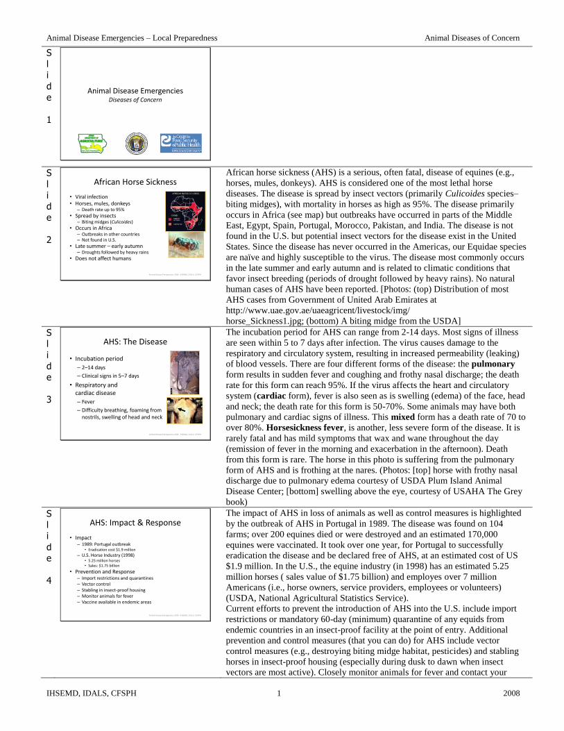


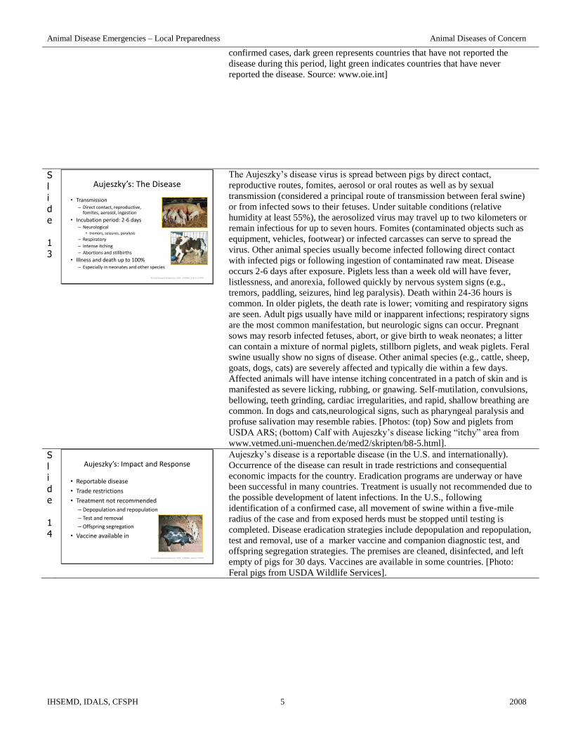

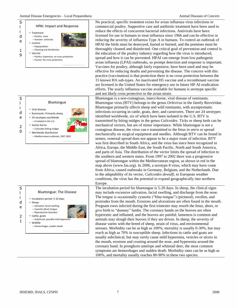

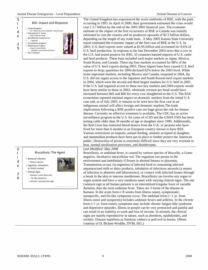


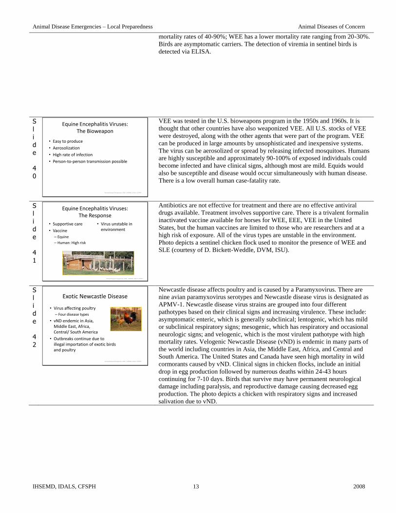

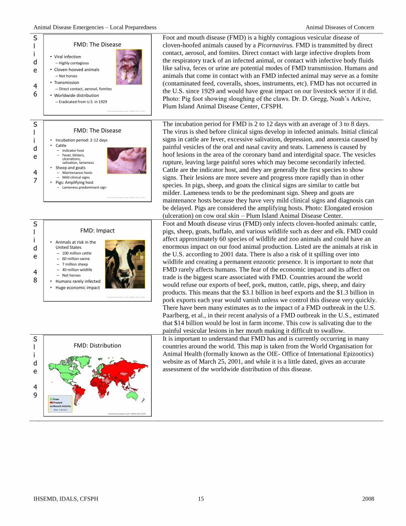
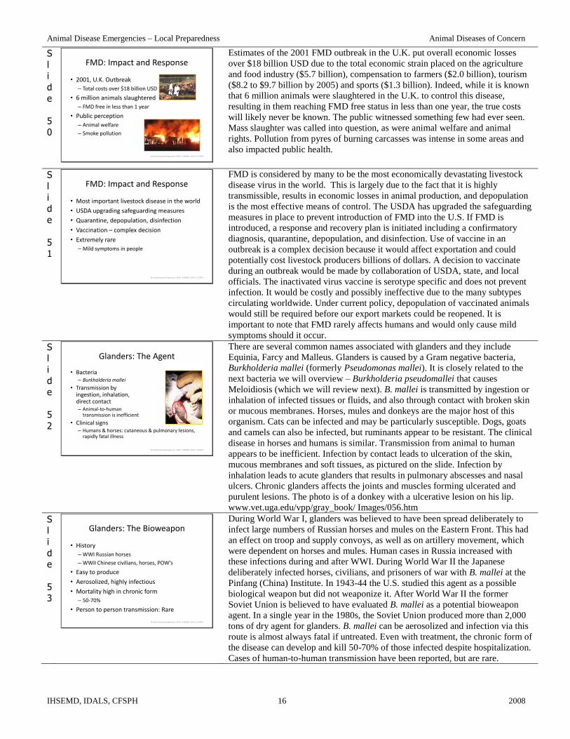


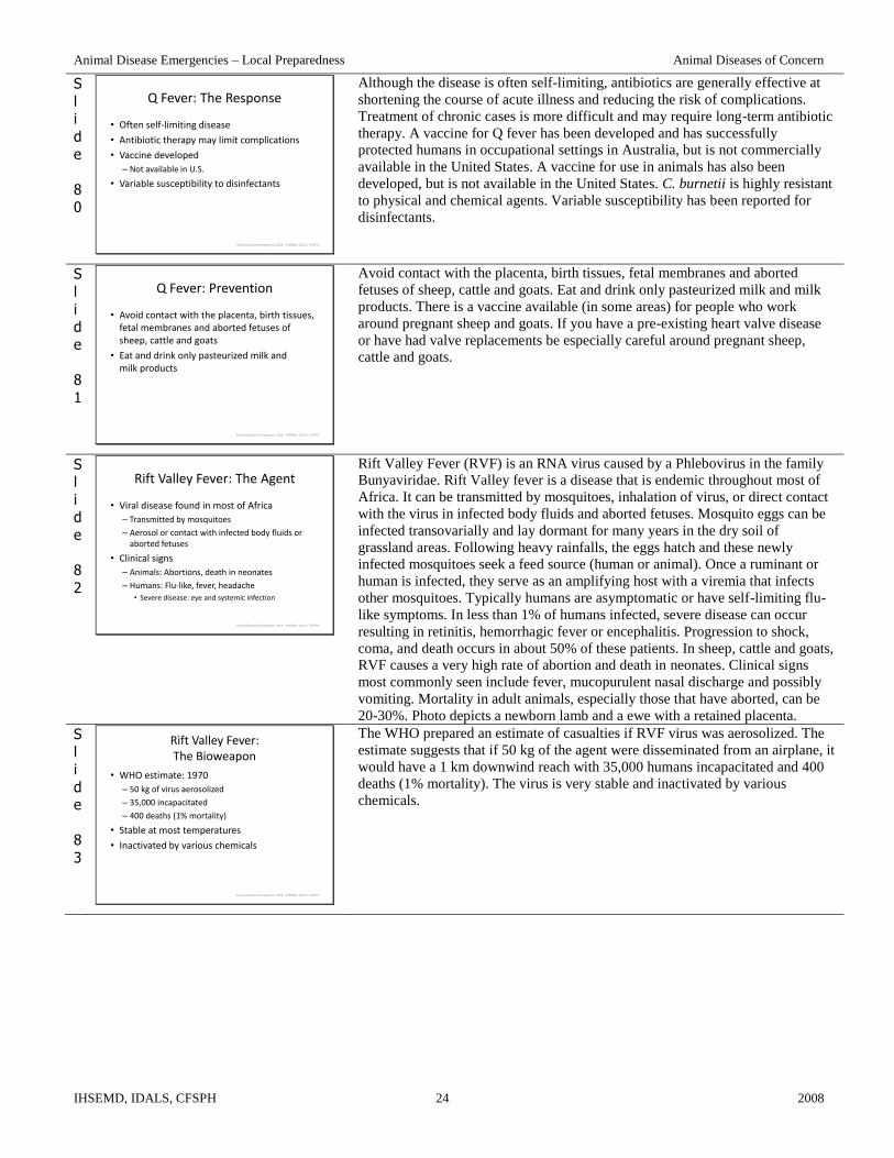


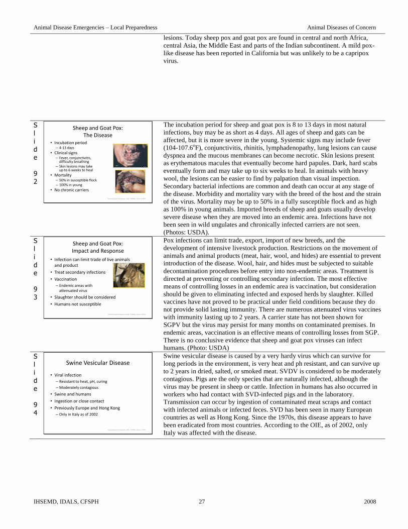


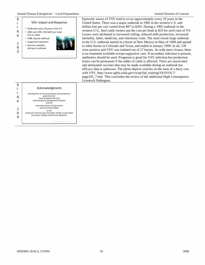


![Untitled-1 []Clinical Diagnostic Methods . Endodontic Emergencies Diseases of Dental Pulp Diseases of Periradicular Tissues Pulpotomy and Apexification Rationale of Endodontic Treatment](https://static.documents.pub/doc/80x56/5e77e90e3c586d38116f8a30/untitled-1-clinical-diagnostic-methods-endodontic-emergencies-diseases-of.jpg)


